Investigation of nuclear nano-morphology marker as a biomarker for cancer risk assessment using a mouse model
←
→
Page content transcription
If your browser does not render page correctly, please read the page content below
Investigation of nuclear nano-
                 morphology marker as a biomarker for
                 cancer risk assessment using a mouse
                 model

                 Rajan K. Bista
                 Shikhar Uttam
                 Douglas J. Hartman
                 Wei Qiu
                 Jian Yu
                 Lin Zhang
                 Randall E. Brand
                 Yang Liu

Downloaded From: https://www.spiedigitallibrary.org/journals/Journal-of-Biomedical-Optics on 07 Oct 2021
Terms of Use: https://www.spiedigitallibrary.org/terms-of-use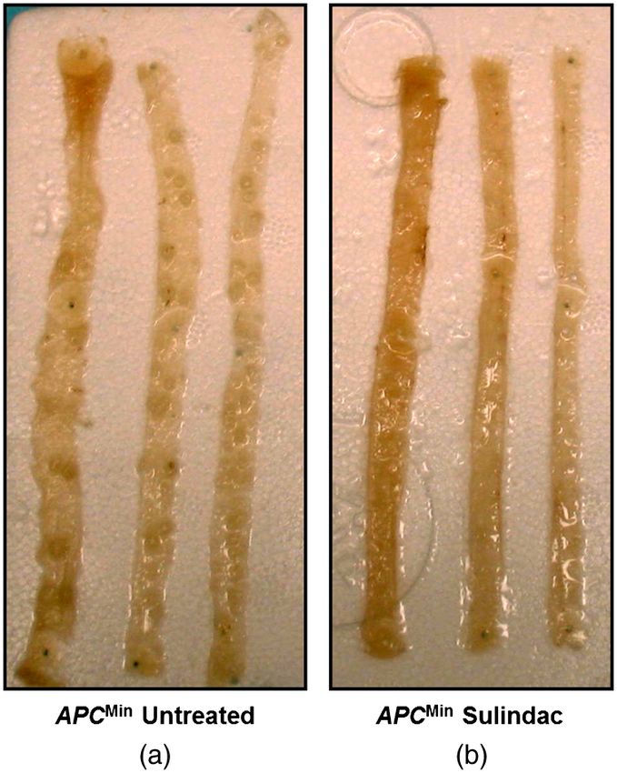
Journal of Biomedical Optics 17(6), 066014 (June 2012)

                Investigation of nuclear nano-morphology marker as a
                biomarker for cancer risk assessment using a mouse model
                Rajan K. Bista,a Shikhar Uttam,a Douglas J. Hartman,b Wei Qiu,b Jian Yu,b Lin Zhang,c Randall E. Brand,d and Yang Liua
                a
                  University   of   Pittsburgh, Department of Medicine and Bioengineering, Biomedical Optical Imaging Laboratory, Pittsburgh, Pennsylvania 15232
                b
                  University   of   Pittsburgh School of Medicine, Department of Pathology, Pittsburgh, Pennsylvania 15213
                c
                  University   of   Pittsburgh Cancer Institute, Department of Pharmacology and Chemical Biology, Pittsburgh, Pennsylvania 15213
                d
                  University   of   Pittsburgh, Department of Medicine, Division of Gastroenterology, Hepatology and Nutrition, Pittsburgh, Pennsylvania 15232

                                Abstract. The development of accurate and clinically applicable tools to assess cancer risk is essential to define
                                candidates to undergo screening for early-stage cancers at a curable stage or provide a novel method to monitor
                                chemoprevention treatments. With the use of our recently developed optical technology—spatial-domain low-
                                coherence quantitative phase microscopy (SL-QPM), we have derived a novel optical biomarker characterized
                                by structure-derived optical path length (OPL) properties from the cell nucleus on the standard histology and cytol-
                                ogy specimens, which quantifies the nano-structural alterations within the cell nucleus at the nanoscale sensitivity,
                                referred to as nano-morphology marker. The aim of this study is to evaluate the feasibility of the nuclear nano-
                                morphology marker from histologically normal cells, extracted directly from the standard histology specimens,
                                to detect early-stage carcinogenesis, assess cancer risk, and monitor the effect of chemopreventive treatment.
                                We used a well-established mouse model of spontaneous carcinogenesis—ApcMin mice, which develop multiple
                                intestinal adenomas (Min) due to a germline mutation in the adenomatous polyposis coli (Apc) gene. We found that
                                the nuclear nano-morphology marker quantified by OPL detects the development of carcinogenesis from histolo-
                                gically normal intestinal epithelial cells, even at an early pre-adenomatous stage (six weeks). It also exhibits a good
                                temporal correlation with the small intestine that parallels the development of carcinogenesis and cancer risk. To
                                further assess its ability to monitor the efficacy of chemopreventive agents, we used an established chemopreven-
                                tive agent, sulindac. The nuclear nano-morphology marker is reversed toward normal after a prolonged treatment.
                                Therefore, our proof-of-concept study establishes the feasibility of the SL-QPM derived nuclear nano-morphology
                                marker OPL as a promising, simple and clinically applicable biomarker for cancer risk assessment and evaluation of
                                chemopreventive treatment. © 2012 Society of Photo-Optical Instrumentation Engineers (SPIE). [DOI: 10.1117/1.JBO.17.6.066014]
                                Keywords: biomarker; phase microscopy; nano-morphology; optical path length; mouse model.
                                Paper 12021 received Jan. 10, 2012; revised manuscript received Apr. 16, 2012; accepted for publication Apr. 23, 2012; published
                                online Jun. 4, 2012.

                1     Introduction                                                                  assessment, especially during the early-stage development of
                Identification of effective biomarkers that can accurately predict                  carcinogenesis. The conventional morphology only detects
                cancer risk or serve as surrogates for preventive or therapeutic                    structural alterations at the micron scale, which are relatively
                efficacy has become increasingly important, as more treatment                       insensitive to various molecular alterations associated with car-
                options become available. However, the limited resource avail-                      cinogenesis. The recent advances in molecular technologies
                ability, high cost, and the inherent side-effects of many treatments                have identified a series of carcinogenesis-associated molecular
                preclude their use in all the affected individuals. Effective                       biomarkers that may serve as more sensitive and reliable bio-
                risk-assessment biomarkers can help to identify those indivi-                       markers for cancer risk assessment and detecting the early-
                duals who are truly at highest risk of cancer, and also help in                     stage carcinogenesis. Despite the great potential of these
                reducing the harm from overtreatment. Further, surrogate                            approaches, they can be confounded by intrinsic heterogeneity
                biomarkers for preventive or therapeutic efficacy represent                         in tumorigenesis or multitude of separate molecular mechanisms
                potential intermediate endpoints for short-term intervention                        underlying carcinogenesis.
                studies with preventive and therapeutic agents to measure the                           While the micron-scale morphologic abnormalities may not
                progress of treatment.                                                              be present early in the disease process, there are nanoscale mole-
                    Carcinogenesis involves a complex series of molecular                           cular alterations in “normal” tissue early in the development of
                events that involve various distinct or interconnected pathways                     carcinogenesis. The nanoscale internal structural manifestations
                toward invasive cancer. Conventional pathology remains the                          of various molecular events associated with carcinogenesis
                gold-standard in cancer detection and various cell morphologi-                      referred to as nano-morphology markers, have shown promise
                cal characteristics often serve as predictors for cancer risk.                      to serve as common shared characteristics for accurate cancer
                However, they often have a limited accuracy in cancer risk                          risk assessment despite the inherent heterogeneity.1 The recent
                                                                                                    development in optical techniques has allowed the detection of
                                                                                                    these nanoscale structural properties.1–9 These changes occur at
                Address all correspondence to: Yang Liu, University of Pittsburgh, Department of
                                                                                                    a scale of about 20 to 1000 times smaller than what conventional
                Medicine and Bioengineering, Biomedical Optical Imaging Laboratory, Depart-
                ment of Medicine and Bioengineering, University of Pittsburgh, Pittsburgh, Penn-
                sylvania 15232.; Tel: 412-623-3751; Fax: 412-623-7828; E-mail: liuy@pitt.edu        0091-3286/2012/$25.00 © 2012 SPIE

                Journal of Biomedical Optics                                                  066014-1                                          June 2012   •   Vol. 17(6)

Downloaded From: https://www.spiedigitallibrary.org/journals/Journal-of-Biomedical-Optics on 07 Oct 2021
Terms of Use: https://www.spiedigitallibrary.org/terms-of-use
Bista et al.: Investigation of nuclear nano-morphology marker as a biomarker : : :

                optical microscope can detect. For example, partial-wave spec-                     sex-matched C57BL ApcMin mice were sacrificed at the age
                troscopy has shown that changes in the nano-structural proper-                     of six weeks. A second set of three C57BL Apc wild-type
                ties could be highly sensitive for the detection of molecular                      mice and three age-matched C57BL ApcMin mice were sacri-
                alterations associated with carcinogenesis prior to the occur-                     ficed at the age of 4.5 months. Those ApcMin mice at the
                rence of phenotype.2,3,7,10,11 Moreover, other light scattering                    age of six weeks had only a few microscopically-visible
                techniques have been employed to measure subcellular structure                     micro-adenomas; whereas those ApcMin mice at the age of
                and provide noninvasive early detection of chemotherapy-in                         4.5-month had developed visible multiple adenomatous polyps
                uced apoptosis and to study calcium-induced alterations in mito-                   in their small intestine.
                chondrial morphology.12,13                                                             The small intestines tissue were removed, longitudinally
                    Our group has recently developed a novel optical                               opened and washed with phosphate buffered saline. A segment
                microscope, Spatial-domain low-coherence quantitative phase                        of small intestine (proximal and middle parts) was cut, and the
                microscopy (SL-QPM), that can detect subtle structural changes                     individual slide from each animal was prepared following the
                at sub-nanometer sensitivity, which is about 1000 times more                       standard tissue histology processing protocol with paraffin-
                sensitive than what conventional microscopy can detect.4–6,14                      embedding, sectioning at 4 μm thickness, mounting on a
                We derived nano-morphology markers quantified by nanoscale                         glass slide, paraffin removal and hematoxylin and eosin
                optical path length (OPL) properties from cell nuclei that showed                  (H&E) staining. After staining, the tissue sections were covered
                a superior sensitivity in detecting carcinogenesis from histologi-                 with coverslips using mounting media.
                cally “normal” cells from cancer patients, suggesting that
                SL-QPM is a powerful tool to detect subtle nano-structural                         2.2     Chemopreventive Study
                changes, not appreciated by conventional pathology.4,5
                SL-QPM also has the advantage of being directly applicable                         Two groups of ten-week-old sex-matched ApcMin mice compris-
                on the standard histology and cytology specimens, and has                          ing of six mice in each group were fed with control or experi-
                the potential for clinical translation.                                            mental high-fat AIN93G diet (Dyets) containing 200 ppm
                    In the present study, we evaluate the feasibility of the nano-                 (approximately 20 mg∕kg∕d) of sulindac (Sigma) for two
                morphology markers extracted directly from standard tissue                         weeks and four months as previously described.17,18 Mice were
                histology specimens as novel optical biomarkers for detecting                      sacrificed immediately after the treatment. The number of tumors
                early-stage carcinogenesis, assessing the cancer risk and evalu-                   was counted under the dissection microscope (polyps; >0.5 mm
                ating the efficacy of chemopreventive agents. For this purpose, a                  in diameter). Afterwards, a segment of small intestine was cut and
                well-established animal model—ApcMin mouse model for car-                          then prepared into “Swiss-rolls” and processed following stan-
                cinogenesis is used in this investigation. This animal model has                   dard histology protocol as mentioned earlier.
                adenomatous polyposis coli (Apc) gene mutation that causes
                spontaneous development of multiple intestinal neoplasia                           2.3     Analysis of the Nuclear Nano-Morphology
                (Min),15 primarily in the small intestine. The mutations in the                            Marker from Cell Nucleus
                Apc tumor suppressor gene occur very early in the transforma-
                tion process and are found in a majority of sporadic colorectal                    The nano-morphology marker from the cell nucleus is derived
                tumors as well as in the familial adenomatous polyposis.16 This                    using a recently developed optical microscopy system—SL-
                model has previously been shown to be a robust indicator of                        QPM. The technical development of SL-QPM system has
                tumorigenesis with changes in tumor number observed in                             been described in detail in our previous publications.4,5,20 We
                response to a variety of preventive and therapeutic agents.17                      obtain an OPL map of the individual cell nucleus under obser-
                Further, a well-documented chemopreventive agent—sulindac,                         vation, which is sensitive to subtle structural changes in the cell
                a type of nonsteroidal anti-inflammatory drugs (NSAIDs) has                        nucleus with nanoscale sensitivity.
                been shown to be very effective in reducing the number and                             In brief, SL-QPM system uses reflection-mode common-
                size of polyps and preventing colon cancer in human and rodent                     path interferometer configuration combined with a low-
                models.17–19 These characteristics make the ApcMin mouse model                     coherence light source and spectroscopic detection and records
                an ideal system for our feasibility study. We first analyze the                    a three-dimensional spectroscopic intensity data cube [Iðx; y; zÞ;
                nano-morphology marker from the cell nuclei of normal epithelial                   k ¼ free-space wavenumber, ðx; yÞ corresponds to the specific
                cells in the small intestine at different stages of carcinogenesis to              pixel of the image], predominately the interference signals
                evaluate its ability to assess the cancer risk by its temporal cor-                between the backscattered reference wave and the backscattered
                relations with future neoplasia. We then assess its ability to moni-               sample wave. These interference signals were mathematically
                tor the efficacy of chemopreventive agent sulindac by temporal                     transformed via the pixel-wise Fourier transform of Iðx; y; zÞ
                correlations with the number and size of the tumors.                               along the k direction—after removing the bias term—gives
                                                                                                   us I F ðx; y; z 0 Þ, where z 0 is the OPL. The I F ðx; y; z 0 Þ is then
                                                                                                   used to find the prominent peak corresponding to the OPL of
                2     Materials and Methods
                                                                                                   interest, zp . We then calculate the structure-derived OPL with
                2.1     Animal Model                                                               the equation OPLðx; yÞ ¼ zp þ ∠Iðx; y; z 0 Þjz 0 ¼zp ∕ð2kÞ, where
                                                                                                   ∠Iðx; y; z 0 Þ represents the phase term and the factor 2 in the
                All animal studies were performed in accordance with the in-                       denominator of above relation accounts for the double path
                stitutional Animal Care and Use Committee of the University                        length geometry due to the reflectance-mode configuration and
                of Pittsburgh. All mice were housed in micro isolator cages                        the free-space wavenumber k corresponds to λ0 ¼ 550 nm. The
                in a room illuminated from 7:00 AM to 7:00 PM (12:12-h                             nanoscale structural sensitivity comes from the second term of
                light-dark cycle), with access to water and chow ad libitum.                       this equation. Figure 1 illustrates the data processing steps of
                A total of 24 female mice were included in this study. A first                     this system. We also account for the effect of variation in the
                set of three C57BL Apc wild-type mice and three age- and                           stain-induced structure-derived OPL using a staining correction

                Journal of Biomedical Optics                                               066014-2                                               June 2012   •   Vol. 17(6)

Downloaded From: https://www.spiedigitallibrary.org/journals/Journal-of-Biomedical-Optics on 07 Oct 2021
Terms of Use: https://www.spiedigitallibrary.org/terms-of-use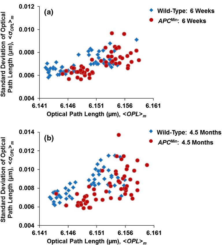
Bista et al.: Investigation of nuclear nano-morphology marker as a biomarker : : :

                  Fig. 1 Schematics of the data processing steps involved in the spatial-domain low-coherence quantitative phase microscopy (SL-QPM) system.

                model.20 As a result, the structure-derived OPL map can be                         the representative conventional bright-field images and corre-
                obtained from each cell nucleus.                                                   sponding structure-derived nuclear OPL maps for three selected
                                                                                                   nuclei of the histologically normal intestinal epithelial cells for
                2.4     Statistical Analysis                                                       (a) a six-week wild-type mouse, (b) a six-week ApcMin mouse,
                                                                                                   and (c) a 4.5-month ApcMin mouse. Although these cells are
                To quantify the statistical nano-morphology characteristics of the                 labeled as “normal” by an expert pathologist (DJH), their
                structure-derived OPL map in the nucleus, we extracted the aver-                   pseudo-color nuclear OPL maps exhibit a progressive change.
                age hOPLi from each cell nucleus as the representative statistical                 The pre-dominant blue color from the cell nucleus of the
                nuclear nano-morphology marker. We analyzed this marker for                        wild-type mouse suggests a smaller value of OPL, compared
                approximately 50 to 60 epithelial cell nuclei from each sample.                    to the nuclear OPL map from the six-week and 4.5-month
                To minimize the variations of cell appearance due to tissue sec-                   mice, suggesting the promise of using nuclear structural
                tioning, we particularly focused on the columnar-shaped epithe-                    abnormalities to detect the subtle structural changes that are
                lial cells having similar morphological features such as intact                    not appreciated by using conventional pathology approach.
                nuclear boundary and no overlap of nucleus as marked region
                in Fig. 2. The statistical analysis was performed using the stu-
                dent’s t-test (Microsoft Excel 2010). Two-sided P values (assum-                   3.2     Statistical Analysis of Nuclear Nano-Morphology
                ing unequal variances) were used for all analyses. A two-sided P                           Marker from the Cell Nucleus
                value of 0.05 or less was considered as statistically significant.
                                                                                                   Based on the above structure-derived nuclear OPL maps of his-
                                                                                                   tologically normal cells, we performed the statistical analysis for
                3     Results                                                                      ∼50 to 60 epithelial cell nuclei for each mouse. As shown in
                We first examined whether the nuclear nano-morphology marker                       Fig. 3, the nuclear nano-morphology marker of hOPLi from his-
                deduced from the structure-derived OPL can be used to predict                      tologically normal epithelial cells (i.e., uninvolved cells) from
                the concurrent and future neoplasm in the well-established animal                  small intestine show a progressive increase from wild-type
                model of colorectal carcinogenesis—ApcMin mice. In this mouse                      mice, to six-week mice, to 4.5-month mice (P ¼ 0.01 between
                model, the majority of the intestinal adenomatous polyps occur in                  six weeks and 4.5 months), which parallels the development of
                the small intestine and tumors occur in a well-established chron-                  colon carcinogenesis. Importantly, there is a statistical signifi-
                ology. The mice at six weeks represent an early-stage carcinogen-                  cance even for early-stage neoplastic changes of six weeks in
                esis without any visible tumors and with only microscopically                      ApcMin mice (P ¼ 0.001) and follows the temporal progression
                visible micro-adenomas; while those at 4.5 months represent                        of intestinal tumorigenesis with more elevated value of nuclear
                an advanced stage of carcinogenesis with multiple macroscopi-                      hOPLi in the advanced neoplastic stage of 4.5 months
                cally visible adenomatous polyps (tumors).                                         (P ¼ 8.1E − 11), suggesting that this nuclear nano-morphology
                                                                                                   marker detects the subtle structural changes beyond the morpho-
                3.1     Structure-Derived Optical Path Length (OPL)                                logically evident tumor (i.e., the histologically normal cells from
                        Map from Cell Nuclei Detects the Concurrent                                ApcMin mice). It is a good indicator to predict a genetic substrate
                        and Future Neoplasm from Normal Intestinal                                 to neoplastic transformation before occurrence of neoplasia.
                        Epithelial Cells                                                           We also found that the values of nuclear hOPLi for the
                                                                                                   wild-type mice at different ages of six weeks and 4.5 months
                We first analyzed the histologically normal epithelial cells from                  are not statistically significant (P ¼ 0.1), suggesting that the
                the small intestine from two sets of age-matched wild-type mice                    nano-morphology markers do not appear to be compromised
                and ApcMin mice at six weeks and 4.5 months. Figure 2 shows                        by confounding factors contributed by the age of the mice.21

                Journal of Biomedical Optics                                               066014-3                                            June 2012   •   Vol. 17(6)

Downloaded From: https://www.spiedigitallibrary.org/journals/Journal-of-Biomedical-Optics on 07 Oct 2021
Terms of Use: https://www.spiedigitallibrary.org/terms-of-use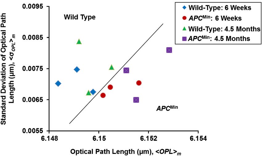
Bista et al.: Investigation of nuclear nano-morphology marker as a biomarker : : :

                Fig. 2 Representative regular bright-field images of H&E stained epithelial cell nuclei and corresponding structure-derived nuclear optical path length
                (OPL) maps for three selected cells derived from (a) wild-type mouse (six weeks), (b) histologically normal appearing cells (uninvolved cells) from six
                weeks ApcMin mouse, and (c) histologically normal appearing cells (uninvolved cells) from 4.5 months ApcMin mouse. The marked area in part (a)
                shows the representative columnar shaped epithelial cells having similar morphological features.

                Journal of Biomedical Optics                                               066014-4                                            June 2012   •   Vol. 17(6)

Downloaded From: https://www.spiedigitallibrary.org/journals/Journal-of-Biomedical-Optics on 07 Oct 2021
Terms of Use: https://www.spiedigitallibrary.org/terms-of-use
Bista et al.: Investigation of nuclear nano-morphology marker as a biomarker : : :
Fig. 3 Statistical analysis of the nuclear nano-morphology marker char-
acterized by structure-derived optical path length (OPL) from the cell
nuclei of wild-type mice and histologically normal (i.e., uninvolved)
appearing intestinal epithelial cell nuclei from gender and age-matched
ApcMin mice at six weeks and 4.5 months. The nuclear OPL exhibits a
progressive increase with the development of carcinogenesis. Approxi-
mately 50 to 60 cell nuclei were analyzed from each mouse.
3.3 Performance of Nuclear Nano-Morphology
Markers
The scatter plots in Fig. 4 present the cell-to-cell variation of
nano-morphology markers [hOPLi and the intra-nuclear stan- Fig. 4 The scatter plots showing the cell-to-cell variation from the his-
tologically normal cells from wild-type and ApcMin mice at (a) six weeks
dard deviation of OPLðσ OPL Þ] in the wild-type and ApcMin and (b) 4.5 months. Despite the intrinsic variation, the histologically
mice at six weeks and 4.5 months. Despite the intrinsic varia- normal cells from the ApcMin mice occupy a distinct regime compared
tion, these histologically normal cells from the ApcMin mice to those from wild-type mice.
occupy a distinct region with minor overlap compared to
those from the wild-type mice. However, such cell-to-cell var-
iation within the same animal is much smaller than the inter- sulindac treatment prevents intestinal tumors by reducing the
group animal variation (wild-type versus ApcMin group), as number of polyps (≥0.5 mm in diameter) by ∼88% (from
quantified by the p-values (P < 0.05) presented in the previous approximately 85 in untreated mice to 10 after 4-month treat-
section. ment, P < 0.0001).17
Further, to use these nuclear nano-morphology markers for Figure 7 demonstrates the statistical analysis of the structure-
future clinical use to assess the risk of developing carcinogen- derived nuclear hOPLi from histologically normal intestinal
esis for each patient, we have to obtain the patient-based char- epithelial cells of untreated ApcMin mice and sulindac-treated
acteristic statistical measure. In this proof-of-concept study with
ApcMin mice for two weeks and four months treatment. For
the animal model, the average values of hOPLi and the intra-
nuclear standard deviation of OPLðσ OPL Þ denoted respectively
as hOPLim and hσ OPL im , were taken from about 50 to 60 cell
nuclei as characteristic subject-specific marker for each mouse.
Figure 5 shows the scatter plot of hOPLim and hσ OPL im for each
mouse at different time points. It is evident that there is a clear
separation between the wild-type mice and ApcMin mice group,
as indicated by the solid line in Fig. 5. All of those mice with the
risk of developing carcinogenesis (even those at the early stage)
are detected by the nuclear nano-morphology markers.
3.4 Chemoprevention Studies with Sulindac
To further confirm the neoplastic relevance of the nuclear nano-
morphology marker quantified by OPL and evaluate its potential
to monitor the efficacy of preventive agents, we investigated
how the nuclear nano-morphology marker from histologically
normal cells changes as a response to the treatment using an Fig. 5 The scatter plot of the individual-animal based characteristic
established chemopreventive agent, sulindac. It has been pre- nano-morphology markers characterized by the mean values
viously shown that dietary supplementation with NSAIDs (hOPLim and hσ OPL im ) of intra-nuclear hOPLi and intra-nuclear standard
deviation of optical path length σ OPL averaged over 50 to 60 cell nuclei
such as sulindac for several months prevent adenoma formation for each wild-type and ApcMin mouse. Evidently, the nuclear nano-mor-
in the small intestine of ApcMin mice.22 We showed that sulindac phology markers can clearly distinguish wild-type mice from ApcMin
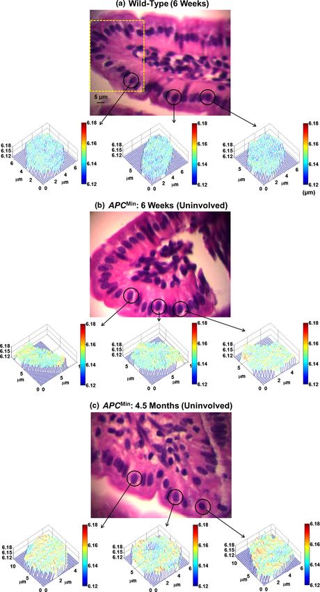
has significantly reduced number of polyps after four-month mice (even at the early-stage carcinogenesis), as indicated by the
treatment in the ApcMin mice, as shown in Fig. 6. The 4-month solid line.
Journal of Biomedical Optics 066014-5 June 2012 • Vol. 17(6)
Downloaded From: https://www.spiedigitallibrary.org/journals/Journal-of-Biomedical-Optics on 07 Oct 2021
Terms of Use: https://www.spiedigitallibrary.org/terms-of-useBista et al.: Investigation of nuclear nano-morphology marker as a biomarker : : :
untreated ApcMin mice, the nuclear hOPLi in the histologically
normal intestinal epithelial cells significantly increases with
time (P ¼ 1.0E − 5); while for sulindac-treated ApcMin mice,
the values of nuclear hOPLi in the normal intestinal epithelial
cells do not show a statistically significant increase over time at
six weeks and four months (P ¼ 0.3). At the early-stage of the
sulindac treatment (two weeks), the normal intestinal epithelial
cells show a slight decrease of nuclear hOPLi (P ¼ 0.4, no sta-
tistical significance) in the ApcMin mice compared to the
untreated mice. But after four months of prolonged treatment
with sulindac, their nuclear hOPLi value significantly decreases,
compared to those untreated ApcMin mice (P ¼ 4.7E − 5), and
reversed to normal. These results are consistent with the finding
that the number of tumors has significantly reduced after the
prolonged treatment of sulindac. These data also further confirm
Fig. 7 Statistical analysis of the nuclear optical path length (OPL) from
that the nuclear nano-morphology marker defined by OPL from the nuclei of intestinal epithelial cells in the ApcMin mice with regular
the histologically normal intestinal epithelial cells is truly due to diet and with sulindac supplement. The progression of nuclear OPL in
the neoplastic changes, rather than other nonspecific changes in the histologically normal (i.e., uninvolved) intestinal epithelial cell
the animal model. The result also shows a potential of using the nuclei significantly slowed down after 4-month of sulindac treatment
nuclear OPL as a surrogate marker to monitor the effect of (P ¼ 0.3) compared to the corresponding untreated ApcMin mice
chemopreventive treatment. (P ¼ 1E − 5). The nuclear OPL from sulindac-treated mice is signifi-
cantly lower than those untreated ApcMin mice (P ¼ 4.7E − 5). Approxi-
mately 50 to 60 cell nuclei were analyzed from each mouse.
4 Discussion
The nuclear nano-morphology marker is derived from a
With the use of a well-established animal model of colon car- recently developed novel optical microscope—spatial-domain
cinogenesis and chemo-preventive agent, we report herein the low-coherence quantitative phase microscopy (SL-QPM). It
significant alteration of the nano-morphology marker character- represents a novel class of cellular characteristics for evaluating
ized by structure-derived OPL from the cell nuclei in the histo- nanoscale structural changes in the cell nucleus that could
logically normal intestinal epithelial cells that precede the accompany the complex accumulation of molecular alterations
development of the phenotype (i.e., adenomas). The progressive during carcinogenesis. SL-QPM takes advantage of the ultra-
change in this nano-morphology marker parallels the progres- high sensitivity of the light interference effect to achieve nanos-
sion of carcinogenesis. Further, this marker is reversed to normal cale sensitivity not attainable with a conventional microscope.4,5
as a response to the treatment of the potent chemopreventive Compared with other biomarkers, the nano-morphology marker
agent sulindac. Hence, such nano-morphology marker repre- has a few major advantages. First, it can be extracted from the
sents a powerful means of detecting an inherited cancer risk, clinical tissue histology specimens (glass-slide-based) prepared
monitoring the progression of carcinogenesis even at the according to standard routine clinical protocols without any
early stage, and for evaluation of response to chemoprevention.
additional processing, thus easily can be integrated with the
existing workflow of current pathology laboratories. Second,
the analysis of nuclear nano-morphology marker does not
require any consumable agents, so there is no additional cost
or effort in preparing the sample.
The nuclear nano-morphology marker shows the ability to
detect the presence of “field effect” in carcinogenesis, or detect
the presence of neoplastic conditions from histologically normal
cells adjacent to or distant from the tumor. For example, for
those cells labeled as “normal” by the expert gastrointestinal
pathologist, their nuclear OPL are significantly elevated in
the ApcMin mice at both early-stage of carcinogenesis of six
weeks without development of any visible tumors and an
advanced neoplastic stage of 4.5 months with multiple adeno-
matous polyps, when compared to those from the wild-type
mice. The field effect in the ApcMin mice results from the
genetic predisposition to the development of carcinogenesis.23
Besides the inherited genetic defects, other significant biochem-
ical and genetic molecular abnormalities have also been reported
to contribute to the altered “cancer field” in the histologically
normal intestinal mucosa of the ApcMin mice, such as altered
β-catenin,24 cell proliferation, apoptosis,25 and metabolic altera-
tions.26 Although the specific biological mechanisms responsi-
Fig. 6 Longitudinally opened sections of small intestine; (a) ApcMin / ble for the detected changes in nuclear OPL are still unknown,
untreated mice and (b) ApcMin /sulindac treated mice. Sulindac signifi- the reported biological processes provide the biological plausi-
cantly reduced the number of polyps after 4-month treatment. bility. One or more of the molecular, cellular and biochemical
Journal of Biomedical Optics 066014-6 June 2012 • Vol. 17(6)
Downloaded From: https://www.spiedigitallibrary.org/journals/Journal-of-Biomedical-Optics on 07 Oct 2021
Terms of Use: https://www.spiedigitallibrary.org/terms-of-useBista et al.: Investigation of nuclear nano-morphology marker as a biomarker : : :
events associated with “field effect” may also be responsible for 2. H. Subramanian et al., “Optical methodology for detecting histologi-
the change in the nuclear nano-morphology marker OPL. For cally unapparent nanoscale consequences of genetic alterations in
biological cells,” Proc. Natl. Acad. Sci. USA 105(51),20118–20123
example, our recent finding revealed that increased DNA con-
(2008).
tent is one of the contributing factors for the increased nuclear 3. H. Subramanian et al., “Nanoscale cellular changes in field carcinogen-
OPL observed in carcinogenesis.14 esis detected by partial wave spectroscopy,” Cancer Res. 69(13), 5357–
The detection of such “field effect” can also be used as an 5363 (2009).
early-stage marker to monitor the progression of carcinogenesis 4. P. Wang et al., “Spatial-domain low-coherence quantitative phase micro-
and predict the future neoplastic risk, as suggested by the tem- scopy for cancer diagnosis,” Opt. Lett. 35(17), 2840–2842 (2010).
poral correlation of the nano-morphology marker of nuclear 5. P. Wang et al., “Nanoscale nuclear architecture for cancer diagnosis
beyond pathology via spatial-domain low-coherence quantitative
OPL with the neoplastic progression. The most significant pro- phase microscopy,” J. Biomed. Opt. 15(16), 066028 (2010).
gression (P ¼ 0.001) occurs between the wild-type mice and the 6. P. Wang et al., “An insight into statistical refractive index properties of
ApcMin mice at the pre-adenomatous stage (six weeks), well pre- cell internal structure via low-coherence statistical amplitude micro-
ceding the development of visible tumor (i.e., adenomatous scopy,” Opt. Express 18(21), 21950–21958 (2010).
polyps). The neoplastic relevance of nuclear OPL is further 7. N. T. Shaked et al., “Quantitative phase microscopy of articular chon-
underscored by its significant reversal to normal (wild-type) drocyte dynamics by wide-field digital interferometry,” J. Biomed. Opt.
15(1), 010505 (2010).
mice, as a response to sulindac treatment that leads to a signifi- 8. I. Itzkan et al., “Confocal light absorption and scattering spectroscopic
cant reduction (∼88%) in the number of tumors. This result sug- microscopy monitors organelles in live cells with no exogenous labels,”
gests that the nano-morphology marker of the histologically Proc. Natl. Acad. Sci. USA 104(44), 17255–17260 (2007).
normal epithelial cells detects the tumor-suppressing ability 9. B. Kemper et al., “Integral refractive index determination of living sus-
of chemopreventive agents and can also serve as an early- pension cells by multifocus digital holographic phase contrast micro-
stage surrogate marker to monitor the efficiency of chemopre- scopy,” J. Biomed. Opt. 12(5), 054009 (2007).
10. H. K. Roy et al., “Optical detection of buccal epithelial nanoarchitec-
ventive agents.
tural alterations in patients harboring lung cancer: implications for
In conclusion, in this proof-of-concept study using a well- screening,” Cancer Res. 70(20), 7748–7754 (2010).
established animal model of carcinogenesis—ApcMin mouse 11. R. K. Bista et al., “Using optical markers of nondysplastic rectal epithe-
model and an efficient chemopreventive agent of sulindac, lial cells to identify patients with ulcerative colitis-associated neopla-
we evaluated the use of a novel nuclear nano-morphology mar- sia,” Inflamm. Bowel Dis. 17(12), 2427–2435 (2011).
ker quantified by structure-derived OPL, extracted directly from 12. K. J. Chalut et al., “Light scattering measurements of subcellular struc-
standard histology specimens, as a potential biomarker to detect ture provide noninvasive early detection of chemotherapy-induced
apoptosis,” Cancer Res. 69(3), 1199–1204 (2009).
the early-stage carcinogenesis, assess the cancer risk and moni- 13. N. N. Boustany, R. Drezek, and N. V. Thakor, “Calcium-induced altera-
tor the efficiency of chemopreventive agents. We demonstrated tions in mitochondrial morphology quantified in situ with optical scatter
that the nuclear nano-morphology marker OPL detects the pre- imaging,” Biophys. J. 83(3), 1691–1700 (2002).
sence of neoplasm from histologically normal cells, or cancer 14. R. K. Bista et al., “Quantification of nanoscale nuclear refractive index
“field effect.” The temporal correlation of the nuclear nano- changes during the cell cycle,” J. Biomed. Opt. 16(7), 070503 (2011).
morphology marker OPL with the neoplastic progression sug- 15. K. W. Kinzler and B. Vogelstein, “Lessons from hereditary colorectal
gests its use as an early-stage biomarker to predict the future cancer,” Cell 87(2), 159–170 (1996).
16. B. Vogelstein and K. W. Kinzler, “Cancer genes and the pathways they
neoplastic risk. Moreover, the substantial reversal of this bio- control,” Nat. Med. 10(8), 789–799 (2004).
marker to normal as a response to the chemopreventive agent 17. W. Qiu et al., “Chemoprevention by nonsteroidal anti-inflammatory
of sulindac suggests its use as a surrogate marker to gauge drugs eliminates oncogenic intestinal stem cells via SMAC-dependent
the chemopreventive efficacy for cancer risk reduction. This apoptosis,” Proc. Natl. Acad. Sci. USA 107(46), 20027–20032 (2010).
marker is currently being evaluated for identifying patients 18. W. Qiu et al., “PUMA suppresses intestinal tumorigenesis in mice,”
who are at an increased risk of developing malignancy and Cancer Res. 69(12), 4999–5006 (2009).
19. S. J. Shiff et al., “Sulindac sulfide, an aspirin-like compound, inhibits
as a surrogate marker for monitoring the effect of chemopreven-
proliferation, causes cell cycle quiescence, and induces apoptosis in HT-
tive agents. If proven successful in clinical studies, it will help in 29 colon adenocarcinoma cells,” J. Clin. Invest. 96(1), 491–503 (1995).
identifying patients with truly high-risk for proper preventive or 20. S. Uttam et al., “Correction of stain variations in nuclear refractive index
therapeutic treatment. This nano-morphology marker is derived of clinical histology specimens,” J. Biomed. Opt. 16(11), 116013
from the standard tissue histology specimens, and therefore (2011).
could be readily translated to clinical settings. 21. L. L. Shen et al., “MGMT promoter methylation and field defect
in sporadic colorectal cancer,” J. Natl. Cancer Inst. 97(18), 1330–
Acknowledgments 1338 (2005).
22. Y. Beazer-Barclay et al., “Sulindac suppresses tumorigenesis in the Min
We gratefully acknowledge the National Institute of Health mouse,” Carcinogenesis 17(8), 1757–1760 (1996).
(R21CA152935), American Cancer Society (RGS-10-124-01- 23. B. J. Braakhuis et al., “A genetic explanation of Slaughter's concept of
CCE), Wallace H. Coulter foundation and University of Pitts- field cancerization: evidence and clinical implications,” Cancer Res. 63
burgh Medical Center for supporting this research. The authors (8), 1727–1730 (2003).
appreciate the help from Kevin Staton in sample preparation. 24. K. Aoki and M. M. Taketo, “Adenomatous polyposis coli (APC): a
multi-functional tumor suppressor gene,” J. Cell Sci. 120(19), 3327–
3335 (2007).
25. S. V. Russello and S. K. Shore, “SRC in human carcinogenesis,” Front
References
Biosci: A J Virtual Library 9, 139–144 (2004).
1. H. K. Roy, T. Hensing, and V. Backman, “Nanocytology for field 26. A. Backshall et al., “Detection of metabolic alterations in non-tumor
carcinogenesis detection: novel paradigm for lung cancer risk stratifica- gastrointestinal tissue of the Apc(Min/+) mouse by (1)H MAS NMR
tion,” Future Oncol. 7(1), 1–3 (2011). spectroscopy,” J. Proteome Res. 8(3), 1423–1430 (2009).
Journal of Biomedical Optics 066014-7 June 2012 • Vol. 17(6)
Downloaded From: https://www.spiedigitallibrary.org/journals/Journal-of-Biomedical-Optics on 07 Oct 2021
Terms of Use: https://www.spiedigitallibrary.org/terms-of-useYou can also read