Hepatic Branch Vagotomy Modulates the Gut-Liver-Brain Axis in Murine Cirrhosis
←
→
Page content transcription
If your browser does not render page correctly, please read the page content below
ORIGINAL RESEARCH
                                                                                                                                                  published: 25 June 2021
                                                                                                                                         doi: 10.3389/fphys.2021.702646

                                                Hepatic Branch Vagotomy Modulates
                                                the Gut-Liver-Brain Axis in Murine
                                                Cirrhosis
                                                Yuan Zhang 1 , Jason D. Kang 1 , Derrick Zhao 1 , Siddartha S. Ghosh 2 , Yanyan Wang 1 ,
                                                Yunling Tai 1 , Javier Gonzalez-Maeso 3 , Masoumeh Sikaroodi 4 , Patrick M. Gillevet 4 ,
                                                H. Robert Lippman 5 , Phillip B. Hylemon 1 , Huiping Zhou 1 and Jasmohan S. Bajaj 6*
                                                1
                                                  Division of Microbiology and Immunology, Central Virginia Veterans Health Care System, Virginia Commonwealth University,
                                                Richmond, VA, United States, 2 Division of Nephrology, Virginia Commonwealth University, Richmond, VA, United States,
                                                3
                                                  Department of Physiology and Biophysics, Virginia Commonwealth University, Richmond, VA, United States, 4 Microbiome
                                                Analysis Center, George Mason University, Manassas, VA, United States, 5 Department of Pathology, Central Virginia
                                                Veterans Health Care System, Richmond, VA, United States, 6 Division of Gastroenterology, Hepatology, and Nutrition,
                                                Central Virginia Veterans Health Care System, Virginia Commonwealth University, Richmond, VA, United States

                                                Background: Cirrhosis and hepatic encephalopathy (HE) are linked with an altered gut-
                             Edited by:         liver-brain axis, however, the relative contribution of hepatic vagal innervation is unclear.
                    Phillipp Hartmann,
   University of California, San Diego,
                                                We aimed to determine the impact of hepatic vagotomy on the gut microbiome, brain,
                          United States         and liver in murine cirrhosis.
                         Reviewed by:
                                                Methods: 10–15-week-old male C57BL/6 mice with and without hepatic vagotomy
                         Angela Jacan,
      Center for Biomarker Research             underwent carbon tetrachloride (CCl4) gavage for 8 weeks. Frontal cortex [inflammation,
        in Medicine (CBmed), Austria            glial/microglial activation, BDNF (brain-derived neurotrophic factor)], liver [histology
                                  Di Lu,
             Zhejiang University, China         including inflammation and steatosis, fatty acid synthesis (sterol-responsive binding
                           Haoche Wei,          protein-1) SREBP-1, insulin-induced gene-2 (Insig2) and BDNF], and colonic mucosal
 University of Bristol, United Kingdom
                                                microbiota (16srRNA microbial sequencing) were evaluated on sacrifice. Conventional
                *Correspondence:
                                                mice with and without cirrhosis were compared to vagotomized counterparts.
                 Jasmohan S. Bajaj
       jasmohan.bajaj@vcuhealth.org
                                                Results: Conventional control vs. cirrhosis: Cirrhosis resulted in dysbiosis,
                   Specialty section:
                                                hepatic/neuro-inflammation with glial/microglial activation, and low brain BDNF vs.
         This article was submitted to          controls. Conventional control vs. vagotomy controls: Vagotomized control mice had
           Gastrointestinal Sciences,
                                                a lower colonic dysbiosis than conventional mice but the rest of the hepatic/brain
               a section of the journal
               Frontiers in Physiology          parameters were similar. Conventional cirrhosis vs. vagotomized cirrhosis: After
             Received: 29 April 2021            vagotomy + cirrhosis, we found lower dysbiosis but continuing neuroinflammation in the
            Accepted: 02 June 2021              absence of glial/microglial activation vs. conventional cirrhosis. Vagotomy + Cirrhosis
            Published: 25 June 2021
                                                groups showed higher hepatic steatosis due to higher SREBP1 and low Insig2
                             Citation:
          Zhang Y, Kang JD, Zhao D,
                                                protein and altered activation of key genes involved in hepatic lipid metabolism
            Ghosh SS, Wang Y, Tai Y,            and inflammation. BDNF levels in the brain were higher but low in the liver in
     Gonzalez-Maeso J, Sikaroodi M,
                                                vagotomy + cirrhosis, likely a protective mechanism.
            Gillevet PM, Lippman HR,
   Hylemon PB, Zhou H and Bajaj JS              Conclusions: Hepatic vagal innervation affects the gut microbial composition, hepatic
    (2021) Hepatic Branch Vagotomy
   Modulates the Gut-Liver-Brain Axis           inflammation and steatosis, and cortical inflammation and BDNF expression and could
                   in Murine Cirrhosis.         be a critical modulator of the gut-liver-brain axis with consequences for HE development.
           Front. Physiol. 12:702646.
    doi: 10.3389/fphys.2021.702646              Keywords: vagotomy, pathobiont, inflammation, BDNF, hepatic encephalopathy, microbiota (16S)

Frontiers in Physiology | www.frontiersin.org                                         1                                            June 2021 | Volume 12 | Article 702646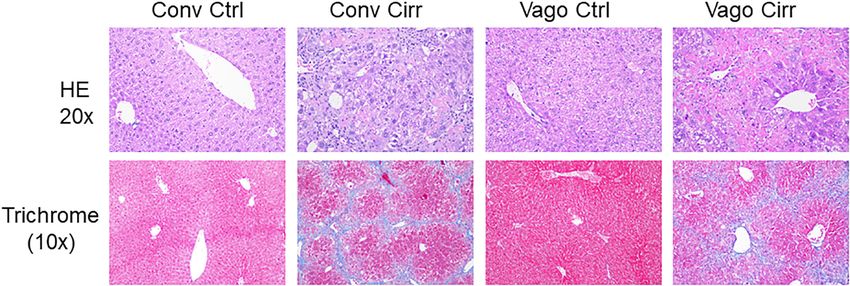
Zhang et al.                                                                                                    Vagotomy, Cirrhosis and Gut-Liver-Brain Changes

INTRODUCTION                                                                          Harada et al., 2014; Metz and Pavlov, 2018). The liver-brain
                                                                                      connection through the hepatic vagus branch could be used to
The gut-liver-brain axis mediates the development and                                 determine the relative contribution of the liver-brain aspect of
progression of complications of cirrhosis, such as hepatic                            the gut-liver-brain axis.
encephalopathy (HE) (Kang et al., 2016a; Ochoa-Sanchez                                   Our aim was to evaluate the effect of isolated hepatic vagotomy
and Rose, 2018). Most therapies for HE are focused on the                             on gut microbiota, liver inflammation, and brain inflammation in
modulation of the gut microbial milieu using laxatives and                            the setting of murine cirrhosis.
antibiotics (Vilstrup et al., 2014). In addition to the microbial
metabolite and barrier impairment, gut-brain axis alterations
could also be modulated through vagal innervation (Cryan et al.,                      MATERIALS AND METHODS
2019). Prior murine studies have shown that transplanted stool
from patients with cirrhosis, but not from healthy controls,                          We used 10–12-week-old C57BL/6 male mice for this experiment
results in neuro-inflammation in germ-free mice (Liu et al.,                          (Figure 1). Cirrhosis was induced in a subgroup of mice
2020). In cirrhosis, the additional impact of hepatic failure and                     using our protocol of CCl4 gavage for 14 weeks (Kang et al.,
inflammation adds to the overall inflammatory milieu that can                         2016a). Another group of 10–12-week-old male C57BL/6 mice
affect brain function (Shawcross et al., 2004, 2011). The hepatic                     with isolated hepatic branch vagotomy were obtained from
branch of the vagus is associated with modulating metabolic                           Charles River laboratories. After acclimatization at the VCU
signals between the brain and the liver (Pocai et al., 2005;                          animal facility, the mice were then again divided into those
                                                                                      followed as controls and those who received 14 weeks of
Abbreviations: HE, hepatic encephalopathy; CCl4, carbon tetrachloride; BDNF,          CCL4 gavage. All mice were sacrificed at week 14 (n = 6
brain derived neurotrophic factor; MCP-1, monocyte chemoattractant protein-1;
                                                                                      per group). At the time of sacrifice, we collected the small
IL, interleukin; IBA1, ionized calcium-binding adaptor molecule 1; GFAP, glial
fibrillary acidic protein; SREBP1, sterol regulatory element-binding protein 1;       intestinal mucosa, large intestinal mucosa, liver, and frontal
INSIG2, insulin induced gene 2; LEfSe, linear discriminant effect size.               cortices from all mice. In the brain, we studied messenger

  FIGURE 1 | Schema of the experiment. CCl4, carbon tetrachloride.

TABLE 1 | Real-time PCR Primers.

Name of gene                      Genebank number                     Forward primer                                  Reverse primer

BDNF                              X55573                              ACGGTCACAGTCCTAGAG                              AGCCTTCCTTGGTGTAAC
Cd11b                             NM_008401                           CGGTAGCATCAACAACAT                              GCATCAAAGAGAACAAGGT
Ces1g                             NM_021456                           CGCCAGAAGACTGAAGATGAGC                          TGGTGCCTTTGGCAGCAACACT
Ces2                              NM_133960.5                         GCTCTCCAAGTGGCACATTTCC                          CAAAGGCAACGTCATCACCATGG
CK-19                             NM_008471                           CGAGATTACAACCACTAC                              GTTCTGTCTCAAACTTGG
Col1αa                            NM_007742.4                         ACATGTTCAGCTTTGTGGACC                           TAGGCCATTGTGTATGCAGC
GFAP                              NM_001131020                        CAACCTGGCTGCGTATAG                              CGAACTTCCTCCTCATAGAT
IBA-1                             AB036423.1                          GTCCTTGAAGCGAATGCTGG                            CATTCTCAAGATGGCAGATC
IL-1b                             NM_008361.4                         GCAACTGTTCCTGAACTCAACT                          GCCCAACATCTTTTGGGGTCCGTCAACT
LPL                               NM_008509                           GTCTAACTGCCACTTCAACC                            CACCCAACTCTCATACATTCC
MCP-1                             NM_011333                           CTTCTGGGCCTGCTGTTCA                             CCAGCCTACTCATTGGGATCA
Sirt1                             NM_019812                           TAGCACTAATTCCAAGTTCTATAC                        TAACATCGCAGTCTCCAA
α-SMA                             NM_007392.3                         TGAAGAGCATCCGACACT                              AGCCTGAATAGCCACATAC

Frontiers in Physiology | www.frontiersin.org                                     2                                      June 2021 | Volume 12 | Article 702646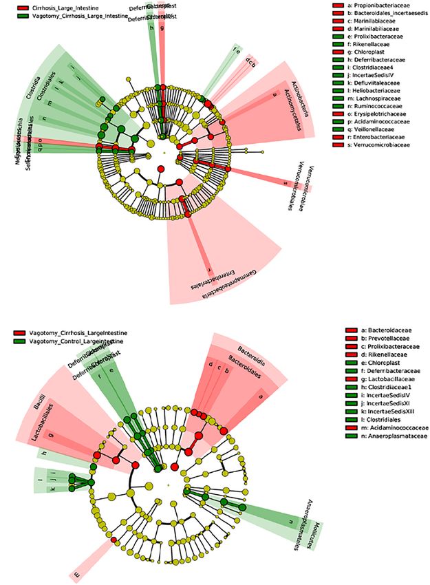
Zhang et al.                                                                                                             Vagotomy, Cirrhosis and Gut-Liver-Brain Changes

RNA (mRNA) expression of (a) neuroinflammation [interleukin                              changes in microbial composition between mouse groups
(IL)-1β, monocyte chemoattractant protein 1 (MCP1)]; (b)                                 using linear discriminant effect size (LEfSe) (Segata et al.,
glial/microglial activation [ionized calcium-binding adaptor                             2011) analysis.
molecule 1 (IBA1) and glial fibrillary acidic protein (GFAP)];                              Approvals were obtained from the Institutional Animal Care
(c) brain regeneration and plasticity [brain-derived neurotrophic                        and Use Committee at VCU before study initiation. All animals
factor (BDNF)]. The liver tissues were processed for histological                        received humane care according to the criteria outlined in the
analysis and isolation of total RNA and total protein lysate.                            Guide for the Care and Use of Laboratory Animals. All authors
The mRNA levels of the key genes involved in hepatic lipid                               had access to the study data and had reviewed and approved the
metabolism and inflammation were measured by real-time                                   final manuscript.
quantitative PCR (RT-qPCR) (Table 1). The protein levels of
SREBP-1 (Purified Mouse Anti-SREBP-1, from BD Pharmingen,
Cat#557036) and Insig2 (Santa Cruz, Cat# sc-34823). Histological
analysis of the hepatic inflammation, steatosis, fibrosis or                             RESULTS
cirrhosis was done by a pathologist in a blinded fashion with
                                                                                         There were no obvious changes in health appearance, weight
hematoxylin and eosin and trichrome staining using the NASH-
                                                                                         changes or sickness behavior noted in any of the mice
Clinical Research Network criteria.
                                                                                         before the sacrifice.
   Large intestinal mucosal microbiota was analyzed using 16S
ribosomal RNA (rRNA) sequencing after DNA was extracted
(Gillevet et al., 2010).                                                                 Frontal Cortex Evaluation
   Statistical analysis: We analyzed continuous data using                               Neuro-Inflammation
unpaired t-tests/ANOVA for mean and SEM and Kruskal–                                     Two neuro-inflammatory markers (IL-6 and MCP-1) were
Wallis/Mann–Whitney for comparing medians. For in vivo                                   significantly higher in conventional cirrhotic mice than controls
studies, in order to determine the group size, we will perform                           (Figure 2). There was a significant increase in IL-1β and
the power calculations to detect a 25% difference at a power                             MCP-1 mRNA expression in mice with vagotomy compared to
of 0.8 and a confidence level of 95% for neuro-inflammation.                             the controls, which marginally changed after the development
Based on the data from an earlier mouse study with the                                   of cirrhosis. Glial (GFAP) and microglial (IBA) markers
standard deviation of 50% for each group, the group sizes                                were higher in cirrhotic mice without vagotomy. However,
equal to or greater than 6 are required. This was also based                             these changes were largely abrogated after vagotomy. BDNF
on our prior study of changes in gut microbiome affecting                                (Cattaneo et al., 2016) was not affected by cirrhosis in the
neuro-inflammation (Liu et al., 2020). Data are presented                                conventional state. Vagotomized mice showed increased frontal
as mean ± SEM unless otherwise specified. We compared                                    cortex expression and BDNF.

  FIGURE 2 | mRNA expression in frontal cortices presented as median and 95% CI. (A) Interleukin 1 β, (B) Monocyte chemoattactant protein 1, (C) Glial fibrillary
  acidic protein and (D) ionized calcium-binding adaptor molecule 1. (E) Brain-derived neurotrophic factor. Conv, conventional; Ctrl, control; Vago-Ctrl, isolated hepatic
  branch vagotomy mice; Vago-Cirr, mice with CCl4 cirrhosis after isolated hepatic branch vagotomy. *p < 0.05–0.01, **p < 0.01–0.001, and ***p < 0.001 on
  Mann–Whitney or Kruskal–Wallis as appropriate.

Frontiers in Physiology | www.frontiersin.org                                        3                                             June 2021 | Volume 12 | Article 702646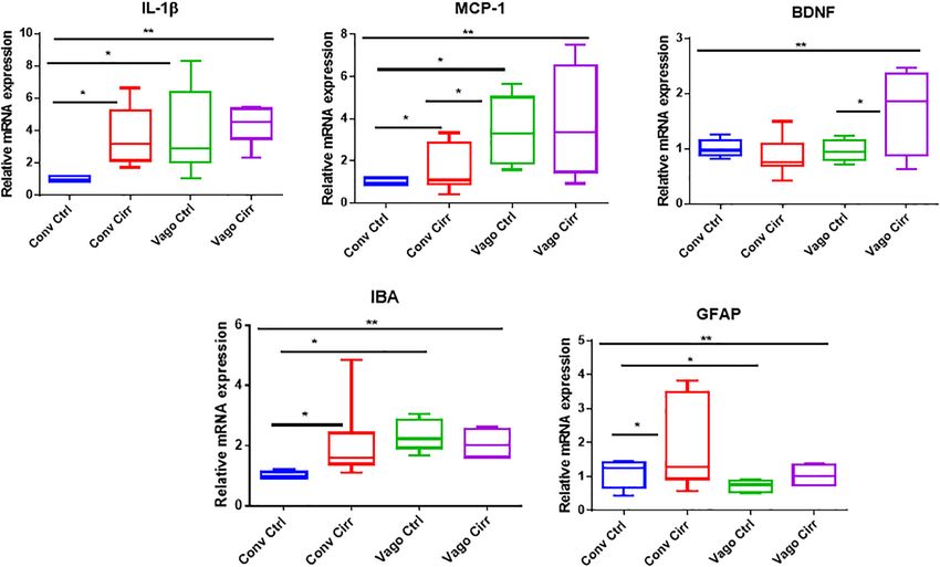
Zhang et al. Vagotomy, Cirrhosis and Gut-Liver-Brain Changes
TABLE 2 | LEfSe changes in colonic mucosal microbiota in Conventional mice and mice with Vagotomy.
LEfSe comparison Higher in controls Higher in cirrhosis
Control vs. Cirrhosis Actinobacteria_Bifidobacteriaceae Actinobacteria _Proprionibacteriaceae
Bacteroidetes_Cryomorphaceae Bacteroidetes_Marinilabiliaceae
Bacteroidetes_Rikenellaceae Cyanobacteri a_Chloroplast
Firmicutes_Acidaminococcaceae Firmicutes_St reptococcaceae
Firmicutes_Heliobacteriaceae Proteobacteri a_Enterobacteriaceae
Firmicutes_Lachnospiraceae Proteobacteri a_Burkholderiaceae
Firmicutes_Peptostreptococcaceae
Tenericutes_Anaeroplasmataceae
Higher in controls Higher in vagotomy controls
Control vs. Vagotomy Proteobacteria_Enterobacteriaceae Firmicutes_Veillonellaceae
control
Firmicutes_Erysipelothricaceae Firmicutes_Lachnospiracae
Firmicutes_Peptostreptococcaceae Firmicutes_Ruminococcaceae
Actinobacteria_Corynebacteriaceae Firmicutes_ClostridialesIncSedXI
Actinobacteria_Bifidobacteriaceae Firmicutes_ClostridialesIncSedIV
Actinobacteria_Coriobacteriacae Actinobacteria_Propionibacteriaceae
Bacteroidetes_Bacteroidaceae Tenericutes_Aneroplasmataceae
Bacteroidetes_Prevotellaceae
Bacteroidetes_Cryomorphaceae
Bacteroidetes_Marinilibiaceae
Bacteroidetes_Flammeovirgiaceae
Bacteroidetes_Flavobacteriaceae
Bacteroidetes_Porphyromonadaceae
Verrucomicrobia_Verrucomicrobiaceae
Higher in cirrhosis Higher in vagotomy cirrhosis
Cirrhosis vs. Vagotomy Cyanobacteria_Chloroplast Bacteroidetes_Rikenellaceae
Cirrhosis
Actinobacteria_Propionibacteriaceae Bacteroidetes_Prolixibacteriaceae
Bacteroidetes_Bacteroidaceae Deferribacters_Deferribacteriaceae
Bacteroidetes_Marinilibilaceae Firmicutes_Lachnospiracae
Firmicutes_Erysipelothricaceae Firmicutes_Ruminococcaceae
Proteobacteria_Enterobacteriaceae Firmicutes_Veillonellaceae
Firmicutes_Heliobacteriaceae
Firmicutes_Acidaminococcaceae
Firmicutes_Clostridiaceae
Firmicutes_Defluviitaleaceae
Microbiota presented as Phylum_Family. LEfSe: Linear Discriminant Analysis Effect Size.
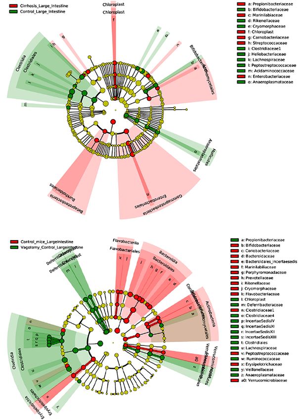
Mucosal Microbial Changes Ruminococcaceae and Rikenellaceae, and lesser relative
An overview of microbial comparisons are shown in Table 2 and abundance of Enterobacteriaceae and Verrucomicrobiaceae
Figure 3. compared to conventional control mice.
Conventional Controls vs. Conventional Cirrhosis Conventional Cirrhosis vs. Vagotomy Cirrhosis
Comparisons Comparisons
As expected, conventional controls had higher potentially Mice with cirrhosis after vagotomy had a higher
beneficial taxa such as Lachnospiraceae and Bifidobacteriaceae representation of beneficial Firmicutes families compared
compared to conventional mice with cirrhosis that to conventional cirrhosis.
demonstrated greater pathobionts such as Enterobacteriaceae
and Streptococcaceae. Liver Findings
Histological Analysis
Conventional Controls vs. Vagotomized Control As expected, mice receiving CCl4 for 12 weeks developed
Comparisons cirrhosis on histology regardless of vagotomy. No
After vagotomy, control mice had greater beneficial taxa changes in histology were seen at 12 weeks between
belonging to Firmicutes, including Lachnospiraceae, conventional control and control vagotomized mice
Frontiers in Physiology | www.frontiersin.org 4 June 2021 | Volume 12 | Article 702646Zhang et al. Vagotomy, Cirrhosis and Gut-Liver-Brain Changes
(Figures 4, 5). The inflammatory grade increased as α-SMA and Col1α were significantly upregulated in vagotomized
expected with cirrhosis development, was lower in the cirrhotic mice.
vagotomized group, but the opposite was seen with the
steatosis grade.
Steatosis
Given the increase in steatosis with vagotomy cirrhotic mice, we
Liver Inflammation and Fibrosis examined the key genes involved in hepatic lipid metabolism
In addition to the histological inflammatory grade, there using real-time RT-PCR and Western blot analyses. The RT-PCR
was a significant increase in mRNA expression of MCP- further showed the upregulation of lipoprotein lipase (LPL) and
1, Cd112b, and CD63 in the mice that developed cirrhosis downregulation of carboxylesterases, Ces1g and Ces2.
in the setting of vagotomy (Figure 6). BDNF expression As shown in Figure 7, the nuclear form of SREBP1
increased in conventional mice with cirrhosis but not in protein levels were significantly increased in the vagotomy-
vagotomized ones. In addition, the mRNA levels of Ck-19, cirrhosis group compared to the conventional cirrhosis group.
FIGURE 3 | Continued
Frontiers in Physiology | www.frontiersin.org 5 June 2021 | Volume 12 | Article 702646Zhang et al. Vagotomy, Cirrhosis and Gut-Liver-Brain Changes
FIGURE 3 | Linear discriminant analysis Effect Size (LEfSe) cladograms of large intestinal mucosal comparisons. Significant differences are color coded. (A)
Conventional controls vs. conventional cirrhosis. (B) Conventional controls vs. vagotomized controls. (C) Conventional cirrhosis vs. vagotomized cirrhosis.
(D) Vagotomized controls vs. vagotomized cirrhosis.
Furthermore, the protein level of Insig 2 was significantly reduced that hepatic vagal innervation can affect the gut-liver-brain
in vagotomy cirrhotic mice. axis in cirrhosis.
Although previous findings suggested that the hepatic branch
of the vagus has a pivotal role in transmitting information
DISCUSSION from the liver to the brain, how this modulates the effects of
cirrhosis remained largely unknown (Pocai et al., 2005; Harada
Connections between the gut, liver, and brain are germane et al., 2014). Mice with vagotomy showed lower dysbiosis with
toward the development and progression of cirrhosis, a higher relative abundance of potential autochthonous taxa
but the role of hepatic vagal output requires greater such as Lachnospiraceaeae and Ruminococcaceae regardless
clarification. Using a mouse model of hepatic branch of whether this was controls or in mice that developed
vagotomy, we found that vagal output from the liver is cirrhosis. This interesting role of the hepatic vagus adds
associated with changes in gut microbial composition, another dimension to the gut-liver axis in addition to the
neuro-inflammation, and plasticity, as well as hepatic previously described factors such as intestinal barrier integrity,
steatosis and inflammation. These results demonstrate bile acid features and bacterial products. The data show
Frontiers in Physiology | www.frontiersin.org 6 June 2021 | Volume 12 | Article 702646Zhang et al. Vagotomy, Cirrhosis and Gut-Liver-Brain Changes FIGURE 4 | Histological evaluation of steatosis and inflammation according to NASH-Clinical Research Network criteria. Conv, conventional; Ctrl, control; Cirr, CCL4 cirrhosis; VagoCtrl, isolated hepatic branch vagotomy mice; VagoCirr, Mice with CCl4 cirrhosis after isolated hepatic branch vagotomy. ***p < 0.001 on Kruskal–Wallis Individual mouse data and median 95% CI are presented. (A) Steatosis grade. (B) Inflammatory grade. FIGURE 5 | Representative liver histological sections in H/E 20X and trichrome 10X showing normal histology in both control groups, cirrhosis on trichrome in both cirrhosis groups and steatosis in Vagotomized cirrhosis liver. Conv, conventional; Ctrl, control; Cirr, CCL4 cirrhosis; VagoCtrl, isolated hepatic branch vagotomy mice; VagoCirr, Mice with CCl4 cirrhosis after isolated hepatic branch vagotomy. that vagal output from the liver enhances the overgrowth Ruminococcaceae. With the appearance of cirrhosis, these of potential pathobionts since vagotomy reduced dysbiosis in changes in Verrucomicrobia were not seen, likely due to the the mucosal microbiome. This is even more striking because cirrhosis state reducing these organisms uniformly regardless hepatic branch vagotomy does not affect gastric acid secretion, of vagotomy. intestinal motility or physical connections between the liver and Prior studies in vagotomized mice in the setting of NAFLD the gut. before cirrhosis have shown greater steatosis, but we extended While the findings of lower dysbiosis after vagotomy these in a cirrhosis model of CCl4 gavage that typically does seem to be at odds with the higher brain inflammation and not demonstrate steatosis. While the exact mechanisms are higher liver steatosis, most of these changes only occurred unclear, there was an upregulation of the activated nuclear after CCl4 gavage and were associated with differential form of SREBP1 (nSREBP1) protein level in the liver after changes in BDNF expression. However, some microbial vagotomy. This increase in nSREBP1 was associated with a taxa belonging to Ruminococcaceae that are typically relative loss of Insig2 after vagotomy in the liver. Insig2 is lower in advancing cirrhosis, are actually associated with an ER stress-responsive gene, which prevents the proteolytic fibrosis and metabolic syndrome in patients with liver processing of SREBP-1c from forming a maturing form, a steatosis (Boursier et al., 2016; Lee et al., 2020). In addition, critical transcriptional regulator of hepatic fatty acid metabolism Verrucomicrobiaceae, which includes the beneficial taxon, (Takaishi et al., 2004). LPL is an important player in regulating Akkermansia muciniphilia were lower in vagotomized controls, lipid metabolism and energy balance. The upregulation of which could also promote hepatic steatosis (Everard et al., LPL has been reported to exacerbate liver fibrosis (Teratani 2013). Following this, we found higher hepatic steatosis et al., 2019). The carboxylesterases plays a critical role in in vagotomized cirrhotic mice, which had higher mucosal hydrolyze a variety of xenobiotic and endogenous compounds Frontiers in Physiology | www.frontiersin.org 7 June 2021 | Volume 12 | Article 702646
Zhang et al.                                                                                                        Vagotomy, Cirrhosis and Gut-Liver-Brain Changes

  FIGURE 6 | Hepatic Expression of BDNF and Genes involved in fatty acid metabolism and inflammation presented as Mean ± SEM. Conv, conventional; Ctrl,
  control; Cirr, CCL4 cirrhosis; VagoCtrl, isolated hepatic branch vagotomy mice; VagoCirr, mice with CCl4 cirrhosis after isolated hepatic branch vagotomy.
  Comparisons using Mann–Whitney test, # /*p < 0.05, ## /**p < 0.01, and ### /***p < 0.0001.

and including lipid esters. Six human CES genes have been                               Despite the increase in Ruminococcaceae in vagotomized
identified. CES1 and CES2 are the two most prominent genes,                          animals that can promote steatosis, vagotomized animals did
which are mainly expressed in the gastrointestinal tract and                         have higher relative abundances of other potentially beneficial
liver. In mice, eight genes belong to Ces1 have been identified                      taxa such as Lachnospiraceae and reduction in pathobionts like
with relatively unique tissue expression patterns. Compared to                       Enterobacteriaceae (Kang et al., 2016a,b, 2017). In prior studies
other Ces1 family members, Ces1g is highly expressed in the                          of germ-free mice colonized with stools from differing human
liver and intestine (Lian et al., 2018). Previous studies have                       phenotypes, there was an increase in hepatic and frontal cortical
shown that deficiency of Ces1g or Ces2a was linked to metabolic                      neuro-inflammation in mice that received stool from patients
diseases. It has been reported that that Ces1g suppresses the                        with cirrhosis (Liu et al., 2020). Other studies demonstrated that
activity of Srebp1c promoter and enhances the degradation of                         susceptibility to alcohol-related liver disease was also modulated
Srebp1 (Xu et al., 2001). In addition, Ces1g inhibits Insig 1                        by the donor of the microbiota, whether it be a different
degradation and de novo lipogenesis. Downregulation of Ces1g                         mouse group or humans (Llopis et al., 2016; Cassard et al.,
may attribute to activation of SREBP1c, leading to lipogenesis.                      2017). Microglial and glial activation are usually associated
Liver specific expression of Ces1g reduces hepatic steatosis                         with cirrhosis-related neuro-inflammation but in vagotomized
(Bahitham et al., 2016). In addition, downregulation of Ces2                         mice, the expression of GFAP and IBA were abrogated even
is associated with NASH disease progression and high-fat-diet-                       with cirrhosis. Therefore, factors other than dysbiosis and
induced steatosis (Li et al., 2016). Hepatic CES2 plays a key                        glial/microglial activation, such as systemic inflammation and
role in fatty acid oxidation and inhibiting lipogenesis. We                          BDNF may be associated with neuroinflammation in mice
also found that the expression of Ces2 was downregulated in                          with hepatic branch vagotomy and cirrhosis (Shawcross et al.,
both conventional and vagotomy cirrhotic mice and vagotomy                           2011; Gorg et al., 2015). BDNF is usually thought of as
further inhibited Ces2 expression. Therefore, these could be the                     a primary neurotrophic molecule but of late studies have
potential mechanisms behind the development of steatosis in                          shown it to be an important part of the liver-brain axis
the liver and lower inflammation after vagotomy and cirrhosis                        (Pocai et al., 2005; Bercik et al., 2011; Cattaneo et al., 2016).
compared to the conventional animals (Gao et al., 2015;                              The role of BDNF is complex since it can modulate insulin
Amir et al., 2020).                                                                  resistance and liver disease in animal models and is found

Frontiers in Physiology | www.frontiersin.org                                    8                                            June 2021 | Volume 12 | Article 702646
Zhang et al.                                                                                                Vagotomy, Cirrhosis and Gut-Liver-Brain Changes

                                                                                  Vagotomy-induced lower hepatic and higher BDNF cortical
                                                                                  expression also suggest that the neurotrophic factors may
                                                                                  have a major role in the gut-liver-brain axis in cirrhosis
                                                                                  (Amir et al., 2020).
                                                                                     Our study was limited since we used isolated hepatic vagotomy
                                                                                  and not a more radical subdiaphragmatic approach (Bercik et al.,
                                                                                  2011). However, the latter approach affects most gastrointestinal
                                                                                  organs and can impact acid secretion and motility, all of which
                                                                                  can confound the results by affecting microbiota independently.
                                                                                  We used the entire frontal cortex given its involvement in
                                                                                  HE but further work will be necessary to characterize the
                                                                                  cell type in which expression of these neural plasticity and
                                                                                  inflammatory markers occurs. We only used the CCl4 model
                                                                                  via gavage because, unlike the bile duct ligated model, it
                                                                                  does not result in microbiota change due to immediate bile
                                                                                  diversion (Fouts et al., 2012; Dhanda et al., 2018). However,
                                                                                  it does not cause major behavioral changes in mice, which
                                                                                  is why we used inflammatory gene expression as the readout
                                                                                  (Butterworth et al., 2009); future behavioral testing models are
  FIGURE 7 | Western Blot expression of Hepatic SREBP1 and Insig2 using           needed (DeMorrow et al., 2021).
  Actin as the reference protein. (A) gels, (B) Mean ± SEM for SREBP1/Actin          We conclude that the parasympathetic innervation of the liver
  ratio, (C) Mean ± SEM for Insig2/Actin Ratio. Conv, conventional; Ctrl,
  control; Cirr, CCL4 cirrhosis; VagoCtrl, isolated hepatic branch vagotomy
                                                                                  modulates hepatic steatosis, neuro-inflammation and dysbiosis
  mice; VagoCirr, mice with CCl4 cirrhosis after isolated hepatic branch          even after the development of cirrhosis using CCl4 gavage.
  vagotomy. ### p < 0.001 and ## p < 0.01 between Conv Cirr and Vago Cirr,        These data that vagal innervation of the liver plays an important
  *p < 0.05 between VagoCtrl and VagoCirr on Mann–Whitney tests.                  role through modulation of BDNF in the gut-liver-brain axis,
                                                                                  which has implications for the pathogenesis of cirrhosis and
                                                                                  associated complications.
in higher liver levels in those with cirrhosis and alcohol-
induced injury (Teillon et al., 2010; Gao et al., 2015; Yang
et al., 2017; Amir et al., 2020). However, BDNF is anti-                          DATA AVAILABILITY STATEMENT
inflammatory in the brain under most circumstances, including
HE (Kang et al., 2016a; Dhanda et al., 2018). Our data showed                     Data are available now at https://www.ncbi.nlm.nih.gov/Traces/
that with the intact vagus, BDNF increased with cirrhosis,                        study/?acc=PRJNA735706.
paralleling prior studies, but this circuit is broken with
vagotomy, where cirrhosis does not lead to BDNF increase
in the liver but does in the brain. This may be a protective
mechanism since the reverse profile i.e., higher liver BDNF                       ETHICS STATEMENT
and lower brain BDNF is found in psychiatric disorders
                                                                                  The animal study was reviewed and approved by Virginia
(Yang et al., 2017).
                                                                                  Commonwealth University IACUC.
    Ultimately, it is striking that most differentiators in the setting
of vagotomy compared to conventional mice only occurred
after the induction of cirrhosis through CCl4. Vagotomized
control mice and conventional control mice were otherwise                         AUTHOR CONTRIBUTIONS
similar in most outcomes related to the brain and liver.
Therefore, despite the relatively lower dysbiosis in vagotomized                  JB and HZ conceptualized and were involved at all levels of the
control mice, they were equally susceptible to CCl4-induced                       study. YZ, JK, DZ, SG, YW, and YT were involved in the animal
cirrhosis. Gavage with CCl4 typically induces toxic cirrhosis                     handling, sacrifice, and experiments. JG-M and PH were involved
with dysbiosis and neuro-inflammation without the diversion                       in experiments design and manuscript revision. HL was involved
of bile flow (Kang et al., 2016a). These interactions were                        in histological analysis. All authors contributed to the article and
enhanced in vagotomized animals, pointing to an important                         approved the submitted version.
role of hepatic parasympathetic innervation in not only fatty
liver as previously described but also in cirrhosis (Gao
et al., 2015; Amir et al., 2020). Moreover, the decoupling                        FUNDING
of the gut-liver-brain axis found due to a lower dysbiosis
but higher neuro-inflammation in vagotomized cirrhosis shows                      This work was partly supported by VA Merit Review
that the neuronal input from the liver may be an important                        I0CX001076, R21TR002024, and R21TR003095 to JB, R01
way station in the gut-brain communication in cirrhosis.                          DK104893, R01DK-057543, R21 AA026629; VA Merit

Frontiers in Physiology | www.frontiersin.org                                 9                                      June 2021 | Volume 12 | Article 702646
Zhang et al. Vagotomy, Cirrhosis and Gut-Liver-Brain Changes
Award I01BX004033 and Research Career Scientist Award ACKNOWLEDGMENTS
(IK6BX004477) to HZ and R01MH084894 and R01MH111940 to
JG-M. None of the funders had any role to play in experiment This work was selected as a poster of distinction for Digestive
design and conduct nor the decision to publish. Disease Week 2020.
REFERENCES Kang, D. J., Hylemon, P. B., Gillevet, P. M., Sartor, R. B., Betrapally, N. S.,
Kakiyama, G., et al. (2017). Gut microbial composition can differentially
Amir, M., Yu, M., He, P., and Srinivasan, S. (2020). Hepatic autonomic nervous regulate bile acid synthesis in humanized mice. Hepatol. Commun. 1, 61–70.
system and neurotrophic factors regulate the pathogenesis and progression of doi: 10.1002/hep4.1020
non-alcoholic fatty liver disease. Front. Med. (Lausanne) 7:62. doi: 10.3389/ Kang, D. J., Kakiyama, G., Betrapally, N. S., Herzog, J., Nittono, H., Hylemon, P. B.,
fmed.2020.00062 et al. (2016b). Rifaximin exerts beneficial effects independent of its ability to
Bahitham, W., Watts, R., Nelson, R., Lian, J., and Lehner, R. (2016). Liver- alter microbiota composition. Clin. Transl. Gastroenterol. 7:e187. doi: 10.1038/
specific expression of carboxylesterase 1g/esterase-x reduces hepatic steatosis, ctg.2016.44
counteracts dyslipidemia and improves insulin signaling. Biochim. Biophys. Lee, G., You, H. J., Bajaj, J. S., Joo, S. K., Yu, J., Park, S., et al. (2020). Distinct
Acta 1861, 482–490. doi: 10.1016/j.bbalip.2016.03.009 signatures of gut microbiome and metabolites associated with significant
Bercik, P., Denou, E., Collins, J., Jackson, W., Lu, J., Jury, J., et al. (2011). The fibrosis in non-obese NAFLD. Nat. Commun. 11:4982.
intestinal microbiota affect central levels of brain-derived neurotropic factor Li, Y., Zalzala, M., Jadhav, K., Xu, Y., Kasumov, T., Yin, L., et al. (2016).
and behavior in mice. Gastroenterology 141, 599–609, 609.e1-3. Carboxylesterase 2 prevents liver steatosis by modulating lipolysis, endoplasmic
Boursier, J., Mueller, O., Barret, M., Machado, M., Fizanne, L., Araujo-Perez, reticulum stress, and lipogenesis and is regulated by hepatocyte nuclear
F., et al. (2016). The severity of nonalcoholic fatty liver disease is associated factor 4 alpha in mice. Hepatology 63, 1860–1874. doi: 10.1002/hep.
with gut dysbiosis and shift in the metabolic function of the gut microbiota. 28472
Hepatology 63, 764–775. doi: 10.1002/hep.28356 Lian, J., Nelson, R., and Lehner, R. (2018). Carboxylesterases in lipid metabolism:
Butterworth, R. F., Norenberg, M. D., Felipo, V., Ferenci, P., Albrecht, J., Blei, from mouse to human. Protein Cell 9, 178–195. doi: 10.1007/s13238-017-
A. T., et al. (2009). Experimental models of hepatic encephalopathy: ISHEN 0437-z
guidelines. Liver Int. 29, 783–788. doi: 10.1111/j.1478-3231.2009.02034.x Liu, R., Kang, J. D., Sartor, R. B., Sikaroodi, M., Fagan, A., Gavis, E. A., et al. (2020).
Cassard, A. M., Gerard, P., and Perlemuter, G. (2017). Microbiota, liver diseases, Neuroinflammation in murine cirrhosis is dependent on the gut microbiome
and alcohol. Microbiol. Spectr. 5. doi: 10.1128/microbiolspec.BAD-0007-2016 and is attenuated by fecal transplant. Hepatology 71, 611–626. doi: 10.1002/
Cattaneo, A., Cattane, N., Begni, V., Pariante, C. M., and Riva, M. A. (2016). The hep.30827
human BDNF gene: peripheral gene expression and protein levels as biomarkers Llopis, M., Cassard, A. M., Wrzosek, L., Boschat, L., Bruneau, A., Ferrere, G.,
for psychiatric disorders. Transl. Psychiatry 6:e958. doi: 10.1038/tp.2016.214 et al. (2016). Intestinal microbiota contributes to individual susceptibility
Cryan, J. F., O’riordan, K. J., Cowan, C. S. M., Sandhu, K. V., Bastiaanssen, T. F. S., to alcoholic liver disease. Gut 65, 830–839. doi: 10.1136/gutjnl-2015-31
Boehme, M., et al. (2019). The microbiota-gut-brain axis. Physiol. Rev. 99, 0585
1877–2013. Metz, C. N., and Pavlov, V. A. (2018). Vagus nerve cholinergic circuitry to the
DeMorrow, S., Cudalbu, C., Davies, N., Jayakumar, A. R., and Rose, C. F. (2021). liver and the gastrointestinal tract in the neuroimmune communicatome. Am.
ISHEN guidelines on animal models of hepatic encephalopathy. Liver Int. doi: J. Physiol. Gastrointest. Liver Physiol. 315, G651–G658.
10.1111/liv.14911 Ochoa-Sanchez, R., and Rose, C. F. (2018). Pathogenesis of hepatic encephalopathy
Dhanda, S., Gupta, S., Halder, A., Sunkaria, A., and Sandhir, R. (2018). Systemic in chronic liver disease. J. Clin. Exp. Hepatol. 8, 262–271.
inflammation without gliosis mediates cognitive deficits through impaired Pocai, A., Obici, S., Schwartz, G. J., and Rossetti, L. (2005). A brain-liver circuit
BDNF expression in bile duct ligation model of hepatic encephalopathy. Brain regulates glucose homeostasis. Cell Metab. 1, 53–61. doi: 10.1016/j.cmet.2004.
Behav. Immun. 70, 214–232. doi: 10.1016/j.bbi.2018.03.002 11.001
Everard, A., Belzer, C., Geurts, L., Ouwerkerk, J. P., Druart, C., Bindels, L. B., et al. Segata, N., Izard, J., Waldron, L., Gevers, D., Miropolsky, L., Garrett, W. S.,
(2013). Cross-talk between Akkermansia muciniphila and intestinal epithelium et al. (2011). Metagenomic biomarker discovery and explanation. Genome Biol.
controls diet-induced obesity. Proc. Natl. Acad. Sci. U.S.A. 110, 9066–9071. 12:R60.
doi: 10.1073/pnas.1219451110 Shawcross, D. L., Davies, N. A., Williams, R., and Jalan, R. (2004). Systemic
Fouts, D. E., Torralba, M., Nelson, K. E., Brenner, D. A., and Schnabl, B. (2012). inflammatory response exacerbates the neuropsychological effects of induced
Bacterial translocation and changes in the intestinal microbiome in mouse hyperammonemia in cirrhosis. J. Hepatol. 40, 247–254. doi: 10.1016/j.jhep.
models of liver disease. J. Hepatol. 56, 1283–1292. doi: 10.1016/j.jhep.2012.01. 2003.10.016
019 Shawcross, D. L., Sharifi, Y., Canavan, J. B., Yeoman, A. D., Abeles, R. D.,
Gao, X., Van Der Veen, J. N., Zhu, L., Chaba, T., Ordonez, M., Lingrell, S., Taylor, N. J., et al. (2011). Infection and systemic inflammation, not
et al. (2015). Vagus nerve contributes to the development of steatohepatitis ammonia, are associated with Grade 3/4 hepatic encephalopathy, but not
and obesity in phosphatidylethanolamine N-methyltransferase deficient mice. mortality in cirrhosis. J. Hepatol. 54, 640–649. doi: 10.1016/j.jhep.2010.
J. Hepatol. 62, 913–920. doi: 10.1016/j.jhep.2014.11.026 07.045
Gillevet, P., Sikaroodi, M., Keshavarzian, A., and Mutlu, E. A. (2010). Quantitative Takaishi, K., Duplomb, L., Wang, M. Y., Li, J., and Unger, R. H. (2004). Hepatic
assessment of the human gut microbiome using multitag pyrosequencing. insig-1 or -2 overexpression reduces lipogenesis in obese Zucker diabetic
Chem. Biodivers. 7, 1065–1075. doi: 10.1002/cbdv.200900322 fatty rats and in fasted/refed normal rats. Proc. Natl. Acad. Sci. U.S.A. 101,
Gorg, B., Karababa, A., Shafigullina, A., Bidmon, H. J., and Haussinger, D. (2015). 7106–7111. doi: 10.1073/pnas.0401715101
Ammonia-induced senescence in cultured rat astrocytes and in human cerebral Teillon, S., Calderon, G. A., and Rios, M. (2010). Diminished diet-induced
cortex in hepatic encephalopathy. Glia 63, 37–50. doi: 10.1002/glia.22731 hyperglycemia and dyslipidemia and enhanced expression of PPARalpha and
Harada, S., Yamazaki, Y., Koda, S., and Tokuyama, S. (2014). Hepatic branch vagus FGF21 in mice with hepatic ablation of brain-derived neurotropic factor.
nerve plays a critical role in the recovery of post-ischemic glucose intolerance J. Endocrinol. 205, 37–47. doi: 10.1677/joe-09-0405
and mediates a neuroprotective effect by hypothalamic orexin-A. PLoS One Teratani, T., Tomita, K., Furuhashi, H., Sugihara, N., Higashiyama, M., Nishikawa,
9:e95433. doi: 10.1371/journal.pone.0095433 M., et al. (2019). Lipoprotein lipase up-regulation in hepatic stellate cells
Kang, D. J., Betrapally, N. S., Ghosh, S. A., Sartor, R. B., Hylemon, P. B., exacerbates liver fibrosis in nonalcoholic steatohepatitis in mice. Hepatol.
Gillevet, P. M., et al. (2016a). Gut microbiota drive the development of Commun. 3, 1098–1112. doi: 10.1002/hep4.1383
neuroinflammatory response in cirrhosis in mice. Hepatology 64, 1232–1248. Vilstrup, H., Amodio, P., Bajaj, J., Cordoba, J., Ferenci, P., Mullen, K. D., et al.
doi: 10.1002/hep.28696 (2014). Hepatic encephalopathy in chronic liver disease: 2014 practice guideline
Frontiers in Physiology | www.frontiersin.org 10 June 2021 | Volume 12 | Article 702646Zhang et al. Vagotomy, Cirrhosis and Gut-Liver-Brain Changes by the American association for the study of liver diseases and the European Conflict of Interest: The authors declare that the research was conducted in the association for the study of the liver. Hepatology 60, 715–735. doi: 10.1002/hep. absence of any commercial or financial relationships that could be construed as a 27210 potential conflict of interest. Xu, J., Teran-Garcia, M., Park, J. H., Nakamura, M. T., and Clarke, S. D. (2001). Polyunsaturated fatty acids suppress hepatic sterol regulatory element-binding Copyright © 2021 Zhang, Kang, Zhao, Ghosh, Wang, Tai, Gonzalez-Maeso, protein-1 expression by accelerating transcript decay. J. Biol. Chem. 276, 9800– Sikaroodi, Gillevet, Lippman, Hylemon, Zhou and Bajaj. This is an open-access 9807. doi: 10.1074/jbc.m008973200 article distributed under the terms of the Creative Commons Attribution License Yang, B., Ren, Q., Zhang, J. C., Chen, Q. X., and Hashimoto, K. (2017). Altered (CC BY). The use, distribution or reproduction in other forums is permitted, provided expression of BDNF, BDNF pro-peptide and their precursor proBDNF in brain the original author(s) and the copyright owner(s) are credited and that the original and liver tissues from psychiatric disorders: rethinking the brain-liver axis. publication in this journal is cited, in accordance with accepted academic practice. No Transl. Psychiatry 7:e1128. doi: 10.1038/tp.2017.95 use, distribution or reproduction is permitted which does not comply with these terms. Frontiers in Physiology | www.frontiersin.org 11 June 2021 | Volume 12 | Article 702646
You can also read