Isolation of Prion with BSE Properties from Farmed Goat
←
→
Page content transcription
If your browser does not render page correctly, please read the page content below
Isolation of Prion with BSE
             Properties from Farmed Goat
           John Spiropoulos, Richard Lockey, Rosemary E. Sallis, Linda A. Terry, Leigh Thorne,
                       Thomas M. Holder, Katy E. Beck, and Marion M. Simmons

     Transmissible spongiform encephalopathies are                humans (5,6). Because of its ability to cross species barriers
fatal neurodegenerative diseases that include variant             and particularly its zoonotic potential, BSE is considered
Creutzfeldt-Jakob disease in humans, scrapie in small             a public health risk, and extensive measures have been
ruminants, and bovine spongiform encephalopathy (BSE)             established to detect and eliminate the disease.
in cattle. Scrapie is not considered a public health risk, but         Scrapie, a naturally occurring TSE affecting small
BSE has been linked to variant Creutzfeldt-Jakob disease.
                                                                  ruminants, has been known for centuries (7) and is not
Small ruminants are susceptible to BSE, and in 2005 BSE
was identified in a farmed goat in France. We confirm
                                                                  considered to pose a public health risk (8). Under experimental
another BSE case in a goat in which scrapie was originally        conditions, however, small ruminants are susceptible to
diagnosed and retrospectively identified as suspected BSE.        BSE, with pathogenesis and clinical signs that are not readily
The prion strain in this case was further characterized by        distinguishable from scrapie (9–12). Additionally, the fact
mouse bioassay after extraction from formaldehyde-fixed           that small ruminants were exposed to BSE-contaminated
brain tissue embedded in paraffin blocks. Our data show           food before the exclusion of meat and bone meal from
that BSE can infect small ruminants under natural conditions      ruminant feedstuffs led to the possibility that sheep and goats
and could be misdiagnosed as scrapie. Surveillance                on commercial farms could be affected by BSE that could
should continue so that another outbreak of this zoonotic         be misdiagnosed as scrapie (13,14). The response to this
transmissible spongiform encephalopathy can be prevented          potential risk was the implementation of extensive statutory
and public health safeguarded.
                                                                  active surveillance, elimination, and breeding for resistance
                                                                  programs in the European Union (EU).

T    ransmissible spongiform encephalopathies (TSEs)
     are fatal diseases characterized by neurodegenerative
changes in the central nervous system that include
                                                                       In 2005, as part of a review of historical TSE-positive
                                                                  cases of sheep and goats in France, a specimen from a goat
                                                                  slaughtered for human consumption in 2002 was reported
vacuolation, gliosis, and accumulation of an abnormal             to be “indistinguishable from a BSE isolate on the basis
isoform (PrPSc) of a naturally occurring host-encoded             of all identification criteria available.” (15). In response
protein (PrPC) (1). According to the prion hypothesis, PrPSc      to this report, 2 retrospective studies were initiated in the
is the major or the sole infectious agent (1). Although this      United Kingdom to analyze archived samples from goat
hypothesis has not received universal acceptance, PrPSc is        cases that were initially diagnosed as scrapie (16,17).
ubiquitous in all known naturally occurring TSEs, and its         Because only fixed material was available, both studies
detection is widely used for their diagnosis.                     had to use differential immunohistochemical analysis
     Bovine spongiform encephalopathy (BSE), a TSE of             (D-IHC), a technique that can discriminate scrapie from
cattle, was first detected in 1986 (2) and has since been         experimentally induced BSE in sheep (18). These studies
linked with emerging TSEs in other species (3,4) including        identified a single case, originally diagnosed in 1990 as
                                                                  scrapie, that had a D-IHC signature indistinguishable from
Author affiliation: Animal Health and Veterinary Laboratories
                                                                  BSE (16).
Agency, Weybridge, Surrey, UK
                                                                       Given the wide phenotypic variance of scrapie in sheep
DOI: http://dx.doi.org/10.3201/eid1712.110333                     and our limited knowledge of this variance in goats, the

                         Emerging Infectious Diseases • www.cdc.gov/eid • Vol. 17, No. 12, December 2011                    2253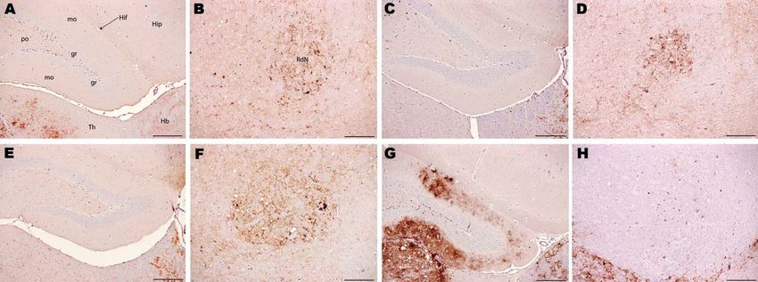
RESEARCH

D-IHC result on its own was insufficient for an unequivocal            Additional controls of fixed and frozen brain tissues from
diagnosis. In accordance with EU regulation 36/2005 (19),         the same source were used to assess the effect of fixation,
the case was referred to the EU Reference Laboratory              processing, and retrieval on the biological properties of the
Strain Typing Expert Group, which recommended further             TSE agents present. All samples included in this study were
investigation by bioassay.                                        from animals showing clinical signs of TSE. These came
     Bioassay is conventionally undertaken by using unfixed       from animals with confirmed TSE sourced through passive
tissues to prepare inocula. Much historical tissue is available   surveillance schemes, with the exception of an ovine BSE
only as formalin fixed or formalin fixed and paraffin wax         case that was produced experimentally (11). Because the
embedded. TSE infectivity persists in such material but           sampling site of the brain may also affect the infectious titer,
with a lower infectious titer than with unfixed frozen tissue     in addition to the above parameters we identified a bovine
(20). However, the potential effects on biological activity,      BSE case for which whole frozen brain stem was available.
and therefore strain characterization, of fixation and            Given the left-right symmetry of PrPSc distribution, which
processing are unknown. Thus, further investigation of this       was verified by IHC analysis of the adjacent rostral and
case required an extensive panel of controls. We report the       caudal coronal levels of the selected sample, we assumed
results of the bioassay analysis and confirm the diagnosis of     that titer did not vary substantially on either side of the
BSE in a goat in the United Kingdom.                              midline. Therefore, the obex was cut sagitally in half.
                                                                  Half was processed histologically and was subsequently
Materials and Methods                                             recovered and rehydrated to replicate the process applied in
                                                                  the fixed samples; the other half was kept frozen. Each half
Sample Preparation                                                was homogenized and inoculated into mice.
     Whenever fixed tissue was used, it had been processed             Each source was administered to 3 panels of wild-type
and embedded in paraffin wax. To recover the fixed,               inbred mice (C57/BL6, RIII, and VM) and a transgenic
paraffin-embedded tissue from the wax blocks, samples were        mouse line (tg388 line was provided by Hubert Laude,
processed in reverse. Specifically, the wax was liquefied         Institut National de la Recherche Agronomique, Jouy-en-
by immersing the tissue blocks in a wax bath preheated to         Josas, France). C57/BL6 and RIII mice share the same PrP
55°C. The samples were then placed in an ether bath and           sequence (PrP-a), but it is believed that RIII alone could be
subsequently were immersed in 100% ethanol to remove the          used to discriminate BSE from other TSEs on the basis of
ether. This process was followed by sequential washes in          lesion profile (LP) data on first passage (5), although this
alcohol solutions of decreasing concentrations to gradually       belief has been challenged (22). VM mice have a different
remove the alcohol and rehydrate the sample. Finally, the         PrP sequence (PrP-b) and have been used to identify BSE
samples were suspended in normal saline (10% wt/vol)              after 2 serial passages on the basis of incubation period (IP)
before homogenization. Unfixed samples were kept frozen at        and LP data (23). The tg338 mouse line overexpresses an
−80°C. After thawing, they were suspended in normal saline        ovine VRQ transgene and has been proved to be susceptible
(10% wt/vol) before homogenization. All homogenates were          to scrapie (24,25) and relatively resistant to BSE (26,27).
examined microbiologically and treated with antimicrobial              Serial passage from the suspected case was initiated
drugs as required. Only microbiologically cleared inocula         only in the VM mouse line because subpassage of BSE in
were used to challenge animals.                                   this line gives rise to the mouse-adapted BSE strain 301V,
                                                                  which has a characteristically short IP that can be used to
Experimental Design                                               discriminate BSE from scrapie (23). The acquired data
     The only tissue available from the 1990 suspected UK         were compared with an experimental ovine BSE case and
case (16) was paraffin wax–embedded brain (supplied by            with a 301V reference strain that were serially passaged in
Martin Jeffrey, Veterinary Laboratories Agency, Lasswade,         VM mice in different studies.
UK). Several sources were used to control for TSE strain,              The number of mice inoculated with each source varied
host species, and tissue condition (i.e., frozen vs. fixed and    from 5 to 20 for each mouse line depending on availability
wax-embedded) to ensure unequivocal interpretation of             of material (Table). Serial passages used 10 mice. Where
the results (Table). Paraffin wax–embedded material from          tissue availability was limited, the mouse lines of choice
2 field cases that were contemporary with the suspected           were RIII and tg338.
case but gave a scrapie profile on D-IHC (16,17) and an
experimental caprine BSE case (supplied by Nora Hunter,           Animal Procedures
Roslin Institute, Edinburgh, UK) were used to control for             Because of the small amount of available material,
strain variation in material that had been handled and stored     only intracerebral inoculations (20 μL of 10% of brain
in the same way and for a similar time as material from the       homogenate in normal saline) were performed (22,28).
suspected case (21).                                              For secondary passage, VM mice were challenged

2254                     Emerging Infectious Diseases • www.cdc.gov/eid • Vol. 17, No. 12, December 2011
Prion with BSE Properties in Goat

intracerebrally with 20 μL of 1% brain homogenate. Mice                        processed for histopathologic and IHC analysis. All animal
were monitored for signs of clinical disease and euthanized                    procedures were performed in compliance with the Animal
either at specified clinical endpoints (29) or on the basis                    (Experimental Procedures) Act 1986 under license from
of animal welfare justification (intercurrent losses). The                     the UK Home Office and were approved by the local ethics
brains were removed under sterile conditions by using                          committee.
disposable equipment. Each brain was cut parasagittally,
and the smaller fraction was frozen for biochemical                            Histopathologic and IHC Analyses
analysis or serial bioassay; the larger fraction, which                             Sections (3 μm thick) of 4 different coronal levels
included the midline, was fixed in formal saline and                           (frontal, thalamic, midbrain, and medulla) were stained

Table. Bioassay results of first passage (ruminant to mouse) to determine presence of BSE*
                                                                                      Sc
                                                                             No. PrP -positive Clinically positive    Mean (SD)
                                                                               mice/total no.  and H&E-positive incubation period
Sample source and treatment†                    Tissue‡        Mouse line     mice inoculated§        mice         postinoculation, d
Caprine BSE suspected (V459/90) (16)
  Fixed and embedded                        Brainstem and       C57/BL6              5/20               0                  NA
                                              cerebellum           RIII              5/20               3               525 (93)
                                                                   VM                9/20               6               620 (32)
                                                                  tg338              8/10               0                  NA
Natural caprine scrapie 1 (E90/89) (16)
  Fixed and embedded                           Brainstem        C57/BL6              0/20               0                  NA
                                                                   RIII              0/20               0                  NA
                                                                   VM                0/20               0                  NA
                                                                  tg338              6/10               0                  NA
Natural caprine scrapie 2 (84/1549) (17)
  Fixed and embedded                           Brainstem        C57/BL6             18/20              18               506 (26)
                                                                   RIII              8/20               2               413 (98)
                                                                   VM               15/20               6               586 (87)
                                                                  tg338             10/10               5               196 (13)
Experimental caprine BSE (45x48) (21)
  Fixed and embedded                           Brainstem        C57/BL6              2/20               1              588 (NA)
                                                                   RIII              3/20               2                535 (5)
                                                                   VM                6/20               4               592 (51)
                                                                  tg338             10/10               0                  NA
Experimental ovine BSE (PG0341/00) (11)
  Fixed and embedded                            Medulla            RIII              7/20               6               388 (23)
                                                                  tg338               5/5               0                  NA
  Frozen                                        Medulla            RIII              6/20               0                  NA
                                                                  tg338               NA               NA                >785¶
Natural bovine BSE (PG0475/05, UK passive surveillance)
  Fixed and embedded                            Medulla         C57/BL6              2/20               1              713 (NA)
                                                                   RIII              6/20               5               477 (49)
                                                                   VM               13/20               2               578 (35)
                                                                  tg338              7/10               0                  NA
  Frozen                                        Medulla         C57/BL6              7/20               3               535 (76)
                                                                   RIII             10/20               9               431 (29)
                                                                   VM               16/20               3               568 (31)
                                                                  tg338              6/10               0                  NA
Natural ovine scrapie (PG2413/98, UK passive surveillance)
  Fixed and embedded                            Medulla            RIII              0/20               0                  NA
                                                                  tg338               5/5               3                 97 (2)
  Frozen                                        Medulla            RIII              9/20               8               490 (31)
                                                                  tg338             10/10              10                 64 (2)
*BSE, bovine spongiform encephalopathy; PrP, host-encoded prion protein; H&E, hematoxylin and eosin; NA, not applicable; UK, United Kingdom.
†Animal identification numbers are indicated in parentheses and, where available, references are supplied.
‡Brainstem inocula were prepared by pooling separate samples of brainstem (including medulla, midbrain, and thalamus) retrieved from paraffin wax
blocks.
§Positive for PrPSc by using immunohistochemical analysis.
¶Postinoculation time, after which 2 of 10 inoculated mice were still alive at the time of writing; 5 of the 8 mice that died were PrPSc positive when
examined by using immunohistochemical analysis.

                             Emerging Infectious Diseases • www.cdc.gov/eid • Vol. 17, No. 12, December 2011                                             2255
RESEARCH

with hematoxylin and eosin according to standard methods         of the experimental caprine BSE control, but the value
(22,30). Positive histologic diagnosis was based on the          of this comparison and that of the other controls was
identification of TSE-related vacuolation. The intensity         limited because of the generally low ARs observed and the
of vacuolation in 9 gray matter areas was assessed               proportion of positive mice that did not progress to show
semiquantitatively, and the resultant scores were plotted        clinical disease. However, because none of the C57/BL6
against the respective brain areas as described (22,30,31).      or tg338 mice inoculated with fixed brain from the goat
     For IHC evaluation, each section was labeled with a         with suspected BSE showed development of clinical signs
rabbit polyclonal antibody against PrP (Rb486) according         of TSE, comparisons of IP in these recipients could not be
to established methods (22,28). The distribution of              made.
different PrPSc types in the rodent brain at primary passage
can provide a means of identifying different strains in wild     LP and Histopathologic Analysis
type (22,28) and transgenic mice (32). By examination                 It is generally accepted that, during first passage, LPs
of immunolabeled sections, different PrPSc deposits were         from RIII mice can be used to discriminate BSE from
identified and their distribution in different neuronatomic      scrapie (5), though this principle has been challenged (22).
brain areas recorded as described (22,28).                       An LP is considered to be reliable when >5 clinically and
                                                                 pathologically positive mice contribute to the evaluation
Western Blotting Analysis                                        (22,28). Although the ovine and bovine BSE controls
     Western blotting (WB) was applied only for PrP-a            fulfilled this requirement, few of the goat-derived samples
mice (C57/BL6 and RIII) because PrP-b mice inoculated            complied (Table). Therefore, LPs could not be used with
with either scrapie or BSE produce similar banding profiles      any confidence to classify the suspected case.
and cannot be distinguished by this approach (33). Brain              However, the histopathologic lesions observed
homogenates (10% wt/vol for murine samples and 20%               individually in all clinically positive mice that were
wt/vol for ruminant controls) were prepared by using             inoculated with material from the caprine BSE-suspected
ribosylation tubes (Bio-Rad Laboratories, Hercules, CA,          case were consistent with those observed in the known
USA). Dilutions of TSE-positive brain homogenates were           BSE controls, irrespective of whether they came from
prepared in known TSE-negative brain homogenates of the          fixed or frozen tissue, and with previous studies of BSE
same mouse strain. Each brain homogenate was subjected to        isolates (22,30). In addition, they were distinct from
proteinase K digestion as directed by the manufacturer (Bio-     the lesions observed in the scrapie controls. These BSE-
Rad Laboratories) and subsequently prepared for analysis by      specific lesions included minimal vacuolation in the ventral
sodium dodecyl sulfate–polyacrylamide gel electrophoresis        midbrain and the cerebellum, characteristic vacuoles in the
and WB according to the TeSeE WB protocol (Bio-Rad               trigeminal nerve nucleus, and confluent vacuolation in the
Laboratories) with detection of PrPSc by SHA31, epitope          dorsal cochlear nuclei in PrP-a mice (Figure 1) as described
148
   YEDRYYRE155 (proprietary kit reagent) and12B2                 elsewhere (5,22,30).
(provided by Jan Langeveld, Central Veterinary Institute,
Lelystad, the Netherlands), epitope 93WGQGG97 (0.2 μg/           IHC Analysis
mL) and 12% BisS/Tris (Criterion) acrylamide gels (Bio-               Samples from the goat with suspected BSE and
Rad Laboratories) in 3-(N-morpholino)propanesulfonic             samples from the experimentally BSE-infected goat and
acid buffer. Relative band mass was measured by using            experimentally infected sheep generated equivalent PrPSc
Quantity One software (Bio-Rad Laboratories).                    distribution patterns in PrP-a mice (Figure 2, panels
                                                                 A–F), which were clearly distinct from the PrPSc patterns
Results                                                          generated by goat scrapie in the same mice (Figure 2,
                                                                 panels G and H). In PrP-b mice, the suspected goat was
Attack Rate and IP Analysis                                      also indistinguishable from BSE and distinct from scrapie
     As anticipated (20), the use of fixed tissue had a          (Figure 3). The BSE-associated PrPSc distribution pattern
negative effect on attack rate (AR) and IP irrespective of       was identified in all mice that were inoculated with either
TSE source or mouse line (Table). The most reliable data,        frozen or fixed BSE tissues from various sources (Table),
suggesting that fixation causes a decrease in titer, are those   suggesting that the histologic processing and suboptimal
relating to the bovine BSE, where not only the same source       storage conditions of the archived samples do not alter the
but also the same neuroanatomic region was sampled               biologic properties of the agent. These BSE-related patterns
because of the symmetric distribution of PrPSc with respect      were distinct from the classical scrapie samples that were
to the midline of the brainstem.                                 analyzed here or reported (22,25).
     The IP of RIII and VM mice inoculated with material              Clinical stage TSE (Table) did not develop in any of
from the goat with suspected BSE were similar to that            the BSE-challenged tg338 mice. Therefore, the distribution

2256                     Emerging Infectious Diseases • www.cdc.gov/eid • Vol. 17, No. 12, December 2011
Prion with BSE Properties in Goat
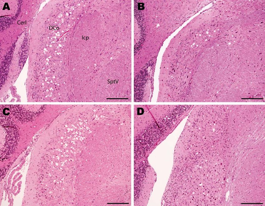
Figure 1. Histopathologic analysis of cochlear nuclei from host-encoded prion protein (PrP)-a mice (C57/BL6) inoculated with (A) fixed
material from the suspected case, (B) fixed material from experimental goat bovine spongiform encephalopathy (BSE), (C) unfixed material
from experimental sheep BSE, and (D) fixed material from experimental goat scrapie. The BSE-challenged mice (A–C) show confluent
vacuolation in the dorsal cochlear nucleus that extends ventrally with increasing lesion severity. Even in mild cases (B) this lesion can
be distinguished from the low-frequency randomly dispersed vacuoles observed in scrapie (D). Note the unaffected nature of the lesion
between fixed (A and B) and unfixed (C) samples. Cerl, cerebellum; DCo, dorsal cochlear nucleus; Icp, inferior cerebellar peduncle; SptV,
spinal tract of the trigeminal nerve. Scale bars = 200 μm.
of spongiform lesions and PrPSc deposits in tg338 mice in demonstrated lower binding with the 12B2 antibody,
which BSE was diagnosed was limited, and intensity of confirming that the proteinase K cleavage site was
the labeling was weak. Despite this finding, where PrPSc indistinguishable from that of ovine BSE. In contrast, mice
distribution was widespread, individual mice challenged inoculated with various scrapie sources demonstrated a
with BSE differed qualitatively from those challenged with 20.1-kDa unglycosylated band and increased reactivity
scrapie (data not shown). with 12B2 (Figure 4).
WB Analysis Secondary Passage Data in PrP-b Mice
When examined by WB, brain tissues from PrP-a After 1 serial passage in PrP-b (VM) mice, the sample
mice that were inoculated with the sample from the from the goat with suspected BSE generated IP of 128
goat with suspected BSE showed PrPSc bands that were ± 4 (mean ± SD) days postinoculation similar to serially
indistinguishable from those of mice inoculated with the passaged ovine BSE (109 ± 4) and the 301V mouse-
various BSE sources (Figure 4). The lower unglycosylated adapted BSE strain (107 ± 6). The comparatively longer
band had a molecular mass of ≈18.8 kDa, and the samples IP generated by that goat sample relative to these mouse-
Emerging Infectious Diseases • www.cdc.gov/eid • Vol. 17, No. 12, December 2011 2257RESEARCH Figure 2. Immunohistochemical analysis of brains of host-encoded prion protein (PrP)-a mice (RIII) inoculated with (A and B) fixed material from the goat with suspected bovine spongiform encephalopathy (BSE), (C and D) fixed material from experimental goat BSE, (E and F) unfixed material from experimental sheep BSE, and (G and H) fixed material from experimental goat scrapie. No PrPSc was detected in the molecular layer of the dentate gyrus in the suspected case (A) and the BSE controls (C and E); in the scrapie control (G) the same area is heavily affected. In the red nucleus, small PrPSc aggregates were observed in the suspected case (B) and in the BSE controls (D and F), whereas the same nucleus seem to be unaffected in the scrapie control despite evident PrPSc deposits in the surrounding area. Hip, hippocampus; Hif, hippocampal fissure; Hb, habenular nuclei; RdN, red nucleus; Th, thalamus; gr, mo, and po, granular, molecular, and polymorph layers, respectively, of the dentate gyrus. Scale bars = 200 μm. adapted BSE isolates is a common observation at second produced by serially passaged experimental ovine BSE and passage; for example, the IP of the serially passaged ovine similar to the 301V strain (Figure 5). After serial passage BSE at second passage was 148 ± 3 days postinoculation. of material from the goat suspected to have BSE in VM In these mice, the LPs were indistinguishable from those mice, the PrPSc patterns observed were indistinguishable Figure 3. Immunohistochemical analysis of brains of host-encoded prion protein (PrP)-b mice (VM) inoculated with (A and B) fixed material from the goat with suspected case, (C and D) fixed material from experimental goat bovine spongiform encephalopathy (BSE), (E and F) unfixed material from experimental sheep BSE, and (G and H) fixed material from experimental goat scrapie. In thalamus, cerebral cortex, and hippocampus the suspected case (A) and the BSE controls (C and E) showed mainly granular PrPSc deposits with comparable distribution. In the scrapie control (G), the predominant PrPSc type was large aggregates and plaques. In the periaqueductal gray matter, mice challenged with the suspected case (B) and with BSE controls (D and F) showed a manifold lower staining intensity of PrPSc labeling compared with the surrounding area, but the scrapie control (H) showed an intense labeling in the ventral periaqueductal region associated with substantial reduction of labeling in all neighboring areas. Aq, aqueduct; CC, corpus callosum; Cer Ctx, cerebral cortex; Hb, habenular nuclei; Rn, raphe nucleus; Pag, periaqueductal gray; Th, thalamus; Or, Py, Rad, and Lm, oriens, pyramidal cell, radiatum, and lacunosum-moleculare layers, respectively, of the hippocampus; gr, mo, and po, granular, molecular, and polymorph layers, respectively, of the dentate gyrus. Scale bars = 200 μm. 2258 Emerging Infectious Diseases • www.cdc.gov/eid • Vol. 17, No. 12, December 2011
Prion with BSE Properties in Goat

                                                                      (22,28,32). This approach generates high-resolution data
                                                                      that appear to be specific to individual TSE strains.
                                                                           The data show that the TSE agents in this study
                                                                      were not altered by the adverse conditions applied to
                                                                      them during histologic procedures. However, titer may
                                                                      decrease, suggesting that the effect of histologic processing
                                                                      is quantitative not qualitative. Therefore, bioassay is a
                                                                      valid approach for identifying BSE in archived histologic
                                                                      material when other techniques are not applicable, as in
                                                                      the current study. Regarding the suitability of different
                                                                      mouse lines for confirming BSE, our data show that any
Figure 4. Western blot analysis of a range of murine transmissible
                                                                      mouse line in which the agent can propagate sufficiently
spongiform encephalopathy–affected brain homogenates in host-         is suitable. An additional requirement at a practical level is
encoded prion protein (PrP)–a (RIII) mice. A) Western blot probed     the ability to characterize the agent on first passage. In this
with SHA31, 15-s exposure time. B) Western blot probed with           respect, use of PrP-a mice is preferable because in addition
12B2, 5-min exposure time. M, biotinylated marker; lane 1, ovine      to AR, IP, histopathologic analysis, and PrPSc patterning,
scrapie field case; lane 2, bovine spongiform encephalopathy
(BSE) field case; lane 3, unchallenged mouse; lane 4, bovine BSE-
                                                                      WB can also be applied to diagnose BSE. In contrast, its
challenged mouse; lane 5, ovine BSE-challenged mouse; lane 6,         application in PrP-b mice is less informative (33).
caprine BSE-challenged mouse; lanes 7 and 8, mice challenged               These methods can also be applied to analyze bioassay
with suspected case; lane 9, caprine scrapie-challenged mouse;        data derived from validated transgenic mouse lines
lane 10, ovine scrapie-challenged mouse. Molecular weights are        that offer the advantage of higher AR and decreased IP,
indicated kDa. Red line indicates 19 kDa unglycosylated band;
yellow line indicates 20 kDa unglycosylated band. Identical results
                                                                      provided that appropriate transgenic lines are selected and
were also obtained with C57/BL6 mice.                                 the TSE source and the donor species under investigation
                                                                      are taken into consideration. In this particular instance,
                                                                      our first choices would have been the use of a mouse line
                                                                      overexpressing a bovine transgene in combination with 1
from those induced by other mouse-adapted BSE isolates                that overexpresses a caprine transgene. At initiation of the
(data not shown).

Discussion
     We confirmed that the agent responsible for TSE in a
UK goat, which was initially reported as scrapie in 1990
and subsequently as suspected BSE in 2006 (16), was a BSE
agent. This conclusion was based on bioassay of nervous
tissue in mice demonstrating similarities of histopathologic
lesions, PrPSc mapping in the brain, and WB of PrPSc with
those of mice inoculated with BSE from various ovine,
caprine, and bovine sources.
     From a method perspective, the data suggest that
AR, IP, and LP are not optimal bioassay parameters for
differentiating TSE sources during first passage because
they represent mean values derived from a group of animals
that have been inoculated with a specific source. Therefore,          Figure 5. Lesion profiles from VM mice after second passage of
a substantial number of animals must die of clinical TSE for          the suspected case, serial passage of an ovine bovine spongiform
                                                                      encephalopathy (BSE) source, and a 301V control. Profiles were
these parameters to be meaningful. This finding is a limiting
                                                                      made on the basis of the lesion score, which is the quantification of
factor in instances in which TSE is diagnosed in only a few           transmissible spongiform encephalopathy–specific vacuolation in 9
animals because of low titer, restricted permissiveness of            neuroanatomical gray matter areas: G1, dorsal medulla nuclei; G2,
specific TSE strains in certain laboratory animals, or both.          cerebellar cortex of the folia including the granular layer, adjacent
These limitations can be overcome by application of IHC               to the fourth ventricle; G3, cortex of the superior colliculus; G4,
                                                                      hypothalamus; G5, thalamus; G6, hippocampus; G7, septal nuclei
and WB to differentiate BSE from scrapie confidently in
                                                                      of the paraterminal body; G8, cerebral cortex (at the level of G4 and
individual mice on first passage. Use of IHC has shown                G5); G9, cerebral cortex (at the level of G7). At least 9 clinically and
that different PrPSc deposits can be identified, and the              histopathologically positive mice contributed to each profile. Error
distribution of each deposit in the brain can be mapped               bars indicate SEM.

                          Emerging Infectious Diseases • www.cdc.gov/eid • Vol. 17, No. 12, December 2011                               2259
RESEARCH study, an established bovinised line was not available to entering the human food chain through consumption of food us, and the data generated from the wild-type mice were products derived from small ruminants. considered sufficient to identify unequivocally the agent Because TSEs in goats are still a problem, particularly strain. Caprine transgenic mouse lines are still under in Mediterranean countries, our data suggest that extensive development and not characterized or widely available. surveillance and breeding schemes must remain in place Instead, we used tg338 mice although they show
Prion with BSE Properties in Goat

 8. Scientific Panel on Biological Hazards. Opinion of the Scientific       25.   Thackray AM, Hopkins L, Spiropoulos J, Bujdoso R. Molecular
    Panel on Biological Hazards on certain aspects related to the risk            and transmission characteristics of primary passaged ovine scra-
    of transmissible spongiform encephalopathies (TSEs) in ovine and              pie isolates in conventional and ovine PrP transgenic mice. J Virol.
    caprine animals. The EFSA Journal. 2007;466:1–10.                             2008;82:11197–207. doi:10.1128/JVI.01454-08
 9. Foster JD, Bruce M, McConnell I, Chree A, Fraser H. Detection           26.   Béringue V, Andréoletti O, Le Dur A, Essalmani R, Vilotte JL, La-
    of BSE infectivity in brain and spleen of experimentally infected             croux C, et al. A bovine prion acquires an epidemic bovine spon-
    sheep. Vet Rec. 1996;138:546–8. doi:10.1136/vr.138.22.546                     giform encephalopathy strain-like phenotype on interspecies trans-
10. Foster JD, Parnham D, Chong A, Goldmann W, Hunter N. Clinical                 mission. J Neurosci. 2007;27:6965–71. doi:10.1523/JNEUROSCI.
    signs, histopathology and genetics of experimental transmission of            0693-07.2007
    BSE and natural scrapie to sheep and goats. Vet Rec. 2001;148:165–      27.   Béringue V, Bencsik A, Le DA, Reine F, Laï TL, Chenais N, et al.
    71. doi:10.1136/vr.148.6.165                                                  Isolation from cattle of a prion strain distinct from that causing
11. Bellworthy SJ, Hawkins SA, Green RB, Blamire I, Dexter G, Dex-                bovine spongiform encephalopathy. PLoS Pathog. 2006;2:e112.
    ter I, et al. Tissue distribution of bovine spongiform encephalopathy         doi:10.1371/journal.ppat.0020112
    infectivity in Romney sheep up to the onset of clinical disease after   28.   Beck KE, Sallis RE, Lockey R, Simmons MM, Spiropoulos J. Ovine
    oral challenge. Vet Rec. 2005;156:197–202.                                    PrP genotype is linked with lesion profile and immunohistochem-
12. Konold T, Bone G, Vidal-Diez A, Tortosa R, Davis A, Dexter G,                 istry patterns following primary transmission of classical scrapie
    et al. Pruritus is a common feature in sheep infected with the BSE            to wild type mice. J Neuropathol Exp Neurol. 2010;69:483–97.
    agent. BMC Vet Res. 2008;4:16. doi:10.1186/1746-6148-4-16                     doi:10.1097/NEN.0b013e3181db2497
13. Kao RR, Gravenor MB, Baylis M, Bostock CJ, Chihota CM, Ev-              29.   Wells GAH, Hawkins SA. Animal models of transmissible spongi-
    ans JC, et al. The potential size and duration of an epidemic of              form encephalopathies: experimental infection, observation and tis-
    bovine spongiform encephalopathy in British sheep. Science.                   sue collection. In: Lehmann S, Grassi J, editors. Techniques in prion
    2002;295:332–5. doi:10.1126/science.1067475                                   research. Basel: Birkhäuser Verlag; 2004. p. 37–71.
14. Gravenor MB, Ryder SJ, Gubbins S, Hunter N, Baylis M, Kao RR.           30.   Green R, Horrocks C, Wilkinson A, Hawkins SA, Ryder SJ. Primary
    Searching for BSE in sheep: interpreting the results so far. Vet Rec.         isolation of the bovine spongiform encephalopathy agent in mice:
    2003;152:298–9. doi:10.1136/vr.152.10.298                                     agent definition based on a review of 150 transmissions. J Comp
15. Eloit M, Adjou K, Coulpier M, Fontaine JJ, Hamel R, Lilin T, et al.           Pathol. 2005;132:117–31. doi:10.1016/j.jcpa.2004.08.002
    BSE agent signatures in a goat. Vet Rec. 2005;156:523–4.                31.   Fraser H, Dickinson AG. The sequential development of the brain le-
16. Jeffrey M, Martin S, Gonzalez L, Foster J, Langeveld JP, Van Zi-              sion of scrapie in three strains of mice. J Comp Pathol. 1968;78:301–
    jderveld FG, et al. Immunohistochemical features of PrP(d) accu-              11. doi:10.1016/0021-9975(68)90006-6
    mulation in natural and experimental goat transmissible spongiform      32.   Baron T, Bencsik A, Biacabe AG, Morignat E, Bessen RA. Pheno-
    encephalopathies. J Comp Pathol. 2006;134:171–81. doi:10.1016/j.              typic similarity of transmissible mink encephalopathy in cattle and
    jcpa.2005.10.003                                                              L-type bovine spongiform encephalopathy in a mouse model. Emerg
17. Dustan BH, Spencer YI, Casalone C, Brownlie J, Simmons MM. A                  Infect Dis. 2007;13:1887–94.
    histopathologic and immunohistochemical review of archived UK           33.   Groschup MH, Kuczius T, Junghans F, Sweeney T, Bodemer W,
    caprine scrapie cases. Vet Pathol. 2008;45:443–54. doi:10.1354/               Buschmann A. Characterization of BSE and scrapie strains/isolates.
    vp.45-4-443                                                                   Arch Virol Suppl. 2000;(16):217–26.
18. Jeffrey M, Martin S, Gonzalez L, Ryder SJ, Bellworthy SJ, Jackman       34.   Wilesmith JW, Ryan JB, Arnold ME, Stevenson MA, Burke PJ. De-
    R. Differential diagnosis of infections with the bovine spongiform            scriptive epidemiological features of cases of bovine spongiform
    encephalopathy (BSE) and scrapie agents in sheep. J Comp Pathol.              encephalopathy born after July 31, 1996 in Great Britain. Vet Rec.
    2001;125:271–84. doi:10.1053/jcpa.2001.0499                                   2010;167:279–86. doi:10.1136/vr.c4552
19. European Commission. Commission Regulation (EC) No 36/2005              35.   The TSE community reference laboratory strain typing expert group
    of 12 January 2005 amending Annexes III and X to Regulation (EC)              (STEG). Summary of the STEG opinion on two caprine isolates
    No 999/2001 of the European Parliament and of the Council as re-              (08–357 from France; G08–1469 from the UK) referred to the group,
    gards epidemio-surveillance for transmissible spongiform encepha-             2008 [cited 2011 Oct 3]. http://www.defra.gov.uk/vla/science/docs/
    lopathies in bovine, ovine and caprine animals [cited 2011 Oct 7].            sci_tse_rl_steg1008.pdf
    http://ec.europa.eu/food/food/biosafety/tse_bse/docs/r05-36.pdf         36.   González L, Martin S, Sisó S, Konold T, Ortiz-Peláez A, Phelan L, et
20. Brown P, Rohwer RG, Green EM, Gajdusek DC. Effect of chemicals,               al. High prevalence of scrapie in a dairy goat herd: tissue distribution
    heat, and histopathologic processing on high-infectivity hamster-             of disease-associated PrP and effect of PRNP genotype and age. Vet
    adapted scrapie virus. J Infect Dis. 1982;145:683–7. doi:10.1093/             Res. 2009;40:65. doi:10.1051/vetres/2009048
    infdis/145.2.683                                                        37.   Lantier I, Bertbon P, Leroux H, Rossignol C, Lacroux C, Torres JM,
21. Foster J, McKelvey W, Fraser H, Chong A, Ross A, Parnham D, et                et al. Ovine and bovine PRNP transgenic mice allow discrimination
    al. Experimentally induced bovine spongiform encephalopathy did               between scrapie and BSE in co-infected mice and sheep. Prion 2009.
    not transmit via goat embryos. J Gen Virol. 1999;80:517–24.                   2009;97.
22. Beck KE, Chaplin M, Stack M, Sallis RE, Simonini S, Lockey R,           38.   Bellworthy SJ, Dexter G, Stack M, Chaplin M, Hawkins SA, Sim-
    et al. Lesion profiling at primary isolation in RIII mice is insuffi-         mons MM, et al. Natural transmission of BSE between sheep within
    cient in distinguishing BSE from classical scrapie. Brain Pathol.             an experimental flock. Vet Rec. 2005;157:206.
    2010;20:313–22. doi:10.1111/j.1750-3639.2009.00273.x
23. Bruce ME, Boyle A, Cousens S, McConnell I, Foster J, Goldmann           Address for correspondence: John Spiropoulos, Animal Health and
    W, et al. Strain characterization of natural sheep scrapie and com-
                                                                            Veterinary Laboratories Agency, Weybridge, Surrey KT15 3NB, UK;
    parison with BSE. J Gen Virol. 2002;83:695–704.
24. Vilotte JL, Soulier S, Essalmani R, Stinnakre MG, Vaiman D,             email: john.spiropoulos@ahvla.gsi.gov.uk
    Lepourry L, et al. Markedly increased susceptibility to natural
    sheep scrapie of transgenic mice expressing ovine PrP. J Virol.           All material published in Emerging Infectious Diseases is in
    2001;75:5977–84. doi:10.1128/JVI.75.13.5977-5984.2001                     the public domain and may be used and reprinted without
                                                                              special permission; proper citation, however, is required.

                             Emerging Infectious Diseases • www.cdc.gov/eid • Vol. 17, No. 12, December 2011                                       2261
You can also read