Role of Neutrophils in Preventing and Resolving Acute
←
→
Page content transcription
If your browser does not render page correctly, please read the page content below
INFECTION AND IMMUNITY, Dec. 2007, p. 5663–5668                                                                                             Vol. 75, No. 12
0019-9567/07/$08.00⫹0 doi:10.1128/IAI.01542-06
Copyright © 2007, American Society for Microbiology. All Rights Reserved.

                Role of Neutrophils in Preventing and Resolving Acute
                                  Fungal Sinusitis䌤†
        Tobias E. Rodriguez,1,2 Nicole R. Falkowski,1 Jack R. Harkema,3 and Gary B. Huffnagle1,2*
Division of Pulmonary and Critical Care Medicine1 and Departments of Internal Medicine and of Microbiology and Immunology,2
  University of Michigan Medical School, Ann Arbor, Michigan, and Department of Pathobiology and Diagnostic Investigation,
                                      Michigan State University, East Lansing, Michigan3
                     Received 25 September 2006/Returned for modification 3 November 2006/Accepted 31 August 2007

             Acute fungal sinusitis (AFS) is a devastating disease of the paranasal sinuses afflicting immunocompromised

                                                                                                                                                                 Downloaded from http://iai.asm.org/ on February 5, 2021 by guest
          individuals. Knowledge about this disease is limited to clinical observations because there are no animal
          models in which to study the pathogenesis of the infection. Our goal was to develop a murine model of AFS and
          examine the role of neutrophils in host defense within the nasal cavity. Female C57BL/6 mice were depleted of
          neutrophils using anti-Gr-1 monoclonal antibody from day ⴚ1 to day 5 postinfection to initiate a transient
          neutropenia within the mice. At day 0, Aspergillus fumigatus conidia were administered intranasally. The
          untreated Aspergillus-exposed group had significant neutrophil recruitment by day 3, but by day 7 the leukocyte
          numbers had returned to unexposed levels. There was not a significant influx of mononuclear cells at either
          time point. In contrast, beginning at day 3 postinfection and continuing through day 7, anti-Gr-1-treated mice
          had increased cellular recruitment consisting of banded neutrophils at day 3 and mature neutrophils at day
          7. Hyphal masses developed only in the anti-Gr-1-treated mice (25 to 36%) but only during the period of
          treatment. When the treatment was discontinued, hyphal masses could no longer be detected in the nasal
          cavities of these mice. In contrast, cyclophosphamide treatment did not induce neutropenia, and the nasal
          cavity remained free of hyphal masses. These studies demonstrate the feasibility of using this model to study
          AFS and implicate neutrophils in protection of the sinuses against acute Aspergillus infection and in clearance
          of established hyphal masses.

   Acute fungal sinusitis (AFS), also known as fulminant sinus-                 inflammatory response in rodents coexposed to lipopolysac-
itis, is a rapid, invasive disease of the paranasal cavities. It                charide and ozone, but the role of these cells in an active sinus
develops primarily in immunocompromised individuals, such                       infection has yet to be evaluated (6, 25).
as bone marrow transplant and AIDS patients (5, 21). The                           Prior to these studies, there was no report of an animal
infection is characterized by rapid hyphal transformation                       model of fungal sinusitis in physically intact animals. Other
within the paranasal sinus cavities. This is followed by fungal                 murine studies focused on the pathology of bacterial sinusitis
invasion of the surrounding mucosa, musculature, vasculature,                   and used physical alterations of the nasal cavity to cause dis-
and, in severe cases, cranium. Patients who do not receive                      ease. For example, one model of chronic bacterial rhinosinus-
proper medical attention can suffer from bone and tissue loss                   itis used a Merocel sponge to cause a blockage within the nasal
and, if left untreated, death.                                                  cavity in order to facilitate an infection (9). Although this study
   Recent research about AFS has been limited to clinical                       did show bacterial colonization and ensuing inflammation, the
observations because there are not currently animal models of                   infection was not localized to a sinus cavity and aspects of the
this disease. Much of what is understood about the innate                       innate immune response were not addressed. The focus of our
response to fungal infections in the nasal cavity is inferred from              studies was to determine whether neutrophils protect the sinus
correlatives drawn from other tissue sites. For example, within                 cavity from Aspergillus infection. We did this by developing a
the lungs, fungal conidium challenge stimulates leukocyte re-                   novel murine model of AFS. In our studies, the nasal cavities
cruitment into the airspaces. The leukocytes, primarily macro-                  of the mice were not physically altered, but the mice were
phages and neutrophils, internalize resting or swollen conidia                  rendered neutropenic, a common predisposing condition for
and use mechanisms such as respiratory burst to combat infec-                   the development of AFS. The mice were then exposed to
tions (2, 3, 10–12, 22, 24). It is believed that macrophages and                Aspergillus fumigatus intranasally to determine if the absence of
neutrophils may have a similar response to fungi within the                     neutrophils was sufficient for fungal colonization and subse-
nasal cavity, although there have been no murine models that                    quent development of AFS. The neutropenia was then discon-
demonstrate this (18, 20). Neutrophils help mediate the nasal                   tinued to determine whether neutrophils play a role in resolv-
                                                                                ing established hyphal masses.

   * Corresponding author. Mailing address: Division of Pulmonary and
                                                                                                       MATERIALS AND METHODS
Critical Care Medicine, Department of Internal Medicine, University of
Michigan Medical School, Ann Arbor, MI 48109-0642. Phone: (734) 936-               Mice. Female C57BL/6 mice (Harlan Sprague Dawley, Inc.) were housed
9369. Fax: (734) 764-2665. E-mail: ghuff@umich.edu.                             under specific-pathogen-free conditions in enclosed filter top cages. Food and
   † Supplemental material for this article may be found at http://iai          sterile water were given ad libitum. The mice were maintained by the Unit for
.asm.org/.                                                                      Laboratory Animal Medicine at the University of Michigan (Ann Arbor, MI),
   䌤
     Published ahead of print on 17 September 2007.                             and protocols were approved by an animal institutional review board.

                                                                         5663
5664 RODRIGUEZ ET AL. INFECT. IMMUN.
Induction of temporary neutropenia within mice. Temporary neutropenia was
established within mice to allow development of an invasive fungal infection.
Mice were injected intraperitoneally (i.p.) with 100 g of anti-Gr-1 antibody
(RB6-8C5 ascites; raised at Taconic Biotechnology, Germantown, NY) 1 day
prior to intranasal fungal exposure. Three additional i.p. injections were given to
the mice at days 1, 3, and 5 postinfection to efficiently deplete the mature
neutrophil population in treated mice. Once anti-Gr-1 treatment was discontin-
ued at day 5, mature neutrophils began to return, and by day 7 the numbers
corresponded to those in a wild-type, uninfected animal.
Immunosuppression in mice using cyclophosphamide. Animals were treated
with 150 mg/kg of cyclophosphamide (Sigma, St. Louis, MO) via i.p. injection 3
days prior to intranasal exposure. In order to maintain immunosuppression,
subsequent injections were then given every 3 days.
Intranasal inoculation of A. fumigatus conidia. A. fumigatus ATCC 13073 was
grown on Sabouraud dextrose agar (Difco) for 14 days. Spores (conidia) were
harvested by washing plates with sterile 0.1% Tween 80, followed by filtration of
Downloaded from http://iai.asm.org/ on February 5, 2021 by guest
the suspension through two layers of sterile gauze to remove hyphae. For infec-
tion, the spores were washed in nonpyrogenic saline (Abbott Laboratories, Chi-
cago, IL), counted with a hemocytometer, and diluted to obtain 108 spores/ml in FIG. 1. Hyphal colonization in untreated and anti-Gr-1 treated
sterile nonpyrogenic saline in order to prepare the final inoculum, 106 conidia/
mice infected with A. fumigatus. The percent hyphal colonization rep-
mouse administered in 5 l/nostril (total volume, 10 l/mouse). Prior to intra-
resents the fraction of animals in which hyphal colonization of the
nasal cavity was detected. Histology samples were analyzed for the
nasal inoculation, mice were anesthetized by i.p. injection of a ketamine-xylazine
presence of hyphal masses within the maxillary sinuses and nasal cavity
solution (2.5 mg of ketamine [Fort Dodge Animal Health, Fort Dodge, IA]/
(n ⱖ 8/group/time point).
mouse plus 0.1 g of xylazine [Lloyd Laboratories, Shenandoah, IA]/mouse).
Nasal leukocyte isolation. Mice were euthanized with CO2. The heads of the
mice were removed, and the sinuses were exposed via a transverse cut along the
skull. The samples then were enzymatically digested for 40 min at 37°C with 15
ml of digestion buffer (RPMI 1640, 10% fetal calf serum, antibiotics, 1 mg/ml
RESULTS
collagenase [Boehringer Mannheim Biochemicals, Chicago, IL], 30 g/ml DNase
[Sigma Chemical Co., St. Louis, MO]) per sample. Samples were removed from
To determine the effect of neutrophil depletion on the de-
the resulting cell suspensions and dispersed by drawing the cells up and down velopment of AFS, mice were made temporarily neutropenic
through the bore of a 10-ml syringe. Each cell suspension was then pelleted, and by administration of anti-Gr-1 at days ⫺1, 1, 3, and 5. Mice
erythrocytes were lysed by incubation in ice-cold NH4Cl buffer (0.829% NH4Cl, were inoculated intranasally with A. fumigatus at day 0 and
0.1% KHCO3, 0.0372% Na2EDTA [pH 7.4]; Sigma). Excess RPMI 1640 was then analyzed histologically for the presence of hyphal masses
added to make the solution isotonic, and the cells were pelleted and resuspended
(Fig. 1 to 3). The anti-Gr-1 depletion protocol was effective in
in complete medium (RPMI 1640, 10% fetal calf serum [Life Technologies], 5 ⫻
10⫺5 M 2-mercaptoethanol, sodium pyruvate, nonessential amino acids, glu- significantly reducing the amount of neutrophils present in the
tamine, antibiotics [Sigma]). Cell concentrations were determined by counting nasal cavity and spleen following intranasal exposure to A.
the cells after trypan blue staining. fumigatus at day 3 postinfection (Fig. 4; see Fig. S1 in the
Splenocyte isolation. Spleens were excised, and cells were dispersed with the supplemental material).
plunger of a 3-ml syringe. Erythrocytes were lysed using NH4Cl buffer, and cells
Mice treated with anti-Gr-1 antibody developed invasive
were resuspended in complete medium (RPMI 1640, 5% fetal calf serum, 2
mmol/liter L-glutamine, 50 mol/liter 2-mercaptoethanol, 100 U/ml penicillin,
hyphal masses that were visible in histology sections. The
100 g/ml streptomycin sulfate). masses were localized to the maxillary sinus cavities at days 3
Histological analysis. Samples were fixed in 10% neutral buffered formalin and 7. By day 3 postinfection, 36% of treated mice had devel-
and decalcified prior to sectioning (4). Slides were then stained with hematoxylin oped hyphal masses. By day 7, masses were seen in 25% of
and eosin for visualization, with Alcian Blue-periodic acid-Schiff stain for treated mice (Fig. 1). Conversely, untreated mice infected with
visualization of intraepithelial mucosubstances, and with Gomori methenamine
A. fumigatus showed no signs of hyphal formation at any time
silver for visualization of the fungi.
Cytokine and chemokine analysis. Nasal cavities were excised using microsur- point.
gical techniques and kept in RNAlater (Ambion, Austin, TX) at ⫺20°C until We could not continue to treat mice with anti-Gr-1 antibody
RNA isolation was performed. For reverse transcription-PCR, total RNA was past day 7. This is because the mice mounted an immune
isolated from the septa using Trizol reagent (Life Technologies, Gaithersburg, response (serum sickness) against the anti-Gr-1 monoclonal
MD) as outlined in the Trizol protocol. After amplification, samples were sep-
antibody, which is a rat antibody. We continued to monitor
arated on a 2% agarose gel containing 5 l/100 ml ethidium bromide (10 mg/ml;
Sigma), and bands were visualized and photographed using UV transillumina-
mice after they recovered from the transient neutropenia. At
tion. The primer sequences used are as follows: for KC, forward primer 5⬘-TG days 14 and 28 postinfection, there were no signs of hyphal
AGCTGCGCTGTCAGTGCCT-3⬘ and reverse primer 5⬘-AGAAGCCAGCGT colonization in histological sections from either untreated or
TCACCAGA-3⬘; for tumor necrosis factor alpha, forward primer 5⬘-CCTGTA previously anti-Gr-1-treated mice (data not shown).
GCCCACGTCGTAGC-3⬘ and reverse primer 5⬘-AGCAATGACTCCAAAGT We next examined whether there were changes to the nasal
AGACC-3⬘; for gamma interferon, forward primer 5⬘-CTACCTCAGACTCTT
TGAAGTCT-3⬘ and reverse primer 5⬘-CAGCGACTCCTTTTCCGCTT-3⬘; for
architecture that were caused by hyphal colonization and in-
MIP-2, forward primer 5⬘-GCTGGCCACCAACCACCAGG-3⬘ and reverse duction of AFS. All histological analysis was confined to the
primer 5⬘-AGCGAGGCACATCAGGTACG-3⬘; for interleukin-6 (IL-6), for- maxillary sinuses and the areas of the nasal cavity nearest
ward primer 5⬘-GACAAAGCCAGAGTCCTTCAGAGAG-3⬘ and reverse them. Untreated, Aspergillus-exposed mice did not have pa-
primer 5⬘-CTAGGTTTGCCGAGTAGATCTC-3⬘; and for -actin, forward thology indicative of AFS (Fig. 2 and 3). All cavities and
primer 5⬘-TGGAATCCTGTGGCATCCATGAAAC-3⬘ and reverse primer 5⬘-
airways were intact and free of fungal masses, although some
TAAAACGCAGCTCAGTAACAGTCCG-3⬘. Kodak Molecular Imaging soft-
ware was used to quantify the intensity of the bands, and the net intensity was minor inflammation was seen in the subepithelial space. In
used to for comparisons between groups. Net intensity is the background-sub- contrast, anti-Gr-1-treated mice had hyphal masses with le-
tracted pixel values in the band rectangle. sions centered predominantly in the maxillary sinus cavitiesVOL. 75, 2007 MURINE AFS 5665
Downloaded from http://iai.asm.org/ on February 5, 2021 by guest
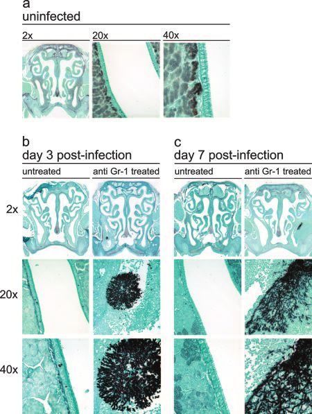
FIG. 2. Hematoxylin and eosin staining of the mouse nasal cavity. FIG. 3. Gomori methenamine silver staining of the mouse nasal
Mice were either not treated or treated with anti-Gr-1 and then intra- cavity. Mice were either not treated or treated with anti-Gr-1 and then
nasally infected with A. fumigatus as outlined in the text. At days 3 and intranasally infected with A. fumigatus as outlined in the text. At days
7 postinfection, the sinuses were removed, fixed, sectioned, and stained 3 and 7 postinfection, the sinuses were removed, fixed, sectioned, and
with hematoxylin and eosin to differentially stain leukocytes. (a) Un- stained with Gomori methenamine silver, which stains polysaccharides
treated, uninfected control animal. (b) Untreated and anti-Gr-1- black, for fungal visualization. (a) Untreated, uninfected control ani-
treated mice infected with A. fumigatus at day 3 postinfection. (c) mal. (b) Untreated and anti-Gr-1-treated mice infected with A. fumiga-
Untreated and anti-Gr-1-treated mice infected with A. fumigatus at day tus at day 3 postinfection. (c) Untreated and anti-Gr-1-treated mice
7 postinfection. The 20⫻ and 40⫻ images were derived from the infected with A. fumigatus at day 7 postinfection. The 20⫻ and 40⫻
infected maxillary sinus cavities shown in the 2⫻ images of sections. images were derived from the infected maxillary sinus cavities shown in
the 2⫻ images of sections.
(Fig. 2 and 3). The lesions were asymmetrical and were found
in either the left or right maxillary sinus cavity but never in day 7, there was no mucus production in these sites (see Fig. S2
both cavities. All lesions were characterized by a marked, py- in the supplemental material).
ronecrotizing fungal sinusitis with acute rhinitis that varied in The next objective was to analyze the kinetics of leukocyte
severity. At day 3 postinfection, all hyphal masses were local- recruitment into the nasal cavity following infection. The nasal
ized to the maxillary sinuses and there was some invasion of cavity was excised and enzymatically digested as outlined in
the underlying tissue, resulting in necrosis and an influx of Materials and Methods. The cells in the cellular infiltrates
inflammatory cells (Fig. 2b and 3b). were enumerated and then cytospun onto slides, stained, and
By day 7, the pathology had worsened, and there was exac- analyzed. At day 3 postinfection, there was a significant in-
erbation of the necrosis and corresponding inflammation. Hy- crease in mature neutrophil recruitment in untreated, infected
phal invasion into the outer musculature of the cranium was mice compared to uninfected mice (Fig. 4). In contrast, there
seen, and portions of the initial hyphal mass had become de- were no mature neutrophils at day 3 postinfection in the nasal
tached. The hyphal fragments then colonized other areas of cavities of anti-Gr-1-treated mice. However, a significant num-
the nasal cavity, resulting in additional inflammation and dras- ber of banded neutrophils were recruited to the infection
tic architectural changes in both the maxillary sinuses and nasal within the nasal cavity in anti-Gr-1-treated mice (Fig. 4). By
cavity (Fig. 2c and 3c). At day 7, untreated Aspergillus-exposed day 7, a small number of banded neutrophils were still present;
mice still had no pathology; all cavities were clear of debris, however, a substantial majority of the infiltrate was now ma-
inflammation, and hyphae. ture neutrophils (the last injection of anti-Gr-1 was on day 5).
Mucus production was also disrupted in tissue and mucosa The number of neutrophils in this group returned to unin-
where hyphal masses had become invasive. Using a stain to fected levels by days 14 and 28 (data not shown). Thus, the
highlight mucus (Alcian Blue-periodic acid-Schiff stain), there kinetics of mature neutrophil influx in both groups correlated
was a significant decrease in mucus production at the sites of with fungal clearance, and if neutrophil influx was prevented by
hyphal masses in the treated mice at day 3 postinfection. By anti-Gr-1 treatment, hyphal masses developed.5666 RODRIGUEZ ET AL. INFECT. IMMUN.
Downloaded from http://iai.asm.org/ on February 5, 2021 by guest
FIG. 5. Nasal cavity chemokine profiles of A. fumigatus-infected
mice. Mice were either not treated, infected with A. fumigatus (A.
fum), or treated with anti-Gr-1 and infected with A. fumigatus (anti-
Gr1). The nasal cavities were excised from mice using microsurgical
techniques. RNA was isolated from the samples, and reverse trans-
criptase PCR was performed in order to analyze localized chemokine
expression. (A) After visualization, a representative band was taken
from each gel in order to compare groups (n ⫽ 4). Kodak Molecular
Imaging software was used to quantify the intensity of the bands.
(B) Net intensity values expressed as a mean ratio (chemokine/-actin)
FIG. 4. Differential analysis of nasal leukocyte recruitment. Fol- for each group (n ⫽ 4).
lowing leukocyte isolation from nasal cavity digests, cytospin slides
were made and stained with Wright-Giemsa stain to visualize cell
populations. (A) Mature neutrophil cell counts. (B) Immature
(banded) neutrophil cell counts. (C) Mononuclear cell counts. An
(CXCL1) and MIP-2 (CXCL2) and the inflammatory cytokine
asterisk indicates a P value of ⬍0.05 and a dagger indicates a P value IL-6 in the nasal cavity at day 3 postinfection (Fig. 5). The
of ⬍0.01 as determined by Student’s t test (n ⫽ 6 mice per group). levels of expression of KC, MIP-2, and IL-6 were all elevated
PMNs, polymorphonuclear leukocytes. following Aspergillus infection, and the induction was not af-
fected by treatment with anti-Gr-1. These data demonstrate
that mature neutrophils are not required for expression of
Since Gr-1 can also be expressed on a subpopulation of these cytokines.
recently recruited monocytes (16), we also analyzed mononu- The last objective of these studies was to determine if cyclo-
clear cell recruitment in the mice following exposure to As- phosphamide could also induce the development of invasive
pergillus. There was no statistically significant change in mono- hyphal masses within the nasal cavities of mice exposed to
nuclear cells in either untreated or anti-Gr-1-treated mice at Aspergillus. Cyclophosphamide depletes circulating T cells, B
days 3 and 7 postinfection compared to uninfected mice (Fig. cells, and mononuclear cells, but it does not interfere with
4). Thus, mononuclear cells are not a major component of the neutrophil development. Mice were treated with 150 mg/kg of
inflammatory infiltrate in the nasal cavity following Aspergillus cyclophosphamide via i.p. injection 3 days prior to intranasal
exposure. exposure. Subsequent injections were administered every 3
We next examined expression of the CXC chemokines KC days in order to maintain immunosuppression. Mice were thenVOL. 75, 2007 MURINE AFS 5667
analyzed at days 3, 7, 14, and 21 postinfection. Cyclophospha- may be extremely important during chronic fungal infection.
mide did not affect splenic neutrophil numbers at day 3 postin- However, the anti-Gr-1 and cyclophosphamide results clearly
fection, while anti-Gr-1 caused a significant decrease (see Fig. demonstrate that Gr-1⫹ neutrophils are critical for innate host
S1 in the supplemental material). At all time points, develop- defense in the sinuses during acute fungal exposure.
ment of hyphal masses was not seen within the nasal cavities of Expression of the neutrophil chemotactic factors KC and
cyclophosphamide-treated mice (data not shown). In every MIP-2 was elevated in the nasal cavity following Aspergillus
specimen analyzed, small numbers of leukocytes were seen in exposure. Patients with AFS often have elevated levels of the
the nasal cavities of treated mice; however, hyphal masses were CXC chemokines IL-8, which is a functional homologue of KC,
never seen in the sinus or nasal cavities. Thus, protection and MIP-2 (23). Increased levels of CXC chemokines follow-
against AFS depends on a cyclophosphamide-resistant cell ing Aspergillus exposure have also been reported in murine
population, and since anti-Gr-1 treatment ablated protection, models of invasive aspergillosis (3, 12–14). Additionally, trans-
these results suggest that the primary cell involved in host genic overexpression of KC in the lungs was able to improve
defense in the nasal cavity is the neutrophil. the outcome following pulmonary aspergillus infection (15).
Thus, it is very likely that CXC chemokines are the major
Downloaded from http://iai.asm.org/ on February 5, 2021 by guest
DISCUSSION chemotactic signals for neutrophil influx into the nasal cavity
following Aspergillus infection.
We examined the protective role of neutrophils against A. In conclusion, while much is known about host defense
fumigatus in AFS by developing a novel, reproducible model of against Aspergillus in the lungs and systemically, relatively little
the disease. In our studies, mice made temporarily neutropenic is known about host defense against this mold in the sinuses.
were not protected against fungal colonization and subsequent Furthermore, little is known about the effect of hyphal coloni-
development of AFS. Previous studies of sinusitis pathogenesis zation on nasal architecture and function. The studies pre-
have relied on alterations to the nasal architecture or physical sented here demonstrate the feasibility of this model for study-
impairments to allow disease progression. In the studies de- ing AFS and implicate neutrophils in protection of the sinuses
scribed here, the nasal cavity of mice was not altered and the from Aspergillus infection and in clearance of established hy-
mice were exposed to the fungus through a physiologically phal masses. The approach presented here can also be used to
relevant route of infection. In both anti-Gr-1-treated and un- study the effect of virus-induced inflammation on the develop-
treated experimental groups, inflammation or fungal coloniza- ment of AFS. Viral infections can cause inflammation of the
tion was always localized to the nasal cavity and did not dis- ostia, the sinuses, and the surrounding mucosa. Such inflam-
seminate to the lungs (data not shown). mation has been hypothesized to provide an environment con-
Prior to this investigation, knowledge of the innate immune ducive to fungal colonization by disrupting normal regulation
response against invasive aspergillosis in the nasal cavity was of innate immunity and nasal physiology within the paranasal
based on clinical observations or extrapolation from murine region (1, 7). Thus, using models which do not disrupt the
models of pulmonary disease. Although many correlations can underlying architecture of the nasal cavity holds significant
be inferred between AFS and pulmonary invasive aspergillosis, promise for identifying the etiologic factors that lead to fungal
there are significant differences between these diseases. The sinusitis.
most noteworthy difference is the formation of the large hyphal
masses seen in our model of AFS, which are never seen in ACKNOWLEDGMENTS
pulmonary invasive aspergillosis. Even in neutropenic mice
We thank Galen Toews and Jeffery Terrell for their invaluable
with severe cellular infiltrate, small hyphal fragments are all scholarly contributions to this study.
that is visualized within the lungs of the mice (3, 14). One This work was supported in part by grants R01AI064479 (to G.B.H.)
explanation for this difference is that alveolar macrophages are and T32AI007528 (to T.E.R.) from the National Institute of Allergy
responsible for ingesting resting conidia, thereby preventing and Infectious Diseases.
hyphal transformation and germination within the lungs (8, 11,
REFERENCES
19, 22, 26). These observations can also account for the inabil-
ity of cyclophosphamide to induce AFS in our studies. Many 1. Baraniuk, J. 1994. Physiology of sinusitis, p. 19–39. In H. M. Druce (ed.),
Sinusitis: pathophysiology and treatment, vol. 1. Marcel Dekker, Inc., New
murine models of pulmonary invasive aspergillosis use cyclo- York, NY.
phosphamide in order to immunosuppress the animals and 2. Braedel, S., M. Radsak, H. Einsele, J. P. Latge, A. Michan, J. Loeffler, Z.
Haddad, U. Grigoleit, H. Schild, and H. Hebart. 2004. Aspergillus fumigatus
allow initiation of fungal infection. The nasal cavity does not antigens activate innate immune cells via Toll-like receptors 2 and 4. Br. J.
have resident macrophages that reside on the mucosal surfaces Haematol. 125:392–399.
within the open air spaces. Without resident airway cells to 3. Cenci, E., A. Mencacci, C. Fe d’Ostiani, C. Montagnoli, A. Bacci, G. Del
Sero, S. Perito, F. Bistoni, and L. Romani. 1998. Cytokine- and T-helper-
ingest inhaled resting conidia, hyphal transformation may oc- dependent immunity in murine aspergillosis. Res. Immunol. 149:445–454.
cur within the nasal cavity. Thus, the nasal cavity must rely on (Discussion, 149:504–505.)
an influx of neutrophils to control hyphal growth. 4. Farraj, A. K., J. R. Harkema, and N. E. Kaminski. 2004. Allergic rhinitis
induced by intranasal sensitization and challenge with trimellitic anhydride
The results presented here definitively demonstrate that but not with dinitrochlorobenzene or oxazolone in A/J mice. Toxicol. Sci.
Gr-1⫹ cells are required for protection of the nasal cavity from 79:315–325.
5. Ferguson, B. J. 2000. Definitions of fungal rhinosinusitis. Otolaryngol. Clin.
Aspergillus infection. In addition to neutrophils, other cells, N. Am. 33:227–235.
such as a subpopulation of macrophages and plasmacytoid 6. Harkema, J. R., and J. G. Wagner. 2005. Epithelial and inflammatory re-
dendritic cells, also express Gr-1 (16, 17). This study did not sponses in the airways of laboratory rats coexposed to ozone and biogenic
substances: enhancement of toxicant-induced airway injury. Exp. Toxicol.
exclude the potential positive contribution of these cells to host Pathol. 57(Suppl. 1):129–141.
defense in the upper airways, and we hypothesize that the cells 7. Herbert, R. A., and J. R.Leininger. 1999. Nose, larynx and trachea, p. 259–5668 RODRIGUEZ ET AL. INFECT. IMMUN.
292. In R. Maronpot (ed.), Pathology of the mouse. Cache River Press, 17. Nakano, H., M. Yanagita, and M. D. Gunn. 2001. CD11c⫹ B220⫹ Gr-1⫹
Vienna, IL. cells in mouse lymph nodes and spleen display characteristics of plasma-
8. Ibrahim-Granet, O., B. Philippe, H. Boleti, E. Boisvieux-Ulrich, D. Grenet, cytoid dendritic cells. J. Exp. Med. 194:1171–1178.
M. Stern, and J. P. Latge. 2003. Phagocytosis and intracellular fate of 18. Peeters, D., M. J. Day, and C. Clercx. 2005. An immunohistochemical study
Aspergillus fumigatus conidia in alveolar macrophages. Infect. Immun. 71: of canine nasal aspergillosis. J. Comp. Pathol 132:283–288.
891–903. 19. Philippe, B., O. Ibrahim-Granet, M. C. Prevost, M. A. Gougerot-Pocidalo,
9. Jacob, A., B. T. Faddis, and R. A. Chole. 2001. Chronic bacterial rhinosinusitis: M. Sanchez Perez, A. Van der Meeren, and J. P. Latge. 2003. Killing of
description of a mouse model. Arch. Otolaryngol. Head Neck Surg. 127:657– Aspergillus fumigatus by alveolar macrophages is mediated by reactive oxi-
664. dant intermediates. Infect. Immun. 71:3034–3042.
10. Latgé, J. P. 2001. The pathobiology of Aspergillus fumigatus. Trends Micro- 20. Pitzurra, L., S. Bellocchio, A. Nocentini, P. Bonifazi, R. Scardazza, L. Gallucci,
biol. 9:382–389. F. Stracci, C. Simoncelli, F. Bistoni, and L. Romani. 2004. Antifungal immune
11. Levitz, S. M., M. E. Selsted, T. Ganz, R. I. Lehrer, and R. D. Diamond. 1986. reactivity in nasal polyposis. Infect. Immun. 72:7275–7281.
In vitro killing of spores and hyphae of Aspergillus fumigatus and Rhizopus 21. Pleis, J., and R. Coles. 2003. Summary health statistics for U.S. adults:
oryzae by rabbit neutrophil cationic peptides and bronchoalveolar macro-
National Health Interview Survey, 1999. National Center for Health Statis-
phages. J. Infect. Dis. 154:483–489.
tics. Vital Health Stat. 10:1–145.
12. Mehrad, B., T. A. Moore, and T. J. Standiford. 2000. Macrophage inflam-
22. Roilides, E., H. Katsifa, and T. J. Walsh. 1998. Pulmonary host defences
matory protein-1 alpha is a critical mediator of host defense against invasive
against Aspergillus fumigatus. Res. Immunol. 149:454–465. (Discussion, 149:
pulmonary aspergillosis in neutropenic hosts. J. Immunol. 165:962–968.
13. Mehrad, B., R. M. Strieter, T. A. Moore, W. C. Tsai, S. A. Lira, and T. J. 523–524.)
Downloaded from http://iai.asm.org/ on February 5, 2021 by guest
Standiford. 1999. CXC chemokine receptor-2 ligands are necessary compo- 23. Rudack, C., W. Stoll, and C. Bachert. 1998. Cytokines in nasal polyposis,
nents of neutrophil-mediated host defense in invasive pulmonary aspergil- acute and chronic sinusitis. Am. J. Rhinol. 12:383–388.
losis. J. Immunol. 163:6086–6094. 24. Schaffner, A., H. Douglas, and A. Braude. 1982. Selective protection against
14. Mehrad, B., R. M. Strieter, and T. J. Standiford. 1999. Role of TNF-alpha conidia by mononuclear and against mycelia by polymorphonuclear phago-
in pulmonary host defense in murine invasive aspergillosis. J. Immunol. cytes in resistance to Aspergillus. Observations on these two lines of defense
162:1633–1640. in vivo and in vitro with human and mouse phagocytes. J. Clin. Investig.
15. Mehrad, B., M. Wiekowski, B. E. Morrison, S. C. Chen, E. C. Coronel, D. J. 69:617–631.
Manfra, and S. A. Lira. 2002. Transient lung-specific expression of the 25. Wagner, J. G., J. A. Hotchkiss, and J. R. Harkema. 2001. Effects of ozone
chemokine KC improves outcome in invasive aspergillosis. Am. J. Respir. and endotoxin coexposure on rat airway epithelium: potentiation of toxicant-
Crit. Care Med. 166:1263–1268. induced alterations. Environ. Health Perspect. 109(Suppl. 4):591–598.
16. Mordue, D. G., and L. D. Sibley. 2003. A novel population of Gr-1⫹-acti- 26. Waldorf, A. R., S. M. Levitz, and R. D. Diamond. 1984. In vivo bronchoal-
vated macrophages induced during acute toxoplasmosis. J. Leukoc. Biol. veolar macrophage defense against Rhizopus oryzae and Aspergillus fumiga-
74:1015–1025. tus. J. Infect. Dis. 150:752–760.
Editor: A. CasadevallYou can also read