Genetic Loss of LCK Kinase Leads to Acceleration of Chronic Lymphocytic Leukemia - Frontiers
←
→
Page content transcription
If your browser does not render page correctly, please read the page content below
ORIGINAL RESEARCH
                                                                                                                                         published: 02 September 2020
                                                                                                                                        doi: 10.3389/fimmu.2020.01995

                                            Genetic Loss of LCK Kinase Leads to
                                            Acceleration of Chronic Lymphocytic
                                            Leukemia
                                            Melanie Märklin 1,2 , Alexander R. Fuchs 1 , Claudia Tandler 2 , Jonas S. Heitmann 1,2 ,
                                            Helmut R. Salih 2 , Joseph Kauer 3 , Leticia Quintanilla-Martinez 4 , Stefan Wirths 1 ,
                                            Hans-Georg Kopp 1,5 and Martin R. Müller 1,6*
                                            1
                                             Department of Hematology, Oncology and Clinical Immunology and Rheumatology, University of Tübingen, Tübingen,
                                            Germany, 2 Clinical Collaboration Unit Translational Immunology, German Cancer Consortium (DKTK), University Hospital
                                            Tübingen, Tübingen, Germany, 3 Department of Immunology, Interfaculty Institute for Cell Biology, University of Tübingen,
                                            Tübingen, Germany, 4 Department of Pathology, University of Tübingen, Tübingen, Germany, 5 Department of Molecular
                                            Oncology and Thoracic Oncology, Robert-Bosch-Hospital Stuttgart, Stuttgart, Germany, 6 Department of Hematology,
                                            Oncology and Immunology, Klinikum Region Hannover, KRH Klinikum Siloah, Hanover, Germany

                                            Most patients with chronic lymphocytic leukemia (CLL) exhibit an indolent disease course
                                            and unresponsive B cell receptors (BCRs) exemplified by an anergic phenotype of their
                                            leukemic cells. In up to 5% of patients, CLL transforms from an indolent subtype to an
                                            aggressive form of B cell lymphoma (Richter’s syndrome), which is associated with worse
                           Edited by:
                       Nurit Hollander,     disease outcome and severe downregulation of NFAT2. Here we show that ablation of
              Tel Aviv University, Israel   the tyrosine kinase LCK, which has previously been characterized as a main NFAT2 target
                       Reviewed by:         gene in CLL, leads to loss of the anergic phenotype, thereby restoring BCR signaling,
                   Dimitar G. Efremov,
      International Centre for Genetic
                                            which results in an acceleration of CLL. Our study identifies LCK as a main player in
  Engineering and Biotechnology, Italy      mediating BCR unresponsiveness and its role as a crucial regulator of anergy in CLL.
                   Riccardo Bomben,
  Centro di Riferimento Oncologico di       Keywords: CLL, NFAT2, NFATc1, LCK, Richter’s transformation, anergy, BCR
                  Aviano (IRCCS), Italy

                 *Correspondence:
                      Martin R. Müller
                                            INTRODUCTION
                martin.mueller@krh.eu
                                            Chronic lymphocytic leukemia (CLL) is characterized by a clonal expansion of mature
                   Specialty section:
                                            CD5+ CD19+ B cells and constitutes the most common leukemia in adults. By the introduction
         This article was submitted to      of monoclonal antibodies targeting CD20 (rituximab, obinutuzumab and ofatumumab) (1, 2)
Cancer Immunity and Immunotherapy,          and small molecule inhibitors (ibrutinib, idelalisib & venetoclax) (3–5) treatment options have
               a section of the journal     been substantially improved. Nevertheless, CLL is still considered an incurable disease. While
              Frontiers in Immunology       the majority of patients exhibit an indolent disease course and do not require therapy for many
           Received: 14 April 2020          years, other patients show aggressive disease phenotype with early progression and requirement
            Accepted: 23 July 2020          for therapy. A severe progression of the disease is termed Richter’s syndrome, which constitutes a
     Published: 02 September 2020           transformation to an aggressive lymphoma and occurs in about 5% of CLL patients (6).
                             Citation:         Several prognostic factors like unmutated immunoglobulin heavy chain (IGHV) gene loci, high
    Märklin M, Fuchs AR, Tandler C,         expression of ZAP70 and CD38 as well as certain cytogenetic abnormalities (e.g., del17p) have
    Heitmann JS, Salih HR, Kauer J,         been associated with dismal treatment outcome (7). Progression of the disease was also shown
     Quintanilla-Martinez L, Wirths S,
                                            to be highly dependent on the signaling capacity of the B cell receptor (BCR) [for review (8, 9)].
    Kopp H-G and Müller MR (2020)
Genetic Loss of LCK Kinase Leads to
                                            Previous studies demonstrated a strong tendency for indolent CLL cases to exhibit anergic features
Acceleration of Chronic Lymphocytic         like surface IgM (sIgM) downregulation and reduced BCR signaling, in line with elevated basal
 Leukemia. Front. Immunol. 11:1995.         intracellular Ca2+ and constitutive ERK1/2 phosphorylation (10–13). NFAT2 (NFATc1) is known
    doi: 10.3389/fimmu.2020.01995           as an important Ca2+ dependent transcription factor in lymphocyte development [for review (14)]

Frontiers in Immunology | www.frontiersin.org                                      1                                          September 2020 | Volume 11 | Article 1995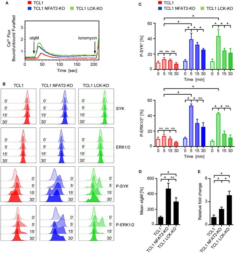
Märklin et al.                                                                                                      LCK Deletion Promotes CLL Acceleration

and has been shown to be overexpressed and constitutively                           Germany. Mice were used on the C57BL/6 background (CRL
activated in a subset of CLL patients (15, 16). We and others                       027) and maintained under specific pathogen-free conditions.
further reported that indolent CLL cells show a high degree of                      Homozygous TCL1 NFAT2fl/fl CD19Cre (TCL1 NFAT2-KO) mice
NFAT2 expression correlating with non-responsive, i.e., anergic                     with B cell-specific deletion of NFAT2 and TCL1 NFAT2fl/fl
BCRs (15, 16).                                                                      (TCL1) mice without NFAT2 deletion served as controls. Both
   Using the Eµ-TCL1 mouse model, which is the most widely                          cohorts were crossed with the LCK-KO strain to obtain the
used animal model to study pathophysiology, clonal evolution                        experimental cohorts exhibiting a germline deletion of LCK
and drug sensitivity in CLL (17–19), we could characterize that                     kinase. All mice were age and sex-matched and were sacrificed
CD5+ CD19+ leukemic cells from Eµ-TCL1 transgenic mice                              at the indicated time points by overdosing inhalation anesthesia.
possess many important features of anergic CLL cells. Although                      Mice exhibiting clinical signs of disease or >20% weight loss
this model has limitations, we demonstrated that CD5+ CD19+                         were removed early from the experimental cohort. Animal
leukemic cells showed impaired Ca2+ mobilization capacity as                        experiments were performed with the authorization of the
well as a reduced expression level of sIgM when compared                            Institutional Animal Care and Use Committee of the University
to physiological B cells. We further reported that these cells                      of Tübingen according to German federal and state regulations
exhibit a constitutive activation of NFAT2 and ERK1/2 (16).                         following the ARRIVE guidelines (34).
We also recently demonstrated that loss of NFAT2 in the B
cell compartment causes an aggressive course of CLL, enhances                       Flow Cytometry
BCR signaling and results in the selection of unmutated BCRs                        Cell suspensions of different organs were prepared as previously
and leads to a highly proliferative disease in a murine Eµ-                         described (16) and stained with fluorophore labeled mAbs. For
TCL1 leukemia model (16, 20). This was mirrored by low                              detailed information see Supplementary Material.
NFAT2 expression in patients with Richter transformation (16).
Since anergy is a hallmark of CLL (9, 21), we established a                         Ca2+ Measurements
gene signature contributing to the anergic state of CLL cells,                      Splenic B cells were freshly isolated by density gradient
which includes Cbl-b, Grail, Egr2 and the tyrosine kinase Lck.                      centrifugation and stained as previously described (16). In
Chromatin-immunoprecipitation studies in primary human CLL                          brief, cells were stained with CD19-FITC and CD5-APC mAb
cells further resulted in the first report of Lck as a NFAT2 target                 for 20 min, washed and loaded with 10 µg/ml FuraRed and
gene in CLL (16, 22).                                                               0.02% Pluronic F127 (both from Thermo Fisher) for 25 min
   The LCK kinase, originally discovered in T cells (23), was                       at 30◦ C. Baseline was recorded in 4 mM Ca2+ Krebs-Ringer
found to be expressed in both physiological and malignant B                         solution for 30 s and Ca2+ mobilization was assessed after
cells (24–26). In physiological B cells, LCK has been shown                         stimulation of the cells with 10 µg/mL α-IgM F(ab′ )2 (Jackson
to display opposing functions by potentiating or suppressing                        ImmunoResearch). After 3 min of recording, 1 µM Ionomycin
BCR signaling (27, 28), while in CLL patients, LCK expression                       was added as a positive control. Increases in free intracellular
identifies a subpopulation with aberrant BCR signaling (29).                        Ca2+ were measured in real-time with the Canto II cytometer.
Several previous studies did not detect a correlation between LCK                   To determine the Ca2+ flux, the ratio of bound and unbound
expression and clinical outcome (30–32). Overall, the role of LCK                   FuraRed was calculated with the FlowJo software.
in CLL is still not completely understood. Our previous finding                     Phospho Flow Analysis
that Lck is a direct target gene of NFAT2 in CLL and the essential                  Splenic B cells were stained with CD19-BV510 and CD5-
role of LCK in acceleration and transformation of CLL (16, 20,                      APC mAb for 15 min, washed and diluted in FCS. Cells were
22) prompted us to investigate its precise function in disease                      stimulated with 10 µg/mL α-IgM (Southern Biotech) for the
pathophysiology. To this end, we employed a combination of the                      indicated time points at 37◦ C. Cells were immediately fixed and
Eµ-TCL1 transgenic mouse model and the LCK knockout model                           permeabilized with the PerFix EXPOSE Kit (Beckman Coulter)
to further demonstrate that LCK deletion leads to acceleration                      according to the manufacturer’s instructions. Intracellular
and reversal of the anergic phenotype of CLL. In summary our                        staining of ERK1/2/P-ERK1/2(T202/Y204) (1:400/1:800) and
data confirm that LCK plays a major role in maintaining the                         SYK/P-SYK(Y525/526) (both 1:200) and respective isotype
anergic phenotype in CLL.                                                           controls was performed, followed by detection with an α-
                                                                                    rabbit F(ab′ )2 -PE conjugate (1:250) (all from Cell Signaling
MATERIALS AND METHODS                                                               Technology) and measured with the LSRFortessa cytometer
                                                                                    (BD Bioscience).
Mice
NFAT2fl/fl CD19-Cre (NFAT2-KO) mice and transgenic Eµ-                              Real-Time RT-PCR
TCL1 mice [kindly provided by C. M. Croce (33)] have                                RNA isolation and qRT-PCR was performed as previously
been described previously (16). B6.129S2-Lcktm1Mak /J (LCK-KO)                      described (16). Primer sequences were shown in
mice were kindly provided by L. Simeoni from Magdeburg,                             Supplementary Table 1.

Abbreviations: BCR, B cell receptor; CLL, chronic lymphocytic leukemia; IGHV,
                                                                                    Western Blot
immunoglobulin heavy chain; NFAT, nuclear factor of activated T cells; SEM,         Western blotting of isolated B cells was performed with a LI-COR
standard error of the mean.                                                         Odyssey imaging system as previously described (16). Primary

Frontiers in Immunology | www.frontiersin.org                                   2                                 September 2020 | Volume 11 | Article 1995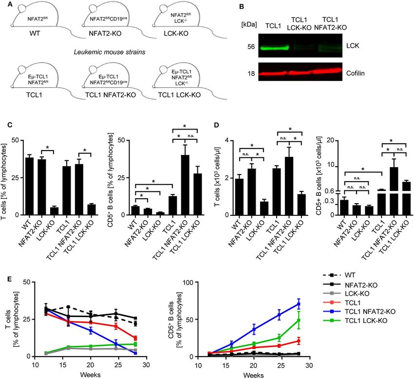
Märklin et al. LCK Deletion Promotes CLL Acceleration
antibodies for LCK (#2752, 1:1,000) and Cofilin (D3F9, 1:2,000) multipotent transcription factor like NFAT2, knockout of a single
(both from Cell signaling) were used for detection. kinase mimics the respective phenotype.
Further characterization of the underlying mechanism
showed that in vivo proliferation, as assessed by BrdU
Immunohistochemistry incorporation was diminished in LCK-deficient T cells in the
Spleens were fixed in 4% formalin, embedded in paraffin
leukemic cohorts (Figures 2A,B). CD5+ B cells of TCL1 NFAT2-
and immunohistochemistry and microscopy was performed as
KO and TCL1 LCK-KO cohorts showed significantly higher
previously described (16).
proliferation rates compared to the TCL1 group, pointing to a
differing role for LCK in T and B cells. The observed B cell
Statistical Analysis expansion was in line with the increased spleen size of TCL1 mice
For statistical analysis GraphPad Prism 8 was used. Mean values with NFAT2 or LCK KO compared to TCL1 mice with intact
and standard error of the mean (SEM) are shown. The 95% expression of these genes (Figure 2C). Immunohistochemistry
confidence level was used and p-values were calculated with analyses in the TCL1 group showed a normal architecture of
an unpaired two-tailed Student’s t-test in the case of normally the spleen with mild expansion of the white pulp, while the red
distributed data. Significance of not normally distributed data pulp showed a subtle lymphocytic infiltrate of B cells (B220),
was calculated with a paired two-tailed Wilcoxon matched-pairs with mature chromatin without nucleoli and low mitotic activity
signed-rank test. An unpaired analysis of variance (ANOVA) was as demonstrated by Ki-67 staining (Figure 2D). In contrast,
used to analyze the differences among group means. Significance spleens in the TCL1 NFAT2-KO group showed mild to severe
of survival data was calculated by using a Log-rank (Mantel- atrophy of the white pulp and infiltration of B cells with mature
Cox) test. Significant p-values p < 0.05 were marked with ∗ . chromatin, conspicuous nucleoli and scant cytoplasm in the red
pulp. Ki-67 staining revealed a higher proliferation rate in the
spleens compared to the TCL1 group. The TCL1 LCK-KO group
RESULTS AND DISCUSSION showed atrophy of the white pulp and expansion of the red
pulp, which was infiltrated by small size B lymphocytes (B220).
To analyze how the genetic loss (KO) of LCK impacts disease Of note, decreased B220 expression in NFAT2 deficient mice
progression, we bred a conditional NFAT2-KO strain expressing was already shown before (16) and B cell identity was shown
the TCL1 oncogene with germline LCK-KO mice (Figure 1A). with additional staining of CD79a (Supplementary Figure 1).
LCK deletion in B cells was confirmed by Western blot analysis The infiltrating small lymphoid cells displayed scant cytoplasm
(Figure 1B). We analyzed a new cohort of the previously and mature chromatin. CD3 staining revealed the atrophy of the
published TCL1 and the TCL1 NFAT2-KO mice in comparison white pulp. In both KO cohorts, the proliferation rate was very
with the newly generated TCL1 LCK-KO mice in a new set high as revealed by Ki-67 immunohistochemistry. Of note, Ki-
of experiments. 67 staining in the TCL1 LCK-KO group was additionally seen
As it was already known that LCK-KO mice show in the red pulp reflecting reactive hematopoiesis. Taken together,
compromised T cell compartments, we first analyzed the T we observed comparable malignant infiltration and disruption of
cells of different KO strains. As expected, LCK deletion leads to the spleen architecture in both knockout cohorts, which provides
a severe reduction of T cells, independent of leukemia induction further evidence for the significance of LCK as disease-modifying
in the TCL1 expressing cohort (Figures 1C,D). NFAT2 deletion factor in CLL.
also leads to decreased CD5+ B cell populations compared to The combined data observed with peripheral blood and
WT mice with intact NFAT2 expression. LCK-KO mice show spleens clearly demonstrate the acceleration of CLL due to the
a further decreased CD5+ B cell subset, indicating an essential genetic loss of LCK kinase. Prompted by our previously published
role for both NFAT2 and LCK in CD5+ B cell development. data (16) we performed long term survival analyses with the
Interestingly, while TCL1 NFAT2-KO mice exhibit an increased LCK deficient mouse cohorts (Figure 2E). Compared to healthy
CD5+ B cell population compared to TCL1 mice as described WT mice LCK-KO control mice showed a significantly reduced
before (16), TCL1 LCK-KO mice show a similarly expanded median life span during the observation period of 350 days, due
CD5+ B cell compartment, comparable to the TCL1 NFAT2-KO to their lack of T cells and the resulting immunodeficiency (239
mice (Figures 1C,D). This suggests that knockout of LCK can, days vs. not reached). For this reason, TCL1 mice with LCK
at least in part, substitute for the deletion NFAT2, thereby deletion were thoroughly monitored and mice which died from
emphasizing the essential role of LCK as a main player in CLL. infection instead of leukemic progression were excluded from the
Analyses over an extended time period of 28 weeks analysis. Comprehensive statistical analyses of TCL1 and TCL1
confirmed these data and again demonstrated that T cell NFAT2-KO mice obtained in the previous study (16) and the
compartments are virtually not affected during leukemia newly generated LCK deficient cohorts revealed no significant
development, but as expected decreased exclusively due to CD5+ difference regarding the median survival of TCL1 mice with
B cell expansion in the different mouse cohorts (Figure 1E, NFAT2 deletion compared to TCL1 mice with LCK deletion (201
Supplementary Table 2). A slightly lower expansion of CD5+ B vs. 163 days), whereas TCL1 mice with intact LCK expression
cells in TCL1 LCK-KO mice compared to TCL1 NFAT2-KO mice survived significantly longer than TCL1 LCK-KO mice (325 vs.
indicates that LCK is not the exclusive player in CLL acceleration. 163 days) (Figure 2F). However, we are aware of the confounded
However, it should be highlighted that in contrast to deleting a effects of the germline LCK-KO, which could be avoided by
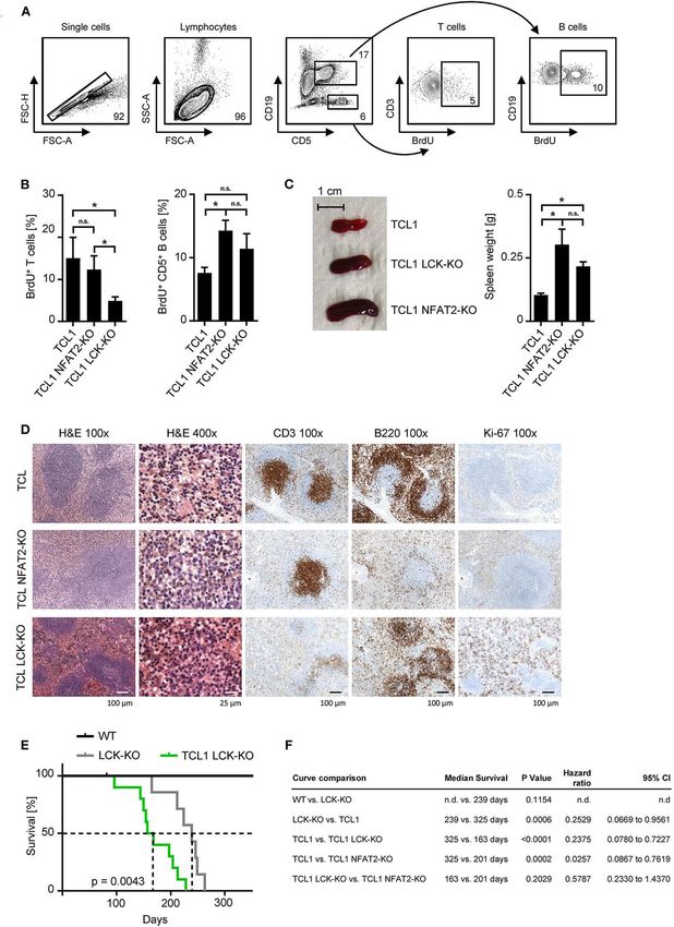
Frontiers in Immunology | www.frontiersin.org 3 September 2020 | Volume 11 | Article 1995Märklin et al. LCK Deletion Promotes CLL Acceleration FIGURE 1 | LCK deletion leads to increased CD5+ B cells in TCL1 leukemic mice. (A) Mouse strain schemes of all mice utilized in this study are depicted. (B) CD19+ splenic B cells from TCL1, TCL1 LCK-KO, and TCL1 NFAT2-KO mice were isolated and LCK protein expression was assessed by western blotting. Cofilin was used as loading control. One exemplary result out of three is shown. (C) Peripheral blood of mice with the indicated genotype at the age of 23 weeks was analyzed for CD3+ T cells (left) and CD5+ B cells (right) by flow cytometry (CD3+ T: WT/NFAT2-KO/TCL1/TCL1 NFAT2-KO n = 5; LCK-KO n = 8; TCL1 LCK-KO n = 10 per group; CD5+ B cells: TCL1 LCK-KO n = 6; LCK-KO n = 7; WT n = 10; NFAT2-KO n = 9; TCL1/TCL1 NFAT2-KO n = 8 per group). (D) Absolut numbers of CD3+ T cells (left) and CD5+ B cells (right) in the peripheral blood of mice with the indicated genotype at the age of 23 weeks (CD3+ T: NFAT2-KO/LCK-KO/TCL1 n = 8; WT/TCL NFAT2-KO n = 9; TCL1 LCK-KO n = 10 per group; CD5+ B cells: WT/NFAT2-KO/LCK-KO/TCL1 LCK-KO n = 7; TCL1 n = 6; TCL1 NFAT2-KO n = 9 per group). (E) Expansion of CD3+ T cells and CD5+ B cells in the peripheral blood of mice with the respective genotypes was assessed at the indicated time points (WT/NFAT2-KO n = 5; LCK-KO n = 9; TCL1/TCL1 LCK-KO n = 11; TCL1 NFAT2-KO n = 10 per group at the beginning of the observation). Multiple comparisons of the different groups at week 28 is shown in Supplementary Table 2. n.s., not significant; *p < 0.05. adoptive transfer experiments in the future. Nevertheless, these is an early kinase proximal to the BCR and is strongly active data implies that loss of LCK leads to decreased survival of in TCL1 mice exhibiting a more indolent disease course (16, leukemic animals, which is most likely due to increased cell 32). To test whether the BCR of TCL1 LCK-KO also displayed proliferation and acceleration of CLL progression. a hyperresponsive phenotype, as we have previously observed Differential B cell receptor (BCR) signaling was hypothesized for TCL1 NFAT2-KO mice, we performed Ca2+ mobilization as potential mechanism underlying these observations, since LCK analyses (Figure 3A). Stimulation of the BCR with an anti-IgM Frontiers in Immunology | www.frontiersin.org 4 September 2020 | Volume 11 | Article 1995
Märklin et al.                                                                                                                LCK Deletion Promotes CLL Acceleration

  FIGURE 2 | LCK deletion leads to acceleration of leukemia and decreased survival of TCL1 transgenic mice. (A) Proliferation of CD3+ T cells and CD5+ B cells in the
  peripheral blood was assessed. Mice were injected with 10 mM BrdU i.p. and peripheral blood cells were harvested after 48 h. Cells were stained with αBrdU and
  measured by flow cytometry. One exemplary result of the TCL1 LCK-KO group with gating strategy is shown. (B) Pooled data of the BrdU analysis for the indicated
  genotypes at the age of 20 weeks (n = 5 per group). (C) Spleen sizes of representative animals of the indicated genotypes at an age of 25 weeks are shown and
  average spleen weight of TCL1 (n = 9), TCL1 NFAT2-KO (n = 8), and TCL1 LCK-KO (n = 6), mice at the age of 25 weeks is shown. (D) H & E staining of
  paraffin-embedded spleen sections of one representative TCL1, TCL1 LCK-KO, and TCL1 NFAT2-KO mouse out of three at an age of 25 weeks (left panels) and
  immunohistochemical staining for CD3, B220 and Ki-67 (right panels). (E) Kaplan-Meier survival plot of WT (n = 7), LCK-KO (n = 7) and TCL1 LCK-KO (n = 10) mice
  is depicted. Median survival is indicated with dashed lines. (F) Log-rank test of the Kaplan-Meier analysis of WT, LCK-KO and TCL1 LCK-KO mice in the present study
  compared to the previous published TCL1 and TCL1 NFAT2-KO cohorts (16) with median survival, hazard ratio and the 95% confidence interval (CI) is shown. (n.d.,
  not determined). n.s., not significant; *p < 0.05.

Frontiers in Immunology | www.frontiersin.org                                     5                                         September 2020 | Volume 11 | Article 1995
Märklin et al.                                                                                                                LCK Deletion Promotes CLL Acceleration

  FIGURE 3 | Loss of LCK restores BCR signaling in TCL1 transgenic mice. (A) Ca2+ flux analysis of splenic CD5+ B cells from TCL1, TCL1 NFAT2-KO, and TCL1
  LCK-KO mice at an age of ∼18 weeks after stimulation with 10 µg/mL αIgM is depicted. Ionomycin (1 mM) was added as positive control. One representative result of
  three experiments is shown. (B,C) After staining of splenocytes (∼20 weeks old mice) for CD19 and CD5, cells were stimulated with 10 µg/mL αIgM for the indicated
  time points and BCR activation was assessed by intracellular staining of SYK/P-SYK(Y525/526) and ERK1/2/P-ERK1/2(T202/Y204) in CD5+ B cells with flow
  cytometry. (B) One representative result out of four independent experiments is shown. (C) Pooled result of P-SYK+ and P-ERK1/2+ CD5+ B cells at the indicated
  time points of TCL1, TCL-NFAT2-KO and TCL LCK-KO mice (TCL1 n = 6, TCL1 NFAT2-KO/TCL1 LCK-KO n = 5) is shown. (D) IgM surface expression on B cells
  from TCL1, TCL1 NFAT2-KO, and TCL1 LCK-KO mice was determined by flow cytometry (TCL1/TCL1 LCK-KO n = 8, TCL1 NFAT2-KO n = 7 per group, ∼18 weeks
  old). Mean expression levels of IgM were normalized to TCL1 mice. One representative result of four experiments is shown. (E) Relative gene expression in splenic B
  cells of Prdm1 in TCL1 NFAT2-KO and TCL1 LCK-KO was normalized to mice normalized TCL1 mice. Relative gene expression was calculated by ratio of Prdm1 to
  Actin expression from (TCL1/TCL1 NFAT2-KO n = 5, TCL1 LCK-KO n = 4 per group, ∼18 weeks old). n.s., not significant; *p < 0.05.

Frontiers in Immunology | www.frontiersin.org                                     6                                         September 2020 | Volume 11 | Article 1995
Märklin et al.                                                                                                                LCK Deletion Promotes CLL Acceleration

antibody revealed a significantly decreased Ca2+ flux capacity                        ETHICS STATEMENT
of TCL1 mice, which were previously characterized to exhibit
an anergic phenotype (16), whereas TCL1 mice lacking LCK                              The animal study was reviewed and approved by Institutional
expression showed increased Ca2+ mobilization comparable to                           Animal Care and Use Committee of the University of Tübingen.
TCL1 NFAT2-KO mice. To further elucidate the downstream                               Written informed consent was obtained from the owners for the
signaling of the BCR, SYK and ERK1/2 activation were analyzed                         participation of their animals in this study.
in splenocytes by phosphorylation flow cytometry. Stimulation
with an anti-IgM antibody showed induction of P-SYK and                               AUTHOR CONTRIBUTIONS
P-ERK1/2 in CD5+ B cells of TCL1 NFAT2-KO and TCL1
LCK-KO mice over time, while total protein levels were not                            MM, AF, JH, CT, SW, LQ-M, and MMü designed and
affected. In contrast, TCL1 mice exhibit a significantly less                         performed the experiments. MM, JH, JK, LQ-M. and MMü
responsive BCR which results in just very weak induction of                           analyzed data and wrote the manuscript. MRM, HS, and
P-SYK and P-ERK1/2 (Figures 3B,C). Of note, specificity of                            H-GK designed research, analyzed data, and provided important
this assay could be shown by the absence of any P-ERK1/2                              advice. All authors contributed to the article and approved the
induction in T cells contained in the splenocyte population                           submitted version.
(Supplementary Figure 2).
    Since it was previously described that sIgM levels directly                       FUNDING
correspond with BCR responsiveness (12, 35–37), we analyzed
IgM surface expression levels (Figure 3D). Here, we could                             This work was supported by the DFG grant MU 3340/1-1 and
observe a similar upregulation of sIgM in LCK deficient                               MU3340/1-2 (MRM) and the Deutsche Krebshilfe grant 111134
TCL1 B cells as it has been recently documented for                                   (HS, MRM) and 70113496 (MM).
TCL1 NFAT2-KO B cells, thus supporting the concept of
hyperresponsive BCRs in aggressive CLL. Anergic CLL cells                             ACKNOWLEDGMENTS
have also been characterized to exhibit depression of Prdm1
(Blimp-1) expression (16, 38). In LCK deficient TCL1 mice,                            We thank Carlo M. Croce for providing the Eµ-TCL1 mice,
we observed a substantial increase of Prdm1 gene expression                           K. Mark Ansel for providing the conditional NFAT2 knockout
compared to TCL1 mice (Figure 3E), which supports our data                            mice and L. Simeoni for providing the LCK KO mice. We
on BCR hyperresponsiveness and aggressive disease acceleration                        thank Alexandra Poljak for excellent technical assistance and the
caused by LCK knockout.                                                               Flow Cytometry Core Facility Berg of the University Hospital
    In summary, our data clearly support the hypothesis that LCK                      Tübingen for their excellent technical support. We thank Irene
is a critical regulator of the anergic phenotype in CLL and that                      Gonzalez-Menendez from the Institute of Pathology Tübingen
its loss leads to hyperresponsive BCRs and disease acceleration in                    for performing immunohistochemistry analyses of paraffin-
the Eµ-TCL1 mouse model.                                                              embedded spleens.

                                                                                      SUPPLEMENTARY MATERIAL
DATA AVAILABILITY STATEMENT
                                                                                      The Supplementary Material for this article can be found
The raw data supporting the conclusions of this article will be                       online at: https://www.frontiersin.org/articles/10.3389/fimmu.
made available by the authors, without undue reservation.                             2020.01995/full#supplementary-material

REFERENCES                                                                             5. Jain N, Keating M, Thompson P, Ferrajoli A, Burger J, Borthakur G, et al.
                                                                                          Ibrutinib and venetoclax for first-line treatment of CLL. N Engl J Med. (2019)
 1. Goede V, Fischer K, Busch R, Engelke A, Eichhorst B, Wendtner                         380:2095–103. doi: 10.1056/NEJMoa1900574
    CM, et al. Obinutuzumab plus chlorambucil in patients with CLL                     6. Swords R, Bruzzi J, Giles F. Recent advances in the diagnosis and therapy of
    and coexisting conditions. New Engl J Med. (2014) 370:1101–                           Richter’s syndrome. Med Oncol. (2007) 24:17–32. doi: 10.1007/BF02685899
    10. doi: 10.1056/NEJMoa1313984                                                     7. Hallek M, Cheson BD, Catovsky D, Caligaris-Cappio F, Dighiero G, Dohner
 2. Hallek M, Fischer K, Fingerle-Rowson G, Fink AM, Busch R, Mayer                       H, et al. iwCLL guidelines for diagnosis, indications for treatment, response
    J, et al. Addition of rituximab to fludarabine and cyclophosphamide in                assessment, and supportive management of CLL. Blood. (2018) 131:2745–
    patients with chronic lymphocytic leukaemia: a randomised, open-label,                60. doi: 10.1182/blood-2017-09-806398
    phase 3 trial. Lancet. (2010) 376:1164–74. doi: 10.1016/S0140-6736(10)6            8. Stevenson FK, Forconi F, Packham G. The meaning and relevance of B-
    1381-5                                                                                cell receptor structure and function in chronic lymphocytic leukemia. Semin
 3. Byrd JC, Furman RR, Coutre SE, Flinn IW, Burger JA, Blum KA,                          Hematol. (2014) 51:158–67. doi: 10.1053/j.seminhematol.2014.05.003
    et al. Targeting BTK with ibrutinib in relapsed chronic lymphocytic                9. Packham G, Krysov S, Allen A, Savelyeva N, Steele AJ, Forconi
    leukemia. New Engl J Med. (2013) 369:32–42. doi: 10.1056/NEJMoa1                      F, et al. The outcome of B-cell receptor signaling in chronic
    215637                                                                                lymphocytic leukemia: proliferation or anergy. Haematologica. (2014)
 4. Furman RR, Sharman JP, Coutre SE, Cheson BD, Pagel JM, Hillmen P, et al.              99:1138–48. doi: 10.3324/haematol.2013.098384
    Idelalisib and rituximab in relapsed chronic lymphocytic leukemia. New Engl       10. Mockridge CI, Potter KN, Wheatley I, Neville LA, Packham G, Stevenson
    J Med. (2014) 370:997–1007. doi: 10.1056/NEJMoa1315226                                FK. Reversible anergy of sIgM-mediated signaling in the two subsets

Frontiers in Immunology | www.frontiersin.org                                     7                                         September 2020 | Volume 11 | Article 1995
Märklin et al. LCK Deletion Promotes CLL Acceleration
of CLL defined by VH-gene mutational status. Blood. (2007) 109:4424– cells and in chronic lymphocytic leukemia B cells. Blood. (1998) 91:3390–
31. doi: 10.1182/blood-2006-11-056648 6. doi: 10.1182/blood.V91.9.3390.3390_3390_3396
11. Duhren-von Minden M, Ubelhart R, Schneider D, Wossning T, Bach 27. Dal Porto JM, Burke K, Cambier JC. Regulation of BCR signal transduction
MP, Buchner M, et al. Chronic lymphocytic leukaemia is driven by in B-1 cells requires the expression of the Src family kinase Lck. Immunity.
antigen-independent cell-autonomous signalling. Nature. (2012) 489:309– (2004) 21:443–53. doi: 10.1016/j.immuni.2004.07.018
12. doi: 10.1038/nature11309 28. Ulivieri C, Valensin S, Majolini MB, Matthews RJ, Baldari CT. Normal B-1
12. Apollonio B, Scielzo C, Bertilaccio MT, Ten Hacken E, Scarfo L, Ranghetti P, cell development but defective BCR signaling in Lck-/- mice. Eur J Immunol.
et al. Targeting B-cell anergy in chronic lymphocytic leukemia. Blood. (2013) (2003) 33:441–5. doi: 10.1002/immu.200310019
121:3879–88, S1–8. doi: 10.1182/blood-2012-12-474718 29. Theofani E, Alexis S, Costeas P, Andriopoulos C, Feleskoura G, Zikos
13. Muzio M, Apollonio B, Scielzo C, Frenquelli M, Vandoni I, Boussiotis V, P, et al. Ectopic Lck expression in CLL demarcates intratumoral
et al. Constitutive activation of distinct BCR-signaling pathways in a subset subpopulations with aberrant B-cell receptor signaling. Blood Adv. (2018)
of CLL patients: a molecular signature of anergy. Blood. (2008) 112:188– 2:877–82. doi: 10.1182/bloodadvances.2017015321
95. doi: 10.1182/blood-2007-09-111344 30. Paterson JC, Tedoldi S, Craxton A, Jones M, Hansmann ML, Collins G,
14. Muller MR, Rao A. NFAT, immunity and cancer: a transcription factor comes et al. The differential expression of LCK and BAFF-receptor and their role
of age. Nat Rev Immunol. (2010) 10:645–56. doi: 10.1038/nri2818 in apoptosis in human lymphomas. Haematologica. (2006) 91:772–80.
15. Le Roy C, Deglesne PA, Chevallier N, Beitar T, Eclache V, Quettier 31. Till KJ, Allen JC, Talab F, Lin K, Allsup D, Cawkwell L, et al.
M, et al. The degree of BCR and NFAT activation predicts clinical Lck is a relevant target in chronic lymphocytic leukaemia cells whose
outcomes in chronic lymphocytic leukemia. Blood. (2012) 120:356– expression variance is unrelated to disease outcome. Sci Rep. (2017)
65. doi: 10.1182/blood-2011-12-397158 7:16784. doi: 10.1038/s41598-017-17021-w
16. Märklin M, Heitmann JS, Fuchs AR, Truckenmüller FM, Gutknecht 32. Talab F, Allen JC, Thompson V, Lin K, Slupsky JR. LCK is an important
M, Bugl S, et al. NFAT2 is a critical regulator of the anergic mediator of B-cell receptor signaling in chronic lymphocytic leukemia cells.
phenotype in chronic lymphocytic leukaemia. Nat Commun. (2017) Mol Cancer Res. (2013) 11:541–54. doi: 10.1158/1541-7786.MCR-12-0415-T
8:755. doi: 10.1038/s41467-017-00830-y 33. Bichi R, Shinton SA, Martin ES, Koval A, Calin GA, Cesari R,
17. Holler C, Pinon JD, Denk U, Heyder C, Hofbauer S, Greil R, et al. et al. Human chronic lymphocytic leukemia modeled in mouse by
PKCbeta is essential for the development of chronic lymphocytic leukemia targeted TCL1 expression. Proc Natl Acad Sci USA. (2002) 99:6955–
in the TCL1 transgenic mouse model: validation of PKCbeta as a 60. doi: 10.1073/pnas.102181599
therapeutic target in chronic lymphocytic leukemia. Blood. (2009) 113:2791– 34. Kilkenny C, Browne WJ, Cuthill IC, Emerson M, Altman DG. Improving
4. doi: 10.1182/blood-2008-06-160713 bioscience research reporting: the ARRIVE guidelines for reporting animal
18. Simonetti G, Bertilaccio MT, Ghia P, Klein U. Mouse models in the study research. PLoS Biol. (2010) 8:e1000412. doi: 10.1371/journal.pbio.1000412
of chronic lymphocytic leukemia pathogenesis and therapy. Blood. (2014) 35. Goodnow CC, Crosbie J, Jorgensen H, Brink RA, Basten A. Induction of
124:1010–9. doi: 10.1182/blood-2014-05-577122 self-tolerance in mature peripheral B lymphocytes. Nature. (1989) 342:385–
19. Zaborsky N, Gassner FJ, Hopner JP, Schubert M, Hebenstreit D, Stark R, 91. doi: 10.1038/342385a0
et al. Exome sequencing of the TCL1 mouse model for CLL reveals genetic 36. Merrell KT, Benschop RJ, Gauld SB, Aviszus K, Decote-Ricardo D,
heterogeneity and dynamics during disease development. Leukemia. (2019) Wysocki LJ, et al. Identification of anergic B cells within a wild-
33:957–68. doi: 10.1038/s41375-018-0260-4 type repertoire. Immunity. (2006) 25:953–62. doi: 10.1016/j.immuni.200
20. Müller DJ, Wirths S, Fuchs AR, Märklin M, Heitmann JS, Sturm M, 6.10.017
et al. Loss of NFAT2 expression results in the acceleration of clonal 37. Quach TD, Manjarrez-Orduno N, Adlowitz DG, Silver L, Yang H, Wei C, et al.
evolution in chronic lymphocytic leukemia. J Leukoc Biol. (2019) 105:531– Anergic responses characterize a large fraction of human autoreactive naive
8. doi: 10.1002/JLB.2AB0218-076RR B cells expressing low levels of surface IgM. J Immunol. (2011) 186:4640–
21. Woyach JA. Survival of the weak (signalers): anergy in CLL. Blood. (2013) 8. doi: 10.4049/jimmunol.1001946
121:3781–3. doi: 10.1182/blood-2013-03-489849 38. Duckworth A, Glenn M, Slupsky JR, Packham G, Kalakonda
22. Fuchs AR, Märklin M, Heitmann JS, Futterknecht S, Haap M, Wirths S, et al. N. Variable induction of PRDM1 and differentiation in chronic
A chromatin immunoprecipitation assay to identify novel NFAT2 target genes lymphocytic leukemia is associated with anergy. Blood. (2014)
in chronic lymphocytic leukemia. J Vis Exp. (2018). doi: 10.3791/58270 123:3277–85. doi: 10.1182/blood-2013-11-539049
23. Marth JD, Peet R, Krebs EG, Perlmutter RM. A lymphocyte-
specific protein-tyrosine kinase gene is rearranged and overexpressed Conflict of Interest: The authors declare that the research was conducted in the
in the murine T cell lymphoma LSTRA. Cell. (1985) 43(2 Pt absence of any commercial or financial relationships that could be construed as a
1):393–404. doi: 10.1016/0092-8674(85)90169-2 potential conflict of interest.
24. Von Knethen, Abts H, Kube D, Diehl V, Tesch H. Expression
of p56lck in B-cell neoplasias. Leuk Lymphoma. (1997) 26:551– Copyright © 2020 Märklin, Fuchs, Tandler, Heitmann, Salih, Kauer, Quintanilla-
62. doi: 10.3109/10428199709050891 Martinez, Wirths, Kopp and Müller. This is an open-access article distributed
25. Abts H, Jucker M, Diehl V, Tesch H. Human chronic lymphocytic leukemia under the terms of the Creative Commons Attribution License (CC BY). The use,
cells regularly express mRNAs of the protooncogenes lck and c-fgr. Leuk Res. distribution or reproduction in other forums is permitted, provided the original
(1991) 15:987–97. doi: 10.1016/0145-2126(91)90103-Z author(s) and the copyright owner(s) are credited and that the original publication
26. Majolini MB, D’Elios MM, Galieni P, Boncristiano M, Lauria F, Del Prete in this journal is cited, in accordance with accepted academic practice. No use,
G, et al. Expression of the T-cell-specific tyrosine kinase Lck in normal B-1 distribution or reproduction is permitted which does not comply with these terms.
Frontiers in Immunology | www.frontiersin.org 8 September 2020 | Volume 11 | Article 1995You can also read