Initial Single-Site Surgical Experience with SMILE: A Comparison of Results to FDA SMILE, and the Earliest and Latest Generation of LASIK
←
→
Page content transcription
If your browser does not render page correctly, please read the page content below
Ophthalmol Ther
https://doi.org/10.1007/s40123-018-0137-7

 ORIGINAL RESEARCH

Initial Single-Site Surgical Experience with SMILE:
A Comparison of Results to FDA SMILE,
and the Earliest and Latest Generation of LASIK
Majid Moshirfar . Michael S. Murri . Tirth J. Shah . Steven H. Linn .
Yasmyne Ronquillo . Orry C. Birdsong . Phillips C. Hoopes Jr.

Received: May 13, 2018
Ó The Author(s) 2018

ABSTRACT                                                   postoperative uncorrected distance visual acuity
                                                           (UDVA), corrected distance visual acuity
Introduction: The primary objective was to                 (CDVA), manifest sphere, manifest cylinder,
show our initial surgical single-site experience           intraoperative complications, and preoperative
with small incision lenticule extraction (SMILE)           and postoperative visual symptoms were col-
after the official enrollment in March 2017 fol-           lected. We compared our findings to the results
lowing Food and Drug Administration (FDA)                  from the FDA SMILE study, and to the three
approval for simple myopia in late 2016 in the             earliest (1999–2000) and three of the most
United States and, subsequently, compare our               updated (2013–2016) platforms for LASIK.
results to the earliest and most advanced gen-             Results: The cumulative UDVA was 20/20 and
eration of excimer platforms for laser-assisted            20/40 or better in 74% and 100% of patients,
in situ keratomileusis (LASIK) surgery.                    respectively. The intended target refraction was
Methods: This was a retrospective single-site              within ± 0.5 and ± 1.00 D in 80% and 93% of
study of 68 eyes from 35 patients who had                  cases, respectively. The prevalence of dry eyes
SMILE surgery. The patients’ preoperative and              decreased by nearly half from 1-week to the
                                                           6-month postoperative interval. Patients noted
Enhanced digital features To view enhanced digital         improvement in glare (17%), halos (17%), fluctu-
features for this article go to https://doi.org/10.6084/   ation (25%), and depth perception (8%) at the
m9.figshare.6572846.                                       6-month interval compared to preoperative levels.
                                                           Conclusions: This study’s findings are consis-
M. Moshirfar (&)  M. S. Murri                             tent with current SMILE reports. Notably, the
Department of Ophthalmology and Visual Sciences,
John A. Moran Eye Center, University of Utah
                                                           results are superior to the earliest generation of
School of Medicine, Salt Lake City, UT, USA                LASIK, however inferior to the latest excimer
e-mail: cornea2020@me.com                                  platforms. SMILE does meet the efficacy and
                                                           safety criteria met by FDA; however, there is a
M. Moshirfar
Utah Lions Eye Bank, Murray, UT, USA                       definite need for further improvement to reach
                                                           the superior refractive outcomes produced by
M. Moshirfar  S. H. Linn  Y. Ronquillo                  the latest generation of LASIK platforms.
O. C. Birdsong  P. C. Hoopes Jr.
HDR Research Center, Hoopes Vision, Draper, UT,
USA                                                        Keywords: Alcon Contoura; SMILE
                                                           astigmatism; SMILE dry eyes; SMILE
T. J. Shah
University of Arizona College of Medicine, Phoenix,        improvements; SMILE vs. LASIK; SMILE
Phoenix, AZ, USA
Ophthalmol Ther

symptoms; Visumax femtosecond laser; VISX            UT, USA. The study included 68 eyes from 35
iDesign; Wavefront                                   patients. All procedures performed in studies
                                                     involving human participants were in accor-
                                                     dance with the ethical standards of the institu-
INTRODUCTION                                         tional and/or national research committee and
                                                     with the 1964 Helsinki declaration and its later
Lenticular extraction of the corneal stroma was      amendments or comparable ethical standards.
first conceived in the late 1990s with the cre-      Informed consent was obtained from all indi-
ation of the lenticule with a picosecond laser       vidual participants included in the study. This
[1, 2] followed by its manual extraction. Later,     study was approved by the Hoopes, Durrie,
Femtosecond Lenticule Extraction (FLEx) was          Rivera Research Review Board. Before providing
innovated, with the purpose of treating patients     written informed consent, all patients were
with severe myopia. With the advent of the           given detailed information regarding each
Visumax femtosecond laser (Carl Zeiss Meditec,       treatment. The preoperative manifest sphere
Jena, Germany), and the refinement of the            and cylinder of our preoperative patient popu-
technique to involve only a 2–3 mm incision,         lation ranged from - 7.50 D to - 2.75 D and
small incision lenticule extraction (SMILE)          - 0.75 D to 0.00 D, respectively. Patients had
became an increasingly viable procedure in           complete refractive screening and normal fun-
mainstream refractive surgery [3, 4]. FDA regis-     doscopic exam prior to enrollment in the study.
tered clinical trials began in 2011 and official     All candidates were healthy and appropriate
trials for SMILE approval began in the United        refractive surgical candidates for SMILE. Any
States in 2012.                                      patient with cylinder greater than 0.75 D,
    Since that time, numerous international          abnormal corneal topographic findings (i.e.,
studies originating in France, Singapore, Egypt,     keratoconus, pellucid marginal degeneration,
China, Spain, Japan, Germany, Denmark, South         etc.), history of herpes zoster ophthalmicus or
Korea, Turkey, India, and the United Kingdom         herpes simplex keratitis, significant dry eye
have reported on SMILE visual outcomes [4–22].       syndrome refractory to treatment or dry eye
However, with FDA approval in September of           disease, history of diabetes or connective tissue
2016, and official enrollment of patients in the     disease, pregnant or lactating were excluded
United States in early 2017, there is a paucity of   from our study.
data from patients in the United States. Our             SMILE was performed using the Visumax
objective was to report the 6-month efficacy,        500 kHz femtosecond laser (Carl Zeiss Meditec,
safety, predictability, and stability of patients    Jena, Germany) by two surgeons (MM, PCH).
undergoing SMILE surgery from a single surgical      Although the surgeons had no prior SMILE
center. Because there is limited literature          experience, they have each had LASIK experi-
regarding the visual quality after SMILE, our        ence of over 15 years. Follow up appointments
second aim was to collect data concerning            occurred at 1 day, 1 week, 1 month, 3 months,
patient symptoms (i.e., glare, dryness, halos,       and 6 months. At the 1-week interval, we lost
etc.) after surgery. Lastly, given that SMILE is     one patient (two eyes) to follow-up. We had 66
still a relatively new surgery in the United         eyes and 41 eyes of follow-up data at the
States, we compared our findings to those from       3-month and 6-month interval, respectively.
the earliest excimer platforms for LASIK             Some patients have not had their 6-month fol-
(1999–2000) and to the most updated excimer          low-up, thus explaining the difference between
platforms for LASIK (2013–2016).                     the 3-month and 6-month sample size. A few of
                                                     our patients had their 1-year follow-up; how-
                                                     ever, because this was a small subset of our
METHODS                                              cohort (* 10 eyes), we decided to omit these
                                                     data from our study. Data collected included
This retrospective study was conducted at the        patients’ preoperative and postoperative
HDR research center, Hoopes Vision, Draper,          uncorrected distance visual acuity (UDVA),
Ophthalmol Ther

corrected distance visual acuity (CDVA), mani-       6 months), we assessed for the presence or
fest sphere, manifest cylinder, and any compli-      absence of any dry eye symptoms via a physi-
cations during or after surgery. LogMAR visual       cian-interview for a total of 22 patients. For
acuity was calculated with count fingers (CF) set    example, prior to the surgery, each patient was
at logMAR 2.0 per recommendation by Holla-           asked whether or not they have any dry eye
day [23]. Three eyes were excluded from UDVA         symptoms with a ‘‘yes or no’’ response. At the
analysis because their target refraction was         6-month postoperative interval, the same
adjusted for monovision, but were included in        question was asked to the patient with a ‘‘yes or
all other visual and refractive analyses. Surgical   no’’ response. The specific severity of the dry
parameters were set with a cap thickness of          eyes was not obtained. For further analysis, we
120 lm and cap diameter of 7.5 mm. Hinge             then performed corneal staining to assess for
placement was superior with hinge angle at 50        superficial punctate keratitis (SPK) for the same
degrees and side-cut angle of 90 degrees. Len-       22 patients. We assessed SPK via a scoring scale:
ticule diameter was 6.5 mm. Spot separation          0 indicating no staining, 1 ? indicating mild
was 3 lm for the lenticule, 2.5 lm for the len-      staining, 2 ? indicating moderate staining, and
ticule side-cut, 3 lm for the flap, and 2 lm for     3 ? indicating significant staining of cornea.
the flap side cut. Laser-bed energy was set at 145      At the 6-month postoperative visit, a total of
nJ.                                                  24 patients were asked via a physician-interview
    After obtaining our standard refractive          whether their visual symptoms concerning
results, the percent of individuals within 20/20     glares, halos, starbursts, double vision, fluctua-
UDVA,       20/40       UDVA, ± 0.5 D       MRSE,    tion, and depth perception were ‘‘better, worse,
and ± 1.0 D MRSE were compared to those              or not changed’’ from the preoperative levels.
published by the recent FDA SMILE study [24].           We assessed differences in visual outcomes
We also compared our findings to the first           between the first 12 patients and last 12 patients
(1999–2000) and latest generation (2013–2016)        to receive SMILE surgery. The postoperative
of excimer LASIK platforms. The following early      visual outcomes that were assessed include:
generation LASIK platforms were used for com-        manifest sphere, cylinder, and spherical equiv-
parisons: SVS Apex Plus Excimer Laser Worksa-        alence. Lastly, in order to appropriately pool
tion by Summit Technology, Inc. (Waltham,            data from both surgeons, we ensured there was
MA, USA; PMA: P930034/S13) [25], Nidek EC-           no statistically significant difference in the
5000 Excimer Laser System by Nidek Tech-             aforementioned postoperative visual outcomes
nologies, Inc. (Pasadena, CA, USA; PMA:              between surgeons.
P970053/S002) [26], and VISX STAR S2 Excimer
Laser System by AMO Manufacturing (Scotts-           Statistical Analysis
dale, AZ, USA; PMA: P990010) [27]. We chose
these specific earlier excimer models because        Data was collected and analyzed using Data-
they represented the most popular platforms at       graph-Med (Wendelstein, Germany) and
that time point in the United States. The fol-       Microsoft Excel software. Astigmatic data was
lowing newest LASIK platforms were used for          calculated using Dr. Peyman’s online Astigmatic
comparisons: STAR S4 Excimer Laser System            Vector Analyzer [31]. Standard refractive graphs
with iDesign Advanced WaveScan Studio Sys-           were created using Standard Graphs for
tem by VISX, Inc. (Milpitas, CA, USA; PMA:           Reporting Refractive Surgery (London Clinic)
P930016/S045) [28], Nidek EC-5000 Excimer            [32]. The postoperative sphere, cylinder, and
Laser System by Nidek, Inc. (Gamagori, Aichi,        spherical equivalence of the first 12 patients was
Japan; PMA: P970053/S011) [29], and Allegretto       compared to the last 12 patients using a two-
Wave Eye-Q Excimer Laser by Alcon Laborato-          pair Wilcoxon signed-rank test. The same test
ries, Inc. (Fort Worth, TX, USA; PMA:                was used to compare results between different
020050/S12) [30].                                    surgeons. A p value less than 0.05 was consid-
    At the preoperative appointment and each         ered statistically significant.
follow-up visit (1 week, 1 month, 3 months, and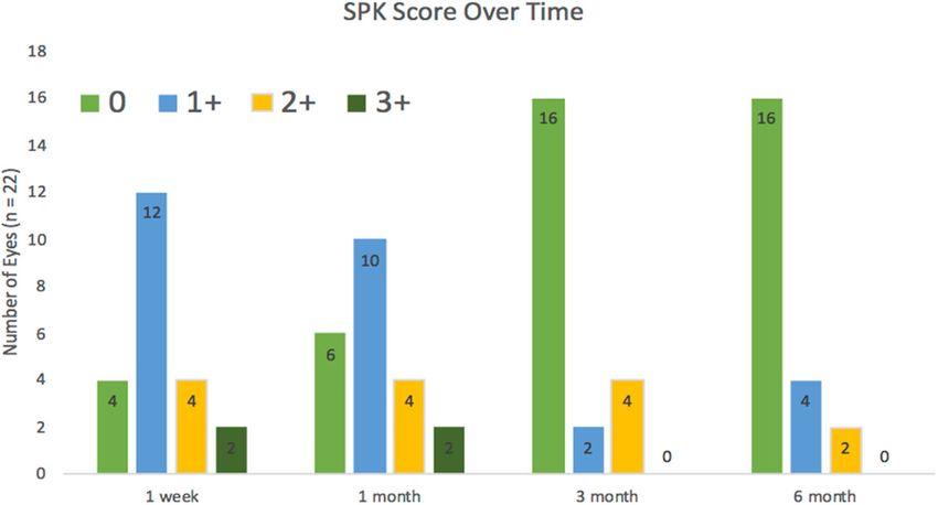
Ophthalmol Ther

RESULTS                                                      patients had no change and no eye had a
                                                             decrease in CDVA. The safety index was 1.12.
The average patient age was 31 years with a                     With regards to predictability, the intended
standard deviation of 7 and a range from 23 to               target refraction was within ± 0.5 and ± 1.00 D
49 years of age. At 6 months, we had 21 right                in 80% and 93% of cases, respectively. We
eyes and 20 left eyes from 63% female and 37%                noticed there was an induction of astigmatism.
male patients. Average logMAR UDVA                           For instance, refractive astigmatism increased in
6 months after surgery was 0.05 ± 0.13 and                   the 0.51–0.75 D surgical subgroup (2% preop-
spherical equivalent (SE) 6 months after surgery             erative to 17% postoperative) and 0.76–1.00 D
was - 0.25 ± 0.53 D (Table 1). The 6-month                   surgical subgroup (0% preoperative to 7.3%
UDVA, manifest sphere, manifest SE, and CDVA                 postoperative). We also noticed no pattern in
were all significantly improved (p \ 0.0001;                 the refractive astigmatism angle of error. About
Table 1). The manifest cylinder significantly                half the eyes had a positive angle of error and
increased from - 0.23 D at preoperative to                   vice versa.
- 0.49 D at 6 months postoperative (p \ 0.0001;                 Lastly, concerning stability, 7% of patients
Table 1). The standard refractive surgery graphs             had greater than 0.5 D change in refraction over
are shown in Fig. 1.                                         the postoperative period from 1 month to
   With regards to efficacy, cumulative UDVA                 6 months. Overall, the greatest improvement in
was 20/20, 20/25, or 20/40 or better in 74%,                 visual acuity and refraction was seen in the first
87%, and 100% of patients, respectively. The                 month postoperatively without significant
UDVA at 6 months was the same or better than                 changes in the 3-month and 6-month follow-
pre-operative CDVA in 74% of cases and within                up.
one line of CDVA in 87% of cases. The efficacy
index was 0.89. At 6 months, 37% of patients                 Results vs. FDA SMILE Data
gained one Snellen line of CDVA.
   Concerning safety, when comparing pre-op-                 Visual acuity outcomes in our study were UDVA
erative CDVA to postoperative CDVA, 63% of                   of 20/20 and 20/25 or better in 74% and 83% of
                                                             patients and 20/40 or better in 98% of patients.

Table 1 Describes changes in standard visual outcomes from preoperative to the 1-month, 3-month, and 6-month interval
                  Pre-operative (n = 68)    1-month                  3-month                  6-month
                                            Postoperative            Postoperative            Postoperative
                                            (n = 66)                 (n = 62)                 (n = 41)
UDVA              1.46 ± 0.33 (0.88 to      0.09 ± 0.16 (- 0.10 to 0.05 ± 0.14 (- 0.10 to 0.05 ± 0.13 (- 0.12 to
 (logMAR)           2.00)                     0.80)                  0.54)                  0.48)a
CDVA              0                         - 0.01 ± 0.06 (- 0.10 - 0.02 ± 0.05 (- 0.10 - 0.05 ± 0.06 (- 0.12
 (logMAR)                                    to 0.18)              to 0.18)              to 0.00)a
Manifest SE (D) - 5.16 ± 1.34 (- 7.50       - 0.19 ± 0.53 (- 1.50 - 0.21 ± 0.46 (- 1.75 - 0.26 ± 0.53 (- 1.63
                 to - 2.75)                  to 1.38)              to 1.13)              to 1.25)a
Manifest sphere   - 5.05 ± 1.32 (- 7.50     0.01 ± 0.54 (- 1.25 to 0.00 ± 0.48 (- 1.50 to - 0.01 ± 0.58 (- 1.50
 (D)               to - 2.75)                 1.75)                  1.75)                 to 1.75)a
Manifest          - 0.23 ± 0.22 (- 0.75     - 0.41 ± 0.44 (- 2.50 - 0.42 ± 0.36 (- 1.50 - 0.49 ± 0.30 (- 1.50
 cylinder (D)      to 0.00)                  to 0.25)              to 0.00)              to 0.00)a
The data represent the mean ± standard deviation (range)
a
  Indicates statistically significant findings (p \ 0.05)
Ophthalmol Ther Fig. 1 Standard refractive surgery graphs These can be compared to recent FDA pre-mar- population, patients were within ± 0.5 ket approval (PMA) data for SMILE in the Uni- and ± 1.00 D of target refraction in 80% and ted States [24] in which 87.5% and 99.7% of 93% of cases, respectively. FDA approval data patients achieved UDVA of 20/20 and 20/40 or reported values of 93% within ± 0.5 D and better, respectively (Table 2). In our study 98.5% within ± 1.00 D.
Ophthalmol Ther

Table 2 A comparison of our SMILE results to those found by the FDA, in addition to the early generation (1999–2000)
and most updated excimer platforms (2013–2016)
                          % within 20/20 UDVA % within 20/40 UDVA % within – 0.5 D % within 1 1.0 D
Our SMILE results         74                         98                      80                   93
FDA SMILE results         87.5                       99.7                    93                   98.5
Current Excimer Laser Platform
 VISX iDesign (2016)      91.7                     100                       69                   93
 Alcon Contoura (2013) 88.9                          98.7                    95                  100
 Nidek EC-5000 (2013) 88.7                         100                       91                   99
Early Excimer Laser Platform
 SVS Apex Plus (1998)     46.9                       92.1                    61                   84
 Nidek EC-5000 (2000) 47.4                           84.4                    67                   90
 VISX STAR S2 (1999) 54.1                            95.4                    72.5                 90.6

Results vs. Early Generation LASIK                          Reported Visual Symptoms
Platforms
                                                            Our findings showed a decrease in the percent-
The results showed that overall our SMILE                   age of dry eyes over time. Our findings showed
results had significantly more percentage of                100%, 88%, 45%, and 45% of reported dry eyes
eyes within 20/20 UDVA, 20/40 UDVA, ± 0.5                   at the 1-week, 1-month, 3-month, and 6-month
D, and ± 1.0 D than those recorded by the early             intervals, respectively (n = 22 eyes). Notably,
platforms. An average of 51% and 91% of                     preoperative levels of dry eyes were 45%, thus
patients had UDVA under 20/20 and 20/40,                    indicating that at 3 months, the prevalence of
respectively (Fig. 2) compared to 74% and 98%               dry eyes returned to baseline. To correlate
of our SMILE patients, respectively.                        reported dry eyes with exam findings, patients
                                                            had decreased 1? , 2? , and 3? SPK staining
Results vs. Latest Generation LASIK                         over time. The number of eyes without SPK
Platforms                                                   increased from four in the first week to 16 at the
                                                            6-month interval (n = 22 eyes; Fig. 3). Addi-
This study reported 20/20 UDVA or better for                tionally, we used a physician-centered ques-
91.7%, 88.9%, and 88.7% of patients and 20/40               tionnaire to assess other visual symptoms at the
vision or better for 100%, 98.7%, and 100% of               6-month postoperative interval to assess whe-
patients in the Visx iDesign, Alcon Contoura,               ther these symptoms have ‘‘improved, wors-
and Nidek EC-5000, respectively. In addition,               ened, or not changed’’ from preoperative levels
achieved refraction was within ± 0.5 and ±                  (Fig. 4). We found that symptoms of glare
 1.00 D of target refraction in 69% and 93% for             (Fig. 4a), halos (Fig. 4b), fluctuation (Fig. 4c),
Visx, 91% and 99% for Nidek CATz, and 95%                   and depth perception (Fig. 4d) were better in 17,
and 100% in the Alcon Contoura, respectively.               17, 25, and 8% of eyes at 6 months than pre-
An average of 89% and 99% of patients had                   operative, respectively (n = 24 eyes). We found
UDVA under 20/20 and 20/40, respectively                    8% of eyes had worsening of glare (Fig. 4a),
(Fig. 2).                                                   halos (Fig. 4b), starburst (Fig. 4c), double vision
                                                            (Fig. 4d), fluctuation (Fig. 4e), and depth per-
                                                            ception (Fig. 4f). Importantly, this 8% is
Ophthalmol Ther
Fig. 2 Comparing the average UDVA between our SMILE results, the FDA SMILE results, and the earliest and latest
excimer platforms for LASIK
Fig. 3 Changes in superficial punctate keratitis (SPK) scoring from 1-week, 1-month, 3-month, and 6-month postoperative
intervals (n = 22)
composed of one patient (two eyes), who Visual Outcome Differences
reported worsening of all aforementioned between Surgeons
symptoms. It is likely this patient had these
symptoms due to moderate SPK and dryness There was no statistically significant differences in
with residual astigmatism. postoperative sphere, cylinder, and spherical
equivalence between the two surgeons (p [ 0.05).Ophthalmol Ther
Fig. 4 Patients comparing visual symptoms at 6 months as b halos, c starbursts, d double vision, e fluctuation, f depth
‘ better, worse, or no change’’ from their preoperative levels perception
(n = 24). The following symptoms were compared: a glare,
Visual Outcome Differences between First better visualization and uncomplicated removal
12 and Last 12 Patients of the lenticule without further sequelae. There
were no other intraoperative complications
The last 12 patients to receive SMILE were found including cap perforation, incomplete incision
to have spherical equivalence closer to emme- creation, or lenticular remnants. Postopera-
tropia (p = 0.0058). tively, two eyes (5%) in one patient developed
diffuse lamellar keratitis (DLK), which resolved
Complications with topical corticosteroid treatment. Three
eyes (7%) from two patients required postoper-
ative enhancement after the initial 6 months:
One patient experienced a difficult lenticule
one patient required unilateral limbal relaxing
extraction and was subsequently taken from the
incisions (LRI) for correction of postoperative
laser suite to the operating microscope for
cylinder (- 1 D) and two eyes from the sameOphthalmol Ther
patient with DLK required bilateral photore- visual outcomes. Notably, our findings showed
fractive keratectomy (PRK) for undercorrection that SMILE induced astigmatism in a small
of myopic spherical refraction. There were no subset of our cohort with preoperative cylinder
occurrences of keratectasia or epithelial down- greater than 0.26 D. Although we don’t believe
growth at the postoperative 6-month visit. this was the case in our study, a possible and
often cited complication of SMILE is an inad-
vertent lenticule remnant from incidental tear-
DISCUSSION ing that can induce irregular astigmatism
[35, 36]. Other than this complication, there
Historically, we remember when earlier excimer was limited prior literature addressing any
generation platforms for LASIK surgery pro- induction of astigmatism with SMILE; however,
duced subpar results with nearly less than 50% past publications have consistently shown
of our patients achieving 20/20 UDVA at some SMILE inferior to LASIK with regards to astig-
later postoperative interval. Our SMILE results matic corrections [8, 37–39]. There are quite a
were superior to these first generation excimer few reasons why SMILE has a tendency to pro-
LASIK platforms from 1999 to 2000, yet inferior duce these suboptimal outcomes. One is the
to the visual outcomes from the newest lack of an automated cyclotorsion alignment
advanced LASIK platforms from 2013 to 2016. that can correct for any unintentional move-
We believe these results must be viewed in the ment of the head and body under the laser [37].
light of years of surgical and computational Ganesh et al. [15] showed in his SMILE cohort a
refinement for LASIK. In fact, a recent review cyclotorsion of more than 5° in approximately
conducted by Sandoval et al. [33] documented 20% of eyes and demonstrated improved astig-
the continuous improvement in LASIK out- matic outcomes via using preoperative limbal
comes since the first approval of LASIK tech- marks as a guide to reduce cyclotorsion [37].
nology in the late 1990s, corresponding with The other reason is a lack of an automated
technological and treatment advances. centration control in the Visumax excimer
As one of the first single-site experiences platform [24] by which small misalignments
with SMILE in the United States following the can lead to slight decentration and may impact
FDA approval, our preliminary results were astigmatism. Until Visumax develops an auto-
similar to other SMILE studies. For instance, mated centration and cyclotorsion control
Sekundo et al. [34] found that 83.5% and 97.6% software, we believe the technique of manually
of patients had 20/20 and 20/40 vision or better, demarcating the limbus may help improve
respectively. Another study by Shah et al. [5] astigmatic outcomes.
found that 62% and 95% of patients were 20/20 When compared to the recent FDA SMILE
and 20/40 or better, respectively. Other results results, our visual outcomes were comparable,
summarized in a 2014 review by Moshirfar et al. although not as strong. The patients in our
[4] include ranges of 39–96% within 20/20 and study had negligible preoperative myopic and
83–100% within 20/40 vision. Since that review, astigmatic differences from the patient popula-
a number of new studies have also been pub- tion in the FDA study (- 5.05 versus - 4.76 D
lished [6–14, 16–22] to which our findings were and - 0.23 versus - 0.19 D, respectively). A
consistent with UDVA results. Although one more likely explanation of the differing results
study [4] reported as high as 96% within 20/20 may be due to limited experience of surgeons in
and 100% within 20/25 UDVA, their patient our study. The retrospective analysis and a small
cohort had an average preoperative SE of - sample size were our primary limitations. Given
2.61 D compared to the SE - 5.16 D in our study that we had 61 eyes at our 3-month analysis and
population. 41 eyes in our 6-month analysis, a larger sample
In the United States, SMILE has not been size would have provided us with a greater sta-
approved for astigmatic correction [24]. There- tistical power. A recent study by Titiyal et al.
fore, it is likely that the presence of astigmatism [40] highlighted the significantly reduced sur-
in our study population affected our overall gical complications and speedier visual recoveryOphthalmol Ther for the surgeon after the initial 50 cases. The would have provided us with a greater statistical FDA study had nearly five times as many cases power. The learning curve associated with as our study (329 versus 68, respectively). Thus, SMILE along with the use of multiple surgeons the larger number of cases in the FDA study may have slightly affected our overall out- compared to ours may have helped improve comes. Furthermore, instead of using standard- their visual outcomes by a small margin. Since ized questionnaires to report subjective visual our visual outcomes are consistent to other symptoms, patients were asked about their published SMILE results, it may be that the FDA symptoms during the physician–patient study, with their larger number of cases, repre- encounter. Our study was only able to collect sents the significant potential improvement in this data from half our initial sample size. Thus, visual outcomes simply from gaining more a limitation of this technique is that physicians experience. may forget to ask certain questions or document Dry eyes continues to be one of the most certain findings. Additionally, there are two significant reported visual symptom postopera- important biases in play with such an interview. tively in refractive surgery [41]. Because SMILE One is the observer-expectancy bias in which a is minimally invasive on the anterior corneal physician is less likely to document a certain surface, it can be argued that SMILE should visual symptom if he or she expects a positive theoretically produce fewer dry eye symptoms outcome. The other is the Hawthorne effect, than LASIK. Our analysis highlights that dry where patients may under report postoperative eyes were most significantly reported early in visual symptoms in an effort to please their the postoperative interval and decreased in physician. Having patients fill out an additional severity with time. Patient reported outcomes questionnaire anonymously through an inde- from the newest LASIK platforms in our previ- pendent agency, away from their physician, ous study have illustrated a similar trend [42]. A may offset some of these biases. We strongly meta-analysis by Denoyer et al. [43] comparing recommend the use of validated questionnaires dry eye disease after SMILE versus LASIK, (i.e., Ocular Surface Disease Index and National determined that SMILE had a lower incidence of Eye Institute’s Refractive error Quality of Life dry eye disease, and consequently, an improved [44–46]) in all postoperative refractive surgery quality of life [43]. In their study, 23% more follow-ups since a standardized comparison will patients in the LASIK group needed to use eye aid surgeons in choosing the appropriate drops for their dry eyes than in the SMILE group refractive surgery technique to optimize patient at the 6-month postoperative interval [43]. outcomes and will help guide future direction Additionally, our analysis shows the prevalence for improvement. of halos, glares, fluctuation, and depth percep- tion were improved in a subset of our patient Improvements cohort. We could not find any other studies demonstrating an improvement of these symp- Current studies utilizing even lower laser energy toms after SMILE surgery. In fact, there is lim- levels show that improved visual acuity and ited data overall regarding patient reported decreased aberrations are possible [47]. For outcomes following SMILE. Given that the instance, Li et al. [48] performed a recent ret- ultimate goal in any refractive surgery is to rospective study demonstrating that laser improve quality of life, we feel future direction energy at 125 nJ with a spot-track-distance of should consistently include results of visual 4.5 um was the optimal combination in pro- symptoms as part of the overall assessment on ducing superior postoperative UDVA results in certain platforms and/or surgical techniques. comparison to our results which were based on There are several limitations to our study. laser energy of 145 nJ. Furthermore, experi- The retrospective analysis and a small sample mentation of SMILE with an infusion of a bal- size were our primary limitations. Given that we anced salt solution may decrease coma and had 61 eyes at our 3-month analysis and 41 eyes improve astigmatic correction [49]. Surgical in our 6-month analysis, a larger sample size
Ophthalmol Ther

experience may also play a role in faster visual          of the work, and have given final approval to
recovery. With more time and experience,                  the version to be published.
specific surgeon nomograms may help reduce
the need for enhancements and potential                     Disclosures. Majid Moshirfar, Michael S.
undercorrection as well. When comparing our               Murri, Tirth J. Shah, Steven H. Linn, Yasmyne
first 12 cases with our last 12 cases, spherical          Ronquillo, Orry C. Birdsong and Phillips C.
equivalence in the last 12 cases were closer to           Hoopes Jr have nothing to disclose.
emmetropia, thus demonstrating that the pro-
cedure may improve outcomes with more sur-                    Compliance with Ethical Guidelines. All
geon experience [16, 50]. As more patient data is         procedures performed in studies involving
analyzed, software will become more refined               human participants were in accordance with
with improved adjustments for higher myopia               the ethical standards of the institutional and/or
since these patients tend to have undercorrec-            national research committee and with the 1964
tion [13, 14, 51].                                        Helsinki declaration and its later amendments
                                                          or comparable ethical standards. Informed
                                                          consent was obtained from all individual par-
CONCLUSION                                                ticipants included in the study.

SMILE has progressively become a more preva-                 Open Access. This article is distributed
lent refractive surgery since its first clinical trials   under the terms of the Creative Commons
in 2011. This single-site experience shows that           Attribution-NonCommercial 4.0 International
SMILE achieved good visual acuity and refrac-             License (http://creativecommons.org/licenses/
tive outcomes. Overall, it performed favorably            by-nc/4.0/), which permits any noncommer-
when compared to FDA PMA data and existing                cial use, distribution, and reproduction in any
international SMILE outcomes. Our findings                medium, provided you give appropriate credit
were superior to early generation excimer plat-           to the original author(s) and the source, provide
forms for LASIK, however inferior to the latest           a link to the Creative Commons license, and
excimer platforms, partially due to the inability         indicate if changes were made.
to correct for low astigmatism. With upgraded
software and laser technology, and increased
surgeon experience with the procedure, SMILE
outcomes will likely improve over time similar            REFERENCES
to past refractive procedures as demonstrated by
our study.                                                1.   Krueger RR, Juhasz T, Gualano A, Marchi V. The
                                                               picosecond laser for nonmechanical laser in situ
                                                               keratomileusis. J Refract Surg. 1998;14(4):467–9.

ACKNOWLEDGEMENTS                                          2.   Ito M, Quantock AJ, Malhan S, Schanzlin DJ,
                                                               Krueger RR. Picosecond laser in situ keratomileusis
                                                               with a 1053-nm Nd:YLF laser. J Refract Surg.
                                                               1996;12(6):721–8.
   Funding. This study was funded by an
unrestricted Grant from Research to Prevent               3.   Reinstein DZ, Archer TJ, Gobbe M. Small incision
                                                               lenticule extraction (SMILE) history, fundamentals
Blindness (RPB), 360 Lexington Avenue, 22nd
                                                               of a new refractive surgery technique and clinical
Floor New York, NY 10017. No support was                       outcomes. Eye Vis. 2014;1(1):3. https://doi.org/10.
received for the publication of this article.                  1186/s40662-014-0003-1.

   Authorship. All named authors meet the                 4.   Moshirfar M, McCaughey MV, Reinstein DZ, Shah
                                                               R, Santiago-Caban L, Fenzl CR. Small-incision len-
international Committee of Medical Journal                     ticule extraction. J Cataract Refract Surg.
Editors (ICMJE) criteria for authorship for this               2015;41(3):652–65. https://doi.org/10.1016/j.jcrs.
manuscript, take responsibility for the integrity              2015.02.006.
Ophthalmol Ther
5. Shah R, Shah S, Sengupta S. Results of small incision 15. Ganesh S, Gupta R. Comparison of visual and
lenticule extraction: all-in-one femtosecond laser refractive outcomes following femtosecond laser-
refractive surgery. J Cataract Refract Surg. assisted LASIK with SMILE in patients with myopia
2011;37(1):127–37. https://doi.org/10.1016/j.jcrs. or myopic astigmatism. J Refract Surg.
2010.07.033. 2014;30(9):590–6. https://doi.org/10.3928/
1081597X-20140814-02.
6. Yildirim Y, Olcucu O, Alagoz C, et al. Visual and
refractive outcomes of photorefractive keratectomy 16. Kim JR, Hwang HB, Mun SJ, Chung YT, Kim HS.
and small incision lenticule extraction (SMILE) for Efficacy, predictability, and safety of small incision
myopia. J Refract Surg. 2016;32(9):604–10. https:// lenticule extraction: 6-months prospective cohort
doi.org/10.3928/1081597X-20160602-02. study. BMC Ophthalmol. 2014. https://doi.org/10.
1186/1471-2415-14-117.
7. Reinstein DZ, Carp GI, Archer TJ, Gobbe M. Out-
comes of small incision lenticule extraction 17. Xu Y, Yang Y. Small-incision lenticule extraction
(SMILE) in low myopia. J Refract Surg. for myopia: results of a 12-month prospective
2014;30(12):812–8. https://doi.org/10.3928/ study. Optom Vis Sci. 2015;92(1):123–31. https://
1081597X-20141113-07. doi.org/10.1097/OPX.0000000000000451.
8. Khalifa MA, Ghoneim A, Shafik Shaheen M, Aly 18. Hansen RS, Lyhne N, Grauslund J, Vestergaard AH.
MG, Piñero DP. Comparative analysis of the clinical Small-incision lenticule extraction (SMILE): out-
outcomes of SMILE and wavefront-guided LASIK in comes of 722 eyes treated for myopia and myopic
low and moderate myopia. J Refract Surg. astigmatism. Graefe’s Arch Clin Exp Ophthalmol.
2017;33(5):298–304. https://doi.org/10.3928/ 2016;254(2):399–405. https://doi.org/10.1007/
1081597X-20170222-01. s00417-015-3226-5.
9. Zhu X, Zou L, Yu M, Qiu C, Chen M, Dai J. Com- 19. Chansue E, Tanehsakdi M, Swasdibutra S, McAlin-
parison of postoperative visual quality after SMILE den C. Safety and efficacy of VisuMaxÒ circle pat-
and LASEK for high myopia: a 1-year outcome. PLoS terns for flap creation and enhancement following
One. 2017. https://doi.org/10.1371/journal.pone. small incision lenticule extraction. Eye Vis.
0182251. 2015;2(1):21. https://doi.org/10.1186/s40662-015-
0031-5.
10. Burazovitch J, Naguzeswski D, Beuste T, Guillard M.
Predictability of SMILE over four years in high 20. Kamiya K, Shimizu K, Igarashi A, Kobashi H. Visual
myopes. J Fr Ophtalmol. 2017;40(6):e201–9. and refractive outcomes of femtosecond lenticule
https://doi.org/10.1016/j.jfo.2017.05.001. extraction and small-incision lenticule extraction
for myopia. Am J Ophthalmol. 2014. https://doi.
11. Liu YC, Rosman M, Mehta JS. Enhancement after org/10.1016/j.ajo.2013.08.011.
small-incision lenticule extraction. incidence, risk
factors, and outcomes. Ophthalmology. 21. Blum M, Täubig K, Gruhn C, Sekundo W, Kunert
2016;124(6):813–21. https://doi.org/10.1016/j. KS. Five-year results of small incision lenticule
ophtha.2017.01.053. extraction (ReLEx SMILE). Cornea. 2016. https://
doi.org/10.1136/bjophthalmol-2015-306822.
12. Kim JR, Kim BK, Mun SJ, Chung YT, Kim HS. One-
year outcomes of small-incision lenticule extraction 22. Shen Z, Shi K, Yu Y, Yu X, Lin Y, Yao K. Small
(SMILE): mild to moderate myopia vs. high myopia. incision lenticule extraction (SMILE) versus fem-
BMC Ophthalmol. 2015. https://doi.org/10.1186/ tosecond laser-assisted in situ keratomileusis (FS-
s12886-015-0051-x. LASIK) for myopia: a systematic review and meta-
analysis. PLoS One. 2016;11(7):e0158176. https://
13. Wu W, Wang Y, Zhang H, Zhang J, Li H, Dou R. doi.org/10.1371/journal.pone.0158176.
One-year visual outcome of small incision lenticule
extraction (SMILE) surgery in high myopic eyes: 23. Holladay JT. Visual acuity measurements. J Cataract
retrospective cohort study. BMJ Open. Refract Surg. 2004;30(2):287–90. https://doi.org/10.
2016;6(9):e010993. https://doi.org/10.1136/ 1016/j.jcrs.2004.01.014.
bmjopen-2015-010993.
24. Carl Zeiss Meditec Inc. Summary of safety and
14. Fernández J, Valero A, Martı́nez J, Piñero DP, effectiveness data (SSED): Femtosecond Laser Sys-
Rodrı́guez-Vallejo M. Short-term outcomes of small- tem for refractive correction; Visumax Femtosec-
incision lenticule extraction (SMILE) for low, med- ond Laser.
ium, and high myopia. Eur J Ophthalmol. 2016.
https://doi.org/10.5301/ejo.5000849. 25. United States Food and Drug Administration.
Summary of Safety and Effectiveness Data
(P930034/S13). https://www.accessdata.fda.gov/Ophthalmol Ther
cdrh_docs/pdf/P930034S013B.pdf. Accessed 15 May 2018;66(1):10–9. https://doi.org/10.4103/ijo.IJO_
2018. 761_17.
26. United States Food and Drug Administration. 37. Alió del Barrio JL, Vargas V, Al-Shymali O, Alió JL.
Summary of Safety and Effectiveness Data Small incision lenticule extraction (SMILE) in the
(P970053/S002). https://www.accessdata.fda.gov/ correction of myopic astigmatism: outcomes and
cdrh_docs/pdf/P970053S002B.pdf. Accessed 15 limitations—an update. Eye Vis. 2017;4(1):26.
May 2018. https://doi.org/10.1186/s40662-017-0091-9.
27. United States Food and Drug Administration. 38. Chan TCY, Ng ALK, Cheng GPM, et al. Vector
Summary of Safety and Effectiveness Data analysis of astigmatic correction after small-inci-
(P990010). https://www.accessdata.fda.gov/cdrh_ sion lenticule extraction and femtosecond-assisted
docs/pdf/P990010B.pdf. Accessed 15 May 2018. LASIK for low to moderate myopic astigmatism. Br J
Ophthalmol. 2016;100(4):553–9. https://doi.org/
28. United States Food and Drug Administration. 10.1136/bjophthalmol-2015-307238.
Summary of Safety and Effectiveness Data
(P930016/S045). https://www.accessdata.fda.gov/ 39. Zhang J, Wang Y, Chen X. Comparison of moder-
cdrh_docs/pdf/P930016S045B.pdf. Accessed 15 ate-to high-astigmatism corrections using wave-
May 2018. front-guided laser in situ keratomileusis and small-
incision lenticule extraction. Cornea. 2016;35(4):
29. United States Food and Drug Administration. 523–30. https://doi.org/10.1097/ICO.00000000000
Summary of Safety and Effectiveness Data 00782.
(P970053/S011). https://www.accessdata.fda.gov/
cdrh_docs/pdf/P970053S011B.pdf. Accessed 15 40. Titiyal JS, Kaur M, Rathi A, Falera R, Chaniyara M,
May 2018. Sharma N. Learning curve of small incision lentic-
ule extraction. Cornea. 2017;36(11):1377–82.
30. United States Food and Drug Administration. https://doi.org/10.1097/ICO.0000000000001323.
Summary of safety and effectiveness data (P020050/
S12). https://www.accessdata.fda.gov/cdrh_docs/ 41. Toda I. LASIK and the Ocular Surface. Cornea. 2008.
pdf2/P020050S012B.pdf. Accessed 15 May 2018. https://doi.org/10.1097/ico.0b013e31817f42c0.
31. Peyman G. Ophthalmology calculator. http://www. 42. Moshirfar M, Shah TJ, Skanchy DF, Linn SH, Durrie
drpeyman.ir/Ophthalmology_Calculator.htm. DS. Meta-analysis of the FDA reports on patient-
Retreived 10 May 2018. reported outcomes using the three latest platforms
for LASIK. J Refract Surg. 2017;33(6):362–8. https://
32. Waring GO, Reinstein DZ, Dupps WJ Jr, Kohnen T, doi.org/10.3928/1081597X-20161221-02.
Mamalis N, Rosen ES, Koch DD, Obstbaum SA,
Stulting RD. Standardized graphs and terms for 43. Denoyer A, Landman E, Trinh L, Faure JF, Auclin F,
refractive surgery results. J Refract Surg. Baudouin C. Dry eye disease after refractive surgery:
2011;27(1):7–9. comparative outcomes of small incision lenticule
extraction versus LASIK. Ophthalmology.
33. Sandoval HP, Donnenfeld ED, Kohnen T, et al. 2015;122(4):669–76. https://doi.org/10.1016/j.opht
Modern laser in situ keratomileusis outcomes. ha.2014.10.004.
J Cataract Refract Surg. 2016;42:1224–34. https://
doi.org/10.1016/j.jcrs.2016.07.012. 44. Schiffman RM. Reliability and validity of the ocular
surface disease index. Arch Ophthalmol.
34. Sekundo W, Kunert KS, Blum M. Small incision 2000;118(5):615. https://doi.org/10.1001/archopht.
corneal refractive surgery using the small incision 118.5.615.
lenticule extraction (SMILE) procedure for the cor-
rection of myopia and myopic astigmatism: results 45. Mangione CM, Lee PP, Pitts J, Gutierrez P, Berry S,
of a 6 month prospective study. Br J Ophthalmol. Hays RD. Psychometric properties of the National
2011;95(3):335–9. https://doi.org/10.1136/bjo. Eye Institute visual function questionnaire (NEI-
2009.174284. VFQ). Arch Ophthalmol. 1998;116(11):1496–504.
35. Dong Z, Zhou X. Irregular astigmatism after fem- 46. McDonnell PJ, Mangione C, Lee P, et al. Respon-
tosecond laser refractive lenticule extraction. siveness of the national eye institute refractive error
J Cataract Refract Surg. 2013;39(6):952–4. https:// quality of life instrument to surgical correction of
doi.org/10.1016/j.jcrs.2013.04.016. refractive error. Ophthalmology. 2003;110(12):
2302–9. https://doi.org/10.1016/j.ophtha.2003.02.
36. Ganesh S, Brar S, Arra RR. Refractive lenticule 004.
extraction small incision lenticule extraction: a new
refractive surgery paradigm. Indian J Ophthalmol.Ophthalmol Ther
47. Ji YW, Kim M, Kang DSY, et al. Lower laser energy separation in small-incision lenticule extraction for
levels lead to better visual recovery after small-in- myopic astigmatism. Med. 2017;96(30):e7409.
cision lenticule extraction: prospective randomized
clinical trial. Am J Ophthalmol. 2017;179:159–70. 50. Taneri S, Kießler S, Rost A, Dick B. Experience with
https://doi.org/10.1016/j.ajo.2017.05.005. introduction of SMILE: learning phase of our first
200 treatments. Klin Monbl Augenheilkd. 2016.
48. Li L, Schallhorn JM, Ma J, Cui T, Wang Y. Energy https://doi.org/10.1055/s-0042-114040.
Setting and visual outcomes in SMILE: a retrospec-
tive cohort study. J Refract Surg. 2018;34(1):11–6. 51. Jin HY, Wan T, Wu F, Yao K. Comparison of visual
https://doi.org/10.3928/1081597X-20171115-01. results and higher-order aberrations after small
incision lenticule extraction (SMILE): High myopia
49. Liu T, Zhu X, Chen K, Bai J. Visual outcomes after vs. mild to moderate myopia. BMC Ophthalmol.
balanced salt solution infiltration during lenticule 2017. https://doi.org/10.1186/s12886-017-0507-2.You can also read