Type A aortic dissection in aneurysms having modelled pre-dissection maximum diameter below 45 mm: should we implement current guidelines to ...
←
→
Page content transcription
If your browser does not render page correctly, please read the page content below
European Journal of Cardio-Thoracic Surgery 00 (2020) 1–6                                                                                ORIGINAL ARTICLE
doi:10.1093/ejcts/ezaa351

                                                                                                                                                                          CONVENTIONAL AORTIC
    Cite this article as: Tozzi P, Gunga Z, Niclauss L, Delay D, Roumy A, Pfister R et al. Type A aortic dissection in aneurysms having modelled pre-dissection maximum
    diameter below 45 mm: should we implement current guidelines to improve the survival benefit of prophylactic surgery? Eur J Cardiothorac Surg 2020; doi:10.1093/
    ejcts/ezaa351.

                                                                                                                                                                               SURGERY
Type A aortic dissection in aneurysms having modelled pre-dissection
  maximum diameter below 45 mm: should we implement current

                                                                                                                                                                          Downloaded from https://academic.oup.com/ejcts/advance-article/doi/10.1093/ejcts/ezaa351/5917268 by guest on 21 December 2020
 guidelines to improve the survival benefit of prophylactic surgery?
     Piergiorgio Tozzi a,*, Ziyad Gungaa, Lars Niclauss a, Dominique Delay a, Aurelian Roumya,
Raymond Pfister a, Sebastien Colombiera, Francesco Patella b, Salah Dine Qanadlic and Matthias Kirsch                                                                 a

a
  Cardiac Surgery, Centre Hospitalier Universitaire Vaudois, Lausanne University, Lausanne, Switzerland
b
  General Practitioner, Losone, Switzerland
c
  Radiology Department, Centre Hospitalier Universitaire Vaudois, Lausanne University, Lausanne, Switzerland

* Corresponding author. Cardiac Surgery Centre, Hospitalier Universitaire Vaudois, Rue du Bugnon 46, Lausanne 1011, Switzerland. Tel: 0041 213142308; e-mail:
  piergiorgio.tozzi@chuv.ch (P. Tozzi).

Received 21 May 2020; received in revised form 3 August 2020; accepted 11 August 2020

Abstract
OBJECTIVES: Current guidelines recommend prophylactic replacement of the ascending aorta at an aneurysmal diameter of >55 mm to
prevent acute Type A aortic dissection (TAAD) in non-Marfan patients. Several publications have challenged this threshold, suggesting that
surgery should be performed in smaller aneurysms to prevent this devastating disease. We reviewed our experience with measuring aortic
size at the time of TAAD to validate the existing recommendation for prophylactic ascending aorta replacement.
METHODS: All patients who had been admitted for TAAD to our emergency department from 2014 to 2019 and underwent ascending
aorta replacement were included. Marfan patients were excluded. The maximum diameter of the dissected aorta was measured
preoperatively using CT scan. We estimated the aortic diameter at the time of dissection to be 7 mm smaller than the measured maximum
diameter of the dissected aorta (modelled pre-dissection diameter).

C The Author(s) 2020. Published by Oxford University Press on behalf of the European Association for Cardio-Thoracic Surgery.
V
This is an Open Access article distributed under the terms of the Creative Commons Attribution Non-Commercial License (http://creativecommons.org/licenses/by-
nc/4.0/), which permits non-commercial re-use, distribution, and reproduction in any medium, provided the original work is properly cited. For commercial re-use,
please contact journals.permissions@oup.com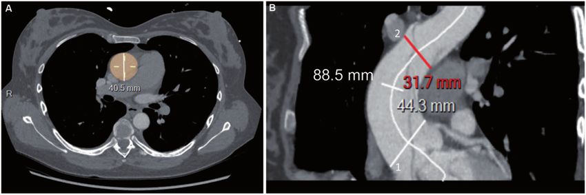
2 P. Tozzi et al. / European Journal of Cardio-Thoracic Surgery
RESULTS: Overall, 102 patients were included. Of these, 67 were male (65.6%) and 35 were female (34.4%), and the cohort’s mean age was
65 ± 12.1 years. In addition, 66% were treated for arterial hypertension. The mean maximum modelled pre-dissection diameter was
39.6 ± 4.8 mm: 39.1 ± 5.1 mm in men and 40.7 ± 2.8 mm in women (P = 0.1). The cumulative 30-day mortality rate was 19.6% (20/102).
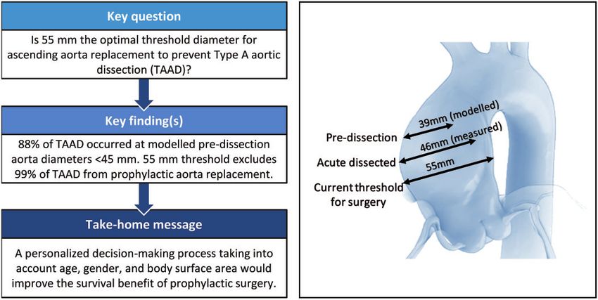
CONCLUSIONS: TAAD occurred at a modelled aortic diameter below 45 mm in 87.7% of our patients. Therefore, the current aortic diam-
eter threshold of 55 mm excludes 99% of patients with TAAD from prophylactic replacement of the ascending aorta. The maximum
diameter of the ascending aorta warrants reappraisal and this parameter should be a distinct part of a personalized decision-making pro-
cess that also takes into account age, gender and body surface area to establish the surgical indication for preventive aorta replacement
aimed to improve the survival benefit of this procedure.
Keywords: Aortic aneurysm • Aortic dissection • Acute aortic syndrome • Aortic surgery
Downloaded from https://academic.oup.com/ejcts/advance-article/doi/10.1093/ejcts/ezaa351/5917268 by guest on 21 December 2020
We have herein reviewed our experience of modelling pre-
ABBREVIATIONS dissection aortic size to possibly validate existing recommenda-
tions for prophylactic ascending aorta replacement.
BSA Body surface area
TAAD Type A aortic dissection
METHODS
INTRODUCTION We retrospectively evaluated clinical data, including demograph-
ics and cardiovascular risk factors, of 117 patients who consecu-
Acute aortic dissection of the ascending aorta (Stanford classifica- tively underwent surgical treatment for TAAD in our institute and
tion Type A) is a devastating disease that has an annual incidence received at least a graft on the ascending aorta from January
of 2.9/100 000 [1]. Mortality rates of medically managed Type A 2014 to October 2019. Patients with a clinical diagnosis of
acute aortic dissection (TAAD) are 20% at 24h after presentation, Marfan syndrome or connective-tissue disorders, iatrogenic or
30% at 48h, 40% at a week and 50% at a month after presenta- traumatic dissection, or bicuspid aortic valve were excluded.
tion [2, 3]. Therefore, surgical treatment is indicated immediately Within this cohort, we identified patients who had received ad-
upon TAAD diagnosis. Despite major improvements in surgical equate computed tomography angiography upon emergency de-
partment admission and we retrieved their clinical data. The
techniques, cerebral perfusion protection strategies, and
following parameters were collected: date of birth, height,
perioperative care, surgical mortality remains high, even in high-
weight, gender and diagnosis of arterial hypertension. We
volume centres with the 30-day mortality of 7.6% and the opera-
defined the treatment of resistant systemic hypertension as
tive mortality of 9.5% [2]. These rates have not significantly
requiring at least 3 antihypertensive drugs, such as beta-blockers,
changed over time for patients receiving immediate and ad-
angiotensin-converting enzyme inhibitors, angiotensin II receptor
equate surgical treatment.
antagonists, vasodilators or calcium channel blockers.
Hospital-based databases only include patients who reach re-
Preoperative trans-oesophageal echocardiography was per-
ferral centres alive, and they exclude some 50% of TAAD
formed to exclude the presence of a bicuspid aortic valve.
patients who die at home or prior to hospital admission. This
Amidst our cohort, the maximum diameter and length of the
therefore leads to underestimating overall disease incidence
dissected aorta were measured on pre-operatory contrast-me-
and mortality [3].
dium CT scan. Centre line reconstructions were generated using
Current guidelines recommend prophylactic ascending
the Carestream Health SA—Switzerland software package. Next,
aorta replacement at an aneurysm diameter of >55 mm in an
the maximum aortic diameter including true and false lumens,
effort to prevent acute TAAD (class I, level C), in the absence and outer to outer aortic wall diameter, was measured on axial
of connective-tissue disorders [4]. These guidelines are transverse images perpendicular to the central line on the
essentially based on expert consensus and retrospective obser- aortic segment between the sinotubular junction and origin of
vational studies. The milestone paper, which was published in the brachiocephalic trunk. Based on our assumption, this is the
1997, is a single-centre study of a heterogeneous 76-patient co- most appropriate segment to study in patients without Marfan
hort [5]. syndrome or connective-tissue disorders and a tricuspid aortic
In recent years, several publications have challenged the >55- valve.
mm threshold. Every single study that has looked at pre- Finally, the aortic length was measured on centre line recon-
dissection diameters has found that only a small percentage of structions starting from the plane corresponding to the sinotubu-
TAAD patients actually had an aortic aneurysm above 55 mm. lar junction to the plane immediately proximal to the origin of
This comprises all major international registries, such as the the brachiocephalic artery (Figure 1). The manual identification
International Registry for Aortic Dissections and German Registry of the centre line was required in case of either no or low con-
for Acute Aortic Dissection type A [6, 7]. Based on a cohort of trast enhancement of the false channel.
343 patients, Rylski et al. modelled the pre-dissection aortic To model the pre-dissection aortic dimension, we considered
diameter from the dissected aorta by subtraction of the average the 2 most recent publications on the effects of TAAD on aortic
diameter increase rate of 30% (according to the results of a study geometry changes and increases in aortic diameter: namely, the
of human aortic geometry changes due to dissection). These study by Rylski, in which the induced average increase in the
authors concluded that >90% failed to meet the guidelines for mid-ascending aortic diameter was 13 ± 7mm (+32%) [8], and
elective ascending aorta replacement [8]. the study by Mansour, in which the pre-dissected aortic
P. Tozzi et al. / European Journal of Cardio-Thoracic Surgery                                                       3

                                                                                                                                                                             CONVENTIONAL AORTIC
                                                                                                                                                                                  SURGERY
                                                                                                                                                                             Downloaded from https://academic.oup.com/ejcts/advance-article/doi/10.1093/ejcts/ezaa351/5917268 by guest on 21 December 2020
Figure 1: Centre line reconstructions of preoperative CT scan with contrast medium. (A) Patient 4: the maximum diameter of the dissected aorta was measured includ-
ing true and false lumens, outer to outer aortic wall diameter, on axial transverse images perpendicular to the central line. (B) Patient 37: measure of the length of the
ascending aorta. The ascending aorta begins at the plane corresponding to the sinotubular junction (white line 1) and extends to the plane immediately proximal to
the origin of the brachiocephalic artery (red line 2): 88.5 mm corresponds to the length of the dissected ascending aorta.

diameter was estimated to be 7 mm (-20%) smaller than the
measured dissected aorta [9]. We decided to arbitrarily apply
Mansour’s criteria to our cohort, so as to estimate pre-
dissection aortic dimensions and identified it as a modelled
pre-dissection diameter.
   To compare our results with normal aortic size limits, the
modelled pre-dissection ascending aorta diameters were com-
pared between genders among 4 age categories (65) and referred to gender-specific, age-specific and
body surface area (BSA)-adjusted normal aortic diameters, as
reported in a study involving 2952 patients by Wolak et al. [10].
   The statistics have been presented as frequencies and percen-
tages for categorical variables and as mean and standard devi-
ation for continuous variables. For comparison of the continuous
variables, the Mann–Whitney rank sum test was used. Categorical
variables were compared using the Chi-squared test. In the event
of small group sizes (n < 5), Fisher’s exact t-test was used.                           Figure 2: Distribution of pre-dissected maximum ascending aorta diameters,
   The study protocol was reviewed and accepted by our local                            according to size and gender. The pre-dissected (modelled) diameter was
ethics committee (authorization number 2020-01054), and we                              obtained by measuring the maximum aortic diameter of the dissected ascend-
                                                                                        ing aorta of -7 mm, according to Mansour et al. results [9]. Grey columns cor-
received written informed consent from each patient.
                                                                                        respond to female gender. TAAD: Type A aortic dissection.

RESULTS
In total, 117 patients underwent surgical replacement of the
ascending aorta, 28 (23.9%) of whom also received a prosthetic
aortic valve and 13 (11.1%) of whom underwent a Bentall pro-
cedure. Fifteen patients (12.8%) were excluded from the study: 8
were Marfan patients, whereas a preoperative CT scan could not
be found for the remaining 7. Of the 102 patients who were
included, there were 67 men (65.6%) and 35 women (34.4%), and
their mean age was 65 ± 12.1 years. Overall, 67 patients (66%)
were treated for arterial hypertension and 54% suffered
treatment-resistant hypertension. The cohort’s mean height was
173 ± 9 cm, and the mean weight was 80 ± 17.8 kg. The mean
maximum modelled pre-dissection diameter was 39.6 ± 4.8 mm,
which was 39.1 ± 5.1 mm in men and 40.7 ± 2.8 mm in women
(P = 0.1) (Figure 2). The mean length of the ascending aorta was                        Figure 3: Scatter plot of dissected ascending aorta length. Aortic length was
                                                                                        measured on centre line reconstructions starting from the plane corresponding
87 ± 22 mm (Figure 3). The mean intensive care unit stay was                            to the sinotubular junction to the plane immediately proximal to the origin of
8 ± 0 days (range: 1–48 days). The operative and 30-day cumula-                         the brachiocephalic artery. One hundred twenty millimetres is the cut-off value
tive mortality rate was 19.6% (20/102). In total, 10 patients died                      for calculating the Tübingen Aortic Pathoanatomy score [11].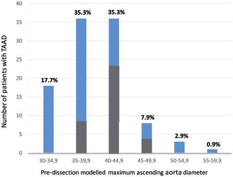
4                                           P. Tozzi et al. / European Journal of Cardio-Thoracic Surgery

due to neurological complications (10%), 9 died due to cardio-               ascending aortic aneurysm [15]. In our cohort, only 9 (8.8%)
vascular complications (haemorrhage, heart failure and intestinal            patients had a pre-dissection aortic diameter larger than 55 mm.
ischaemia) and the remaining 1 died from multiple organ failure.                An analysis of 3400 patients by the Yale Aortic Institute
None of the enrolled patients exhibited bicuspid aortic valves.              Database, which contains data on patients who had, by chance,
Pathological examination of the resected dissected aorta revealed            an aortic CT scan just before aortic dissection, enabled Mansour
anomalies in all patients. Specifically, in 35 patients (34%) we             et al. [9] to claim that the pre-dissection ascending aortic size is
found cystic media necrosis characterized by the local disappear-            7.65 mm smaller, just before dissection occurred. This is because
ance of elastic fibres in the arterial media, a reduction in smooth          rapid separation within the aortic media likely causes acute aortic
muscle cells and an increase in proteoglycans; 67 patients (64%)             wall weakness, which should theoretically trigger an immediate
clearly presented only fragmentation of elastic fibres at the                increase in aortic diameter. The reported threshold for the diam-

                                                                                                                                                     Downloaded from https://academic.oup.com/ejcts/advance-article/doi/10.1093/ejcts/ezaa351/5917268 by guest on 21 December 2020
Elastica van Gieson stain.                                                   eter’s increase due to dissection has a 21% variation coefficient
                                                                             upon measurements. This means that for a dissected aortic diam-
                                                                             eter of 60 mm, for example the pre-dissection diameter was be-
DISCUSSION                                                                   tween 48 and 53 mm. Other authors have reported more
                                                                             aggressive increases in aortic diameter due to acute dissection. In
Prophylactic replacement of the ascending aorta is the only ef-              an elegant study, Rylski et al. reported that the maximum diam-
fective strategy for preventing TAAD. As with all preventive treat-          eter of the ascending aorta increased by 12.8 mm (32%) from
ments, a risk/benefit analysis appears crucial to determine the              40.1 mm (36.6 mm; 45.3 mm) to 52.9 mm (46.1 mm; 58.6 mm)
appropriate timing for the surgical procedure. Unlike coronary               (+12.8 mm; +32%; P < 0.001). A similar but less pronounced in-
and cerebral vascular diseases, data on TAAD risk factors, inci-             crease in dimensions was observed at the sinotubular junction
dence, and outcome are limited and no prospective population-                level (+6.9 mm; p < 0.001). In contrast, the sinus of Valsalva diam-
based studies have been published as yet [3]. Several retrospect-            eter did not significantly increase [13].
ive studies have highlighted the correlation between dissection                 If we apply the smallest correction of -7mm to our cohort, 51
and arterial hypertension. Indeed, hypertension that is resistant            (50%) TAAD patients probably displayed an aortic diameter that
to medication has been shown to be the most significant modifi-              was smaller than 40 mm, whereas 89 (87.7%) exhibited an aortic
able risk factor for acute aortic dissection [3, 11]. The high rate of       diameter that was smaller than 45 mm at the time of dissection.
treatment-resistant hypertension in TAAD patients is most prob-              This suggests that the size criterion for resection of ascending
ably due to decreased aortic compliance, which is associated                 aorta aneurysm warrants reappraisal. In the personalized medi-
with fragility of the aortic wall. In our experience, 54% of patients        cine era, for small aneurysms, aortic size cannot remain the only
suffered from treatment-resistant hypertension, and all these sub-           and absolute criterion for proposing the surgical replacement of
group patients had an aortic diameter of smaller than 40 mm                  the ascending aorta. One possibility to improve the parameter’s
upon dissection, without any significant difference in the inter-            accuracy is to consider the aortic diameter with respect to age,
gender variance of aortic diameters.                                         gender and BSA. In Table 1, we have stratified our cohort accord-
   Although the exact sequence of events leading to TAAD                     ing to gender, age and BSA at the time of dissection. We then
remains poorly understood, several studies have demonstrated                 compared the modelled pre-dissection diameter to the ‘normal’
common pathways of medial degeneration. These comprise elas-                 ascending aorta diameter for the same class of age, gender and
tic fibre fragmentation and smooth muscle cell necrosis, which               BSA, as reported by Wolak et al. [10]. Surprisingly, all patients
both lead to the progressive loss of aortic wall integrity and wall          exhibited a modelled pre-dissection diameter between 10% and
delamination [12, 13]. None of our patients displayed a histologi-           34% larger than it was supposed to be, according to Wolak’s
cally normal aortic wall. Histological examination of the resected           data. This suggests that dissection occurs in dilated aorta with re-
dissected aorta demonstrated elastic fibre fragmentation in all              spect to the standard diameter even when the absolute value is
patients and cystic media necrosis in 34% of them. This supports             below the accepted criteria for surgical resection. We have inte-
the hypothesis, which has been extensively described in the lit-             grated these findings into a personalized decision-making pro-
erature, that spontaneous aortic dissection does not occur in                cess where an increase of at least 10% of patient aortic size with
normal aortic walls [12]. Information on pathological aortic wall            respect to ‘normal’ size for same class of age, gender and BSA
would play a key role in the decision-making process of prevent-             represents an element in favour of early surgery. For example, in
ive aortic replacement. Nevertheless, existing non-invasive inves-           a 70-year-old male with a BSA of 2.2 m2, an ascending aorta of
tigational tools (ultrasounds, CT, and magnetic-resonance                    38 mm could be considered normal, whereas in a 45-year-old
imaging) are unable to characterize aortic wall histology.                   woman with a BSA of 1.6 m2, this would signify an indication for
   The ascending aortic transverse diameter is the only accepted             surgical replacement because the aorta is 30% larger than what is
morphological risk factor for TAAD and indication for preventive             considered as normal aortic diameter, this being 29.6 ± 2.8.
surgery. However, this is a poor predictor of the timing and loca-              Recently, the Aortic Institute at Yale New Haven Hospital pub-
tion of dissection events. Historical studies using mathematical             lished a risk stratification analysis involving their 780-patient co-
models to simulate aortic dissection underline how a dilatation              hort using regression models. Patients were stratified into 4
phase of the ascending aorta (growing aorta) plays a key role in             categories of yearly risk complications, based on their height-
producing an intimal tear, which is followed by the acute dissec-            based aortic height index, which is defined as aortic size/height
tion process [14]. However, size alone only seems to be a good               ratio. Using code colours and nomograms, the authors illustrated
predictor of ascending aortic dissection for diameters over                  that a 17-cm-tall man with a 45-mm ascending aorta exhibits a
60 mm, which seems to be the hinge point for natural complica-               7% annual risk of aortic dissection, rupture or death. The group
tions of aortic aneurysm [15, 16]. Approximately one-third of                concluded that indexing absolute aortic diameter to anthropo-
patients with aortic dissection exhibit a normal-sized or minimal-           metric measurements allows for providing individualized risk
ly enlarged aorta, and only 10% of patients display a true                   classification with satisfactory results [17] and that surgical
P. Tozzi et al. / European Journal of Cardio-Thoracic Surgery 5
CONVENTIONAL AORTIC
Table 1: Gender-, age- and BSA-related ascending aorta diameter upper limits, according to Wolak et al. [10] compared to our cohort
modelled pre-dissection data
SURGERY
Age (years) BSA (m2) Ascending normal (mm)*, n = 2952 Ascending pre-dissection Diameter difference between
modelled (mm), n = 102 normal and modelled (%)
Female, n = 1147 Male, n = 1805 Female, n = 35 Male, n = 67 Female Male
65 2.1 32.8 36.8 ± 2.8 – 41.5 ± 4.5 – +13.0
For our data, continuous variables were expressed as mean and standard deviation. An isolated value means that there was only 1 patient. Positive value in the
diameter difference column indicates the diameter increase in the modelled pre-dissected aorta with respect to normal aorta.
*Data from Wolak et al.
BSA: body surface area.
treatment should be recommended whenever patient-specific mortality rate for emergency ascending aorta replacement in the
surgical mortality appears to be lower than the estimated risk of most experienced hands was estimated at 22% in 1997. Notably,
aortic dissection or death. this has not wavered over the last 2 decades, and was 16.9% in
Other authors have underlined the role of aortic elongation in the best series [1, 11, 19]. In our cohort, we observed a 19.6% cu-
TAAD genesis. Unlike the circumferential aorta dilatation, its lon- mulative mortality rate for 30-day stays. These data should tip
gitudinal dilatation, which is also defined as elongation, has been the scales towards earlier surgery because the risk of TAAD has
largely neglected in pathogenic TAAD models. Recently, Kruger been proven to be greater than that of elective surgery. The
et al. [11] reviewed their experience on 140 patients and pro- number needed to treat could be calculated to match historical
posed the Tübingen Aortic Pathoanatomy (TAIPAN) score to esti- data from our experiences. To illustrate, a man with a 45–49-mm
mate the TAAD risk. They combined the ascending aorta ectasia ascending aorta, without any connective-tissue disorders, and a
and aorta elongation (defined as the central distance between tricuspid aortic valve exhibits an average annual death risk for
the sinotubular junction and brachiocephalic trunk) to generate aortic dissection of about 4.7% [20]. The mortality rate for elective
the following scoring system: ascending aorta diameter 55 mm = 2; ascending aorta length 120 mm = 1. This scoring system was able to identify ity rate by 3.7%, which translates into an number needed to treat
23% of pre-TAAD patients and at least twice as many pre-TAAD of 27 (the formal calculation is 100/3.7 = 27). Accordingly, we
patients as the diameter alone. Based on this retrospective mor- should treat 27 patients with 45–49 mm ascending aorta aneur-
phological analysis, the authors recommended prophylactic ysm to save 1 life.
ascending aorta replacement at >_2 points [11]. When applying However, about 7% of entry tears in TAAD are located in the
the TAIPAN score to our TAAD patient cohort, only 4 (3.4%) aortic arch [19], and it is likely that the prophylactic surgical re-
would have been an indication for elective ascending aorta re- placement of the ascending aorta is not able to prevent the dis-
placement (Figure 3). However, our measurements corresponded section of the aortic arch and descending aorta.
to the length of the dissected aorta and not to that of the pre- We are aware that this study holds several methodological lim-
dissected aorta, which should be used to calculate the TAIPAN itations, particularly its retrospective single-centre study design
score. There are no models to quantify the changes in aortic without any control group, the use of imprecise estimation of
length due to acute aortic dissection as there are for aortic diam- aortic diameter reduction (measurements were performed by a
eter changes. team of radiologists and surgeons without assessing intra-
As previously mentioned, surgical decision-making requires an observer and interobserver variabilities) and the possibility of
accurate risk/benefit analysis. We must balance the reduced diameter increases due to dissection differing among patients.
TAAD incidence, as a result of earlier surgery, against the opera- Moreover, we did not take into account TAAD-related morbidity,
tive risk of prophylactic surgery in an asymptomatic patient. such as non-fatal neurological complications and renal function
Surgical mortality for elective ascending aorta replacement in impairment, both of which affect the quality of life and could po-
otherwise healthy patients was estimated at 9% in 1997, when tentially be avoided with elective surgery. Although all these limi-
the threshold value of 55 mm was proposed. However, this esti- tations could bias the conclusions, in our experience, the current
mated figure has meanwhile decreased to 1% [11, 18]. The aortic diameter threshold of 55 mm excludes 99% of TAAD6 P. Tozzi et al. / European Journal of Cardio-Thoracic Surgery
patients from prophylactic replacement of the ascending aorta. Cardiology (ESC). ESC Committee for Practice Guidelines. Eur Heart J
Therefore, the absolute diameter of 55 mm deserves reappraisal. 2014 1;35:2873–926.
[5] Coady MA1, Rizzo JA, Hammond GL, Mandapati D, Darr U, Kopf GS et
We should go beyond the guidelines and move towards a more al. What is the appropriate size criterion for resection of thoracic aortic
personalized therapeutic approach that tailors treatment accord- aneurysms? J Thorac Cardiovasc Surg 1997;113:476–91.
ing to a multitude of parameters, in order to provide survival [6] Evangelista A, Isselbacher EM, Bossone E, Gleason TG, Eusanio MD,
benefit by replacing the aneurysmal segment of the ascending Sechtem U et al. Insights From the international registry of acute aortic
dissection: a 20-year experience of collaborative clinical research.
aorta before the catastrophic event occurs. An international Circulation 2018;137:1846–60.
taskforce should adapt existing recommendations on surgical [7] Boening A, Karck M, Conzelmann LO, Easo J, Krüger T, Rylski B et
treatment of ascending aortic aneurysm to align with the most al. German registry for acute aortic dissection type A: structure,
recent scientific evidence, including recommending prophylactic results, and future perspectives. Thorac Cardiovasc Surg 2017;65:
Downloaded from https://academic.oup.com/ejcts/advance-article/doi/10.1093/ejcts/ezaa351/5917268 by guest on 21 December 2020
77–84.
intervention at smaller aortic sizes in selected patients. [8] Rylski B, Branchetti E, Bavaria JE, Vallabhajosyula P, Szeto WY, Milewski
R et al. Modeling of predissection aortic size in acute type A dissection:
Conflict of interest: none declared.
more than 90% fail to meet the guidelines for elective ascending re-
placement. J Thorac Cardiovasc Surg 2014;148:944–8.
Author contributions [9] Mansour AM, Peterss S, Zafar MA, Rizzo JA, Fang H, Charilaou P et al.
Prevention of aortic dissection suggests a diameter shift to a lower aortic
size threshold for intervention. Cardiology 2018;319:139–46.
Piergiorgio Tozzi: Conceptualization; methodology; writing—original draft [10] Wolak A, Gransar H, Thomson LE, Friedman JD, Hachamovitch R,
preparation. Ziyad Gunga: Conceptualization, methodology, data curation. Gutstein A et al. Aortic size assessment by noncontrast cardiac com-
Lars Niclauss: Resources. Dominique Delay: Resources. Aurelian Roumy: puted tomography: normal limits by age, gender, and body surface area.
Data curation. Raymond Pfister: Data curation. Sebastien Colombier: Data JACC Cardiovasc Imaging 2008;1:200–9.
curation. Francesco Patella: Writing—review & editing. Salah Dine Qanadli: [11] Kruger T, Boburg RS, Lescan M, Oikonomou A, Schneider W, Vöhringer
Methodology, review & editing. Matthias Kirsch: Methodology, review & L et al. Aortic elongation in aortic aneurysm and dissection: the
editing. Tübingen Aortic Pathoanatomy (TAIPAN) project. Eur J CardioThorac
Surg 2018;54: 26–33.
[12] Nakashima Y. Pathogenesis of aortic dissection: elastic fiber abnormal-
Reviewer information ities and aortic medial weakness. Ann Vasc Dis 2010;3:28–36.
[13] Rylski B, Blanke P, Beyersdorf F, Desai ND, Milewski RK, Siepe M et al.
European Journal of Cardio-Thoracic Surgery thanks Luca Di Marco, Tobias How does the ascending aorta geometry change when it dissects? J Am
Krüger and the other, anonymous reviewer(s) for their contribution to the Coll Cardiol 2014;63:1311–9.
[14] Thubrikar MJ1, Agali P, Robicsek F. Wall stress as a possible mechanism
peer review process of this article.
for the development of transverse intimal tears in aortic dissections.
J Med Eng Technol 1999;23:127–34.
[15] Elefteriades JA. Natural history of thoracic aortic aneurysms: indications
REFERENCES for surgery, and surgical versus nonsurgical risks. Ann Thorac Surg 2002;
74:S1877–80.
[1] Mészáros I, Mórocz J, Szlávi J, Schmidt J, Tornóci L, Nagy L et al. [16] Neri E, Barabesi L, Buklas D, Vricella LA, Benvenuti A, Tucci E et al.
Epidemiology and clinicopathology of aortic dissection. Chest 2000;117: Limited role of aortic size in the genesis of acute type A aortic dissection.
1271–8. Eur J CardioThorac Surg 2005;28:857–63.
[2] Abe T, Yamamoto H, Miyata H, Miyata H, Motomura N, Tokuda Y et al. [17] Zafar MA, Li Y, Rizzo JA, Charilaou P, Charilaou P, Saeyeldin A et al.
Patient trends and outcomes of surgery for type A acute aortic dissection in Height alone, rather than body surface area, suffices for risk estimation
Japan: an analysis of more than 10 000 patients from the Japan in ascending aortic aneurysm. J Thorac Cardiovasc Surg 2018;155:
Cardiovascular Surgery Database. Eur J CardioThorac Surg 2020;57:660–7. 1938–50.
[3] Howard D, Banerjee A, Fairhead J, Perkins J, Silver LE, Rothwell PM. [18] Ziganshin BA, Zafar MA, Elefteriades JA. Descending threshold for
Population-based study of incidence and outcome of acute aortic dis- ascending aortic aneurysmectomy: is it time for a “left-shift” in guide-
section and premorbid risk factor control. 10-year results from the lines? J Thorac Cardiovasc Surg 2019;157:37–42.
Oxford Vascular Study. Circulation 2013;127:2031–7. [19] Sievers HH, Rylski B, Czerny M, Baier ALM, Kreibich M, Siepe M et al.
[4] Erbel R, Aboyans V, Boileau C, Bossone E, Di Bartolomeo R, Eggebrecht Aortic dissection reconsidered: type, entry site, malperfusion classifica-
H et al. 2014 ESC Guidelines on the diagnosis and treatment of aortic tion adding clarity and enabling outcome prediction. Interact
diseases: document covering acute and chronic aortic diseases of the CardioVasc Thorac Surg 2020 1;30:451–7.
thoracic and abdominal aorta of the adult. The Task Force for the [20] Pinard A, Jones GT, Milewicz DM. Genetics of thoracic and abdominal
Diagnosis and Treatment of Aortic Diseases of the European Society of aortic diseases. Circ Res 2019;124:588–606.You can also read