Journal of Physical Medicine Rehabilitation Studies & Reports
←
→
Page content transcription
If your browser does not render page correctly, please read the page content below
Journal of Physical Medicine
Rehabilitation Studies & Reports

Research Article                                                                                                                   Open     Access

Analysis of Gait Parameters in Patients after Triple-Bundle Achilles
tendon Reconstruction – Prospective Evaluation of 60 Patients at 3,
6 and 12 Months Post op
Magdalena Syrek1, 4 *, Urszula E Zdanowicz2, Robert Smigielski3, Michal Staniszewski4

Carolina Medical Center, Physiotherapy Department, Pory 78, 02-757 Warsaw, Poland
1

Carolina Medical Center, Orthopaedic and Sports Traumatology Department, Pory 78, 02-757 Warsaw, Poland
2

McGowan Institute for Regenerative Medicine, University of Pittsburgh, 450 Technology Drive, Suite 300, Pittsburgh, PA 15219-3110, USA

Biological Treatment Centre, Orthopaedics Department, LIFE Institute, Grzybowska 43a, 00-855, Warsaw, Poland
3

Józef Piłsudski University of Physical Education in Warsaw, Department of Physical Education, Marymoncka 34, 00-809, Warsaw, Poland
4

*Corresponding author
Magdalena Syrek, Carolina Medical Center, Physiotherapy Department, Pory 78, 02-757 Warsaw, Poland; Józef Piłsudski University of Physical
Education in Warsaw, Department of Physiotherapy, Marymoncka 34, 00-809, Warsaw, Poland E-mail: magda.syrek@gmail.com

Received: September 05, 2021; Accepted: September 13, 2021; Published: September 15, 2021

Background                                                                       end-to-end suture techniques lead to more successful outcome
The Achilles tendon is formed by collagen fibers running from the                with reduced rate of complications when compared to approach
lateral and medial head of the gastrocnemius muscle and situated                 focused on tendon augmentation [15,16]. Jaakkola et al. designed a
deeper soleus muscle. The fascicles are twisted so the fibers from               study to compare the tensile strength of ruptured Achilles tendons
the medial head of the gastrocnemius muscle are located posteriorly              repaired using either the triple-bundle technique or the Krakow
(superficially) and the fibers from the lateral head are located                 locking loop technique [17]. The difference in average rupture
anteriorly (deeply). Thus, the fibers from the soleus muscle are                 load for the triple-bundle technique and the average rupture load
located in the central and medial part of the tendon [1]. These units,           for the Krakow locking loop technique represented a statistically
depending on the side, rotate as left- or right-handed screws (eg, left          significant superiority in favour of the triple-bundle technique. The
Achilles tendon rotates against clockwise). The significance of the              same researcher conducted a study to evaluate the triple-bundle
torsion stems from the fact it occurs at the site where concentrated             technique for acute Achilles tendon rupture repair followed by
pressure applies potentially due to rupture enabling forces produced             early (14 days) postoperative ankle range of motion, compared to
where all the tendon bundles meet. Independently, it is possible to              non-operative treatment with delayed ankle range of motion [18].
distinguish each separate Achilles tendon musculotendinous unit                  Operative treatment reduced immobilization time, allowed safe
during a thorough, detailed dissection [2].                                      early-return to weight bearing, and diminished risk of re-rupture
                                                                                 compared to non-operative treatment.
Achilles tendon is the strongest one in the human body and has to
withstand forces of up to 3,800 N [3]. The treatment of Achilles                 The goal of the rehabilitation is to allow the patient to fully
tendon ruptures should not only focus on restoring Achilles tendon               recover their strength and function within the shortest period.
length but also the original strength of the whole muscle-tendon                 Rehabilitation after rupture of the Achilles tendon is slow and it
unit [4,5]. The best treatment is still debated [6,7]. There are                 may last longer than 3 months [19]. Despite most of the patients
papers supporting non-operative treatment, traditional open repair               reporting good long-term results, a significant number of patients
as well as percutaneous repair [8-11]. Rehabilitation protocols                  experience persistent symptoms for years [20]. Abnormal gait is a
also vary significantly, with some data suggesting success with                  sign of an incomplete recovery process [20,21]. Spatial and kinetic
early mobilization, particularly in young active patients [12-14].               data collected in a full gait analysis can assess gait abnormalities,
                                                                                 including those caused by injuries, and the relationships between
The goal of operative treatment is to restore full biomechanical                 plantar flexor muscle-tendon unit properties and walking patterns
function of the tendon (or as similar to physiology as possible) and             during rehabilitation [20-22].
to restore the ability to correct transmission of contractile forces,
which is possible only by careful reconstruction of the three-bundle             The aim of the study was to present changes in walk parameters
structure of Achilles tendon [2]. There is no absolute consensus                 in patients at three, six and twelve months after surgical triple-
about the best type of surgical repair: some evidence suggests that              bundle Achilles tendon reconstruction.
J PhyMed Rehab Stud Rep, 2021                                                                                                             Volume 3(3): 1-7
Citation: Magdalena Syrek, Urszula E Zdanowicz, Robert Smigielski, Michal Staniszewski (2021) Analysis of Gait Parameters in Patients after Triple-Bundle Achilles
tendon Reconstruction – Prospective Evaluation of 60 Patients at 3, 6 and 12 Months Post op. Journal of Physical Medicine Rehabilitation Studies & Reports.
SRC/JPMRS/151.
Material and methods the cast and substitute it with a Walker boot with two wedges
Participants (4 cm) placed under the heel [28]. Partial weight bearing, up to
Sixty patients (50 men; 10 women) presenting with complete 25% of patients’ body mass, was permitted. At the beginning of
unilateral rupture of the Achilles tendon, treated by two senior the week eight, one wedge was removed and active motion was
surgeons were included in this prospective study. Demographic permitted from 5° of dorsiflexion to 30° plantarflexion. Partial
data can be seen in Table 1. The rupture involved the left and right weight bearing was increased to 50% of patients’ body mass. After
limb with 30 patients in each group. ten weeks patients started walking with full weight. Twelve weeks
Table 1 post-op another control ultrasound was performed along with a
Variables Mean SD Range clinical examination. Based on gathered findings the decision was
made by the surgeon to remove the Walker boot and allow normal
Age (years) 32 6,2 25-51
footwear. The patients performed mobility and theraband strength
Body mass (kg) 84,2 11,3 65-120 exercises, walking forward, backward, side-wards and stairs up
Height (cm) 180,3 5,8 168-193 and down progressively. Jumping on the trampoline and wobble-
board exercises began after 4 months into rehabilitation. Normal
BMI (kg/m2) 25,9 3,1 20-32
sport activity was allowed to be resumed after seven months. All
Male:female 50:10 patients followed the same rehabilitation protocol.
Side Left 30 Right 30
Measurements
All patients underwent Achilles tendon reconstruction at a mean The protocol of our study consisted of three assessments for the
time of 6±4 days post injury followed by immediate, uniform study group: three, six and twelve months after the surgery. Each
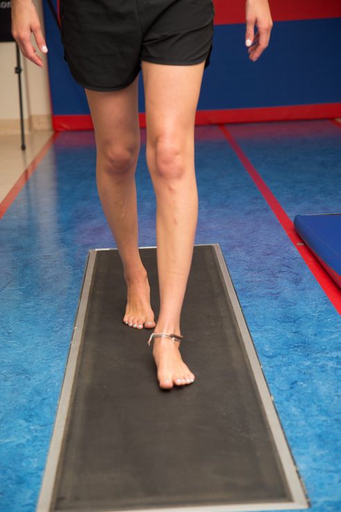
rehabilitation protocol described in a manuscript written by the evaluation included gait parameters analysis (Figure 1).
author of this publication [23]. The general summary of the
protocol has also been presented in the next subsection. Inclusion
criteria were: 25-60 years of age, a healthy contralateral leg with
physiological, clinically determined, alignment of the knee, foot
and ankle, no neuromuscular impairments including muscle
dystrophies, no other post-traumatic injuries or osteoarthritis of
the knee, foot and ankle of the treated leg. Exclusion criteria were:
previous surgical treatment of Achilles tendon, heart diseases,
liver or kidney insufficiency, oncological problem, haematological
diseases, immunological diseases, dermatological diseases,
infection, obesity, hyperlaxity.
The diagnosis of Achilles tendon rupture was initially made
clinically on the basis of palpable defect at the side of the rupture
and positive Thompson test which unveiled a lack of any apparent
plantar flexion. An ultrasound scan and magnetic resonance were
performed in all cases to confirm the diagnosis.
Figure 1
This study was a part of a project in developing a novel Scaffold-
based Tissue engineering Approaches to healing and Regeneration Gait analysis was conducted using the ZEBRIS FDM3 (Zebris
of Tendons and ligaments (START) and was approved by the Medical GmbH, Germany) platform of dimensions 314 x 62 x
appropriate Ethics Committee; all subjects gave their informed 2.1 cm with 17,024 sensors and registration frequency of 120
written consent before entering the study. Hz. The platform was connected to the WinFDM software for
gait analysis, which records consecutive walks. Each participant
Rehabilitation programme walked along the platform three times with a self-preferred speed.
The rehabilitation programme consisted of three sessions per week The evaluation report included spatial parameters: foot rotation
during six months (mean 6,3±1,9 months, which comes down (deg), step length (cm), stance phase (%), swing phase (%) and
to the average of 73±5 sessions per subject). All patients were kinetic parameters: max heel force (N) and max toe force (N).
discharged two days after the surgery if the condition of the patient
had not been complicated by other factors (mean hospitalization Participant self-reported level of function was evaluated using the
2,3 days). During first six weeks the patients were kept non-weight Achilles tendon total rupture score (ATRS) at four and a half (six
bearing and partially immobilized (the cast was split into two parts, weeks after Walker boot was removed), six and twelve months
allowing its removal during supervised physiotherapy sessions) post-op. It is a patient-reported instrument with high reliability,
They began passive and concentric motion of the ankle ranging validity and sensitivity for measuring the outcome related to
from 10° to 30° plantarflexion. Six weeks post-surgery all patients symptoms and physical activity after treatment in patients with
underwent a follow-up ultrasound. Each follow-up ultrasound a total Achilles tendon rupture and can be a good predictor in
examination of a patient in the program included the following patient’s ability to return to sports after one year after an injury
parameters: morphologic characteristics, structure, color Doppler [29,30]. It consists of 10 questions concerning symptoms (first
vascularization, tendon’s gliding within paratenon and mobility. four) and physical activity like walking, running and jumping
The parameters were chosen based on literature combined with the (see APPENDIX). A patient scoring 0 implies major limitations/
experience of the radiologist specializing in orthopedic ultrasound symptoms and 10 no limitations or symptoms. Answers from
MRI assessment [25-27]. The findings from ultrasound combined all 10 questions are added to a total score, with 100 being the
with clinical evaluation gave grounds to make a decision to remove maximum score.
J PhyMed Rehab Stud Rep, 2021 Volume 3(3): 2-7Citation: Magdalena Syrek, Urszula E Zdanowicz, Robert Smigielski, Michal Staniszewski (2021) Analysis of Gait Parameters in Patients after Triple-Bundle Achilles
tendon Reconstruction – Prospective Evaluation of 60 Patients at 3, 6 and 12 Months Post op. Journal of Physical Medicine Rehabilitation Studies & Reports.
SRC/JPMRS/151.
ATRS
(Achilles Tendon Total Rupture Score)
All questions refer to your limitations/difficulties related to your injured Achilles tendon. Answer every question by grading your
limitations/symptoms from 0-10.
Remember (0= Major limitations and 10= No limitations).
Please circle the number that matches your level of limitation
1. Are you limited due to decreased strength in the calf/Achilles tendon/foot?
0 1 2 3 4 5 6 7 8 9 10(No limitations)
2. Are you limited due to fatigue in the calf/Achilles tendon/foot?
0 1 2 3 4 5 6 7 8 9 10(No limitations)
3. Are you limited due to stiffness in the calf/Achilles tendon/foot?
0 1 2 3 4 5 6 7 8 9 10(No limitations)
4. Are you limited due to pain in the calf/Achilles tendon/foot?
0 1 2 3 4 5 6 7 8 9 10(No limitations)
5. Are you limited during activities of daily living?
0 1 2 3 4 5 6 7 8 9 10(No limitations)
6. Are you limited when walking on uneven surfaces?
0 1 2 3 4 5 6 7 8 9 10(No limitations)
7. Are you limited when walking quickly up the stairs or up a hill?
0 1 2 3 4 5 6 7 8 9 10(No limitations)
8. Are you limited during activities that include running?
0 1 2 3 4 5 6 7 8 9 10(No limitations)
9. Are you limited during activities that include jumping?
0 1 2 3 4 5 6 7 8 9 10(No limitations)
10. Are you limited in performing hard physical labour?
0 1 2 3 4 5 6 7 8 9 10(No limitations)
Statistical analysis
The recorded data was statistically analyzed using the STATISTICA package (data analysis software system), Version 12 (StatSoft,
Inc., 2014). The Shapiro-Wilk W test revealed that all the demographics and gait parameters data had a normal distribution; therefore,
parametric tests were applied in further analysis. The significance of the changes in the temporal and spatial gait parameters and
the significance of differences between affected and non-affected lower limbs were evaluated with the repeated measures ANOVA
test and the post-hoc Tukey’s test (HSD). ATRS scores were evaluated with Friedman ANOVA for nonparametric one-way repeated
measures analysis of variance. The significance level was set at p < 0.05 for all tests.
Results
The spatial and kinetic parameters after three, six, and twelve months after the surgery are shown in Table 2. After three months (T1)
the measurements of step length (understood as a distance between the point of initial contact of one foot and the point of initial
contact of the opposite one), stance phase, swing phase and max heel force have shown significant differences when compared to
measurements taken after six (T2) and twelve months (T3) in the affected leg. Based on own observations and experience the researchers
established a set of spatial and kinetic parameters meant to indicate the level of function restoration. Using these parameters the
efficacy of completed rehabilitation protocol was measured six months after the surgery when comparing patients after T2 and T3
(pb) evaluation we observed no significant differences for any gait parameters in affected and non-affected leg.
J PhyMed Rehab Stud Rep, 2021 Volume 3(3): 3-7Citation: Magdalena Syrek, Urszula E Zdanowicz, Robert Smigielski, Michal Staniszewski (2021) Analysis of Gait Parameters in Patients after Triple-Bundle Achilles
tendon Reconstruction – Prospective Evaluation of 60 Patients at 3, 6 and 12 Months Post op. Journal of Physical Medicine Rehabilitation Studies & Reports.
SRC/JPMRS/151.
Table 2
T1 T2 T3 pa pb pc
T1 vs T2 T2 vs T3 T1 vs T3
foot rotation (deg)
AL 8,49±3,46 8,82±3,33 8,97±3,49 0,954 0,998 0,807
NAL 10,87±5,72 8,81±3,84 8,92±3,79Citation: Magdalena Syrek, Urszula E Zdanowicz, Robert Smigielski, Michal Staniszewski (2021) Analysis of Gait Parameters in Patients after Triple-Bundle Achilles
tendon Reconstruction – Prospective Evaluation of 60 Patients at 3, 6 and 12 Months Post op. Journal of Physical Medicine Rehabilitation Studies & Reports.
SRC/JPMRS/151.
Discussion adequately perform their function during the heel- and toe-lift
The results of the present study show that the key goal of the phases – a compensation mechanism is used to move forward.
rehabilitation which is quick yet safe Achilles’ tendon strength and The body’s weight is shifted forward, thus ‘‘inducing’’ the next
function restoration is possible to achieve but requires a proper step. This, in turn, increases the load on the forefoot [31].
post-operative evaluation protocol combined with immediate
physical therapy programme including immediate, supervised Similarly to the study described by Garrido et al. [39] we have
mobilization of the ankle joint. observed an increase in recorded maximum toe force that would be
a subject of continuous improvement in time. Garrido’s study also
There are few reports in the literature concerning the use of shows that asymmetry regarding propulsion and take-off phases
triple-bundle repair technique [17,18,31], which was used in is substantial during the first year after surgery, progressing to
all cases covered by the study. The surgeons who performed all normality after this period of time. These results do not correspond
the reconstructions in the current study were also the creators with the previous research by Naim et al. [44,40] who studied the
of the technique and based on their medical recommendations evolution of patients’ recovery between 8 and 48 months after
the immediate mobilization was introduced and the presented surgery. In their study, albeit no statistically significant differences
rehabilitation protocol developed. between the injured leg and the healthy one were found when the
reaction forces of the forefoot against the floor were measured,
The study was designed to quantify the gait abnormalities found yet still the difference in values was noted. At the same time no
after complete unilateral rupture of the Achilles tendon. As significant variations were observed in relation to plantar flexion
expected, at three months after surgery (when the Walker boot in ankle, the period of stepping phase, twisting period on foot and
was removed), step length and swing phase were significantly base reaction pressure on heel between the intact and traumatic sides
shorter and stance phase longer in affected leg [32,22,33]. The which might indicate a significant elongation in Achilles tendon.
muscle weakness and the resulting loss of symmetry within the gait
cycle produce a less energy efficient walking pattern [21]. This is Significant attribute and uniqueness of this study is that it has been
consistent with the limited walking distance and diffuse aching in based on a uniform rehabilitation protocol completed by all patients.
the calf that affects many patients. Our study points to the close Contrary to rehabilitation programs of Achilles tendon presented in
relationship between the need for healing progress’ monitoring the literature, where full weight bearing in a functional split synthetic
and the development of rehabilitation protocol. The continuous cast in equinus was permitted immediately following surgery, our
assessment enables physiotherapists to boost the rehabilitation patients started weight bearing later (at six weeks post-op) and were
process in a safe way by following the medical recommendations additionally secured from re-injury by a Walker boot worn for next
of radiologists and clinicians based on follow-up examinations. the six weeks. The rehabilitation protocol was being commenced
The continuous cooperation between doctors and physiotherapists two days post-op in all cases and consisted of plantar strengthening
is paramount for patient’s safety and recovery, as well as it open exercises. The non-weight bearing time was fulfilled with manual
doors for new rehabilitation programmes and strategies without therapy sessions that decreased joint stiffness and tendon adhesions
risking re-injury. The remodeling phase takes about six weeks, [34]. The low values of questions in ATRS, related to running and
with decreased cellularity, collagen and glycosaminoglycan jumping, were correlated with late permission of dynamic exercises
synthesis. What follows is the tissue consolidation stage that during rehabilitation process due to Achilles tendon tissue’s healing.
begins at week six and continues up to week ten of rehabilitation. Despite of long immobilization, patients have achieved the same or
In this period, tissue experience structural changes: from cellular to better scores in ATRS and recovered correct walking patterns after
fibrous, hence patients are allowed to start partial weight bearing. six and twelve months after surgery [42-44].
Increased loading prepares Achilles tendon for further rebuilding.
After ten weeks into rehabilitation process, the maturation stage Although the treatment was limited to only one type of surgery
begins, with gradual tissue change from fibrous to scar-like tendon and the follow-up period is limited, the early results are promising.
over the course of one year [34]. In the future longer follow-up is to be considered and compared
with the groups of patients undergoing different kinds of surgical
Measurements of the maximum heel force conducted three months repairs as well as to non-surgically treated ruptures.
after the surgery show much lower values compared to similar
evaluation performed after six and twelve months period. Increase Conclusions
in the maximum toe force on the affected side has been frequently In conclusion, we coincide with other authors [45-47], that
observed to co-occur with the decrease in patients’ heel force. the open three-bundle Achilles tendon reconstruction provides
Such deviation between measurements performed in different good results in the treatment of acute ruptures of the Achilles
rehabilitation stages may be explained by the heel pain that affects tendon, restoring the ankle mobility, as well as gait strength and
patients, particularly in the early stages of rehabilitation. The stability. Despite its relative technical complexity resulting from
second gait analysis, taken six months after the injury, shows the inclusion of partial gait analysis (not including 3D analysis),
improvement of all parameters. These findings conflict with work the protocol with all related monitoring procedures i.e. ultrasound
from Costa and colleagues who reported a significant deficit in and clinical evaluation, shows great potential of becoming a tool
peak forefoot pressure compared to non-affected side. Meanwhile, that would objectively assess the progression of patients’ recovery.
the affected sides’ functional deficits found by Follak et al. manifest
in a decreased active heel lift during the swing phase shortly after In our experience, spatial and kinetic gait analysis was a useful tool
toe lift [21,35]. This is most likely stemming from the fact that the that allowed us to study and quantify the function after Achilles
weaker the calf muscle strength the worse propulsion initiating the tendon ruptures treated with an open technique.
swing phase. These results displayed a marked deficit in plantar
flexor strength in relation to estimations of maximum strength Nevertheless, we believe that stronger recommendations about
which are corroborated by other authors, who also found a strength management of this condition will require a large randomized
deficit of plantar flexors using isokinetics [36-38]. According to controlled trial comparing the different operative and conservative
the literature, the weakened plantar flexors – which could not strategies.
J PhyMed Rehab Stud Rep, 2021 Volume 3(3): 5-7Citation: Magdalena Syrek, Urszula E Zdanowicz, Robert Smigielski, Michal Staniszewski (2021) Analysis of Gait Parameters in Patients after Triple-Bundle Achilles
tendon Reconstruction – Prospective Evaluation of 60 Patients at 3, 6 and 12 Months Post op. Journal of Physical Medicine Rehabilitation Studies & Reports.
SRC/JPMRS/151.
References years after acute Achilles tendon rupture, Knee Surg Sports
1. P Szaro, G Witkowski, R Smigielski, P Krajewski, B Ciszek, Traumatol Arthrosc. 19: 1385-1393.
et al. (2009) Fascicles of the adult human Achilles tendon - an 20. M L Costa, D Kay, S T Donell, (2005) Gait abnormalities
anatomical study, Ann Anat. 191: 586-593. following rupture of the tendo Achillis: a pedobarographic
2. R Śmigielski (2008) Management of partial tears of the gastro- assessment, J Bone Joint Surg Br. 87: 1085-1088.
soleus complex, Clinics in Sports Medicine 27: 219-229. 21. R Don, A Ranavolo, A Cacchio, M Serrao, F Costabile, et
3. C N Maganaris, M V Narici, N Maffulli (2008) Biomechanics al. (2007) Relationship between recovery of calf-muscle
of the Achilles tendon, Disabil Rehabil 30: 1542-1547. biomechanical properties and gait pattern following surgery
4. S P Magnusson, M V Narici, C N Maganaris, M Kjaer (2008) for achilles tendon rupture, Clin Biomech (Bristol, Avon).
Human tendon behaviour and adaptation, in vivo, J Physiol 22: 211-220.
586: 71-81. 22. I M Garrido, J C Deval, M N Bosch, D H Mediavilla, V
5. P Sadoghi, C Rosso, V valderrabano, A Leithner, P Vavken, et P Garcia, et al. (2010) Treatment of acute Achilles tendon
al. (2012) Initial Achilles tendon repair strength-synthesized ruptures with Achillon device: clinical outcomes and kinetic
biomechanical data from 196 cadaver repairs, Int Orthop. gait analysis, Foot Ankle Surg. 16: 189-194.
39: 1947-1951. 23. A Rosińska, M Syrek (2016) carolina pl carolina.pl/START.
6. A Gigante, A Moschini, A Verdenelli, M Del Torto, S Ulisse, [online] https://carolina.pl/wp-content/uploads/2016/09/
et al. (2008) Open versus percutaneous repair in the treatment Start_Rehabilitacja-Sciegna-Achillesa_publikacja.pdf.
of acute Achilles tendon rupture: a randomized prospective 24. S Gitto, A Guja Draghi, Ch Bortolotto, F Draghi (2016)
study, Knee Surg Sports Traumatol Arthrosc. 16: 204-209. Sonography of the Achilles tendon after complete rupture
7. B D Twaddle, P Poon (2007) Early motion for Achilles tendon repair. What the radiologist should know, J Ultrasound Med.
ruptures: is surgery important? A randomized, prospective 35: 2529-2536.
study, Am J Sports Med. 35: 2032-2038. 25. B Ciszkowska-Łysoń, R Śmigielski, U Zdanowicz, M
8. M Weber, M Niemann, R Lanz, T Muller (2003) Nonoperative Drwięga, M Bień, et al. (2010) Role of ultrasonography in
treatment of acute rupture of the achilles tendon: results of a diagnosis of Achilles tendon partial ruptures, Congress ECR
new protocol and comparison with operative treatment, Am 2010, Poster nr: C-2380 [online] https://dx.doi.org/10.1594/
J Sports Med. 31: 685-691. ecr2010/C-2380.
9. S Riedl, L Sandberger, K Nitschmann, PJ Meeder, (2002) 26. A Rosińska, R Śmigielski, B Ciszkowska-Łysoń (2015)
Suture of fresh Achilles tendon rupture. Comparison of open Significant role of ultrasound imaging as biofeedback
with percutaneous suture technique, Chirurg. 73: 607-614. tool allowing for initial design and further modification of
10. F Ceccarelli, L Berti, L Giuriati, M Romagnoli, S Giannini, et original physiotherapy protocol created for patients who
al. (2007) Percutaneous and minimally invasive techniques of underwent Achilles tendon reconstruction, Congress ECR
Achilles tendon repair, Clin Orthop Relat Res. 458: 188-193. 2015, Poster nr: C-2190 [online] https://dx.doi.org/10.1594/
11. J R Lansdaal, J C Goslings, M Reichart, G A Govaert, K M ecr2015/C-2190.
van, et al. (2007) Scherpenzeel, R. Haverlag, K.J. Ponsen, The 27. B Ciszkowska-Łysoń, U Zdanowicz, A Rosińska, M Syrek,
results of 163 Achilles tendon ruptures treated by a minimally R Smigielski, et al. (2018) Establishing MR and ultrasound
invasive surgical technique and functional aftertreatment, based model of a properly healing Achilles tendon allowing
Injury 38: 839-844. to properly determine the healing stage of the structure:
12. N Maffulli, C Tallon, J Wong, K Peng Lim, R Bleakney, et al. preliminary findings, Congress ECR Poster nr: C-3209
(2003) No adverse effect of early weight bearing following [online] https://dx.doi.org/10.1594/ecr2018/C-3209.
open repair of acute tears of the Achilles tendon, J Sports 28. S Gitto, A Guja Draghi, Ch Bortolotto, F Draghi (2016)
Med Phys Fitness. 43: 367-379. Sonography of the Achilles Tendon After Complete Rupture
13. J Wong, V Barrass, N Maffulli, (2002) Quantitative review of Repair: What the Radiologist Should Know, J Ultrasound
operative and nonoperative management of achilles tendon Med. 35: 2529-2536.
ruptures, Am J Sports Med. 30: 565-575. 29. K Nilsson-Helander, R Thomeé, KG Silbernagel, P Thomeé,
14. D M van der Eng, T Schepers, J C Goslings, N W Schep E Faxén, et al. (2007) The Achilles tendon Total Rupture
(2013) Rerupture rate after early weightbearing in operative Score (ATRS): development and validation, Am J Sports
versus conservative treatment of Achilles tendon ruptures: a Med. 35: 421-426.
meta-analysis, J Foot Ankle Surg. 52: 622-628. 30. M S Hansen, M Christensen, T Budolfsen, T F Østergaard, T
15. T Nyyssönen, H Saarikoski, J P Kaukonen, P Lüthje, H Kallemose, et al. (2016) A Troelsen, K.W. Barfod, Achilles
Hakovirta, et al. (2003) Simple end-to-end suture versus tendon Total Rupture Score at 3 months can predict patients’
augmented repair in acute Achilles tendon ruptures: a ability to return to sport 1 year after injury, Knee Surg Sports
retrospective comparison in 98 patients, Acta Orthop Scand. Traumatol Arthrosc. 24: 1365-1371.
74: 206-208. 31. R K Marti RK, C van der Werken, P R Schütte, T J Bast (1983)
16. S Giannini, M Girolami, F Ceccarelli, F Catani, S Stea, et Operative repair of ruptured Netherlands J Surg. 35: 61-68.
al. (1994) Surgical repair of achilles tendon ruptures using 32. A P Chan, Y Y Chan, D T Fong, PY Wong, H Y Lam, et
polypropylene braid augmentation 15: 372-375. al. (2011) Clinical and biomechanical outcome of minimal
17. J L Jaakkola, W C Hutton, J L Beskin, G P Lee (2000) Achilles invasive and open repair of the Achilles tendon, Sports Med
tendon rupture repair: biomechanical comparison of the triple Arthrosc Rehabil Ther Technol. 20; 3: 32.
bundle technique versus the Krakow locking loop technique, 33. K G Silbernagel, R Willy, I Davis (2012) Preinjury and
21: 14-17. postinjury running analysis along with measurements of
18. J L Jaakkola, J L Beskin, L H Griffith, G Cernansky (2001) strength and tendon length in a patient with a surgically
Early ankle motion after triple bundle technique repair vs. repaired Achilles tendon rupture, J Orthop Sports Phys Ther.
casting for acute Achilles tendon rupture, 22: 979-984. 42: 521-529.
19. N Olsson, K Nilsson-Helander, J Karlsson, B I Eriksson, 34. P Sharma, N Maffulli (2005) Tendon injury and tendinopathy:
R Thomée, et al. (2011) Major functional deficits persist 2 healing and repair, J Bone Joint Surg Am. 87: 187-202.
J PhyMed Rehab Stud Rep, 2021 Volume 3(3): 6-7Citation: Magdalena Syrek, Urszula E Zdanowicz, Robert Smigielski, Michal Staniszewski (2021) Analysis of Gait Parameters in Patients after Triple-Bundle Achilles
tendon Reconstruction – Prospective Evaluation of 60 Patients at 3, 6 and 12 Months Post op. Journal of Physical Medicine Rehabilitation Studies & Reports.
SRC/JPMRS/151.
35. N Follak, D Ganzer, H Merk (2002) The utility of gait analysis et al. (2015) Increased unilateral tendon stiffness and its effect
in the rehabilitation of patients after surgical treatment of on gait 2-6 years after Achilles tendon rupture, Scand J Med
Achilles tendon rupture, Eur J Orthop Surg Traumatol. 12: Sci Sports. 25: 860-867.
90-95. 42. N Olsson, KG Silbernagel, BI Eriksson, M Sansone, A
36. H Alfredson, T Pietilä, L Ohberg, R Lorentzon (1998) Achilles Brorsson, et al. (2013) Stable surgical repair with accelerated
tendinosis and calf muscle strength. The effect of short-term rehabilitation versus nonsurgical treatment for acute Achilles
immobilization after surgical treatment, Am J Sports Med. tendon ruptures: a randomized controlled study, Am J Sports
26: 166-171. Med. 41: 2867-2876.
37. B R Mandelbaum, M S Myerson, R Forster (1995) Achilles 43. U Kim, Y S Choi, GC Jang, YR Choi (2017) Early
tendon ruptures. A new method of repair, early range of rehabilitation after open repair for patients with a rupture of
motion, and functional rehabilitation, Am J Sports Med. 23: the Achilles tendon, Injury. 48: 1710-1713.
392-395. 44. J A Zellers, D H Cortes, P Corrigan, L Pontiggia, K G
38. M Syrek, R Śmigielski, M Staniszewski, M Wychowański Silbernagel, et al. (2018) Side-to-side differences in Achilles
(2019) The characteristics of plantar flexors and dorsiflexors tendon geometry and mechanical properties following achilles
strength in patients after complete three-bundle Achilles tendon rupture, Muscles Ligaments Tendons J. 7: 541-547.
tendon reconstruction, Advances in Rehabilitation. 2: 37-45. 45. J D Calder, T S Saxby, Early (2005) active rehabilitation
39. I M Garrido IM, J C Deval, M N Bosch, D H Mediavilla, following mini-open repair of Achilles tendon rupture: a
VP Garcia, et al. (2010) Treatment of acute Achilles tendon prospective study, Br J Sports Med. 39: 857-859.
ruptures with Achillon device: clinical outcomes and kinetic 46. H G Jung, K B Lee, S G Cho, T R Yoon (2008) Outcome of
gait analysis, Foot Ankle Surg. 16: 189-194. achilles tendon ruptures treated by a limited open technique,
40. F Naim, A Simşek, S Sipahioğlu, E Esen, G Cakmak, et al. Foot Ankle Int. 29: 803-807.
(2005) Evaluation of the surgical results of Achilles tendon 47. R Bevoni, A Angelini, G D’Apote, L Berti, I Fusaro, et al.
ruptures by gait analysis and isokinetic muscle strength (2014) Long term results of acute Achilles repair with triple-
measurements, Acta Orthop Traumatol Turc. 39: 1-6. bundle technique and early rehabilitation protocol, Injury
41. A N Agres, G N Duda, T J Gehlen, A Arampatzis, W R Taylor, 45: 1268-1274.
Copyright: ©2021 Magdalena Syrek. This is an open-access article distributed
under the terms of the Creative Commons Attribution License, which permits
unrestricted use, distribution, and reproduction in any medium, provided the
original author and source are credited.
J PhyMed Rehab Stud Rep, 2021 Volume 3(3): 7-7You can also read