Portable single-lead electrocardiogram device is accurate for QTc evaluation in hospitalized patients
←
→
Page content transcription
If your browser does not render page correctly, please read the page content below
Portable single-lead electrocardiogram device is
accurate for QTc evaluation in hospitalized patients

María Mónica Marín O, MD,* Angel  Alberto García P, MD, MSc, PhD(c), FACC,*
Oscar Mauricio Munoz V, MD, PhD, Julio César Castellanos R, MD,‡
                  ~              †

Edward Caceres M, MD, MSc,* David Santacruz P, MDx
From the *Department of Cardiology, Pontificia Universidad Javeriana, Hospital Universitario San Ignacio,
Bogota, Colombia, †Department of Internal Medicine, Hospital Universitario San Ignacio, Pontificia
                            a, Colombia, ‡Hospital Universitario San Ignacio, Bogota, Colombia,
Universidad Javeriana, Bogot
    x
and Division of the Cardiac Electrophysiology Training Program, Pontificia Universidad Javeriana,
Bogota, Colombia.

BACKGROUND Many commonly used drugs can prolong the QTc in-                      441.65 6 40.3 ms, P 5 .15). Bland–Altman analysis showed no
terval (QTc), which can lead to potentially life-threatening arrhyth-            significant difference in QTc values (average difference of
mias. In the current era of the COVID-19 pandemic, it is worth                   -0.797, 95% limits of agreement:-13.179; 11.585). Lin’s concor-
mentioning that the disease itself and several drugs used for its                dance coefficient showed an excellent agreement (0.988,
treatment have been associated with QTc prolongation.                            P , .001). Concordance between the 2 devices for determining
                                                                                 QTc prolongation was excellent (kappa .0.90).
OBJECTIVE To evaluate the agreement and clinical precision of a
portable single-lead electrocardiogram (ECG) device to measure                   CONCLUSION ECG recordings obtained with KM-1L allow an accu-
the QTc interval compared to the standard 12-lead ECG.                           rate QTc interval assessment. Considering its simplicity of use,
                                                                                 this approach has advantages over conventional ECG and can pro-
METHODS In sequential tests, QTc of ECG recordings obtained with                 vide an alternative for the evaluation of QTc in hospitalized pa-
the KardiaMobile (KM-1L) device (AliveCor, San Francisco, CA) were               tients, during the current time of the COVID-19 pandemic and
compared to QTc obtained with conventional 12-lead ECG. Agree-                   beyond.
ment was evaluated using Bland-Altman plots and Lin’s concordance
coefficient. Consistency between the 2 devices in determining QTc                 KEYWORDS COVID-19; Electrocardiography; KardiaMobile; Mobile
prolongation (QTc 470 ms in males or 480 ms in females) was                    applications; QT/QTc interval; Smartphone; Single-lead ECG device;
evaluated with kappa statistics.                                                 Ventricular arrhythmias
RESULTS A total of 128 patients with a presumed or confirmed                      (Heart Rhythm O2 2021;2:382–387) © 2021 Heart Rhythm Society.
diagnosis of COVID-19 admitted to a university hospital were                     Published by Elsevier Inc. This is an open access article under the CC
included. QTc intervals measured with KM-1L were similar to                      BY-NC-ND license (http://creativecommons.org/licenses/by-nc-
QTc measured with conventional ECG (442.45 6 40.5 vs                             nd/4.0/).

Introduction                                                                        Rapid expansion of the COVID-19 pandemic has driven a
Prolongation of QTc interval may cause potentially fatal car-                    worldwide use of a variety of drugs for first-line therapy and
diac arrhythmias, such as torsades de pointes (TdP).1 Multi-                     prophylaxis. Pharmacological treatments have included off-
ple risk factors have been associated with QTc prolongation,                     label use of anti-inflammatory, antiviral, and antiparasite
including congenital QTc prolongation, female sex, age                           drugs, among others, some with the potential risk of QTc in-
greater than 65 years, ischemic cardiomyopathy, severe                           terval prolongation. Such drugs are potential inductors of
bradycardia, electrolyte imbalance, and liver/kidney insuffi-                     TdP and ventricular fibrillation, increasing the risk of sudden
ciency. Also, more than 170 drugs may prolong QTc interval,                      cardiac death. SARS-CoV-2 infection also increases cyto-
including antiarrhythmics, antipsychotics, and antifungal                        kine levels, especially IL-6. IL-6 directly blocks the human
agents.2 In patients with risk factors, QTc interval monitoring                  ether-a-go-go-related gene (hERG) potassium ion channel,
with 12-lead derivation electrocardiogram (ECG) is recom-                        with the consequent prolongation of action potential duration
mended to prevent fatal arrhythmia due to QTc prolongation.                      and delay in phase 3 of repolarization, causing QTc prolon-
                                                                                 gation and risk of TdP development.3,4 Performing 12-lead
                                                                                 ECG in patients with suspected or confirmed COVID-19 in-
Address reprint requests and correspondence: Dr María Mónica Marín O,
Pontifical Javeriana University, Cra. 7 # 40 - 62, Bogota, Colombia. E-mail      creases the risk of infection for patients without this disease
address: marin-maria@javeriana.edu.co.                                           and for healthcare workers.5,6

2666-5018/© 2021 Heart Rhythm Society. Published by Elsevier Inc. This is an open access article               https://doi.org/10.1016/j.hroo.2021.06.005
under the CC BY-NC-ND license (http://creativecommons.org/licenses/by-nc-nd/4.0/).
Marín O et al Portable 1L ECG Device for QTc Evaluation 383
classified. Consequently, the aforementioned committee
KEY FINDINGS approved the exemption of informed consent (Approval
code: 10/2020). This study was done in accordance with the
- Electrocardiogram (ECG) recordings obtained with standards specified in the International Council for Harmoniza-
portable single-lead devices are feasible and allow ac- tion Guidelines for Good Clinical Practice and the principles of
curate assessment of the QTc interval. In this study, the Declaration of Helsinki.
we obtained a very good correlation of the QTc interval A portable single-lead electrocardiographic KM device
measured with the KardiaMobile 1L device (AliveCor, was used. The device is small and lightweight, is available
San Francisco, CA) compared to the standard ECG. for iOS and Android platforms, and can be connected
- The advantage of the KardiaMobile 1L device is the wirelessly to smartphones. The device consists of 2
simplicity of its use, so it can be used by both health- conductive plates (stainless steel electrodes) to make con-
care personnel and patients themselves, and could be tact with the patient’s fingers and record a bipolar lead and
applied for ambulatory monitoring, which is likely to a software application named KardiaÒ (AliveCor, San
increase over time even after the pandemic has sub- Francisco, CA).
sided. To perform a recording, 1 finger of each hand (regardless
of which fingers) must rest on the electrodes of the Kardia-
- An interesting finding is that concordance was lower in Mobile device (AliveCor, San Francisco, CA) for at least
patients with SARS-CoV-2 infection compared to pa- 30 seconds. This technique automatically produces an elec-
tients without the infection. This group of patients trocardiographic recording in D1 in the Kardia app. The
also presented the greatest variation in the heart rates recording is saved in PDF format, labeled, and sent by e-
obtained by the different evaluation methods, which mail for analysis.11,12
may be related to autonomic anomalies that have For each patient a KM tracing and a conventional 12-lead
been described in these patients in autonomic tests. ECG were performed consecutively, and then compared. For
each tracing, average QTc was calculated using Bazett’s for-
mula, with 3 consecutive QTs and the corresponding R-R in-
To reduce the risk of SARS-CoV-2 infection, easy-to- tervals.
use alternatives such as portable single-lead ECG devices In conventional ECG, D1, D2, and V5 derivations were
(eg, KardiaMobile [KM], iWatch) have been proposed for used to measure QT intervals. Three expert investigators per-
evaluating QTc. Such devices have proven to be accurate formed blind measurements and defined QT and QTc values
in detecting atrial fibrillation compared to 12-lead ECG by consensus.
(gold standard) and have been used for this purpose in Figure 1 presents an example of tracing with a KM device
clinical practice.7–10 However, information on the and with a conventional ECG in 1 patient of the study.
diagnostic accuracy of portable single-lead ECG devices
is limited, not only for COVID-19 patients but also for
the general population. Definitions and statistical analysis
The goal of the present study is to evaluate the numerical General characteristics of the population were analyzed using
and clinical precision of a portable single-lead ECG device to descriptive statistics. The difference among QTc values was
measure QTc interval as compared to the standard 12-lead analyzed using a paired t test to determine the mean differ-
ECG for its use in hospitalized patients. We include patients ence.
with confirmed or suspected SARS-CoV-2 infection. The Bland–Altman analysis was used to test the agree-
ment between QTc measured with conventional 12-lead
ECG and portable ECG device.13 Additionally, the Lin’s
Methods concordance coefficient was calculated.14 Numerical preci-
This is a prospective study of a diagnostic test carried out at sion was defined as the proportion of QT and QTc measure-
Hospital Universitario San Ignacio, in Bogota D.C., Colombia, ments where KardiaMobile value had less than 10 ms of
from June to November 2020. The study included hospitalized difference with conventional ECG values. Clinical precision
adult patients with probable or confirmed COVID-19 (poly- was calculated through concordance estimation by confor-
merase chain reaction test). Patients with atrial fibrillation mity between QT and QTc values measured by KM or con-
rhythm, external electrical stimulation (pacemakers, cardiac ventional ECG, assuming the conventional ECG as
defibrillators, and cardiac resynchronization therapy), ventric- reference standard. For each measurement method, 2 cate-
ular assist devices, or extracorporeal membrane oxygenation gories were created based on the existence or absence of
therapy were excluded. The Ethics and Research Committee QTc interval prolongation (QTc 470 ms in postpubertal
of the Faculty of Medicine of Pontificia Universidad Javeriana males or 480 ms in postpubertal females). Concordance be-
and Hospital Universitario San Ignacio approved the study. tween diagnostic methods was analyzed using kappa statis-
According to Colombian legislation, taking an ECG or using tics, with a level of alpha significance of 0.05. For patients
the KM device are classified as minimal- or no-risk procedures, available for analysis, a power higher than 80% was calcu-
and therefore our proposed investigation was also thus lated. All the statistical calculations were performed using384 Heart Rhythm O2, Vol 2, No 4, August 2021
Figure 1 Conventional electrocardiogram (ECG) and electrocardiographic tracing, obtained with KardiaMobile (KM-1L) device (AliveCor, San Francisco,
CA), of a patient admitted in Hospital Universitario San Ignacio. Top: 12-lead ECG showing a heart rate of 69 beats/min and a QTc of 583 ms. Bottom: Record
taken with KM-1L showing a heart rate of 69 beats/min and a QTc of 583ms. In both records, QT interval prolongation is visible.
StataCorp 2020 statistical package (StataCorp. 2020. Stata Results
Statistical Software: Release 16. StataCorp LP, College Records of 128 patients were evaluated, 47.7% men and
Station, TX). 52.3% women. The average age was 60.3 years. Cardiopathy
was the most frequent cause of hospitalization (31.3%). His-
Table 1 Characteristics of the patients at baseline
tory of heart failure, use of potentially QT interval–
Characteristics Results (N 5 128) prolonging drugs, and history of high blood pressure were
Sex, n (%) present in 41.4%, 50.8%, and 60.9% of patients, respectively.
Male 61 (47.7%) SARS-CoV-2 was confirmed in 25.8 % of patients. Table 1
Female 67 (52.3%) presents the characteristics of these patients.
Age, years (mean 6 SD) 60.3 6 17.4 The uncorrected QT interval averages were statistically
Indication of hospitalization, n (%)
different, but not clinically, between KM and conventional
Cardiopathy 40 (31.3%)
Infectious disease 37 (28.9%) ECG (409.0 6 51.1 vs 413.3 6 52.5 ms, P 5 .006). Values
Hematologic disease 15 (11.7%) of the QTc interval were practically the same for both devices
Oncologic condition 8 (6.3%) (442.45 6 40.5 vs 441.65 6 40.3 ms, P 5 .15).
Gastrointestinal disease 4 (3.1%)
Vascular disease 4 (3.1%)
Neuropathy 3 (2.3%) Agreement between conventional 12-lead ECG and
Rheumatological disease 1 (0.8%) portable ECG device
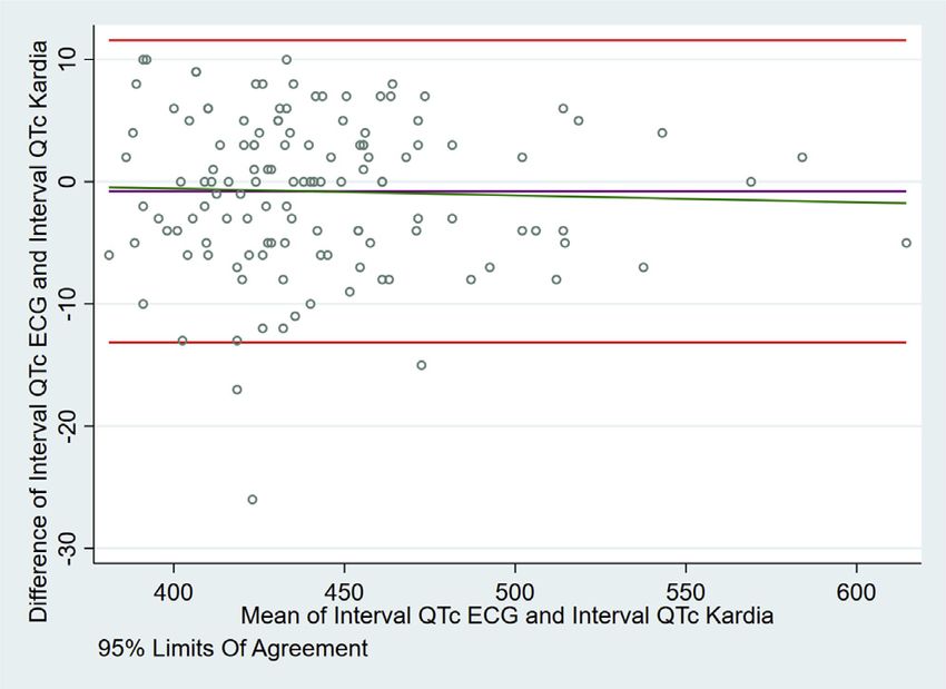
Others 16 (12.5%) The Bland–Altman analysis showed no significant differ-
SARS-CoV-2 infection, n (%) ence in QTc values between conventional 12-lead ECG
Confirmed 33 (25.8%)
Ruled out 95 (74.2%) and portable ECG device, with an average difference of
History of heart failure, n (%) 53 (41.4%) -0.797 (95% limits of agreement: -13.179; 11.585), indi-
History of blood hypertension, n (%) 78 (60.9%) cating suitable agreement between the 2 measurements
Drugs used for COVID-19, n (%) (Figure 2). Lin’s concordance coefficient showed
Lopinavir/ritonavir 1 (0.8%) an excellent agreement (0.988, 95% CI 0.983;0.992,
Hydroxychloroquine 2 (1.6%)
Azithromycin 22 (17.2%) P , .001).
None 103 (80.5%)
Potentially QT-prolonging drugs, n (%) 65 (50.8%) Numerical precision of KardiaMobile device
COVID-19 5 coronavirus disease 2019; SARS-CoV-2 5 severe acute res- Numerical precision, defined as the proportion of measure-
piratory syndrome coronavirus 2. ments in which QT interval measurement from KardiaMobileMarín O et al Portable 1L ECG Device for QTc Evaluation 385
Table 2 Subgroup analysis in function of KardiaMobile numerical
precision compared to conventional electrocardiogram
Numerical
precision†
Characteristics N Cases Percentage P
Number 128 119 93.0%
Sex
Male 61 56 91.8%
Female 67 63 94.0% .623
SARS-CoV-2 infection
Probable and confirmed 40 34 85.0%
Ruled out 88 85 96.6% .017‡
Heart failure history
Yes 53 49 92.5%
No 75 70 93.3% .848
Blood hypertension history
Figure 2 Bland–Altman analysis. No significant difference in QTc values Yes 78 74 94.9%
between conventional 12-lead electrocardiogram (ECG) and portable ECG No 50 45 90.0% .293
device are observed. Potentially QT-prolonging drugs
Yes 65 60 92.3%
No 63 59 93.7% .766
had less than 10 ms of difference with the measurement in
Data are presented as number and percentage.
conventional ECG, was 71.9% for uncorrected QT interval P value obtained through c2 test.
and 93% for QTc. The patient with the greatest variation in †
Numerical precision was defined as the proportion of KardiaMobile (Alive-
heart rate had a difference of 10 beats between the 2 assess- Cor, San Francisco, CA) QT interval measurements with less than 10 ms dif-
ment methods. ference from conventional electrocardiogram measurements.
‡
P , .05.
Numerical precision compared by subgroups only found a
difference in the SARS-CoV-2 infection category. Numerical
precision was 85% in this group of patients vs 96.6% in cases A recent study in the COVID-19 pandemic evaluated 100
in which infection was ruled out (P 5 .017). The greatest consecutive patients recruited in the ambulatory setting or in
variation in the heart rates obtained in the different evaluation the emergency room. An ECG tracing obtained with a smart
methods was observed in the group of patients with probable watch yielded corrected QT interval measurements that were
or confirmed COVID-19. No differences were found in cate- adequate for reading in 85% of patients when the device was
gories of sex, history of heart failure, blood hypertension, and worn on the left wrist. That value rose to 94% when the po-
use of QT-prolonging medications (Table 2). sition of the smart watch was modified to improve the quality
of electrocardiographic tracing and amplitude of the T wave.
Once the optimal position was found, concordance between
Concordance by conformity among tests devices was excellent, similar to the results in our study.17
Table 3 presents analysis of concordance between the KM
device and the conventional ECG to determine presence of
QTc prolongation. Prolongation corresponded to a QTc value
480 ms in female patients and 470 ms in male patients. Table 3 Estimates of concordance by conformity of QTc interval
measurement between KardiaMobile and conventional
Concordance was excellent in both groups of patients, being
electrocardiogram
slightly superior in male compared to female (kappa 5 0.946
and 0.901, respectively) (Table 3). Conventional ECG
Prolonged Nonprolonged
KardiaMobile QTc QTc Total
Discussion Male (QTc 470 ms)
Our study evaluated the agreement between a portable ECG Prolonged QTc 11 (18.0%) 0 (0%) 11 (18%)
device and the conventional ECG for QTc interval evaluation Nonprolonged QTc 1 (1.7%) 49 (80.3%) 50 (82%)
and the numerical and clinical precision of the device. The Total 12 (19.7%) 49 (80.3%) 61 (100%)
study found an excellent agreement and no statistically sig- Kappa: 0.946 6 0.127
nificant differences in the QTc interval measurement using Female (QTc 480 ms)
Prolonged QTc 5 (7.5%) 0 (0%) 5 (7.5%)
these different methods. Nonprolonged QTc 1 (1.5%) 61 (91%) 62 (92.5%)
Previous studies evaluating portable ECG device preci- Total 6 (9.0%) 61 (91%) 67 (100%)
sion for QTc were limited by the small number of evaluated Kappa: 0.901 6 0.122
patients or by the inclusion of healthy subjects and hospital- Data are shown by number (percentage of total patients in the group).
ized ill patients.15,16 Even so, the results were similar to those Kappa value 6 standard error is also shown.
in this study. ECG 5 electrocardiogram.386 Heart Rhythm O2, Vol 2, No 4, August 2021 Frisch and colleagues18 reported the evaluation of a baroreceptor stimulation as well as the regulation of cardio- KardiaMobile-6L ECG device with the accompanying Kar- vascular hormones.21–31 Postural changes from supine to diaStation tablet application to evaluate the QTc in an inpa- sitting or standing are also known to affect QT/QTc tient setting including 6 patients (3 of them with COVID- intervals or QT dispersion. Stretching of the QT interval 19), to evaluate if they were able to record their own ECG may partly explain these findings. This is the phenomenon tracings at least once without any assistance. They found that occurs when the QT interval is not shortened in that the device had the ability to provide reliable QT/QTc in- proportion to the shortening of the R-R interval (when terval measurements. Hospitalized patients were able to heart rate increases). Taking into account that the perform recordings when requested after receiving simple in- measurement of the QT interval is directly related to the R- structions at the time of first use. Unlike this 6-lead device, R interval, several mathematical formulas have been we used a single-lead device and found excellent agreement designed to “correct” the QT interval and adapt it to the compared to the conventional 12-lead ECG when evaluating heart rate or, what is the same, to the R-R interval. QTc using the Bazett formula, suggesting that a simpler de- Therefore, the clinically useful QT interval is the corrected vice could be equally reliable. Future studies are needed to QT interval.32–34 assess whether patients themselves, as reported by Frisch, When the graph obtained by applying the Bland–Altman could perform the measurement with a single-lead device. analysis was analyzed, 3 outliers were identified close to A portable ECG device with 6 leads has been evaluated in the limits of agreement, suggesting significant differences be- settings different from COVID-19. An artificial intelligence– tween the QTc measured by the device and the conventional enabled 12-lead ECG algorithm to determine the QTc was 12-lead ECG. The patients with these findings are part of the evaluated in 686 patients with genetic heart disease (50% group of patients with SARS-CoV-2 infection and they are with long QT syndrome). A strong agreement was observed the ones with the greatest differences in the heart rate values between deep neural network–predicted QTc values derived obtained by the different evaluation methods. from manual ECG (mECG) tracings and those annotated This study is, to our knowledge, the largest reported to from 12-lead ECGs by a QT expert (-0.45 6 24.73 ms), date to assess the accuracy and concordance of the KM de- with values very similar to our findings.19 These results sug- vice compared to the conventional 12-lead ECG. Our study gest that QTc measured with wearable devices could be is also among the first to evaluate the validity of the single- applied for ambulatory surveillance, which is likely to in- lead KM device specifically for the measurement of the crease over time, even after the pandemic has subsided. QTc interval. Numerical precision of KM was similar for categories of Regarding the limitations of our study, we must empha- sex, history of heart failure, blood hypertension, and use of size that it was carried out in a single center and with a single QT-prolonging drugs. An interesting finding is that concor- device. Furthermore, the number of patients with QTc inter- dance was lower in patients with confirmed SARS-CoV-2 val prolongation was relatively small. Further studies are infection compared to patients without the infection. This is required to evaluate the characteristics of KM in other clinical the first time this situation has been described. One possible settings and to strengthen the conclusions in patients with explanation is that the greatest variation in the heart rates ob- prolonged QTc. Noisy recordings have been reported with tained by the different evaluation methods was observed in the use of the KM device, which can also be a limitation. this group of patients. Autonomic abnormalities on auto- In our protocol, it was considered necessary to repeat the re- nomic tests have been described in these patients, and could cordings when noise was identified to obtain clear and inter- be related to the changes in the heart rates observed in our pretable traces. For an anterior precordial lead, the device can study.20 This may be owing to abnormalities related to the be placed on the lower left side of the chest, just below the viral infection or the clinical status of patients. It is necessary pectoral muscle.35 This way of obtaining the record was to evaluate these hypotheses in the future in other studies. not included in our protocol, but it can be a strategy to solve Differences were also identified between the QT and QTc in- the difficulty of dealing with noisy traces. tervals, which can also be explained by the variations be- The results of this study demonstrate that the single-lead tween the heart rate values described. It is important to KM has adequate precision and agreement in compare with mention that it was necessary to modify the position in which 12-lead ECG, justifying its clinical use to assess the QTc in- the electrocardiographic record was taken with the KM de- terval. Because it is quick and convenient to use, KM is ideal vice, from supine to sitting in some patients in our study, to for reducing the exposure time of healthcare workers in the improve the quality of the tracing obtained. Variation of heart COVID-19 pandemic. Further studies should evaluate rate has been associated with postural changes. Resting heart whether these characteristics are observed in outpatients or rate is faster in upright postures such as standing and sitting, in low-complexity institutions. compared with the lying position. In a recent study, it was described that postural change from supine to sitting in- creases heart rate by 10 beats per minute in both sexes, and postural change from supine to standing increases heart rate Acknowledgments by 30 beats per minute in females and males. The mecha- The authors are especially grateful to the nursing staff of the nisms proposed for such differences have been related to Cardiology Unit of Hospital Universitario San Ignacio.
Marín O et al         Portable 1L ECG Device for QTc Evaluation                                                                                                                 387

Funding Sources                                                                            10.   Torfs T, Smeets CJ, Geng D, et al. Clinical validation of a low-power and wear-
                                                                                                 able ECG patch for long term full-disclosure monitoring. J Electrocardiol 2014;
Hospital Universitario San Ignacio provided financial re-                                         47:881–889.
sources for this research.                                                                 11.   AliveCor. How-to-set-up, http://www.alivecor.com. Accessed May 2020.
                                                                                           12.   Saxon LA. Ubiquitous wireless ECG recording: a powerful tool physicians should
                                                                                                 embrace. J Cardiovasc Electrophysiol 2013;24:480–483.
Disclosures                                                                                13.   Bland JM, Altman D. Statistical methods for assessing agreement between two
                                                                                                 methods of clinical measurement. Lancet 1986;327:307–310.
The authors have no conflicts to disclose. The director of                                  14.   Lawrence I, Lin KA. A concordance correlation coefficient to evaluate reproduc-
Hospital Universitario San Ignacio provided the device                                           ibility. Biometrics 1989;255–268.
used for this project.                                                                     15.   Garabelli P, Stavrakis S, Albert M, et al. Comparison of QT interval readings in
                                                                                                 normal sinus rhythm between a smartphone heart monitor and a 12-lead ECG for
                                                                                                 healthy volunteers and inpatients receiving sotalol or dofetilide. J Cardiovasc
Authorship                                                                                       Electrophysiol 2016;27:827–832.
                                                                                           16.   Chung EH, Guise KD. QTC intervals can be assessed with the AliveCor heart
All authors attest they meet the current ICMJE criteria for                                      monitor in patients on dofetilide for atrial fibrillation. J Electrocardiol 2015;
authorship.                                                                                      48:8–9.
                                                                                           17.   Strik M, Caillol T, Ramirez FD, et al. Validating QT-interval measurement using
                                                                                                 the Apple Watch ECG to enable remote monitoring during the COVID-19
Patient Consent                                                                                  pandemic. Circulation 2020;416–418.
The Ethics and Research Committee approved the exemption                                   18.   Frisch DR, Frankel ES, Farzad DJ, et al. Initial experience in moni-
                                                                                                 toring QT intervals using a six-lead contactless mobile electrocardio-
of informed consent (Approval code: 10/2020).                                                    gram in an inpatient setting. J Innov Card Rhythm Manag 2021;
                                                                                                 12:4433–4440.
                                                                                           19.   Giudicessi JR, Schram M, Bos JM, et al. Artificial intelligence–enabled assess-
Ethics Statement                                                                                 ment of the heart rate corrected QT interval using a mobile electrocardiogram de-
The Ethics and Research Committee of the Faculty of Med-                                         vice. Circulation 2021;143:1274–1286.
icine of Pontificia Universidad Javeriana and Hospital Uni-                                 20.   Shouman K, Vanichkachorn G, Cheshire WP, et al. Autonomic dysfunction
                                                                                                 following COVID-19 infection: an early experience. Clin Auton Res 2021;1–10.
versitario San Ignacio approved the study. This study was                                  21.                  sakova M, Smetana P, et al. Sex differences in heart rate responses
                                                                                                 Hnatkova K, Si
done in accordance with the standards specified in the Inter-                                     to postural provocations. Int J Cardiol 2019;297:126–134.
national Council for Harmonization Guidelines for Good                                     22.   Borst C, Wieling W, Van Brederode JF, et al. Mechanisms of initial heart rate
                                                                                                 response to postural change. Am J Physiol 1982;243:H676–H681.
Clinical Practice and the principles of the Declaration of Hel-                            23.   Ewing DJ, Hume L, Campbell IW, et al. Autonomic mechanisms in the initial
sinki.                                                                                           heart rate response to standing. J Appl Physiol 1980;49:809–814.
                                                                                           24.   Fortrat JO, Formet C, Frutoso J, et al. Even slight movements disturb analysis of
                                                                                                 cardiovascular dynamics. Am J Physiol 1999;277:H261–H267.
References                                                                                 25.   Kamegai M, Kristensen MS, Warberg J, et al. Carotid baroreflexes and plasma
 1. Al-Khatib SM, LaPointe NMA, Kramer JM, et al. What clinicians should know                    vasopressin in humans during head-up tilt. Am J Physiol 1992;
    about the QT interval. JAMA 2003;289:2120–2127.                                              263:R318–R323.
 2. Vandael E, Vandenberk B, Vandenberghe J, et al. Risk factors for QTc-prolonga-         26.   Pump B, Christensen NJ, Videbaek R, et al. Left atrial distension and antiortho-
    tion: systematic review of the evidence. Int J Clin Pharm 2017;39:16–25.                     static decrease in arterial pressure and heart rate in humans. Am J Physiol 1997;
 3. Elsaid O, McCullough PA, Tecson KM, et al. Ventricular fibrillation storm in co-              273:H2632–H2638.
    ronavirus 2019. Am J Cardiol 2020;135:177–180.                                         27.   Pump B, Gabrielsen A, Christensen NJ, et al. Mechanisms of inhibition of vaso-
 4. Lazzerini PE, Laghi-Pasini F, Acampa M, et al. IL-6 (interleukin 6) blockade and             pressin release during moderate antiorthostatic posture change in humans. Am J
    heart rate corrected QT interval prolongation in COVID-19. Circ Arrhythm Elec-               Physiol 1999;277:R229–R235.
    trophysiol 2020;13:e008791.                                                            28.   Pump B, Kamo T, Gabrielsen A, et al. Central volume expansion is pivotal for
 5. Asensio E, Acunzo R, Uribe W, et al. Recomendaciones para la medición del in-                sustained decrease in heart rate during seated to supine posture change. Am J
    tervalo QT durante el uso de medicamentos para el tratamiento de infección por               Physiol 2001;281:H1274–H1279.
    COVID–19. Sociedad Latinoamericana del Ritmo Cardiaco (LAHRS); 2020,                   29.   Saborowski F, Krahe-Fritsch G, Krakau M, et al. The effects of orthostasis on the
    https://www.sociedadsadec.org.ar/wp-content/uploads/2020/04/RECOMEND                         ventricular-evoked response. Europace 2000;2:333–338.
    ACIONES-PARA-LA-MEDICION-DEL-INTERVALO-QT-DURANTE-EL                                   30.   Shamsuzzaman ASM, Sugiyama Y, Kamiya A, et al. Head-up suspension in hu-
    -USO-DE-MEDICAMENTOS-PARA-EL-TRATAMIENTO-DE-INFECCION                                        mans: effects on sympathetic vasomotor activity and cardiovascular responses. J
    -POR-COVID.pdf. Accessed July 6, 2021.                                                       Appl Physiol 1998;84:1513–1519.
 6. Simpson TF, Kovacs RJ, Stecker EC. Risk of Ventricular Arrhythmia Due to Hy-           31.   Jones AYM, Kam C, Lai KW, et al. Changes in heart rate and R-wave amplitude
    droxychloroquine-Azithromycin Treatment for COVID-19. Cardiology Maga-                       with posture. Chin J Physiol 2003;46:63–70.
    zine, https://www.acc.org/latest-in-cardiology/articles/2020/03/27/14/00/ventricular   32.   Davey P. Influence of posture and handgrip on the QT interval in left ventricular
    -arrhythmia-risk-due-to-hydroxychloroquine-azithromycin-treatment-for-covid -19.             hypertrophy and in chronic heart failure. Clin Sci 1999;96:403–407.
    Accessed July 6, 2021.                                                                 33.   Ghuran A, Batchvarov V, Dilaveris P, et al. Reflex autonomic modulation of auto-
 7. Haberman ZC, Jahn RT, Bose R, et al. Wireless smartphone ECG enables large-scale             matically measured repolarization parameters. Pacing Clin Electrophysiol 2000;
    screening in diverse populations. J Cardiovasc Electrophysiol 2015;26:520–526.               23:1973–1976.
 8. Galloway CD, Albert DE, Freedman SB. iPhone ECG application for community              34.   Nakagawa M, Takahashi N, Iwao T, et al. Evaluation of autonomic influences on
    screening to detect silent atrial fibrillation: a novel technology to prevent stroke.         QT dispersion using the head-up tilt test in healthy subjects. Pacing Clin Electro-
    Int J Cardiol 2013;165:193–194.                                                              physiol 1999;22:1158–1163.
 9. Narasimha D, Hanna N, Beck H, et al. Validation of a smartphone-based event            35.   AliveCor. Kardia Mobile Instructions for Use, www.alivecor.com.user-manuals/
    recorder for arrhythmia detection. Pacing Clin Electrophysiol 2018;41:487–494.               kardiamobile-instructions-for-use-en.pdf.
You can also read