Absent Cortical Venous Filling Is Associated with Aggravated Brain Edema in Acute Ischemic Stroke
←
→
Page content transcription
If your browser does not render page correctly, please read the page content below
Published March 18, 2021 as 10.3174/ajnr.A7039

                                                                                                                                   ORIGINAL RESEARCH
                                                                                                                                            ADULT BRAIN

 Absent Cortical Venous Filling Is Associated with Aggravated
                      Brain Edema in Acute Ischemic Stroke
                                          H. Xia,       H. Sun,       S. He,      M. Zhao,   W. Huang,      Z. Zhang,    Y. Xue,      P. Fu, and    W. Chen

      ABSTRACT
      BACKGROUND AND PURPOSE: Predicting malignant cerebral edema can help identify patients who may benefit from appropriate
      evidence-based interventions. We investigated whether absent cortical venous filling is associated with more pronounced early
      brain edema, which leads to malignant cerebral edema.

      MATERIALS AND METHODS: Patients with acute ischemic stroke caused by large-vessel occlusion in the MCA territory who presented
      between July 2017 and September 2019 to our hospital were included. Collateral filling was rated using the modified Tan scale on CTA,
      and good collaterals were defined as a score of 2–3. The Cortical Vein Opacification Score (COVES) was calculated, and absent cortical
      venous filling was defined as a score of 0. Early brain edema was determined using net water uptake on baseline CT images. Malignant
      cerebral edema was defined as a midline shift of $5 mm on follow-up imaging or a massive cerebral swelling leading to decompressive
      hemicraniectomy or death. Multivariate linear and logistic regression models were performed to analyze data.

      RESULTS: A total of 163 patients were included. Net water uptake was significantly higher in patients with absent than in those
      with favorable cortical venous filling (8.1% versus 4.2%; P , .001). In the multivariable regression analysis, absent cortical venous filling
      ( b ¼ 2.04; 95% CI, 0.75–3.32; P ¼ .002) was significantly and independently associated with higher net water uptake. Absent cortical
      venous filling (OR, 14.68; 95% CI, 4.03–53.45; P , .001) and higher net water uptake (OR, 1.29; 95% CI, 1.05–1.58; P ¼ .016) were signifi-
      cantly associated with increased likelihood of malignant cerebral edema.

      CONCLUSIONS: Patients with absent cortical venous filling were associated with an increased early brain edema and a higher risk
      of malignant cerebral edema. These patients may be targeted for optimized adjuvant antiedematous treatment.

      ABBREVIATIONS: COVES ¼ Cortical Vein Opacification Score; MCE ¼ malignant cerebral edema; NWU ¼ net water uptake; AIS ¼ acute ischemic stroke;
      OIT ¼ onset to imaging time; ICAS ¼ intracranial atherosclerosis

M      alignant cerebral edema (MCE) is a lethal complication
       mainly of large infarctions in the MCA territory.1 It usually
occurs within the first 3 days after the onset of stroke and is char-
                                                                                        of death, with a mortality rate approaching 80% with conservative
                                                                                        treatment.3 Three randomized controlled trials provided evide-
                                                                                        nce that early decompressive surgery is an efficient way to
acterized by an abrupt neurologic decline associated with massive                       increase the likelihood of favorable functional outcomes and
swelling of the infarcted brain tissues.2 The subsequent cerebral                       reduce the mortality rate.4 Therefore, early identification of
herniation due to intracranial pressure increase is the main cause                      patients at risk of MCE would facilitate timely selection of appro-
                                                                                        priate evidence-based interventions.
                                                                                            Net water uptake (NWU), a quantitative imaging biomarker
Received July 17, 2020; accepted after revision December 01.
                                                                                        used to estimate water uptake in ischemic stroke lesions, was first
From the Department of Radiology (H.X.), Shaoxing People’s Hospital (Shaoxing
Hospital, Zhejiang University School of Medicine), Shaoxing, Zhejiang, China; and       applied to distinguish stroke onset within and beyond 4.5 hours.5
Department of Radiology (H.S., S.H., M.Z., W.H., Z.Z., Y.X., P.F., W.C.), The First     Mounting evidence suggests the importance of NWU because it
Affiliated Hospital of Wenzhou Medical University, Wenzhou, Zhejiang, China.
This study was funded by the Zhenjiang Provincial Key Laboratory of Aging and
                                                                                        has been reported as a surrogate marker for developing MCE.6
Neurological Disorder Research (LH-001), and the Scientific Technology Planning          Recently, the arterial collateral status has been observed as it
Projects of Wenzhou, China (Y20170216).
                                                                                        relates to the early edema progression rate.7 However, the ab-
Please address correspondence to Weijian Chen, MD, Department of Radiology,
The First Affiliated Hospital of Wenzhou Medical University, Nanbai Xiang St,            sence of cortical venous filling could potentially be a better indi-
Ouhai District, Wenzhou, Zhejiang, 325000, China; e-mail: wyyycwj@163.com               cator of aggravating brain edema than the arterial collateral status
     Indicates open access to non-subscribers at www.ajnr.org                           because the venous system is responsible for approximately 70%
http://dx.doi.org/10.3174/ajnr.A7039                                                    of the cerebral blood volume.8 In a case series including 14
                                                                                                     AJNR Am J Neuroradiol :      2021   www.ajnr.org   1

                                       Copyright 2021 by American Society of Neuroradiology.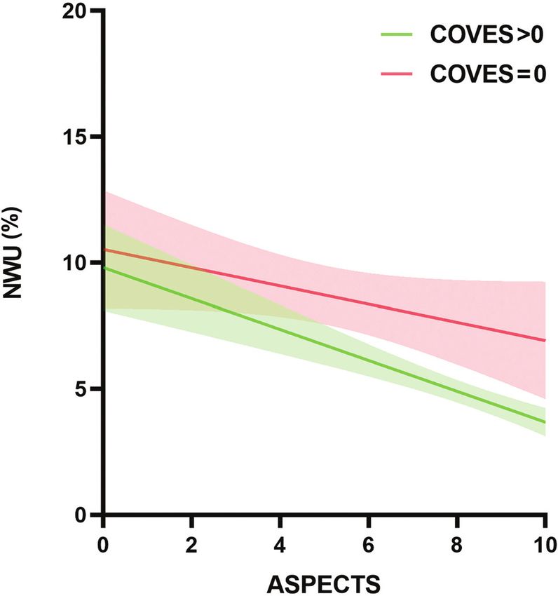
patients, cranial venous outflow abnormalities increased the rate CTA (100 kV; 500 mA; section thickness, 0.625 mm) was performed
of early fatal edema after MCA infarction.9 Nonetheless, studies with acquisition from the aortic arch to the vertex after administra-
on the role of the cortical veins in the development of MCE are tion of 40 mL of an iodinated contrast agent injected at a rate of
limited, and it remains unclear whether absent cortical venous 4.0 mL/s followed by a 30-mL saline flush.
filling is an independent risk factor affecting NWU in patients
with stroke. Image Analysis
Recently, the Cortical Vein Opacification Score (COVES) was Imaging data were independently assessed by 2 radiologists with
introduced as a simple method to assess the cortical venous filling more than 3 years of experience in stroke imaging, who were
status on CTA. It well reflects the intracranial microcirculation blinded to the clinical status and outcome data. The observers
function and can predict whether patients will benefit from endo- were informed about the laterality of symptoms just before evalu-
vascular treatment.10 Thus, the aim of our study was to investi- ation. Any disagreements between the 2 observers were resolved
gate the relationship between absent cortical venous filling, as by a senior medical chief neuroradiologist.
assessed using the COVES, and early brain edema, as assessed The ASPECTS was used to detect early ischemic burden on
using NWU, and whether they can predict the occurrence of NCCT images.11 Early ischemic burden was defined as loss of
MCE. We hypothesized that absent cortical venous filling would gray–white differentiation and tissue hypoattenuation using a
lead to an increase in early brain edema, which in turn leads to narrow window and level settings to maximize the contrast
MCE. between the normal and infarcted brain.
Collateral filling was rated using the modified Tan scale on
MATERIALS AND METHODS baseline CTA maximum intensity projection images as follows: 0
Data supporting the findings of this study are available from the indicated absent collaterals of the occluded MCA territory, 1 indi-
corresponding author on reasonable request. cated collaterals filling .0% but ,50% of the occluded MCA ter-
ritory, 2 indicated filling .50% but ,100% of the occluded MCA
Patients territory, and 3 indicated 100% collateral filling of the occluded
We retrospectively reviewed the data of patients diagnosed with MCA territory. Poor collateral filling was defined as a score of 0–
acute ischemic stroke (AIS) caused by large-vessel occlusion in 1 and good collateral filling as a score of 2–3.12
the MCA territory in our institution from July 2017 to September The COVES was used to assess the venous opacification on
2019. All procedures performed in studies involving human par- baseline CTA source images, as absent (0), moderate (1), or full
ticipants were in accordance with the ethical standards of the (2) in the vein of Labbe, sphenoparietal sinus, and superficial
institutional and national research committee and with the 1964 middle cerebral vein. The sum for the hemisphere ipsilateral to
Helsinki declaration and its later amendments or comparable the occlusion was calculated and ranged between 0 and 6. The ab-
ethical standards. Written informed consent was obtained from sence of cortical vein opacification was defined as a score of 0,
each patient or an appropriate family member. The inclusion cri- and a score .0 was considered favorable as mentioned in a previ-
teria were as follows: 1) known time window from symptom ous study.10
onset to admission imaging of ,8 hours; 2) multimodal CT per- The ischemic lesion NWU was measured by a commercial soft-
formed on admission, which included NCCT, CTP, and CTA; 3) ware, Mistar (Apollo Medical Imaging Technology), using the sin-
AIS with complete occlusion of the distal internal carotid artery, gular value deconvolution with delay and dispersion method. First,
MCA, or both, confirmed by conventional and dynamic CTA previous thresholds were applied to measure the entire ischemic
constructed from CTP; 4) follow-up CT acquired 24–72 hours af- lesion (delay time .3 s) and ischemic core (relative cerebral blood
ter symptom onset; and 5) absence of intracranial hemorrhage, or flow ,30%).13 Next, the ROI was generated automatically in the is-
pre-existing infarction. Baseline clinical characteristics and demo- chemic lesion core. Then the core-based ROI was copied to the
graphic information were recorded including age, sex, NIHSS corresponding NCCT image using Mistar software. The ROI sam-
score, onset to imaging time (OIT), IV administration of tPA, pling of the ischemic core in NCCT was assessed by densitometric
and endovascular treatment. measurements and designated as Dischemic. A mirrored ROI was
placed in the contralateral hemisphere within the normal tissue
Image Acquisition designated as Dnormal. To exclude the voxels of adjacent CSF and
All patients underwent multimodal CT at admission with NCCT, skull, a threshold value of 20–80 HU was chosen. Dischemic and
CTP, and CTA performed on a 64-section CT scanner (Lightspeed Dnormal were then used to calculate the quantitative NWU based
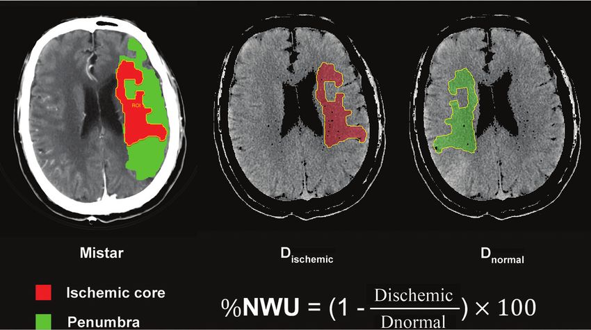
VCT; GE Healthcare). First, NCCT was performed (120 kV; on the formula from Broocks et al14 as shown in Fig 1.
300 mA; section thickness, 5 mm) from the foramen magnum to According to previous studies,15,16 MCE was defined as 1) a
the vertex to detect early ischemic changes and exclude intracranial midline shift of $5 mm at the level of septum pellucidum or 2) a
hemorrhage. If appropriate, CTP (8 cm in the z-axis using the shut- massive cerebral swelling requiring decompressive hemicraniec-
tle mode technique, 80 kV, 375 mA, 0.4 s/rotation, 6-s prescan delay tomy or leading to death.
after injection of contrast medium, 60-s total imaging duration)
with a total of 352 images by 22 consecutive spiral acquisitions was Statistical Analysis
performed immediately after NCCT. A total of 50 mL of iodinated All statistical analyses were performed using SPSS 25.0 (IBM)
contrast agent was administered at a flow rate of 5 mL/s followed and GraphPad Prism (GraphPad Software). Continuous variables
by a saline flush of 50 mL at a rate of 5 mL/s. Subsequently, helical were reported as means 6 SD or median (25th–75th percentile).
2 Xia 2021 www.ajnr.org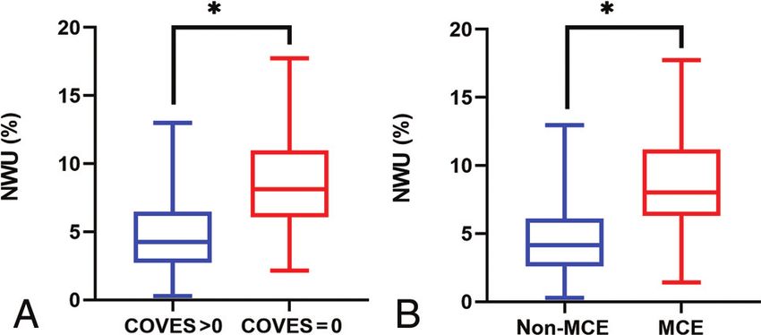
correlation analysis. The relationship
                                                                                                   between the cortical venous filling status
                                                                                                   (COVES was examined as a continuous
                                                                                                   variable and dichotomized into “absent”
                                                                                                   and “favorable” cortical venous filling)
                                                                                                   and early brain edema (NWU on
                                                                                                   NCCT images) was analyzed using
                                                                                                   linear regression. Variables with P ,.1
                                                                                                   in univariable linear regression were
                                                                                                   included in a multivariable linear
                                                                                                   regression analysis. Independent factors
                                                                                                   for MCE were evaluated using a binary
                                                                                                   logistic regression analysis. Factors with
                                                                                                   P ,.1 from the univariate analysis were
                                                                                                   entered into the multivariable logistic
                                                                                                   regression analysis. Interrater reliability
FIG 1. Quantification of NWU. NWU is determined in admission noncontrast CT. The ROI is gen-        of the COVES score was tested by k sta-
erated automatically in the ischemic lesion core, which is measured with Mistar. The mean          tistics. Results with P values of ,.05 are
attenuation of Dischemic and Dnornal are used to calculate NWU based on the formula shown.
                                                                                                   considered significant for all statistical
                                                                                                   analyses.

                                                                                                   RESULTS
                                                                                                   During the study period, data on 207
                                                                                                   consecutive patients with complete
                                                                                                   occlusion of the distal internal carotid
                                                                                                   artery, MCA, or both, undergoing mul-
                                                                                                   timodal CT were collected. A total of
                                                                                                   163 patients met all inclusion criteria
                                                                                                   and were included in the analysis. The
                                                                                                   patient selection process is shown in Fig
                                                                                                   2. The patient characteristics are listed
                                                                                                   in Table 1. The median age was 69
                                                                                                   (IQR 58–78) years, and 103 (63%)
                                                                                                   patients were men. The baseline NIHSS
                                                                                                   score was 11.34 6 5.43, and the median
                                                                                                   OIT was 191 (IQR 116–259) minutes.
                                                                                                   Among all patients, 82 (50%) under-
                                                                                                   went endovascular treatment, and 68
                                                                                                   (42%) received IV thrombolysis. There
                                                                                                   were 112 (69%) patients with good col-
                                                                                                   lateral filling. At baseline, NWU was
                                                                                                   4.9% (IQR 2.98–7.33).
                                                                                                        Comparing patients with absent
                                                                                                   (COVES ¼ 0) versus favorable corti-
                                                                                                   cal venous filling (COVES .0), the
                                                                                                   former had a significantly higher
FIG 2. Flowchart of patient selection.                                                             baseline NIHSS score (13.5 versus 11;
                                                                                                   P ¼ .001), lower ASPECTS (4.5 ver-
                                                                                                   sus 9; P , .001), and a lower rate of
                                                                                                   good collateral filling (22% versus
Categoric variables were reported as numbers (percentages). The        80%; P , .001). No significant differences were found in age,
differences among groups were evaluated using the Student t test       sex, OIT, endovascular treatment, or IV thrombolysis (Table 2).
for normally distributed continuous data, Mann-Whitney U test               Absent cortical venous filling correlated positively with NWU
for non-normally distributed continuous data, and Pearson x 2          (Kendall t ¼ 0.37; 95% CI, 0.27–0.46; P , .001). The collateral
test for categoric data. The correlation between collateral filling,   filling and NWU also showed a significant correlation (Kendall t
cortical venous filling status, and NWU was described by Kendall       ¼  0.30; 95% CI, 0.41 to 0.19; P , .001). Moreover, cortical
                                                                                    AJNR Am J Neuroradiol :     2021   www.ajnr.org      3
Table 1: Patient characteristics according to the presence of MCE
MCE (n = 39) Non-MCE (n = 124)
Patient Characteristic Overall (n = 163) 24% 76% P Value
Age in years, median (IQR) 69 (58–78) 74 (61–81) 69 (57.25–76) .131
Male sex, n (%) 103 (63%) 20 (51%) 83 (67%) .077
Admission NIHSS, mean 6 SD 11.34 6 5.43 14.03 6 5.11 10.49 6 5.27 ,.001
OIT in minutes, median (IQR) 191 (116–259) 201 (128–277) 189.5 (115.25–258.75) .858
IV thrombolysis, n (%) 68 (42%) 12 (31%) 56 (45%) .112
Endovascular treatment, n (%) 82 (50%) 19 (49%) 63 (51%) .820
Good collateral filling, n (%) 112 (69%) 12 (31%) 100 (81%) ,.001
ASPECTS, median (IQR) 9 (7–10) 4 (2–7) 9 (8–10) ,.001
COVES = 0, n (%) 32 (20%) 26 (67%) 6 (5%) ,.001
NWU in %, median (IQR) 4.89 (2.98–7.33) 8.01 (6.29–11.19) 4.15(2.59–6.11) ,.001
Table 2: Patient characteristics according to the COVES
COVES >0 (n = 131) COVES =0 (n = 32)
Patient Characteristic 80% 20% P Value
Age in years, median (IQR) 70 (60–77) 68.5 (53.75–78.75) .383
Male sex, n (%) 84 (64%) 19 (60%) .618
Admission NIHSS, median (IQR) 11 (6–14) 13.5 (10–18) .001
OIT in minutes, median (IQR) 188 (118–261) 205 (110.5–252.75) .910
IV thrombolysis, n (%) 58 (44%) 10 (31%) .180
Endovascular treatment, n (%) 65 (50%) 17 (53%) .722
Good collateral filling, n (%) 105 (80%) 7 (22%) ,.001
ASPECTS, median (IQR) 9 (7–10) 4.5 (2.25–7.75) ,.001
NWU in %, median (IQR) 4.24 (2.73–6.46) 8.11 (6.08–11.00) ,.001
versus 10.49; P , .001) and NWU (8.0% versus 4.2%; P , .001)
(Fig 3), lower ASPECTS (4 versus 9; P , .001), higher rate of
absent cortical venous filling (67% versus 4.8%; P , .001), and
lower rate of good collateral filling (31% versus 81%, P , .001)
compared with patients without MCE.
In the univariable logistic regression analysis, multiple factors
were associated with MCE. The significant variables from the
univariate analysis were entered into the multivariate logistic
regression. Male sex (OR, 0.31; 95% CI, 0.096–0.987; P ¼ .047),
ASPECTS (OR, 0.77; 95% CI, 0.60–0.97; P ¼ .029), absent cortical
FIG 3. Relationship of cortical venous filling, early brain edema, and venous filling (OR, 14.68; 95% CI, 4.03–53.44; P , .001), and
MCE. A, Boxplot shows that NWU was significantly higher in patients NWU (OR, 1.29; 95% CI, 1.05–1.58; P ¼ .016) were significantly
with COVES = 0 than in those with COVES .0. B, Boxplot shows that associated with increased likelihood of MCE after adjustment for
NWU was significantly higher in patients with MCE than in those
without MCE. * indicates P , .001. confounding factors (Table 3).
Interobserver Analysis
venous filling significantly correlated with collateral filling
Interrater reliability for COVES score on baseline CTA source
(Kendall t ¼ 0.50; 95% CI, 0.64 to 0.34; P , .001).
images was substantial (k = 0.75).
Impact of Cortical Venous Filling on Early Brain Edema
NWU was significantly higher in patients with absent cortical ve- DISCUSSION
nous filling than in those with favorable cortical venous filling The aim of our study was to investigate the relationship between
(8.1% versus 4.2%; P , .001) (Fig 3). Furthermore, as seen in absent cortical venous filling and early brain edema assessed
Table 2 and Table 3, in the multivariable linear regression analy- using NWU and its impact on the occurrence of MCE. Our study
ses, the ASPECTS ( b ¼ 0.50; 95% CI, 0.70 to 0.30; revealed that absent cortical venous filling was strongly associated
P , .001) and absent cortical venous filing ( b ¼ 2.04; 95% CI, with higher early brain edema, and both absent cortical venous
0.75 to 3.32; P ¼ .002) were 2 significant factors associated with filling and higher NWU were independent predictors of the risk
NWU adjusted for baseline NIHSS score and collateral filling for the development of MCE in patients with large-vessel occlu-
(Table 3 and Fig 4). sion in the MCA territory.
These results may support our original hypothesis that absent
Factors Associated with MCE cortical venous filling is responsible for the onset of MCE by
A total of 39 (24%) patients developed MCE. As shown in Table inducing early brain edema. The underlying pathophysiological
1, patients with MCE had higher baseline NIHSS score (14.03 mechanisms of MCE may be abnormal venous drainage; the
4 Xia 2021 www.ajnr.orgTable 3: Univariable and multivariable linear regression analyses to predict NWU and univariable and multivariable logistic regres-
sion analyses to predict MCE
Impact on NWU in multivariate regression
Parameter Coefficient 95% CI P Value
Univariable analysis
Age (1-year increase) 0.009 0.03–0.05 .667
Male sex -0.29 1.38–0.79 .597
Admission NIHSS 0.17 0.07–0.26 .001
Onset to imaging time (1 minute) 0.004 0.001–0.009 .147
Good collateral filling -2.90 3.93–1.86 ,.001
ASPECTS -0.70 0.86–0.54 ,.001
COVES = 0 3.99 2.83–5.15 ,.001
Multivariable analysis
Admission NIHSS -0.01 0.08–0.10 .811
Good collateral filling -0.46 1.59–0.67 .423
ASPECTS -0.50 0.70–0.30 ,.001
COVES = 0 2.04 0.75–3.32 .002
Prediction of MCE
Parameter
Univariable analysis
Age (1-year increase) 1.02 0.99–1.05 .300
Male sex 0.52 0.25–1.08 .079
Admission NIHSS 1.14 1.06–1.23 .001
OIT (1 minute) 1.001 0.997–1.004 .733
IV thrombolysis 0.54 0.25–1.16 .115
Endovascular treatment 0.92 0.45–1.89 .820
Good collateral filling 0.11 0.05–0.24 ,.001
ASPECTS 0.59 0.50–0.70 ,.001
COVES = 0 39.33 13.68–113.12 ,.001
NWU 1.58 1.35–1.85 ,.001
Multivariable analysis
Male sex 0.31 0.10–0.99 .047
Admission NIHSS 1.02 0.91–1.35 .730
Good collateral filling 0.75 0.22–2.63 .657
ASPECTS 0.77 0.60–0.97 .029
COVES = 0 14.68 4.03–53.45 ,.001
NWU 1.29 1.05–1.58 .016
subsequent resistance to CSF absorption would lead to increased
venous pressure, which would increase the leakage of fluid into
the perivascular space, leading to brain edema.17 Our results are
in accordance with those of a recent study that investigated the
association between quantitative lesion water uptake and malig-
nant infarction.6 The authors concluded that NWU is a signifi-
cant predictor for developing MCE. We further found that absent
cortical venous filling was significantly associated with higher
early brain edema and could predict the occurrence of MCE.
Thus, these findings can be of great clinical significance in
patients without cortical venous filling and can indicate who
should receive adjuvant treatment with neuroprotectants and
edema prevention therapies such as IV glibenclamide.18
However, whether absent cortical venous filling plays any other
role in the development of MCE needs to be further elucidated.
Previous studies showed that collateral filling is significantly
associated with an early edema progression rate and plays a role
in the development of MCE.7,19 Our univariate analysis showed
that patients with MCE had a significantly lower rate of good col-
lateral filling than those without MCE. However, in the multivari-
ate analysis, this factor could not predict NWU or the occurrence
of MCE. This may be due to the high correlation between cortical
FIG 4. Impact of absent cortical venous filling on early brain edema venous filling and collateral filling (Kendall t = 0.499; 95% CI,
according to baseline ASPECTS. 0.638 to 0.342; P , .001); however, no collinearity was found
AJNR Am J Neuroradiol : 2021 www.ajnr.org 5between them. A previous nonhuman primate model showed this study can be attributed to the large variation in venous anat-
that absent cortical venous filling indicated a less extensive collat- omy, though the 3 cortical veins chosen for the COVES had lesser
eral circulation.20 A recent clinical study that included 228 anatomic variability than that of other veins. Third, NWU mea-
patients with AIS also demonstrated that venous outflow is surement may be imprecise in very small ischemic core volumes.
affected by the reduction of arterial flow.21 Therefore, this sug- However, we improved the precision using the Mistar software to
gests that cortical venous filling can provide additional informa- measure the ischemic core assisting in determining the ROI.
tion on both the occurrence of MCE and arterial collateral status. Furthermore, the recanalization status is a significant predictor of
From our results, we can reasonably infer that cortical veins may the occurrence of MCE. However, in this study, we did not
play a more important role than arterial collateral in the develop- include this factor mainly because many patients without endo-
ment of MCE. These findings prompt us to combine arterial col- vascular treatment were unable to obtain recanalization data
lateral and cortical vein to better evaluate patients with stroke, because of the absence of follow-up vascular imaging. Finally, we
resulting in better treatment. could not determine whether absent cortical venous filling leads
Some clinical factors may also predict MCE. Previous studies to MCE because of early brain edema; we aim to perform a medi-
in Western countries showed that younger age significantly ation analysis to explore this hypothesis more thoroughly in
increased the risk of MCE.22,23 The possible reason for this is that future studies.
the brain volume gradually decreases with increasing age; thus,
younger patients are more likely to develop MCE because there is CONCLUSIONS
a lower intracranial volume to accommodate brain edema.1 We observed that absent cortical venous filling was significantly
However, the results of our study did not show the same findings. associated with early brain edema and the occurrence of MCE.
This discrepancy may be attributed to the differences in the etiol- Early brain edema may be the link between absent cortical venous
ogic composition of patients with stroke between China and filling and MCE. The clinical implication of these results is that
Western countries. Intracranial atherosclerosis (ICAS) is common absent cortical venous filling may assist in the selection of
in the Chinese population, accounting for 33%–50% of AIS cases.24 patients at risk for MCE in whom optimized adjuvant antiedema-
In contrast, AIS is most commonly caused by cardioembolism in tous treatment would be indicated.
Western countries. Previous clinical research showed patients with
cardioembolism tend to be older than those with ICAS25 and are Disclosures: Weijian Chen—RELATED: Grant: Zhenjiang Provincial Key Laboratory
more likely to experience a large hemispheric infarction,26 which is of Aging and Neurological Disorder Research (LH-001), the Scientific Technology
Planning Projects of Wenzhou and China (Y20170216).
the main cause of MCE.27 Our patients may have a higher propor-
tion of ICAS, which makes the effect of age on MCE insignificant.
However, our study did not measure the proportions of each etiol- REFERENCES
1. Hacke W, Schwab S, Horn M, et al. “Malignant” middle cerebral ar-
ogy and the role of etiology in age and MCE because this was not
tery territory infarction: clinical course and prognostic signs. Arch
the primary objective of this study. Another clinical factor, female Neurol 1996;53:309–15 CrossRef Medline
sex, was shown to be an independent predictor of MCE, which is 2. Qureshi AI, Suarez JI, Yahia AM, et al. Timing of neurologic deteri-
consistent with the results of previous studies28,29 indicating that a oration in massive middle cerebral artery infarction: a multicenter
more efficacious stroke management may be needed in female review. Crit Care Med 2003;31:272–77 CrossRef Medline
patients. Although research efforts with a focus on sex differences 3. Berrouschot J, Sterker M, Bettin S, et al. Mortality of space-occupy-
ing (“malignant”) middle cerebral artery infarction under conserv-
in neuroinflammation, neuroprotection, and neuronal cell death
ative intensive care. Intensive Care Med 1998;24:620–23 CrossRef
signaling have recently received more attention,30-32 the reasons Medline
for this phenomenon remain obscure. 4. Vahedi K, Hofmeijer J, Juettler E, et al. Early decompressive surgery
To the best of our knowledge, this is the first study to investi- in malignant infarction of the middle cerebral artery: a pooled anal-
gate the relationship between cortical venous filling, early brain ysis of three randomised controlled trials. Lancet Neurol 2007;6:215–
edema, and MCE. One of the strengths of our study is that early 22 CrossRef Medline
5. Minnerup J, Broocks G, Kalkoffen J, et al. Computed tomography-
brain edema was directly assessed by NWU, which calculates the
based quantification of lesion water uptake identifies patients
water uptake based on the CT attenuation and has been proved within 4.5 hours of stroke onset: a multicenter observational study.
as the criterion standard for histopathologic water uptake volume Ann Neurol 2016;80:924–34 CrossRef Medline
measurement.33,34 In addition, we selected the COVES to evalu- 6. Broocks G, Flottmann F, Scheibel A, et al. Quantitative lesion water
ate intracranial venous filling. This scoring method removes the uptake in acute stroke computed tomography is a predictor of ma-
lignant infarction. Stroke 2018;49:1906–12 CrossRef Medline
deep cerebral venous system and more accurately reflects the sta-
7. Broocks G, Kemmling A, Meyer L, et al. Computed tomography an-
tus of the microcirculation.10,35 Although dynamic CTA may be a giography collateral profile is directly linked to early edema pro-
better way to assess cortical venous filling than conventional gression rate in acute ischemic stroke. Stroke 2019;50:3424–30
CTA,36 this technique requires advanced imaging and complex CrossRef Medline
postprocessing. Our method was simple and convenient to use 8. Ito H, Kanno I, Iida H, et al. Arterial fraction of cerebral blood vol-
and has achieved good interobserver agreement, as previously ume in humans measured by positron emission tomography. Ann
Nucl Med 2001;15:111–16 CrossRef Medline
described.10
9. Yu W, Rives J, Welch B, et al. Hypoplasia or occlusion of the ipsilat-
This study has several limitations. First, it was a retrospective eral cranial venous drainage is associated with early fatal edema of
single-center study, and the number of patients was relatively middle cerebral artery infarction. Stroke 2009;40:3736–39 CrossRef
small; thus, selection bias inevitably existed. Another drawback of Medline
6 Xia 2021 www.ajnr.org10. Jansen IGH, van Vuuren AB, van Zwam WH, et al. Absence of corti- 23. Wu S, Yuan R, Wang Y, et al. Early prediction of malignant brain
cal vein opacification is associated with lack of intra-arterial ther- edema after ischemic stroke. Stroke 2018;49:2918–27 CrossRef Medline
apy benefit in stroke. Radiology 2018;286:643–50 CrossRef Medline 24. Wang Y, Zhao X, Liu L, et al. Prevalence and outcomes of sympto-
11. Pexman JH, Barber PA, Hill MD, et al. Use of the Alberta Stroke matic intracranial large artery stenoses and occlusions in China:
Program Early CT Score (ASPECTS) for assessing CT scans in the Chinese Intracranial Atherosclerosis (CICAS) Study. Stroke
patients with acute stroke. AJNR Am J Neuroradiol 2001;22:1534–42 2014;45:663–69 CrossRef Medline
Medline 25. Lee JS, Hong JM, Lee KS, et al. Endovascular therapy of cerebral ar-
12. Tan IY, Demchuk AM, Hopyan J, et al. CT angiography clot burden terial occlusions: intracranial atherosclerosis versus embolism. J
score and collateral score: correlation with clinical and radiologic Stroke Cerebrovasc Dis 2015;24:2074–80 CrossRef Medline
outcomes in acute middle cerebral artery infarct. AJNR Am J 26. Rebello LC, Bouslama M, Haussen DC, et al. Stroke etiology and col-
Neuroradiol 2009;30:525–31 CrossRef Medline laterals: atheroembolic strokes have greater collateral recruitment
13. Lin L, Bivard A, Kleinig T, et al. Correction for delay and dispersion than cardioembolic strokes. Eur J Neurol 2017;24:762–67 CrossRef
results in more accurate cerebral blood flow ischemic core mea- Medline
surement in acute stroke. Stroke 2018;49:924–30 CrossRef Medline 27. Wartenberg KE. Malignant middle cerebral artery infarction. Curr
14. Broocks G, Flottmann F, Ernst M, et al. Computed tomography- Opin Crit Care 2012;18:152–63 CrossRef Medline
based imaging of voxel-wise lesion water uptake in ischemic brain: 28. Kasner SE, Demchuk AM, Berrouschot J, et al. Predictors of fatal
relationship between density and direct volumetry. Invest Radiol brain edema in massive hemispheric ischemic stroke. Stroke
2018;53:207–13 CrossRef Medline 2001;32:2117–23 CrossRef Medline
15. Cheng Y, Wu S, Wang Y, et al. External validation and modification 29. Jaramillo A, Góngora-Rivera F, Labreuche J, et al. Predictors for ma-
of the edema score for predicting malignant brain edema after acute lignant middle cerebral artery infarctions: a postmortem analysis.
ischemic stroke. Neurocrit Care 2020;32:104–12 CrossRef Medline Neurology 2006;66:815–20 CrossRef Medline
16. Kauw F, Bennink E, de Jong H, et al. Intracranial cerebrospinal fluid 30. Klein SL, Marriott I, Fish EN. Sex-based differences in immune
volume as a predictor of malignant middle cerebral artery infarc- function and responses to vaccination. Trans R Soc Trop Med Hyg
tion. Stroke 2019; May 16 [Epub ahead of print] CrossRef Medline 2015;109:9–15 CrossRef Medline
17. Alperin N, Lee SH, Mazda M, et al. Evidence for the importance of 31. Mirza MA, Ritzel R, Xu Y, et al. Sexually dimorphic outcomes and
extracranial venous flow in patients with idiopathic intracranial inflammatory responses in hypoxic-ischemic encephalopathy. J
hypertension (IIH). Acta Neurochir Suppl 2005;95:129–32 CrossRef Neuroinflammation 2015;12:32 CrossRef Medline
Medline 32. Spychala MS, Honarpisheh P, McCullough LD. Sex differences in
18. Vorasayan P, Bevers MB, Beslow LA, et al. Intravenous glibencla- neuroinflammation and neuroprotection in ischemic stroke. J
mide reduces lesional water uptake in large hemispheric infarc- Neurosci Res 2017;95:462–71 CrossRef Medline
tion. Stroke 2019;50:3021–27 CrossRef Medline 33. Broocks G, Flottmann F, Ernst M, et al. Computed tomography-
19. Horsch AD, Dankbaar JW, Stemerdink TA, et al. Imaging findings based imaging of voxel-wise lesion water uptake in ischemic brain:
associated with space-occupying edema in patients with large mid- relationship between density and direct volumetry. Invest Radiol
dle cerebral artery infarcts. AJNR Am J Neuroradiol 2016;37:831–37 2018;53:207–13 CrossRef
CrossRef Medline 34. Dzialowski I, Klotz E, Goericke S, et al. Ischemic brain tissue water
20. Sasaki M, Honmou O, Radtke C, et al. Development of a middle cer- content: CT monitoring during middle cerebral artery occlusion
ebral artery occlusion model in the nonhuman primate and a and reperfusion in rats. Radiology 2007;243:720–26 CrossRef Medline
safety study of I.V. infusion of human mesenchymal stem cells. 35. Hoffman H, Ziechmann R, Swarnkar A, et al. Cortical vein opacifi-
PloS One 2011;6:e26577 CrossRef Medline cation for risk stratification in anterior circulation endovascular
21. Zhang S, Lai Y, Ding X, et al. Absent filling of ipsilateral superficial thrombectomy. J Stroke Cerebrovasc Dis 2019;28:1710–17 CrossRef
middle cerebral vein is associated with poor outcome after reperfu- Medline
sion therapy. Stroke 2017;48:907–14 CrossRef Medline 36. van den Wijngaard IR, Wermer MJ, Boiten J, et al. Cortical venous
22. Miao J, Song X, Sun W, et al. Predictors of malignant cerebral filling on dynamic computed tomographic angiography: a novel
edema in cerebral artery infarction: a meta-analysis. J Neurol Sci predictor of clinical outcome in patients with acute middle cere-
2020;409:116607 CrossRef Medline bral artery. Stroke 2016;47:762–67 CrossRef
AJNR Am J Neuroradiol : 2021 www.ajnr.org 7You can also read