Properties of macrophages and lymphocytes appearing in rat renal fibrosis followed by repeated injection of cisplatin
←
→
Page content transcription
If your browser does not render page correctly, please read the page content below
FULL PAPER
                                 Pathology

                                 Properties of macrophages and lymphocytes
                                 appearing in rat renal fibrosis followed by
                                 repeated injection of cisplatin
                                 Satoshi MATSUYAMA1)#, Munmun PERVIN1,2)#, Minto NAKAGAWA1),
                                 Takeshi IZAWA1), Mitsuru KUWAMURA1) and Jyoji YAMATE1)*
                                 1)Laboratory  of Veterinary Pathology, Graduate School of Life and Environmental Sciences, Osaka Prefecture
                                   University, 1-58 Rinku-Ourai-Kita, Izumisano-shi, Osaka 598-8531, Japan
                                 2)Department of Pathology, Faculty of Veterinary Science, Bangladesh Agricultural University,

                                   Mymensingh-2202, Bangladesh

                                    ABSTRACT. Properties of macrophages and lymphocytes appearing in renal fibrosis remains
                                    to be investigated. F344 rats were injected once a week with cisplatin (2 mg/kg body weight)
                                    for 8 weeks and examined at post-final injection weeks 1, 3, 6, 9, and 12. Rats developed
                                    progressive renal fibrosis at weeks 1 to 6 as fibrosis-progress phase, and subsequent amelioration
                                    at weeks 9 and 12. CD68+ M1-macrophages and major histocompatibility complex (MHC) class
                                    II+ macrophages remarkably increased persistently, whereas CD163+ M2-macrophages slightly
                                    increased. MHC class II+/CD68+ and MHC class II+/CD163+ macrophages were present, indicating
                                    that MHC class II+ macrophages might have both functions of M1- and M2-macrophages. In the
                                    fibrosis-progress phase, interleukin (IL)-6, tumor necrosis factor (TNF)-α, and interferon (IFN)-γ for
J. Vet. Med. Sci.                   M1-factors, and transforming growth factor (TGF)-β1 and IL-10 for M2-factors tended to increase;
                                    tissue injury by M1 and fibrosis by M2 might have occurred simultaneously. Lots of CD4+ and
83(9): 1435–1442, 2021
                                    CD8+ T cells appeared in close relation with MHC class II+ macrophages, and mainly CD4+ T cells
doi: 10.1292/jvms.21-0341           formed aggregations. In the lymphocyte aggregates collected by laser microdissection, expression
                                    of IL-17A (for Th17 cells) and forkhead box P3 (FoxP3) (for Treg) significantly increased at weeks 1
                                    and 6, respectively; presumably, Th17 cells might be involved in tissue injury, whereas Treg might
Received: 11 June 2021              be related to fibrosis amelioration. These results suggested that macrophages and T cells may
Accepted: 12 July 2021              contribute interrelatedly to renal fibrosis.
Advanced Epub: 		                   KEY WORDS: cisplatin, macrophage, T cell, rat, renal fibrosis
          22 July 2021

   Regardless of initial causes, renal fibrosis is the common final pathway in chronic kidney disease (CKD) [4, 34]. The fibrosis
leads occasionally to renal dysfunction, and the patients with CKD need therapies to improve renal function (such as dialytic
treatment) or transplantation [4]. Renal fibrosis is considered to be the risk factor of cardiovascular disease such as arteriosclerosis
and brain stroke [19]. Therefore, the pathogenesis of renal fibrosis should be clarified, in order to prevent and improve CKD.
Cisplatin (CDDP) is an anti-cancer drug that has nephrotoxicity as a side effect. CDDP damages the epithelium of proximal
renal tubules, especially in the S3 segment, and thereafter, induces renal interstitial fibrosis at the advanced stages [14, 31]. In
renal injury in rats injected by a single injection of CDDP (6 mg/kg body weight (BW)), it is reported that macrophages with
heterogeneous immunophenotypes, and lymphocytes, mainly T cells, participated in the pathology of renal fibrosis [14]. In
the present study, we induced greater renal damage in rats by repeated injections of CDDP (2 mg/kg BW, once a week for 8
weeks), and analyzed the properties of macrophages and lymphocytes appearing after the cessation of the injection. In particular,
we focused on major histocompatibility complex (MHC) class II-expressing macrophages, which may stimulate lymphocyte
infiltration and regulates the fibrosis conditions [8, 12, 14, 35] and MHC class II macrophages are known to play important role in
normal tissue genesis as the modeling (vs. remodeling of fibrosis) [11, 36].

*Correspondence to: Yamate, J.: yamate@vet.osakafu-u.ac.jp
#These authors contributed equally to this work.

©2021 The Japanese Society of Veterinary Science
                  This is an open-access article distributed under the terms of the Creative Commons Attribution Non-Commercial No Derivatives (by-nc-nd)
                  License. (CC-BY-NC-ND 4.0: https://creativecommons.org/licenses/by-nc-nd/4.0/)

                                                                                                                                                  1435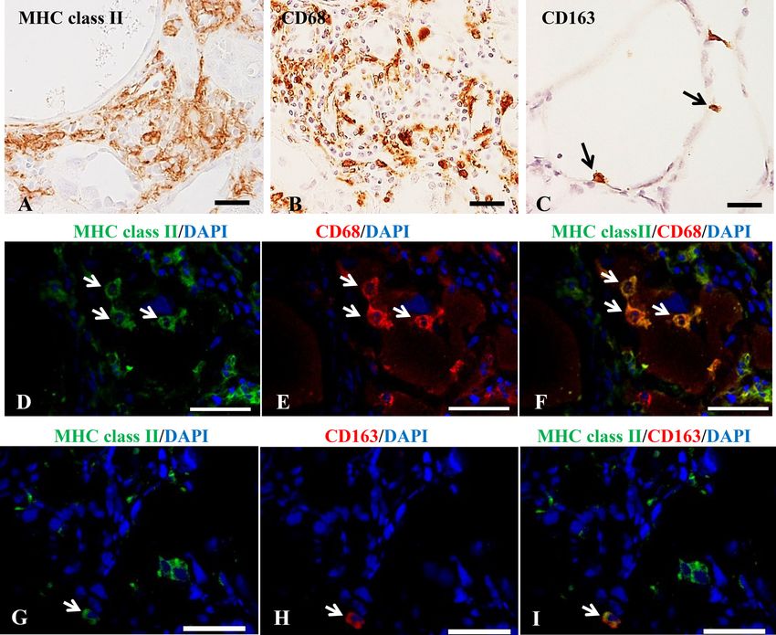
S. MATSUYAMA ET AL.

MATERIALS AND METHODS
Animals and experimental procedures
   A total of twenty-four male F344/DuCrj rats (5 weeks old; 106–146 g; Charles River Japan, Hino, Japan) were housed in an
animal room controlled at 22 ± 2°C and with a 12 hr light-dark cycle and fed a standard diet (DC-8, CLEA, Tokyo, Japan) and
tap water ad libitum. After one-week of acclimatization, twenty rats were injected with CDDP intraperitoneally once a week for 8
weeks at a dose of 2 mg/kg BW. At the post-final injection (PFI) weeks 1, 3, 6, 9 and 12, four rats were examined each. Four rats
as controls received an equal volume of phosphate buffer saline (PBS) in the same manner and were examined at PFI week 1. All
rats were euthanized under deep isoflurane anesthesia.
   Experimental procedures were in agreement with institutional guidelines (approved by the ethical committee of Osaka Prefecture
University; Nos. 23-24 and 25-85) for the Care and Use of Experimental Animals and were conducted in accordance with basic
policies for the conduct of animal experimentation of the Ministry of Health, Labor and Welfare Standards relating to the Care and
Management of Experimental Animals and the Act on Welfare and Management of Animals, Japan.

Histopathology and immunohistochemistry
   Kidneys were immediately fixed in a 10% neutral buffered formalin or periodate-lysine-paraformaldehyde (PLP) solution.
Deparaffinized sections were stained with hematoxylin and eosin (HE) for morphological observations. PLP-fixed deparaffinized
tissue sections were immunostained with mouse monoclonal antibodies specific for CD68 (clone ED1, IgG1, 1:500, AbD Serotec,
Oxford, UK), CD163 (clone ED2, IgG1, 1:300, AbD Serotec), MHC class II (clone OX6, IgG1, 1:1,000, AbD Serotec), CD4
(clone W3/25, IgG1, 1:2,000, AbD Serotec), CD8 (clone OX-8, IgG1, 1:200, AbD Serotec) and CD79α (clone HM57, IgG1κ,
1:5, Nichirei Bioscience Inc. Tokyo, Japan) using Histostainer (Nichirei Bioscience Inc.). Briefly, sections were incubated with
5% skimmed milk for 10 min, followed by 1 hr incubation with the primary antibody. After treatment with 3% H2O2 for 15
min, horseradish peroxidase-conjugated secondary antibody (Histofine Simple Stain MAX PO®; Nichirei Bioscience Inc.) was
applied for 30 min. Positive reactions were visualized with 3, 3′-diaminobezidine tetrahydrochloride (DAB substrate kit, Nichirei
Bioscience Inc.), and the sections were lightly counterstained with hematoxylin. For negative controls, tissue sections were treated
with non-immunized mouse serum instead of the primary antibody.

Reverse-transcription polymerase chain reaction (RT-PCR) and laser microdissection (LMD)
   Renal samples were collected from the affected cortico-medullary junction (corresponding roughly to the outer segment of the
outer medulla) and immediately immersed in RNAlater reagent (Qiagen, Hilden, Germany). Total RNA was extracted with an SV
total RNA isolation system (Promega, Fitchburg, WI, USA) and was reverse transcribed to cDNA with SuperScript VILO reverse
transcriptase (Life technologies, Carlsbad, CA, USA). Real-time PCR was performed with SYBR Green Real-time PCR Master
Mix (Toyobo Co., Ltd., Osaka, Japan) in a PicoReal 96 Real-time PCR system (Thermo Scientific, Waltham, MA, USA). Primers
used were as follows: interleukin (IL)-6 (forward, tccgtttctacctggagtttgtg; reverse, agagcattggaagttggggtag), interferon (IFN)-γ
(forward, tcgcacctgatcactaacttcttc; reverse, cgactccttttccgcttcc), tumor necrosis factor (TNF)-α (forward, tgcctcagcctcttctcattc;
reverse, gctcctctgcttggtggttt), IL-1β (forward, gcaccttcttttccttcatctttg; reverse, tttgtcgttgcttgtctctcctt), transforming growth factor
(TGF)-β1 (forward, cttcagctccacagagaagaactgc; reverse, cacgatcatgttggacaactgctcc), IL-10 (forward, ctgtcatcgatttctcccctgt;
reverse, cagtagatgccgggtggttc), FoxP3 (forward, cttgtttgctgtgcggagac; reverse, tgagggtggcataggtgaaa) and β-actin (forward,
taagaccctctatgccaacac; reverse, ctcctgcttgctgatccacat). For analysis of IL-17A expression, real-time PCR was performed with
Thunderbird Probe qPCR Mix (Toyobo Co., Ltd.) with TaqMan Gene Expression Assays (Rn01757168_m1, Life Technologies).
The data were analyzed using the comparative Ct method (ΔΔCt method) and were normalized to β-actin mRNA as an internal
control. For LMD, 10-micrometer fresh frozen sections of the controls and affected kidneys at PFI weeks 1, 6 and 12 were
prepared on foil slides (Leica Microsystems, Wetzlar, Germany). Sections were fixed in 19:1 ethanol/acetic acid for 3 min and
were stained with 0.01% toluidine blue. The cortico-medullary junction areas of controls and the affected kidneys with lymphocyte
aggregation were confirmed under microscopic observation, and were isolated with the LMD7000 system (Leica Microsystems).
The LMD-isolated samples were subjected to RNA extraction followed by RT-PCR for FoxP3, and IL-17A as mentioned above.

Double immunofluorescence
   To investigate the co-localization between macrophage-specific antigens, double immunofluorescence was carried out using
antibody against MHC class II in combination with CD68 or CD163 antibody in fresh-frozen renal tissue sections (10 µm thick)
from the control and CDDP-injected rats. Briefly, after fixation in acetone: methanol (1:1) for 10 min at 4°C, the sections were
incubated with 10% normal goat serum for 30 min. The sections reacted with the primary antibody overnight at 4°C. After rinsing
with PBS, the sections were incubated for 45 min with the following secondary antibodies: Alexa568/anti-mouse IgG1, Alexa488/
anti-mouse IgG2a (Invitrogen Co., ×1,000). The sections were visualized with Vectashield mounting medium containing 4ˈ,
6-diamidino-2-phenylindole (DAPI; Vector Laboratories Inc.) for nuclear staining and analyzed using a virtual slide scanner (VS-
120, Olympus, Tokyo, Japan).

Statistics and evaluation of immunopositive cells
   Difference between control and CDDP-injected rats were evaluated by Dunnet’s test. Values of P
MACROPHAGES AND LYMPHOCYTES IN RENAL FIBROSIS

no immunopositive cells; ±, a few immunopositive cells, sporadically; +, a small number of immunopositive cells, sporadically or
diffusely; ++, a moderate number of immunopositive cells, diffusely or focally; and +++, a large number of immunopositive cells,
diffusely, focally or aggregately (for lymphocytes) (Tables 1–3). In addition, a semiquantitative evaluation was made for interstitial
fibrosis degree according to that of our previous study [14]: no-fibrosis (−), slight degree (1+), moderate degree (2+), severe degree
(3+) and more severe degree (4+) in the affected cortico-medullary region.

RESULTS
Renal fibrosis followed by repeated injections of CDDP
  Control rats showed no renal lesions (fibrosis degree; –) (Fig. 1A). In CDDP-injected rats at PFI weeks 1 (2+ or 3+), 3 (3+ or
4+), and 6 (3+ or 4+), epithelial cells of proximal renal tubules, especially in the cortico-medullary junction showed necrosis/
desquamation or subsequent regeneration (Fig. 1B and 1C). Fibrotic lesions developed around the damaged renal tubules and
gradually progressed, with the most prominent at PFI week 6 (Fig. 1C and 1D). Afterward, the fibrotic lesions were gradually
ameliorated at PFI weeks 9 (2+) and 12 (1+ or 2+) by regeneration of some renal epithelial cells; however, the fibrotic lesions were

 Fig. 1. Histopathological findings of control (A) and renal fibrosis lesions followed by repeated injection cisplatin (B–F). No histopathological
    change is observed in the kidneys of control rats (A). At post-final injection (PFI) week 1 (B), damaged renal tubules are variously dilated
    and atrophied; regenerating epithelia of these tubules are flattened (B, inset) or cuboidal in shape. At week 6 (C), developing fibrotic lesions
    (C; inset at high magnification) and infiltrating lymphocytes (D) in the fibrotic area are observed diffusely around the cystic dilated tubules.
    At week 12, renal lesion is ameliorated (E) and infiltrating lymphocytes forms aggregation (F). Hematoxylin and eosin stain, A, B, C, and E,
    bar=100 µm; inset of B, bar=30 µm; inset of C, bar=20 µm, D, bar=50 µm; F, bar=40 µm.

J. Vet. Med. Sci. 83(9): 1435–1442, 2021                                                                                                      1437
S. MATSUYAMA ET AL.
Table 1. Semi-quantitative evaluation of major histocompatibility complex (MHC) class II+,
CD68+ and CD163+ macrophages in rat renal fibrosis followed by repeated injection of cisplatin
PFI week 1 3 6 9 12 Control
MHC class II ++ +++ +++ ++ ++ -
CD68 (M1 macrophage) +++ ++ ++ ++ ++ ±
CD163 (M2 macrophage) ± + + + + -
The evaluation methods were mentioned in Materials and Methods. PFI; post-final injection.
not completely resolved (Fig. 1E). There were no marked differences between rats at each point examined. In the present renal
lesions, therefore, roughly, the fibrosis-progress phase was regarded at PFI weeks 1 to 6, whereas the fibrosis-amelioration phase
at PFI weeks 9 and 12. Regenerating tubular epithelial cells were flattened or cuboidal in shape (Fig. 1B, inset) and the damaged
renal tubules were irregularly diluted variously or atrophied at PFI weeks 1, 3, and 6 (Fig. 1B–D). The fibrotic lesions were thin
around the markedly dilated renal tubules (Fig. 1C). In addition, around the damaged renal tubes, infiltration of macrophages
and lymphocytes (as mentioned in detail below) began to be seen sporadically or diffusely as far as PFI week 1; afterward, the
lymphocyte infiltration was increased diffusely or focally with time at PFI weeks 1, 6 and 12 (Fig. 1D and 1F), and occasionally
formed aggregations around the damaged tubules at PFI weeks 6 and 12 (Fig. 1F).
Immunohistochemistry for renal macrophages
The appearance of renal macrophages reacting to MHC class II, CD68, and CD163 is shown in Table 1. MHC class II+
macrophages were seen at a high level throughout the observation period (Table 1), with the greatest at PFI weeks 3 and 6 (Fig.
2A). MHC class II+ macrophages were seen especially around the damaged renal tubes, being accompanied by lymphocytes.
CD68+ macrophages were frequently observed as early as PFI week 1, and they remained increased levels at PFI weeks 3 to 12
(Table 1). The distribution of CD68+ macrophages corresponded almost to that of MHC II+ macrophages; that is, their infiltration
was diffusely or focally seen around the damaged tubules (Fig. 2B). As compared with the appearance of MHC class II+ and CD68+
macrophages, CD163+ macrophages were much less; CD163+ macrophages were slightly increased at PFI weeks 3 to 12, and were
sporadically distributed in the thin fibrotic lesions occurring around the dilated renal tubules at PFI weeks 3 and 6 (Fig. 2C and 2D,
Table1). Such thin fibrotic lesions around the markedly dilated renal tubules have been reported characteristically in CDDP-treated
rat renal fibrosis in our previous study [14], but the degree was more prominent in the present study.
M1/M2-polarization of MHC class II+ macrophages
CD68+ macrophages are regarded as M1 type (classically activated macrophages), whereas M2 macrophages (alternatively
activated macrophages) work through CD163 expression [25, 26, 30]. MHC class II+/CD68+ macrophages increased at PFI weeks
1 to 6 (Fig. 2D–F), showing the peak at PFI week 6, and then, they gradually decreased at PFI weeks 9 and 12 (Table 2). Although
the number was small, MHC class II+/CD163+ macrophages increased at PFI weeks 3 to 12 (Fig. 2G–I). At PFI weeks 1 to 6, MHC
class II+/CD68+ macrophages were seen more predominantly than MHC class II+/CD163+ macrophages, but, at PFI weeks 9 and
12, the appearance level was similar between MHC class II+/CD68+ cells and MHC class II+/CD163+ macrophages (Table 2).
Immunohistochemistry for lymphocytes
CD4+ T cells were consistently seen at the high level throughout the observation period, showing the greatest at PFI weeks 3, 6,
and 9 (Table 3). CD8+ T cells increased at PFI week 1, and afterward, become more severe at PFI weeks 3 to 12 (Table 3). CD4+
T cells were seen focally or aggregately (Fig. 3A), whereas CD8+ T cells tended to be diffusely distributed in the affected cortex-
medullary junction, and some were included in the aggregation (Fig. 3B). On the other hand, the number of CD79α+ B cells were
much smaller during the observation period, in contrast to CD4+ and CD8+ T cells (Table 3), and they were sporadically distributed
only around the affected renal tubules (Fig. 3C).
mRNA expressions of inflammatory factors
Renal expression of IL-6, IFN-γ, and TGF-β1 mRNA showed a significant increase at weeks 1 to 6 (except at PFI week 6 for
IL-6) (figures not shown). TNF-α and IL-10 significantly increased at PFI week 6 and at PFI weeks 6 and 12, respectively. IL-4
and IL-1β did not show any significant change. To investigate the detailed properties of lymphocytes, IL-17A and FoxP3 mRNA
expressions were analyzed using samples of controls and at PFI weeks 1, 6, and 12 obtained by LMD. Expression of IL-17A and
FoxP3 showed a significant increase at PFI week 1 and at PFI week 6, respectively (Fig. 4).
DISCUSSION
Renal fibrosis followed by repeated injections of CDDP
A single injection of CDDP (6 mg/kg BW) induces epithelial degeneration, necrosis and desquamation in the proximal renal
tubules in the cortico-medullary junction, and then, fibrotic lesion develops in the interstitium of the affected areas [14, 29, 33]. In
the present study, CDDP at the low dose of 2 mg/kg BW was injected once a week for 8 weeks, and after the cessation of injection,
J. Vet. Med. Sci. 83(9): 1435–1442, 2021 1438MACROPHAGES AND LYMPHOCYTES IN RENAL FIBROSIS
Fig. 2. Immunohistochemistory (A–C) and immunofluorescence (D–I) for macrophage markers at PFI week 6. Major histocompatibility complex
(MHC) class II+ macrophages are focally observed around the damaged renal tubules (A). CD68+ M1 macrophages are distributed in fibrotic
region (B), whereas CD163+ M2 macrophages are sporadically seen in thin fibrotic lesion around the markedly dilated renal tubules (arrows)
(C). MHC class II+/CD68+ M1 macrophages (D–F) and MHC class II+/CD163+ M2 macrophages (G–I) are present. Arrows indicate the
representative double positive cells. 4’, 6-diamidino-2-phenylindole (DAPI) for nuclear stain. Bar=50 µm.
Table 2. Semi-quantitative analyses of MHC class II+/CD68+ Table 3. Semi-quantitative evaluation of CD4+, CD8+ and CD79α+ lym-
M1- and MHC class II+/CD163+ M2-macrophages in rat phocytes in rat renal fibrosis followed by repeated injection of cisplatin
renal fibrosis followed by repeated injection of cisplatin PFI week 1 3 6 9 12 Control
PFI week 1 3 6 9 12 CD4 T cell ++ +++ +++ +++ ++ -
MHC class II/CD68 ± + ++ + ± CD8 T cell + ++ ++ ++ ++ -
MHC class II/CD163 - ± + + ± CD79α B cell - ± + ± ± -
The evaluation methods were mentioned in Materials and Methods. The evaluation methods are mentioned in Materials and Methods. PFI; post-final
PFI; post-final injection. injection.
the gradually affected kidneys developed interstitial fibrosis at PFI weeks 1 to 6 with the maximum at week 6 as the fibrosis-progress
phase, and then, recovered to some degree (incompletely) at PFI weeks 9 and 12 as the fibrosis-amelioration phase. The appearance
of α-smooth muscle actin-positive myofibroblasts capable of producing collagens has been reported in fibrotic lesions [32]. These
histopathological findings were in agreement with those reported previously [32]. Throughout the observation period, along with
macrophages, lymphocyte reaction was characteristically seen, sometimes forming aggregations as follicles around the damaged
J. Vet. Med. Sci. 83(9): 1435–1442, 2021 1439S. MATSUYAMA ET AL.
renal tubules. Lymphocyte appearance was much greater in the present
low-dose experiment, indicating better samples to analyze the properties
of lymphocytes.
M1/M2 macrophage polarization
In rat renal fibrosis induced by a single injection of CDDP of which
experiment was observed until 20 days after the injection, CD68+
M1 macrophages began to increase after injury of renal tubules and
subsequently, CD163+ M2 macrophages showed a gradual increase with
fibrosis; there was M1/M2-polarization [14]. In the present study, during
the observation period at PFI weeks 1 to 12, CD68+ M1 macrophages
showed a consistent, remarkable increase, and CD163+ M2 macrophages
were also increased, although the appearance of CD163+ M2 macrophages
was much less than CD68+ M1 macrophages (Table 1). Generally, M1
macrophages induce tissue injury and inflammation by producing factors
such as IL-6, TNF-α and IFN-γ, whereas M2 macrophages contribute
to the resolution of inflammation and the promotion of tissue fibrosis
through produced TGF-β1, IL-1β and IL-10 [9, 21, 30]. In the present
study, IL-6, TNF-α and IFN-γ for M1-factors, as well as TGF-β1 and
IL-10 for M2-factors tended to increase mainly at PFI weeks 1, 3 or 6. In
the fibrosis-progress phase, that is, tissue injury by M1 macrophages and
fibrosis by M2 macrophages might have occurred simultaneously. It is also
reported that M1- and M2-macrophages contributed simultaneously to the
development of hepatic pseudolobules (chronic lesions) in rat cirrhosis
induced by repeated injection of thioacetamide, a hepatotoxicant [24].
In the fibrosis-amelioration phase at weeks 9 and 12, there were still
many CD68+ M1 macrophages. CD68 is a member of the lysosomal/
endosomal-associated membrane glycoprotein family, which may play
roles in phagocytosis [15]. CD68+ M1 macrophages seen at PFI weeks
9 and 12 might have contributed to the removal of cell debris for the
amelioration of fibrosis (reparative fibrosis).
MHC class II+ macrophages and lymphocytes
Macrophages reacting to MHC class II were increased consistently
during the observation period; the cells appeared to be the greatest among
macrophage populations (Table 1). MHC class II+ macrophages were
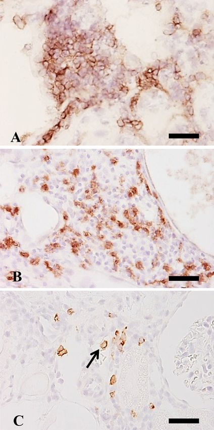
Fig. 3. Immunohistochemistry for lymphocyte markers
also seen in renal fibrosis induced by a single injection of CDDP [14].
at PFI week 6. Many CD4+ T cells are seen in fibrotic
However, their functional properties remain to be investigated. In double region, forming aggregation like follicle (A). CD8+ T
immunofluorescence, there were MHC class II+/CD68+ M1 macrophages cells are diffusely seen in the affected area (B). A few
and MHC class II+/CD163+ M2 macrophages (Table 2), indicating that CD79α+ B cells are sporadically present (arrows) (C).
MHC class II+ macrophages might have both inflammatory and anti- Counterstained with hematoxylin. Bar=20 µm.
Fig. 4. mRNA expressions of interleukin (IL)-17α (A) and forkhead box P3 (FoxP3) (B) by the real-time PCR using samples
of controls, and focal and aggregated lymphocytes at PFI weeks 1, 6 and 12. The expressions were normalized against β-actin
mRNA as internal control gene. Dunnet’s test, *, PMACROPHAGES AND LYMPHOCYTES IN RENAL FIBROSIS
inflammatory functions in renal fibrosis. Macrophages expressing MHC class II are regarded as M1 type in chemical-induced rat
hepatic fibrosis [13, 22, 25, 26], whereas those are polarized toward M2 type in experimentally-induced rat myocardial fibrosis [2].
M1 and M2 macrophages could have expressed MHC class II under unknown microenvironmental conditions [3].
More interestingly, MHC class II+ macrophages were localized mainly around damaged renal tubes throughout the observation
period, apparently in close relation with infiltrating lymphocytes. It is reported that MHC class II expression is related to the
subsequent induction of CD4+ T cells through the complicated immune system [20]. Because CD4+ T cells stimulate macrophages
to generate inflammatory mediators [18], and MHC class II+ macrophages are known to present the processed antigens to CD4+ T
cells [1, 7], CD4+ T cells may act with the relation of MHC class II+ macrophages. In fact, it is likely that CD4+ T cells contribute to
the progress of renal fibrosis [1, 28]. In the present study, additionally, CD8+ T cells also showed an increased number at PFI weeks
3–12, although the appearance of CD8+ T cells was less than CD4+ T cells. In renal fibrosis induced by a single injection, CD4+ and
CD8+ T cells are also seen in the fibrotic lesions [14]; CD8+ T cells may be able to mediate cytotoxicity to tubular cells [23].
Detailed functions of focal or aggregated lymphocytes were analyzed using samples obtained by LMD method at PFI weeks
1, 6 and 12. IL-17A was significantly increased at PFI week 1, and by RT-PCR, increased levels of IL-6 and TGF-β1 were also
confirmed in the fibrosis progression phase as mentioned above. Generally, naïve T cells differentiate to Th17 cells under influence
of IL-6 or TGF-β1 [17]. Th17 cells, which may be mainly involved in inflammation of autoimmune diseases [17], are a subtype of
CD4+ T cells. The focal or aggregated lymphocytes might be capable of producing IL-17A, which could stimulate inflammation
via cytokines such as TNF-α [16, 27]. At PFI week 6, FoxP3 expression significantly increased in focal or aggregated lymphocytes.
FoxP3 is a transcriptional factor and used as a regulatory T cell (Treg) marker. It is considered that Treg could attenuate fibrosis in
tissue remodeling after injury [5, 6]. The increased FoxP3 might have been related to the induction of fibrosis-amelioration phase at
PFI weeks 9 to 12. The timing of increased FoxP3 expression at PFI week 6 in the present study might be a turning point between
the fibrosis-progress and fibrosis-amelioration phases, although a more detailed analysis is needed.
Although CD79α+ B cells were much less in appearance than CD4+ and CD8+ cells, B cells were present during the observation
period, peaking at PFI week 6. A study on mouse unilateral ureteral obstruction model showed that the early-stage accumulation
of B cells in the affected kidney might accelerate monocyte/macrophage infiltration, thereby leading to aggravated fibrosis [1]. The
significance of B cells seen in this renal fibrosis remains to be investigated [14].
In conclusion, rat kidneys damaged by the repeated injection of CDDP developed progressive interstitial renal fibrosis at PFI
weeks 1, 3 and 6, and subsequent amelioration at PFI weeks 9 and 12. CD68+ M1 macrophages showed a consistent increase,
and CD163+ M2 macrophages were also increased but slightly. IL-6, TNF-α and IFN-γ for M1-factors, as well as TGF-β1 and
IL-10 for M2-factors tended to increase mainly at PFI weeks 1, 3 or 6. Tissue injury by M1 macrophages and fibrosis by M2
macrophages might have occurred simultaneously, resulting in renal fibrosis in the fibrosis-progress phase. More interestingly,
MHC class II+ macrophages had functions of both M1 and M2 types in double immunofluorescence analyses, and apparently,
they increased consistently in relation with increased numbers of T cells; these T cells, particularly CD4+ T cells, might act with
MHC class II+ macrophages. The focal or aggregated lymphocytes obtained by LMD showed increased IL-17A (for Th17 cells)
and FoxP3 (a marker of Treg); presumably, Th17 cells, a subtype of CD4+ T cells, might be involved in tissue injury, whereas Treg
might contribute to fibrosis amelioration. It is likely that M1/M2 macrophages and T cells reacting to CD4 and CD8 contribute
complicatedly to renal fibrosis [8, 10]. This study is the first trial to analyze the properties of infiltrating cells in rat renal fibrosis
followed by repeated injection of CDDP. The information would be useful for the understanding of the pathogenesis of CKD in
dogs and cats; in particular, CKD is prevalent in elderly cats [12].
POTENTIAL CONFLICTS OF INTEREST. The authors have nothing to disclose.
ACKNOWLEDGMENTS. This work was supported partly by the JSPS KAKENHI Grant Number19H03130 (to Yamate); the
Platform Project for Supporting Drug Discovery and Life Science Research (Basis for Supporting Innovative Drug Discovery and
Life Science Research (BINDS)) from AMED under Grant Number JP21am0101123 (to Yamate).
REFERENCES
1. Han, H., Zhu, J., Wang, Y., Zhu, Z., Chen, Y., Lu, L., Jin, W., Yan, X. and Zhang, R. 2017. Renal recruitment of B lymphocytes exacerbates
tubulointerstitial fibrosis by promoting monocyte mobilization and infiltration after unilateral ureteral obstruction. J. Pathol. 241: 80–90. [Medline]
[CrossRef]
2. Koga, M., Karim, M. R., Kuramochi, M., Izawa, T., Kuwamura, M. and Yamate, J. 2021. Appearance of heterogeneous macrophages during
development of isoproterenol-induced rat myocardial fibrosis. Toxicol. Pathol. 49: 1048–1061 [CrossRef]. [Medline]
3. Lech, M. and Anders, H. J. 2013. Macrophages and fibrosis: How resident and infiltrating mononuclear phagocytes orchestrate all phases of tissue
injury and repair. Biochim. Biophys. Acta 1832: 989–997. [Medline] [CrossRef]
4. Leung, K. C., Tonelli, M. and James, M. T. 2013. Chronic kidney disease following acute kidney injury-risk and outcomes. Nat. Rev. Nephrol. 9:
77–85. [Medline] [CrossRef]
5. Li, J., Qiu, S. J., She, W. M., Wang, F. P., Gao, H., Li, L., Tu, C. T., Wang, J. Y., Shen, X. Z. and Jiang, W. 2012. Significance of the balance between
regulatory T (Treg) and T helper 17 (Th17) cells during hepatitis B virus related liver fibrosis. PLoS One 7: e39307. [Medline] [CrossRef]
6. Lim, A. I., Tang, S. C., Lai, K. N. and Leung, J. C. 2013. Kidney injury molecule-1: more than just an injury marker of tubular epithelial cells? J.
Cell. Physiol. 228: 917–924. [Medline] [CrossRef]
7. Liu, L., Kou, P., Zeng, Q., Pei, G., Li, Y., Liang, H., Xu, G. and Chen, S. 2012. CD4+ T Lymphocytes, especially Th2 cells, contribute to the
J. Vet. Med. Sci. 83(9): 1435–1442, 2021 1441S. MATSUYAMA ET AL.
progress of renal fibrosis. Am. J. Nephrol. 36: 386–396. [Medline] [CrossRef]
8. Luzina, I. G., Todd, N. W., Iacono, A. T. and Atamas, S. P. 2008. Roles of T lymphocytes in pulmonary fibrosis. J. Leukoc. Biol. 83: 237–244.
[Medline] [CrossRef]
9. Martinez, F. O. and Gordon, S. 2014. The M1 and M2 paradigm of macrophage activation: time for reassessment. F1000Prime Rep. 6: 13.
[Medline] [CrossRef]
10. Martín-Fernández, B., Rubio-Navarro, A., Cortegano, I., Ballesteros, S., Alía, M., Cannata-Ortiz, P., Olivares-Álvaro, E., Egido, J., de Andrés, B.,
Gaspar, M. L., de Las Heras, N., Lahera, V. and Moreno, J. A. 2016. Aldosterone induces renal fibrosis and inflammatory M1-macrophage subtype
via mineralocorticoid receptor in rats. PLoS One 11: e0145946. [Medline] [CrossRef]
11. Matsuyama, S., Karim, M. R., Izawa, T., Kuwamura, M. and Yamate, J. 2018. Immunohistochemical analyses of the kinetics and distribution of
macrophages in the developing rat kidney. J. Toxicol. Pathol. 31: 207–212. [Medline] [CrossRef]
12. McLeland, S. M., Cianciolo, R. E., Duncan, C. G. and Quimby, J. M. 2015. A comparison of biochemical and histopathologic staging in cats with
chronic kidney disease. Vet. Pathol. 52: 524–534. [Medline] [CrossRef]
13. Mosser, D. M. and Edwards, J. P. 2008. Exploring the full spectrum of macrophage activation. Nat. Rev. Immunol. 8: 958–969. [Medline]
[CrossRef]
14. Nakagawa, M., Karim, M. R., Izawa, T., Kuwamura, M. and Yamate, J. 2021. Immunophenotypical characterization of M1/M2 macrophages and
lymphocytes in cisplatin-induced rat progressive renal fibrosis. Cells 10: 257. [Medline] [CrossRef]
15. Nelson, M. P., Christmann, B. S., Dunaway, C. W., Morris, A. and Steele, C. 2012. Experimental Pneumocystis lung infection promotes M2a
alveolar macrophage-derived MMP12 production. Am. J. Physiol. Lung Cell. Mol. Physiol. 303: L469–L475. [Medline] [CrossRef]
16. Nestle, F. O., Kaplan, D. H. and Barker, J. 2009. Psoriasis. N. Engl. J. Med. 361: 496–509. [Medline] [CrossRef]
17. Ogura, H., Murakami, M., Okuyama, Y., Tsuruoka, M., Kitabayashi, C., Kanamoto, M., Nishihara, M., Iwakura, Y. and Hirano, T. 2008. Interleukin-17
promotes autoimmunity by triggering a positive-feedback loop via interleukin-6 induction. Immunity 29: 628–636. [Medline] [CrossRef]
18. Ricardo, S. D., van Goor, H. and Eddy, A. A. 2008. Macrophage diversity in renal injury and repair. J. Clin. Invest. 118: 3522–3530. [Medline] [CrossRef]
19. Schiffrin, E. L., Lipman, M. L. and Mann, J. F. 2007. Chronic kidney disease: effects on the cardiovascular system. Circulation 116: 85–97.
[Medline] [CrossRef]
20. Schmid, A. B., Coppieters, M. W., Ruitenberg, M. J. and McLachlan, E. M. 2013. Local and remote immune-mediated inflammation after mild
peripheral nerve compression in rats. J. Neuropathol. Exp. Neurol. 72: 662–680. [Medline] [CrossRef]
21. Sica, A. and Mantovani, A. 2012. Macrophage plasticity and polarization: in vivo veritas. J. Clin. Invest. 122: 787–795. [Medline] [CrossRef]
22. Tsuji, Y., Kuramochi, M., Golbar, H. M., Izawa, T., Kuwamura, M. and Yamate, J. 2020. Acetaminophen-induced rat hepatotoxicity based on M1/M2-
macrophage polarization, in possible relation to damage-associated molecular patterns and autophagy. Int. J. Mol. Sci. 21: 8998. [Medline] [CrossRef]
23. Wang, Y. and Harris, D. C. 2011. Macrophages in renal disease. J. Am. Soc. Nephrol. 22: 21–27. [Medline] [CrossRef]
24. Wijesundera, K. K., Izawa, T., Murakami, H., Tennakoon, A. H., Golbar, H. M., Kato-Ichikawa, C., Tanaka, M., Kuwamura, M. and Yamate, J.
2014. M1- and M2-macrophage polarization in thioacetamide (TAA)-induced rat liver lesions; a possible analysis for hepato-pathology. Histol.
Histopathol. 29: 497–511. [Medline]
25. Wijesundera, K. K., Izawa, T., Tennakoon, A. H., Golbar, H. M., Tanaka, M., Kuwamura, M. and Yamate, J. 2015. M1-/M2-macrophages contribute to
the development of GST-P-positive preneoplastic lesions in chemically-induced rat cirrhosis. Exp. Toxicol. Pathol. 67: 467–475. [Medline] [CrossRef]
26. Wijesundera, K. K., Izawa, T., Tennakoon, A. H., Murakami, H., Golbar, H. M., Katou-Ichikawa, C., Tanaka, M., Kuwamura, M. and Yamate, J.
2014a. M1- and M2-macrophage polarization in rat liver cirrhosis induced by thioacetamide (TAA), focusing on Iba1 and galectin-3. Exp. Mol.
Pathol. 96: 382–392. [Medline] [CrossRef]
27. Witowski, J., Książek, K. and Jörres, A. 2004. Interleukin-17: a mediator of inflammatory responses. Cell. Mol. Life Sci. 61: 567–579. [Medline]
[CrossRef]
28. Wynn, T. A. 2008. Cellular and molecular mechanisms of fibrosis. J. Pathol. 214: 199–210. [Medline] [CrossRef]
29. Yamamoto, E., Izawa, T., Juniantito, V., Kuwamura, M. and Yamate, J. 2010. Relationship of cell proliferating marker expressions with PGE2
receptors in regenerating rat renal tubules after cisplatin injection. J. Toxicol. Pathol. 23: 271–275. [Medline] [CrossRef]
30. Yamate, J., Izawa, T. and Kuwamura, M. 2016. Histopathological analysis of rat hepatotoxicity based on macrophage functions: in particular, an
analysis for thioacetamide-induced hepatic lesions. Food Saf (Tokyo) 4: 61–73. [Medline] [CrossRef]
31. Yamate, J., Machida, Y., Ide, M., Kuwamura, M., Kotani, T., Sawamoto, O. and LaMarre, J. 2005. Cisplatin-induced renal interstitial fibrosis in
neonatal rats, developing as solitary nephron unit lesions. Toxicol. Pathol. 33: 207–217. [Medline] [CrossRef]
32. Yamate, J., Sato, K., Ide, M., Nakanishi, M., Kuwamura, M., Sakuma, S. and Nakatsuji, S. 2002. Participation of different macrophage populations
and myofibroblastic cells in chronically developed renal interstitial fibrosis after cisplatin-induced renal injury in rats. Vet. Pathol. 39: 322–333.
[Medline] [CrossRef]
33. Yamate, J., Sato, K., Machida, Y., Ide, M., Sato, S., Nakatsuji, S., Kuwamura, M., Kotani, T. and Sakuma, S. 2000. Cisplatin-induced rat renal
interstitial fibrosis; a possible pathogenesis based on the data. J. Toxicol. Pathol. 4: 237–247. [CrossRef]
34. Yamate, J., Okado, A., Kuwamura, M., Tsukamoto, Y., Ohashi, F., Kiso, Y., Nakatsuji, S., Kotani, T., Sakuma, S. and Lamarre, J. 1998.
Immunohistochemical analysis of macrophages, myofibroblasts, and transforming growth factor-β localization during rat renal interstitial fibrosis
following long-term unilateral ureteral obstruction. Toxicol. Pathol. 26: 793–801. [Medline] [CrossRef]
35. Zhang, M. and Zhang, S. 2020. T Cells in Fibrosis and Fibrotic Diseases. Front. Immunol. 11: 1142. [Medline] [CrossRef]
36. Zhang, M. Z., Yao, B., Yang, S., Jiang, L., Wang, S., Fan, X., Yin, H., Wong, K., Miyazawa, T., Chen, J., Chang, I., Singh, A. and Harris, R. C.
2012. CSF-1 signaling mediates recovery from acute kidney injury. J. Clin. Invest. 122: 4519–4532. [Medline] [CrossRef]
J. Vet. Med. Sci. 83(9): 1435–1442, 2021 1442You can also read