Poricoic acid A suppresses TGF β1 induced renal fibrosis and proliferation via the PDGF C, Smad3 and MAPK pathways
←
→
Page content transcription
If your browser does not render page correctly, please read the page content below
EXPERIMENTAL AND THERAPEUTIC MEDICINE 21: 289, 2021

     Poricoic acid A suppresses TGF‑β1‑induced renal fibrosis and
      proliferation via the PDGF‑C, Smad3 and MAPK pathways
                                         QIANG LI, YAO MING, HU JIA and GANG WANG

            Department of Nephrology, Nanjing University of Chinese Medicine, Nanjing, Jiangsu 210023, P.R. China

                                         Received September 4, 2019; Accepted March 17, 2020

                                                         DOI: 10.3892/etm.2021.9720

Abstract. Renal interstitial fibrosis is the most important            Introduction
pathological process in chronic renal failure. Previous studies
have shown that poricoic acid A (PAA), the main chemical               Renal fibrosis is caused by imbalance between synthesis
constituent on the surface layer of the mushroom Poria                 and degradation of the extracellular matrix (ECM) constitu‑
cocos, has protective effects against oxidative stress and             ents, including collagen I, III and IV, as a result of various
acute kidney injury. The present study aimed to investigate            pathological factors, leading to glomerulosclerosis, tubuloint‑
the potential roles of PAA on the pathological process of              erstitial fibrosis, hyalinization and sclerosis of the renal small
renal fibrosis and the associated molecular mechanism. The             vessels (1,2). These cellular and molecular events ultimately
NRK‑49F cell line was treated with transforming growth                 result in parenchymal obstruction and renal failure (3,4). Renal
factor‑β1 (TGF‑β1) with or without PAA or platelet‑derived             fibrosis can also result from a variety of other factors that are
growth factor C (PDGF‑C). Cell Counting Kit‑8 assay,                   associated with kidney injury, including hypertension, inflam‑
western blotting and 5‑ethynyl‑2'‑deoxyuridine immuno‑                 mation, high sugar, high fat and drug damage (5). However,
fluorescence staining were performed to examine cell growth,           previous studies have shown that even if these factors are
protein expression and cell proliferation, respectively. Data          controlled effectively, including blood pressure, blood glucose
from the present study showed that 10 µM PAA attenuated                and drug damage, the process of renal fibrosis remains diffi‑
TGF‑β1‑induced NRK‑49F cell extracellular matrix (ECM)                 cult to prevent (6). Therefore, investigating the molecular
accumulation, fibrosis formation and proliferation. Renal              mechanism underlying the occurrence and development of
fibrosis with the activation of Smad3 and mitogen‑activated            fibrosis, identifying therapeutic agents and target genes that
protein kinase (MAPK) pathways were also inhibited by PAA              can directly interfere with the fibrotic process has become an
treatment. PDGF‑C reversed the inhibitory effects of PAA on            important topic of study in recent years (7‑9).
TGF‑β1‑induced renal fibroblast proliferation and activation               The formation and progression of renal fibrosis is a
of the Smad3/MAPK pathway. The present study suggested                 complex and dynamic process that includes inflammatory
that suppression of TGF‑ β1‑induced renal fibroblast ECM               cell infiltration, fibroblast activation and proliferation, ECM
accumulation, fibrosis formation and proliferation by PAA              accumulation, tubular atrophy and microvascular degen‑
is mediated via the inhibition of the PDGF‑C, Smad3 and                eration (10,11). A number of genes have been reported to be
MAPK pathways. The present findings not only revealed the              involved in this process. Among them, transforming growth
potential anti‑fibrotic effects of PAA on renal fibroblasts, but       factor (TGF)‑β1 is considered to be the most important fibro‑
also provided a new insight into the prevention of fibrosis            genic factor, which initiates the occurrence and accelerates
formation via regulation of the PDGF‑C, Smad3 and MAPK                 the progress of fibrosis (12). In addition, recent reports also
signaling pathways.                                                    found that a number of other factors, including platelet‑derived
                                                                       growth factor (PDGF), Smad3, connective tissue growth
                                                                       factor and angiotensin II, enhanced the constituents of the
                                                                       ECM (13,14). Although the mechanism behind the formation
                                                                       and development of renal fibrosis has been extensively studied,
                                                                       effective treatment strategies has yet to be found (15,16).
                                                                       Therefore, it is of importance to explore the mechanism of
Correspondence to: Dr Qiang Li or Dr Gang Wang, Department of
Nephrology, Nanjing University of Chinese Medicine, 138 Xianlin        renal fibrosis further to find novel potential therapeutic drugs.
Road, Nanjing, Jiangsu 210023, P.R. China                                  Poria cocos Wolf (Polyporaceae) is a traditional Chinese
E‑mail: qianglisci2000@163.com                                         medicine that has been applied for >2,000 years in China that
E‑mail: njboda@sina.com                                                is also widely distributed and easy to obtain because it exists in
                                                                       many plants (17,18). Reported medicinal properties that have been
Key words: poricoic acid A, transforming growth factor‑β1, renal       associated with Poria cocos include diuretic effects, invigorating
fibrosis, platelet‑derived growth factor C, cell proliferation         the spleen, tranquilizes the heart and edema elimination (19‑22).
                                                                       Poricoic acid A (PAA) is one of the main chemical constituents on
                                                                       the surface layer of Poria cocos Wolf. PAA has been documented
2 LI et al: PORICOIC ACID A SUPPRESSES RENAL FIBROBLAST PROLIFERATION
to significantly alleviate oxidative stress and suppress the (cat. no. 1706404; Bio‑rad, Laboratories, Inc.). Subsequently,
increase in inflammatory factors during acute kidney injury (23). the following primary antibodies were used at 1:1,000 dilu‑
Chen et al (24) reported that PAA exerted renoprotective and tion: E‑cadherin (cat. no. 14472S; Cell Signaling Technology,
antifibrotic effects by inhibiting TGF‑β/Smad3 and Wnt/β‑catenin Inc.), collagen I (cat. no. 84336S; Cell Signaling Technology,
signaling pathways, whilst other studies have also demonstrated Inc.), collagen IV (cat. no. 50273S; Cell Signaling Technology,
that PAA could suppress the renin‑angiotensin system (RAS) to Inc.), alpha smooth muscle actin (α‑SMA; cat. no. ab5694;
prevent chronic kidney disease progression (25,26). However, to Abcam), fibronectin (FN; cat. no. 26836S; Cell Signaling
the best of our knowledge, the mechanistic effects of PAA on Technology, Inc.), PDGF‑C (cat. no. abc1392; Sigma‑Aldrich;
renal fibrosis remain to be elucidated. Merck KGaA), phosphorylated (p)‑Smad3 (cat. no. 9520;
The present study aimed to investigate the effects of PAA Cell Signaling Technology, Inc.), Smad3 (cat. no. 9523;
on TGF‑β1‑induced renal fibroblast proliferation and fibrosis Cell Signaling Technology, Inc.), p‑ERK1/2 (cat. no. 4370;
formation in addition to the potential mechanism underlying Cell Signaling Technology, Inc.), ERK1/2 (cat. no. 4695;
the actions of PAA. Information from the present study may Cell Signaling Technology, Inc.), p‑p38 (cat. no. 4511; Cell
provide a novel avenue for the treatment of fibrosis formation Signaling Technology, Inc.), p38 (cat. no. 8690; Cell Signaling
during the development of renal failure. Technology, Inc.) and GAPDH (cat. no. sc‑137179; Santa Cruz
Biotechnology, Inc.). Following incubation with the respective
Materials and methods primary antibodies overnight at 4˚C, the membranes were
washed three times with TBST solution. Subsequently, the
Cell culture and drug treat ment. N R K‑ 49F cells membranes were exposed to horseradish peroxidase‑conju‑
(cat. no. CRL‑1570; American Type Culture Collection), a rat gated goat anti‑rabbit (1:5,000; cat. no. ab6721; Abcam),
renal interstitial fibroblast cell line (27,28), were used to investi‑ goat anti‑mouse (1:5,000; cat. no. ab05719; Abcam), and
gate the effects of PAA on renal fibrosis. Cells were cultured in rabbit anti‑goat (1:5,000; cat. no. ab5755; Abcam) secondary
DMEM (cat. no. 11965‑092; Gibco, Thermo Fisher Scientific, antibodies for 1 h at room temperature prior to treatment
Inc.) supplemented with 10% FBS (cat. no. SH30071.02; with ECL reagent (cat. no. RPN2232; GE Healthcare), using
HyClone; GE Healthcare Life Sciences) in a 5% CO2 atmo‑ autoluminography to visualize protein bands. Protein expres‑
sphere at 37˚C. sion was then quantified using ImageJ (version 1.48V; National
To determine the optimal concentration of PAA (Shanxi Institutes of Health) with GAPDH as the loading control.
Medicine company), cells were exposed to serial concentra‑
tions of PAA (1, 2, 5, 10, 15 and 20 µM) for 24 h at 37˚C in a Cell proliferation assay. To assess kidney cell proliferation,
5%CO2 incubator. The effects of PAA on renal fibrosis were 2x10⁵ NRK‑49F cells/per well in 1ml culture medium were
also determined using recombinant human TGF‑β1 (5 ng/ml; plated onto coverslips in a 6‑well plate and allowed to grow
cat. no. 240‑B; R&D Systems, Inc.) in the presence or absence overnight at 37˚C in a 5% CO2 incubator. Cells were incubated
of recombinant PDGF‑C protein (50 ng/ml; cat. no. 1687‑CC; with Alexa Fluor 555 labeled with 5‑ethynyl‑2'‑deoxyuridine
R&D Systems, Inc.) for 24 h at 37˚C in a 5% CO2 incubator. (EdU; cat. no. A10044; Invitrogen; Thermo Fisher Scientific,
Inc.) at a final concentration of 10 µM for 24 h at 37˚C in
Cell growth assay. NRK‑49F cells were cultured in 96‑well 5% CO2 before being harvested and subjected to treatments in
plates, 100 µl per well with 1x10⁴ cells and exposed to 10 µmol accordance with the Click‑iT®EdU Alexa Fluor® 555 Imaging
PAA for 24 h. Subsequently, 30 µl CCK‑8 solution (Dojindo kit (cat. no. C10353; Invitrogen; Thermo Fisher Scientific, Inc.,
Molecular Technologies, Inc.) was added to each well where the cat. no. C10353). All procedures were performed according
cells were incubated for 4 h at 37˚C. The optical density (OD) to the manufacturer's instructions. Nuclei were stained with
value was detected at a wavelength of 450 nm where a micro‑ DAPI at final a concentration of 3 µM. EdU‑positive cells with
plate reader (Thermo Fisher Scientific, Inc.) was used to DAPI‑labeled nuclei were counted to evaluate cell proliferation.
calculate cell viability and inhibition rate. The detailed calcu‑
lations are as follows: Cell viability (%)=[(As‑Ab)/(Ac‑Ab)] Statistical analysis. Data are presented as the mean ± SD.
x100; inhibition rate (%)=[(Ac‑As)/(Ac‑Ab)] x100; As= OD Statistical analysis was performed using GraphPad Prism 6.0
value of the experimental wells; Ab=OD value of blank wells; (GraphPad Software, Inc.). One‑way ANOVA followed by
Ac=OD value of control wells. Tukey's post hoc test was used for comparisons between
multiple groups. P1 µM reduced NRK‑49FEXPERIMENTAL AND THERAPEUTIC MEDICINE 21: 289, 2021 3 Figure 1. Effects of PAA treatment on NRK‑49F cell viability. (A) The chemical structural formula of PAA. (B) Cytotoxicity of NRK‑49F cells following treat‑ ment with PAA as measured using Cell Counting kit‑8. (C) Cytotoxicity was measured after treatment with PAA at the indicated concentrations. (D) PDGF‑C protein expression was measured by western blotting after treatment with PAA at the indicated concentrations. n=6. *P
4                               LI et al: PORICOIC ACID A SUPPRESSES RENAL FIBROBLAST PROLIFERATION

Figure 2. Effect of PAA on the expression of ECM proteins and TGF‑β1‑induced cell proliferation. (A) Expression of E‑cadherin, collagen I, collagen IV,
α‑SMA and fibronectin, proteins associated with ECM, were measured by western blotting following treatment with PAA, TGF‑ β1 or a combination of
both PAA and TGF‑β1. (B) Cell proliferation was measured after TGF‑β1 and/or PAA treatment using 5‑ethynyl‑2'‑deoxyuridine staining. n=3. *P
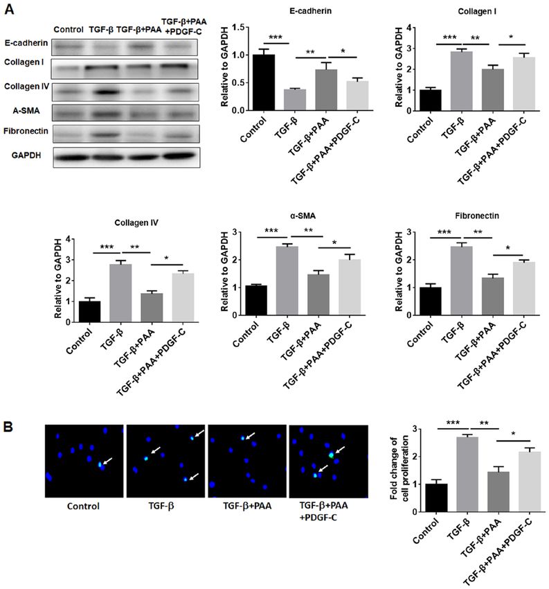
EXPERIMENTAL AND THERAPEUTIC MEDICINE 21: 289, 2021                                                               5

Figure 4. PDGF‑C reverses the inhibitory effects of PAA on the expression of extracellular matrix proteins and cell proliferation in NRK‑49F cells. (A) Effects
of PDGF‑C on the expression of E‑cadherin, collagen I, collagen IV, α‑SMA and fibronectin in PAA‑ and/or TGF‑β1‑treated cells were examined by western
blotting. (B) PDGF‑C reversed the inhibitory effects of PAA on NRK‑49F cell proliferation according to 5‑ethynyl‑2'‑deoxyuridine staining. n=3. *P
6                              LI et al: PORICOIC ACID A SUPPRESSES RENAL FIBROBLAST PROLIFERATION

Figure 5. PDGF‑C treatment reactivates TGF‑β1‑induced Smad and MAPK signaling in NRK‑49F cells. (A) PDGF‑C treatment promoted the expression of
PDGF‑C and phosphorylation of Smad3 in the presence of PAA. (B) PDGF‑C treatment increased ERK and P38 MAPK phosphorylation in the presence
of PAA. n=3. *P
EXPERIMENTAL AND THERAPEUTIC MEDICINE 21: 289, 2021                                                       7

of Smad and MAPK pathways has been reported to signifi‑                    4. Wang J, Yang Q, Nie Y, Guo H, Zhang F, Zhou X and Yin X:
                                                                              Tetrahydrobiopterin contributes to the proliferation of mesangial
cantly reduce renal ECM aggregation and fibrosis formation                    cells and accumulation of extracellular matrix in early‑stage
(52). The present study showed that PAA could suppress the                    diabetic nephropathy. J Pharm Pharmacol 69: 182‑190, 2017.
increased upregulation of Smad and MAPK signaling induced                  5. Klinkhammer BM, Goldschmeding R, Floege J and Boor P:
                                                                              Treatment of renal fibrosis‑turning challenges into opportunities.
by TGF‑β1, which were reversed by the presence of PDGF‑C.                     Adv Chronic Kidney Dis 24: 117‑129, 2017.
    In conclusion, data from the present study suggest that PAA            6. Sandner P and Stasch JP: Anti‑fibrotic effects of soluble guanylate
treatment suppressed TGF‑β1‑induced renal fibroblast ECM                      cyclase stimulators and activators: A review of the preclinical
                                                                              evidence. Respir Med 122 (Suppl 1): S1‑S9, 2017.
accumulation, fibrosis formation and proliferation by inhibiting           7. Wang Y, Cai J, Tang C and Dong Z: Mitophagy in acute kidney
the PDGF‑C, Smad3 and MAPK signaling pathways. PDGF‑C                         injury and kidney repair. Cells 9: pii: E338, 2020.
can block the effect of PAA on TGF‑β1‑induced fibroblasts                  8. Tang J, Goldschmeding R, Samarakoon R and Higgins PJ: Protein
                                                                              phosphatase Mg(2+)/Mn(2+) dependent‑1A and PTEN deregula‑
and promoted the activation of the Smad and MAPK pathways                     tion in renal fibrosis: Novel mechanisms and co‑dependency of
induced by TGF‑β1. These findings revealed the potential of                   expression. FASEB J 34: 2641‑2656, 2020.
PAA application in protecting against the fibrosis of renal                9. Marko L, Park JK, Henke N, Rong S, Balogh A, Klamer S,
                                                                              Bartolomaeus H, Wilck N, Ruland J, Forslund SK, et al: B‑cell
fibroblasts. The present study also provided a new insight into               lymphoma/leukemia 10 (Bcl10) and angiotensin II‑induced
the prevention of fibrosis formation through regulation by                    kidney injury. Cardiovasc Res 116: 1059‑1070, 2019.
PDGF‑C. However, further in vivo studies are necessary to                 10. Li O, Ma Q, Li F, Cai GY, Chen XM and Hong Q: Progress of
                                                                              small ubiquitin‑related modifiers in kidney diseases. Chin Med J
fully clarify the role of PAA in renal fibrosis.                              (Engl) 132: 466‑473, 2019.
                                                                          11. Genovese F, Manresa AA, Leeming DJ, Karsdal MA and
Acknowledgements                                                              Boor P: The extracellular matrix in the kidney: A source of novel
                                                                              non‑invasive biomarkers of kidney fibrosis? Fibrogenesis Tissue
                                                                              Repair 7: 4, 2014.
Not applicable.                                                           12. Castellone MD and Laukkanen MO: TGF‑beta1, WNT, and SHH
                                                                              signaling in tumor progression and in fibrotic diseases. Front
                                                                              Biosci (Schol Ed) 9: 31‑45, 2017.
Funding                                                                   13. van Roeyen CRC, Martin IV, Drescher A, Schuett KA, Hermert D,
                                                                              Raffetseder U, Otten S, Buhl EM, Braun GS, Kuppe C, et al:
No funding was received.                                                      Identification of platelet‑derived growth factor C as a mediator of
                                                                              both renal fibrosis and hypertension. Kidney Int 95: 1103‑1119, 2019.
                                                                          14. Ghayur A, Padwal MK, Liu L, Zhang J and Margetts PJ:
Availability of data and materials                                            SMAD3‑dependent and‑independent pathways in glomerular
                                                                              injury associated with experimental glomerulonephritis. Am J
                                                                              Physiol Renal Physiol 317: F152‑F62, 2019.
The datasets used and/or analyzed during the current study                15. Breyer MD and Susztak K: The next generation of therapeutics
are available from the corresponding author on reasonable                     for chronic kidney disease. Nat Rev Drug Discov 15: 568‑588,
request.                                                                      2016.
                                                                          16. Mencke R, Olauson H and Hillebrands JL: Effects of Klotho on
                                                                              fibrosis and cancer: A renal focus on mechanisms and therapeutic
Author's contributions                                                        strategies. Adv Drug Deliv Rev 121: 85‑100, 2017.
                                                                          17. Li S, Wang Z, Gu R, Zhao Y, Huang W, Wang Z and Xiao W:
                                                                              A new epidioxy‑tetracyclic triterpenoid from Poria cocos Wolf.
QL and GW conceived and designed the experiment. QL, YM                       Nat Prod Res 30: 1712‑1717, 2016.
and HJ performed experiments and collected and analyzed the               18. Lee SR, Lee S, Moon E, Park HJ, Park HB and Kim KH:
data. QL and GW wrote the manuscript. All authors revised                     Bioactivity‑guided isolation of anti‑inflammatory triterpenoids
                                                                              from the sclerotia of Poria cocos using LPS‑stimulated Raw264.7
and approved the manuscript.                                                  cells. Bioorg Chem 70: 94‑99, 2017.
                                                                          19. Li S, Zhang J, Li S, Liu C, Liu S and Liu Z: Extraction and
Ethics approval and consent to participate                                    separation of lactate dehydrogenase inhibitors from Poria cocos
                                                                              (Schw.) Wolf based on a hyphenated technique and in vitro
                                                                              methods. J Sep Sci 40: 1773‑1783, 2017.
Not applicable.                                                           20. Zhao YY, Feng YL, Du X, Xi ZH, Cheng XL and Wei F: Diuretic
                                                                              activity of the ethanol and aqueous extracts of the surface layer of
                                                                              Poria cocos in rat. J Ethnopharmacol 144: 775‑778, 2012.
Patient consent for publication                                           21. Feng YL, Lei P, Tian T, Yin L, Chen DQ, Chen H, Mei Q, Zhao YY
                                                                              and Lin RC: Diuretic activity of some fractions of the epidermis
Not applicable.                                                               of Poria cocos. J Ethnopharmacol 150: 1114‑1118, 2013.
                                                                          22. Zhao YY, Feng YL, Bai X, Tan XJ, Lin RC and Mei Q: Ultra
                                                                              performance liquid chromatography‑based metabonomic study
Competing interests                                                           of therapeutic effect of the surface layer of Poria cocos on
                                                                              adenine‑induced chronic kidney disease provides new insight
                                                                              into anti‑fibrosis mechanism. PLoS One 8: e59617, 2013.
The authors declare that they have no competing interests.                23. Chen DQ, Feng YL, Chen L, Liu JR, Wang M, Vaziri ND and
                                                                              Zhao YY: Poricoic acid A enhances melatonin inhibition of
References                                                                    AKI‑to‑CKD transition by regulating Gas6/AxlNFkappaB/Nrf2
                                                                              axis. Free Radic Biol Med 134: 484‑497, 2019.
                                                                          24. Chen DQ, Cao G, Zhao H, Chen L, Yang T, Wang M, Vaziri ND,
 1. Shiber S, Eliakim‑Raz N and Yair M: Retroperitoneal fibrosis:             Guo Y and Zhao YY: Combined melatonin and poricoic acid A
    Case series of five patients and review of the literature. Rev Bras       inhibits renal fibrosis through modulating the interaction of
    Reumatol Engl Ed 56: 101‑104, 2016.                                       Smad3 and beta‑catenin pathway in AKI‑to‑CKD continuum.
 2. Zhou D and Liu Y: Renal fibrosis in 2015: Understanding the               Ther Adv Chronic Dis 10: 2040622319869116, 2019.
    mechanisms of kidney fibrosis. Nat Rev Nephrol 12: 68‑70, 2016.       25. Wang M, Chen DQ, Chen L, Cao G, Zhao H, Liu D, Vaziri ND,
 3. Hu C, Sun L, Xiao L, Han Y, Fu X, Xiong X, Xu X, Liu Y, Yang S,           Guo Y and Zhao YY: Novel inhibitors of the cellular
    Liu F and Kanwar YS: Insights into the mechanisms involved in             renin‑angiotensin system components, poricoic acids, target
    the expression and regulation of extracellular matrix proteins in         Smad3 phosphorylation and Wnt/β ‑catenin pathway against
    diabetic nephropathy. Curr Med Chem 22: 2858‑2870, 2015.                  renal fibrosis. Br J Pharmacol 175: 2689‑2708, 2018.
8 LI et al: PORICOIC ACID A SUPPRESSES RENAL FIBROBLAST PROLIFERATION
26. Wang M, Chen DQ, Chen L, Liu D, Zhao H, Zhang ZH, 41. McGaraughty S, Davis‑Taber RA, Zhu CZ, Cole TB, Nikkel AL,
Vaziri ND, Guo Y, Zhao YY and Cao G: Novel RAS Inhibitors Chhaya M, Doyle KJ, Olson LM, Preston GM, Grinnell CM, et al:
Poricoic Acid ZG and Poricoic Acid ZH attenuate renal fibrosis Targeting Anti‑TGF‑ β therapy to fibrotic kidneys with a dual
via a Wnt/β ‑catenin pathway and targeted phosphorylation of specificity antibody approach. J Am Soc Nephrol 28: 3616‑3626,
smad3 Signaling. J Agric Food Chem 66: 1828‑1842, 2018. 2017.
27. Tan RJ, Zhou D and Liu Y: Signaling crosstalk between tubular 42. Zhang ZH, He JQ, Zhao YY, Chen HC and Tan NH: Asiatic acid
epithelial cells and interstitial fibroblasts after kidney injury. prevents renal fibrosis in UUO rats via promoting the production
Kidney Dis (Basel) 2: 136‑144, 2016. of 15d‑PGJ2, an endogenous ligand of PPAR‑γ. Acta Pharmacol
28. Tang N, Cunningham K and Enger MD: TGF beta elicits oppo‑ Sin 41: 373‑382, 2020.
site responses in clonal subpopulations of NRK‑49F cells. Exp 43. Bian X, Bai Y, Su X, Zhao G, Sun G and Li D: Knockdown
Cell Res 196: 13‑19, 1991. of periostin attenuates 5/6 nephrectomy‑induced intrarenal
29. Floege J, Eitner F and Alpers CE: A new look at platelet‑derived renin‑angiotensin system activation, fibrosis, and inflammation
growth factor in renal disease. J Am Soc Nephrol 19: 12‑23, 2008. in rats. J Cell Physiol 234: 22857‑22873, 2019.
30. Huang C, Day ML, Poronnik P, Pollock CA and Chen XM: 44. Li X, Ponten A, Aase K, Karlsson L, Abramsson A, Uutela M,
Inhibition of KCa3.1 suppresses TGF‑β1 induced MCP‑1 expres‑ Bäckström G, Hellström M, Boström H, Li H, et al: PDGF‑C is a
sion in human proximal tubular cells through Smad3, p38 and new protease‑activated ligand for the PDGF alpha‑receptor. Nat
ERK1/2 signaling pathways. Int J Biochem Cell Biol 47: 1‑10, Cell Biol 2: 302‑309, 2000.
2014. 45. Eitner F, Bucher E, van Roeyen C, Kunter U, Rong S, Seikrit C,
31. Liu M, Ning X, Li R, Yang Z, Yang X, Sun S and Qian Q: Villa L, Boor P, Fredriksson L, Bäckström G, et al: PDGF‑C
Signalling pathways involved in hypoxia‑induced renal fibrosis. is a proinflammatory cytokine that mediates renal interstitial
J Cell Mol Med 21: 1248‑1259, 2017. fibrosis. J Am Soc Nephrol 19: 281‑289, 2008.
32. Waasdorp M, de Rooij DM, Florquin S, Duitman J and Spek CA: 46. Boor P, Babickova J, Steegh F, Hautvast P, Martin IV, Djudjaj S,
Protease‑activated receptor‑1 contributes to renal injury and Nakagawa T, Ehling J, Gremse F, Bücher E, et al: Role of
interstitial fibrosis during chronic obstructive nephropathy. J Cell platelet‑derived growth factor‑CC in capillary rarefaction in
Mol Med 23: 1268‑1279, 2019. renal fibrosis. Am J Pathol 185: 2132‑2142, 2015.
33. Liu W, Lin S, Cai Q, Zhang L, Shen A, Chen Y, Chu J and 47. Wang Y, Abu‑Asab MS, Yu CR, Tang Z, Shen D, Tuo J, Li X
Peng J: Qingxuan jiangya decoction mitigates renal interstitial and Chan CC: Platelet‑derived growth factor (PDGF)‑C inhibits
fibrosis in spontaneously hypertensive rats by regulating trans‑ neuroretinal apoptosis in a murine model of focal retinal degen‑
forming growth factor‑β1/Smad signaling pathway. Evid Based eration. Lab Invest 94: 674‑682, 2014.
Complement Alternat Med 2017: 1576328, 2017. 48. Chen CL, Chou KJ, Lee PT, Chen YS, Chang TY, Hsu CY,
34. Lawson JS, Liu HH, Syme HM, Purcell R, Wheeler‑Jones CPD Huang WC, Chung HM and Fang HC: Erythropoietin suppresses
and Elliott J: The cat as a naturally occurring model of renal epithelial to mesenchymal transition and intercepts Smad signal
interstitial fibrosis: Characterisation of primary feline proximal transduction through a MEK‑dependent mechanism in pig
tubular epithelial cells and comparative pro‑fibrotic effects of kidney (LLC‑PK1) cell lines. Exp Cell Res 316: 1109‑1118, 2010.
TGF‑β1. PLoS One 13: e0202577, 2018. 49. Grynberg K, Ma FY and Nikolic‑Paterson DJ: The JNK signaling
35. Xu J, Yu TT, Zhang K, Li M, Shi HJ, Meng XJ, Zhu LS and pathway in renal fibrosis. Front Physiol 8: 829, 2017.
Zhu LK: HGF alleviates renal interstitial fibrosis via inhibiting 50. Xu ZJ, Shu S, Li ZJ, Liu YM, Zhang RY and Zhang Y: Liuwei
the TGF‑β1/SMAD pathway. Eur Rev Med Pharmacol Sci 22: Dihuang pill treats diabetic nephropathy in rats by inhibiting of
7621‑7627, 2018. TGF‑β/SMADS, MAPK, and NF‑kB and upregulating expres‑
36. Loeff ler I: MKP2 suppresses TGF‑ β1‑induced epithe‑ sion of cytoglobin in renal tissues. Medicine (Baltimore) 96:
lial‑to‑mesenchymal transition through JNK inhibition. Clin Sci e5879, 2017.
(Lond) 133: 545‑550, 2019. 51. Nakagawa T, Lan HY, Glushakova O, Zhu HJ, Kang DH,
37. Okuda S, Languino LR, Ruoslahti E and Border WA: Elevated Schreiner GF, Böttinger EP, Johnson RJ and Sautin YY: Role
expression of transforming growth factor‑beta and proteoglycan of ERK1/2 and p38 mitogen‑activated protein kinases in the
production in experimental glomerulonephritis. Possible role in regulation of thrombospondin‑1 by TGF‑beta1 in rat proximal
expansion of the mesangial extracellular matrix. J Clin Invest 86: tubular cells and mouse fibroblasts. J Am Soc Nephrol 16:
453‑462, 1990. 899‑904, 2005.
38. Choi HI, Kim DH, Park JS, Kim IJ, Kim CS, Bae EH, Ma SK, 52. Okano K, Hibi A, Miyaoka T, Inoue T, Sugimoto H, Tsuchiya K,
Lee TH and Kim SW: Peroxiredoxin V (PrdxV) negatively regu‑ Akiba T and Nitta K: Inhibitory effects of the transcription factor
lates EGFR/Stat3‑mediated fibrogenesis via a Cys48‑dependent Ets‑1 on the expression of type I collagen in TGF‑β1‑stimulated
interaction between PrdxV and Stat3. Sci Rep 9: 8751, 2019. renal epithelial cells. Mol Cell Biochem 369: 247‑254, 2012.
39. Vincenti F, Fervenza FC, Campbell KN, Diaz M, Gesualdo L,
Nelson P, Praga M, Radhakrishnan J, Sellin L, Singh A, et al: This work is licensed under a Creative Commons
A phase 2, double‑blind, placebo‑controlled, randomized study Attribution-NonCommercial-NoDerivatives 4.0
of fresolimumab in patients with steroid‑resistant primary focal International (CC BY-NC-ND 4.0) License.
segmental glomerulosclerosis. Kidney Int Rep 2: 800‑810, 2017.
40. Trachtman H, Fervenza FC, Gipson DS, Heering P, Jayne DR,
Peters H, Rota S, Remuzzi G, Rump LC, Sellin LK, et al: A
phase 1, single‑dose study of fresolimumab, an anti‑TGF‑ β
antibody, in treatment‑resistant primary focal segmental
glomerulosclerosis. Kidney Int 79: 1236‑1243, 2011.You can also read