Atorvastatin treatment does not abolish inflammatory mediated cardiovascular risk in subjects with chronic kidney disease
←
→
Page content transcription
If your browser does not render page correctly, please read the page content below
www.nature.com/scientificreports
OPEN Atorvastatin treatment does
not abolish inflammatory mediated
cardiovascular risk in subjects
with chronic kidney disease
Renate M. Hoogeveen1, Simone L. Verweij1, Yannick Kaiser1, Jeffrey Kroon1,
Hein J. Verberne2, Liffert Vogt3, Sophie J. Bernelot Moens1 & Erik S. G. Stroes1*
Individuals with chronic kidney disease are at an increased risk for cardiovascular disease. This risk
may partially be explained by a chronic inflammatory state in these patients, reflected by increased
arterial wall and cellular inflammation. Statin treatment decreases cardiovascular risk and arterial
inflammation in non-CKD subjects. In patients with declining kidney function, cardiovascular
benefit resulting from statin therapy is attenuated, possibly due to persisting inflammation. In the
current study, we assessed the effect of statin treatment on arterial wall and cellular inflammation.
Fourteen patients with chronic kidney disease stage 3 or 4, defined by an estimated Glomerular
Filtration Rate between 15 and 60 mL/min/1.73 m2, without cardiovascular disease were included in
a single center, open label study to assess the effect of atorvastatin 40 mg once daily for 12 weeks
(NTR6896). At baseline and at 12 weeks of treatment, we assessed arterial wall inflammation by
18
F-fluoro-deoxyglucose positron-emission tomography computed tomography (18F-FDG PET/CT)
and the phenotype of circulating monocytes were assessed. Treatment with atorvastatin resulted in
a 46% reduction in LDL-cholesterol, but this was not accompanied by an attenuation in arterial wall
inflammation in the aorta or carotid arteries, nor with changes in chemokine receptor expression
of circulating monocytes. Statin treatment does not abolish arterial wall or cellular inflammation
in subjects with mild to moderate chronic kidney disease. These results imply that CKD-associated
inflammatory activity is mediated by factors beyond LDL-cholesterol and specific anti-inflammatory
interventions might be necessary to further dampen the inflammatory driven CV risk in these subjects.
Chronic kidney disease (CKD) is an important cardiovascular (CV) risk factor1,2. Even a modest decrease in renal
function coincides with accelerated atherosclerosis and a significant increase in CV risk1,3. Since atherosclerosis is
a lipid-driven inflammatory disease of the arterial wall, lipid lowering is the cornerstone in treatment and preven-
tion of cardiovascular disease (CVD)4,5. Statin-induced LDL-cholesterol lowering reduces CV risk independent
of baseline LDL-cholesterol levels in subjects with C KD6,7. However, the association between LDL-cholesterol
and CV risk is attenuated compared to the non-CKD population8, resulting in a decreased beneficial impact of
statin therapy as the estimated glomerular filtration rate (eGFR) declines6. Thus, in spite of contemporary lipid
lowering regimens, a substantial residual CV risk remains in CKD p atients9.
Previously, we demonstrated that subjects with mild to moderate CKD have increased arterial wall inflamma-
tion compared to healthy controls as assessed by 18F-FDG (fluordeoxyglucose) positron emission tomography/
computed tomography (PET/CT)10. The systemic nature of this pro-inflammatory state is supported by the
presence of activated circulating monocytes with increased chemokine receptor expression as well as increased
transendothelial migration c apacity10. In patients without CKD, intensive LDL-cholesterol lowering was found
to reduce this inflammatory state, both at the level of the arterial w all11,12 and in circulating m
onocytes13. In
patients with CKD, a variety of specific mediators have been suggested to contribute to inflammatory activation
1
Department of Vascular Medicine, Amsterdam University Medical Centers, University of Amsterdam, Meibergdreef
9, 1105 AZ Amsterdam, The Netherlands. 2Department of Radiology and Nuclear Medicine, Amsterdam
University Medical Centers, University of Amsterdam, Amsterdam, The Netherlands. 3Department of Nephrology,
Amsterdam University Medical Centers, University of Amsterdam, Amsterdam, The Netherlands. *email:
e.s.stroes@amsterdamumc.nl
Scientific Reports | (2021) 11:4126 | https://doi.org/10.1038/s41598-021-83273-2 1
Vol.:(0123456789)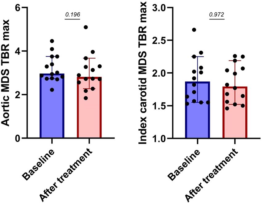
www.nature.com/scientificreports/
beyond the aforementioned lipid particles14,15. Whether statin treatment is capable of dampening inflammation
associated with impaired renal function remains to be established.
Here, we investigated the extent of statin-mediated lowering of inflammatory activity in subjects with CKD.
To this end, we assessed arterial wall inflammation and monocyte phenotype before and after 3 months of potent
statin treatment in subjects with stage 3 or 4 CKD.
Patients and methods
Study design and population. Fourteen patients with chronic kidney disease stage 3 or 4, defined by
an eGFR 15–60 mL/min/1.73 m2 and aged ≥ 50 years, were included in a single center, open label study with
atorvastatin 40 mg once daily for 12 weeks. Exclusion criteria included current statin use, use of drugs altering
cytochrome P450 3A4 metabolism, use of anti-inflammatory drugs, history of CV-events (myocardial infarc-
tion, revascularization, stroke, or peripheral arterial disease), body mass index > 30 kg/m2, malignancy, autoim-
mune disorders, clinically relevant infection (high-sensitivity C-reactive protein > 10 mg/L), diabetes mellitus,
or other inflammatory conditions (NTR6896, registration date 08-Dec-2017). The ethic committee of the Aca-
demic Medical Center approved the study protocol. The study was conducted according to the Declaration of
Helsinki. All study subjects provided written informed consent prior to study enrollment. Based on previous
studies, a sample size of 14 subjects would have a power of 80% to yield a statistically significant difference of 8%
in the most diseased segment (MDS) target-to-background ratio (TBR) (p = 0.05, 2-sided).
18
F‑FDG PET/CT imaging. To assess arterial wall inflammation at baseline and after 12 weeks of atorvasta-
tin treatment, 18F-FDG PET/CT scans were performed on a PET/CT scanner (Biograph mCT Flow, Siemens AG,
Erlangen, Germany). After a fasting period of at least 6 h, 100 MBq of 18F-FDG was administered intravenously.
90 min post-infusion, a low-dose, non-contrast-enhanced CT-scan (40 mAs) was performed for attenuation
correction and anatomic co-registration. As described previously, arterial 18F-FDG uptake was evaluated in the
left and right carotid artery and the ascending and descending a orta16,17. The carotid artery with the highest 18F-
FDG uptake at baseline was identified as the index carotid. Target-to-background ratio (TBR) was calculated
from the ratio of arterial standardized uptake value (SUV) and venous background uptake (arterial SUV/ mean
background SUV)16,17. The venous background activity was derived from the superior vena cava (for aortic
SUV correction) and ipsilateral internal jugular veins (for carotid SUV correction). The most diseased segment
(MDS) was determined by calculating the mean of the maximum TBR of the three adjacent slides with the high-
est TBR (MDS TBR). Readers, blinded for temporal sequence, using dedicated software (Hybrid Viewer version
4.17, Hermes Medical Solutions, Stockholm, Sweden) analyzed the PET/CT images.
Baseline measurements. After overnight fasting, patients visited the hospital for medical history record-
ing, physical examination and blood sampling. Plasma total cholesterol, high-density lipoprotein (HDL) choles-
terol and triglyceride levels were analyzed with commercially available enzymatic methods. LDL-cholesterol was
calculated using the Friedewald formula18.
Flow cytometry. Flow cytometry analysis was performed in whole blood. Red Blood Cells (RBCs) were
lysed using RBC-lysis buffer (Affymetrix, eBioscience, San Diego, CA, USA). Next, the supernatant containing
the lysed RBCs was washed and incubated with the fluorescently labeled antibodies CD14, CD16, CCR2 and
CCR7. Cells were washed and samples were analyzed by flow cytometry (BD FACS Canto II; Becton Dickinson,
Franklin Lakes, New Jersey). Monocytes were classified according to CD14, CD16 and HLA-DR expression. The
expression of the cell surface markers was calculated as delta median fluorescence intensity (∆MFI). Data were
analyzed with dedicated software (FlowJo, LLC, Ashland, OR, USA).
Statistical analyses. Data are presented as mean ± standard deviation (SD) for normally distributed data
and as median [inter-quartile range] (IQR) for skewed data. Categorical variables are expressed as absolute
number and percentage. Paired samples t-tests and Wilcoxon signed rank tests were used on the change after
treatment where appropriate. Two-sided p-values ≤ 0.05 were considered statistically significant. All data were
analyzed with SPSS (IBM SPSS Statistics, version 25, Chicago, IL, USA).
Results
Study population. Fourteen subjects with stage 3–4 CKD, with a mean eGFR of 39 ± 12 and a median
creatinine of 141 µmol/L [106–210] were included in this study. Subjects were aged 62 [59–69] and had a mean
LDL-cholesterol of 3.7 ± 1.1 mmol/L at baseline (Table 1). Six subjects had CKD due to adult dominant polycys-
tic kidney disease (ADPKD), three subjects had hypertensive kidney disease, two subjects had IgA nephropathy
and three subjects had CKD due to unknown factors. Three participants were active smokers, seven were former
smokers, and four never smoked. None of the subjects started smoking nor did any of participants ceased smok-
ing during the study period. Three months of statin treatment significantly lowered total cholesterol (p < 0.001),
LDL-cholesterol (p < 0.001) and triglycerides (p = 0.035), resulting in LDL-cholesterol values of 1.7 ± 0.6 mmol/L
after treatment (Table 2).
Arterial wall inflammation. Median [IQR] baseline MDS TBR was 2.98 [2.75–3.75] for the aorta and 2.00
[1.74–2.25] for the index carotid. Three months of atorvastatin treatment resulted in an aortic MDS TBR of 2.89
[2.42–3.49] and an index carotid MDS TBR of 1.94 [1.77–2.42]. This change was not significant with p-values of
Scientific Reports | (2021) 11:4126 | https://doi.org/10.1038/s41598-021-83273-2 2
Vol:.(1234567890)www.nature.com/scientificreports/
Baseline characteristics N = 14
Age, years 62 [59–69]
Male 6 (42.9)
Systolic blood pressure, mmHg 129 ± 13
Diastolic blood pressure, mmHg 79 ± 11
BMI, kg/m2 26.3 ± 3.7
Smoking
Active 3 (21.4)
Former 7 (50)
Never 4 (28.6)
Creatinine, µmol/L 141 [106–210]
eGFR, mL/min/1.73 m2 39 ± 12
Plasma phosphate, mmol/L 1.0 ± 0.1
Plasma calcium, mmol/L 2.4 ± 0.1
Plasma urea, mmol/L 10.6 [8.9–12.9]
Total protein in urine (portion), g/L 0.07 [0.04–0.34]
Total cholesterol, mmol/L 6.0 ± 1.6
HDL-cholesterol, mmol/L 1.5 ± 0.4
LDL-cholesterol, mmol/L 3.7 ± 1.1
Triglycerides, mmol/L 1.3 [0.9–1.9]
Leukocytes, *109/L 6.2 ± 2.4
hs-CRP, mg/L 2.3 [0.9–5.5]
Glucose, mmol/L 5.7 ± 0.9
Table 1. Baseline characteristics.
Parameter Baseline After treatment p-value
Total cholesterol, mmol/L 6.0 ± 1.6 3.9 ± 1.1 < 0.0001
HDL-cholesterol, mmol/L 1.5 ± 0.4 1.6 ± 0.5 0.515
LDL-cholesterol, mmol/L 3.7 ± 1.1 1.7 ± 0.65 < 0.0001
Triglycerides, mmol/L 1.3 [0.9–1.9] 1.1 [0.6–1.8] 0.035
eGFR, mL/min/1.73m2 39 ± 12 38 ± 13 0.445
hs-CRP, mg/L 2.3 [0.9–5.5] 2.5 [0.7–6.5] 0.975
Table 2. Change in lipid and inflammatory parameters.
0.196 and 0.972 for the aortic and index carotid MDS TBR, respectively (Fig. 1). Additional PET/CT measures
were directionally concordant, but also did not show significant changes after statin treatment (Table 3).
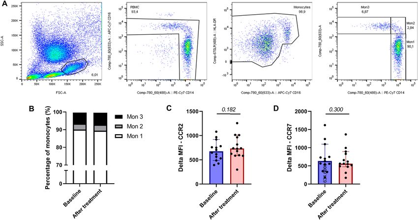
Cellular inflammation. Twelve weeks of atorvastatin did not change monocyte phenotype of freshly iso-
lated monocytes. The distribution across the classical (mon1/CD14++ CD16−), intermediate (mon 2/CD14++,
CD16+), and non-classical (mon 3/CD14+, CD16+) subsets did not change significantly after statin treatment
(p-value 0.406, 0.694 and 0.441, respectively; Fig. 2). In line, CCR2 and CCR7 expression on monocytes (p-value
0.182 and 0.300, respectively; Fig. 2) and monocyte subsets were not affected by statin treatment.
Discussion
This study evaluated the anti-inflammatory effects of statins on arterial wall- and cellular inflammation in subjects
with stage 3–4 CKD. Three months of atorvastatin treatment resulted in a 46% reduction of LDL-cholesterol
levels. Conversely, no effects were seen on MDS TBR of the aorta and carotid arteries or on chemokine receptor
expression of circulating monocytes. These results imply that CKD-associated inflammatory activity is mediated
by factors beyond LDL-cholesterol, and is not attenuated by statin treatment.
Statin‑refractory inflammatory state. In accordance with other subjects with increased CV-risk, sub-
jects with mild to moderate CKD are hallmarked by a pro-inflammatory phenotype, comprising increased arte-
rial wall inflammation and plasma immune cell a ctivation10,19. In the non-CKD CV risk population, lowering of
LDL-cholesterol, by either statins, LDL-cholesterol apheresis or PCSK9ab therapy, coincides with a decrease in
arterial wall inflammation11,12,20 and in plasma immune cell activation13. While the arterial 18F-FDG-uptake in
CKD subjects was comparable with CKD subjects from our earlier study, which showed significantly elevated
Scientific Reports | (2021) 11:4126 | https://doi.org/10.1038/s41598-021-83273-2 3
Vol.:(0123456789)www.nature.com/scientificreports/
Figure 1. Statin treatment does not abolish arterial wall inflammation in CKD subjects. Baseline and after
statin treatment arterial wall inflammation measured as mean of maximum uptake of most diseased segment
target to background ratio (MDS TBR) in the aorta (left) and index carotid (right).
Baseline After treatment p-value
Aortic MDS TBRmax 2.98 [2.75–3.75] 2.89 [2.42–3.49] 0.196
Aortic max TBRmax 2.89 [2.64–3.53] 2.83 [2.36–3.47] 0.208
Index carotid MDS TBRmax 2.00 [1.74–2.25] 1.94 [1.77–2.42] 0.972
Index carotid max TBRmax 1.97 [1.65–2.21] 1.91 [1.72–2.25] 0.972
Table 3. PET/CT parameters.
arterial wall inflammation compared to healthy c ontrols10. The absence of inflammatory improvement may
imply an overriding impact of non-LDL-cholesterol factors mediating inflammatory activation in CKD. Con-
cordantly, the pro-inflammatory phenotype of circulating monocytes in CKD-patients10, comprising increased
CCR2 and CC7 expression, was also not affected by potent statin treatment. In analogy, we recently reported
unchanged arterial wall inflammation and monocyte activation in subjects with marked lipoprotein(a) elevation
following PCSK9ab therapy. In that study, the persistence of an inflammatory state could be attributed to marked
residual Lp(a) elevation following the treatment21.
The inflammatory burden in CKD has been attributed to a complex interaction comprising immunologic,
metabolic and inflammatory components in which resident macrophages and circulating monocytes are major
contributors10,14,15. Pathophysiologic considerations propagating the pro-inflammatory state in CKD include
dysregulation of calcium and phosphate, activation of the renin-angiotensin-aldosteron system, NF-ĸB pathways
and inflammasome activation related to the accumulation of uremic toxins such as trimethylamine N-oxide,
p-cresyl sulfate and indoxyl sulfate derivatives22. Indoxyl sulfate and p-cresyl sulfate are of special interest, since
they are potentially modifiable and associated with both CVD and CKD-progression in the CKD-population.
P-cresyl sulfate and indoxyl sulfate are solely produced by the microbiome of the large intestine 23 and can impair
the intestinal barrier causing translocation of bacteria and bacterial products into the circulation. This change
in microbiome composition, amongst others, causes an increase in bacteria that produce uremic toxins, which
may in part contribute the persistence of systemic inflammation after statin therapy in C KD24.
Therapeutic implications of persistent inflammation in CKD. The mechanism behind the dimin-
ished benefit of statin therapy with progressive renal insufficiency remains to be established. Worsening of kid-
ney function coincides with an increase in both atherosclerotic as well as non-atherosclerotic CV events. Espe-
cially in advanced CKD, a large proportion of CV events can be attributed to non-atherosclerotic disease (e.g.
arrhythmia, valve calcification), which is mostly unaffected by statin therapy25. In this respect, the persistence of
a pro-inflammatory state in CKD patients, despite LDL-cholesterol lowering may provide a clue. Following the
significant CV risk reduction in CVD patients following interleukin (IL)-1β monoclonal antibody treatment,
it is tempting to speculate whether CKD patients need additional specific anti-inflammatory interventions in
order to further reduce their considerable residual CV risk. In support, post-acute coronary syndrome patients
with CKD were observed to have a larger absolute risk reductions following IL-1β antibody compared with non-
CKD patients26. Data evaluating other anti-inflammatory moieties in CKD patients are scarce. Treatment with
allopurinol decreases uric acid and ROS production, thereby decreasing NLRP3 inflammasome activation14,27.
Scientific Reports | (2021) 11:4126 | https://doi.org/10.1038/s41598-021-83273-2 4
Vol:.(1234567890)Scientific Reports |
(2021) 11:4126 |
www.nature.com/scientificreports/
https://doi.org/10.1038/s41598-021-83273-2
Figure 2. Statin treatment does not influence cellular inflammation in subjects with CKD. (A) Flow cytometry monocyte gating strategy. (B) Distribution across the monocyte subtypes before
and after statin treatment. (C) Delta MFI values of CCR2 expression on monocytes before and after statin treatment. (D) Delta MFI values of CCR7 expression on monocytes before and after
statin treatment.
5
Vol.:(0123456789)www.nature.com/scientificreports/
Long-term treatment with allopurinol was reported to attenuate renal function deterioration as well as CVD-
risk in subjects with CKD28,29. Conversely, recent studies argue that urate-lowering treatment with allopurinol
does not slow the decline in eGFR30,31. In line, several other interventions targeting uremic toxins have been
tested, but did not provide clinical benefit32. Preliminary data hint towards interventions decreasing the uremic
toxin load in CKD patients using pre- or probiotics33.
Strengths and limitations. This study is the first study to show the effect of statin treatment on arte-
rial wall and cellular inflammation in the CKD population. However, this study has several limitations. First,
the duration of the intervention with statins was limited. Previously, 18F-FDG-uptake was reported to decrease
within three days after apheresis as well as four weeks after initiation and intensification of statin therapy11. Sec-
ond, smoking might influence the degree of arterial wall inflammation. However, as only three participants were
active smokers, participants were their own control and none of the subjects started or ceased smoking during
this study this is unlikely to have obscured a beneficial effect of statin treatment on arterial wall inflammation.
Finally, the sample size of the study was limited. Since the sample size was in line with previous studies of our
group showing significant reductions in plaque inflammation12, it is unlikely that significant changes in MDS
TBR will be observed when increasing the sample size. Taking these limitations into account, the data from this
study support the presence of other causes beyond LDL-cholesterol which mediate the persistent inflammatory
state in CKD.
Conclusions. Statin treatment does not decrease arterial wall inflammation, nor does it change chemokine
receptor expression of circulating monocytes in subjects with mild to moderate CKD. Specific anti-inflamma-
tory interventions might be necessary to further dampen the inflammatory driven CV-risk in these subjects.
Received: 15 December 2020; Accepted: 27 January 2021
References
1. Go, A. S., Chertow, G. M., Fan, D., McCulloch, C. E. & Hsu, C. Y. Chronic kidney disease and the risks of death, cardiovascular
events, and hospitalization. N. Engl. J. Med. 351, 1296–1305 (2004).
2. Tonelli, M. et al. Risk of coronary events in people with chronic kidney disease compared with those with diabetes: A population-
level cohort study. Lancet 380, 807–814 (2012).
3. Kon, V., Linton, M. F. & Fazio, S. Atherosclerosis in chronic kidney disease: The role of macrophages. Nat. Rev. Nephrol. 7, 45–54
(2011).
4. Libby, P., Ridker, P. M. & Maseri, A. Inflammation and atherosclerosis. Circulation 105, 1135–1143 (2002).
5. Mach, F. et al. ESC/EAS Guidelines for the management of dyslipidaemias: Lipid modification to reduce cardiovascular risk. Eur.
Heart J. 2019, 1–78 (2019).
6. Herrington, W. G. et al. Impact of renal function on the effects of LDL cholesterol lowering with statin-based regimens: A meta-
analysis of individual participant data from 28 randomised trials. Lancet Diabetes Endocrinol. 4, 829–839 (2016).
7. Cholesterol Treatment Trialists’ (CTT) Collaboration et al. Efficacy and safety of more intensive lowering of LDL cholesterol: A
meta-analysis of data from 170,000 participants in 26 randomised trials. Lancet (London, England) 376, 1670–1681 (2010).
8. Tonelli, M. et al. Association between LDL-C and risk of myocardial infarction in CKD. J. Am. Soc. Nephrol. 24, 979–986 (2013).
9. Ridker, P. M., MacFadyen, J., Cressman, M. & Glynn, R. J. Efficacy of rosuvastatin among men and women with moderate chronic
kidney disease and elevated high-sensitivity C-reactive protein. A secondary analysis from the JUPITER (justification for the use
of statins in prevention-an intervention trial evaluating. J. Am. Coll. Cardiol. 55, 1266–1273 (2010).
10. Bernelot Moens, S. J. et al. Arterial and cellular inflammation in patients with CKD. J. Am. Soc. Nephrol. 28, 1278–1285 (2017).
11. Tawakol, A. et al. Intensification of statin therapy results in a rapid reduction in atherosclerotic inflammation: Results of a multi-
center fluorodeoxyglucose-positron emission tomography/computed tomography feasibility study. J. Am. Coll. Cardiol. 62, 909–917
(2013).
12. Hoogeveen, R. M. et al. PCSK9 antibody alirocumab attenuates arterial wall inflammation without changes in circulating inflam-
matory markers. JACC Cardiovasc. Imaging 12, 2571 (2019).
13. Bernelot Moens, S. J. et al. PCSK9 monoclonal antibodies reverse the pro-inflammatory profile of monocytes in familial hyper-
cholesterolaemia. Eur. Heart J. 38, 1584–1593 (2017).
14. Andrade-Oliveira, V., Foresto-Neto, O., Watanabe, I. K. M., Zatz, R. & Câmara, N. O. S. Inflammation in renal diseases: New and
old players. Front. Pharmacol. 10, 1–19 (2019).
15. Castillo-Rodríguez, E. et al. Inflammatory cytokines as uremic toxins: “ni son todos los que estan, ni estan todos los que son”.
Toxins (Basel) 9, 1–21 (2017).
16. Rudd, J. H. F. et al. 18Fluorodeoxyglucose positron emission tomography imaging of atherosclerotic plaque inflammation is highly
reproducible. Implications for atherosclerosis therapy trials. J. Am. Coll. Cardiol. 50, 892–896 (2007).
17. van der Valk, F. M. et al. Thresholds for arterial wall inflammation quantified by 18F-FDG PET imaging: Implications for vascular
interventional studies. JACC Cardiovasc. Imaging 9, 1198–1207 (2016).
18. Friedewald, W. T., Levy, R. I. & Fredrickson, D. S. Estimation of the concentration of low-density lipoprotein cholesterol in plasma,
without use of the preparative ultracentrifuge. Clin. Chem. 18, 499–502 (1972).
19. Barreto, D. V. et al. Plasma interleukin-6 is independently associated with mortality in both hemodialysis and pre-dialysis patients
with chronic kidney disease. Kidney Int. 77, 550–556 (2010).
20. Tahara, N. et al. Simvastatin attenuates plaque inflammation. Evaluation by fluorodeoxyglucose positron emission tomography.
J. Am. Coll. Cardiol. 48, 1825–1831 (2006).
21. Stiekema, L. C. A. et al. Persistent arterial wall inflammation in patients with elevated lipoprotein(a) despite strong low-density
lipoprotein cholesterol reduction by proprotein convertase subtilisin/kexin type 9 antibody treatment. Eur. Heart J. 31, 1–8 (2018).
22. Mihai, S. et al. Inflammation-related mechanisms in chronic kidney disease prediction, progression, and outcome. J. Immunol.
Res. 2018, 2180373 (2018).
23. Rossi, M. et al. Synbiotics easing renal failure by improving gut microbiology (SYNERGY): A randomized trial. Clin. J. Am. Soc.
Nephrol. 11, 223–231 (2016).
24. Anders, H. J., Andersen, K. & Stecher, B. The intestinal microbiota, a leaky gut, and abnormal immunity in kidney disease. Kidney
Int. 83, 1010–1016 (2013).
25. Sarnak, M. J. et al. Chronic kidney disease and coronary artery disease. J. Am. Coll. Cardiol. 74, 1823–1838 (2019).
Scientific Reports | (2021) 11:4126 | https://doi.org/10.1038/s41598-021-83273-2 6
Vol:.(1234567890)www.nature.com/scientificreports/
26. Ridker, P. M. et al. Inhibition of interleukin-1β by canakinumab and cardiovascular outcomes in patients with chronic kidney
disease. J. Am. Coll. Cardiol. 71, 2405–2414 (2018).
27. Mulay, S. R. & Anders, H.-J. Crystal nephropathies: Mechanisms of crystal-induced kidney injury. Nat. Rev. Nephrol. 13, 226–240
(2017).
28. Goicoechea, M. et al. Effect of allopurinol in chronic kidney disease progression and cardiovascular risk. Clin. J. Am. Soc. Nephrol.
5, 1388–1393 (2010).
29. Goicoechea, M. et al. Allopurinol and progression of CKD and cardiovascular events: Long-term follow-up of a randomized clinical
trial. Am. J. Kidney Dis. 65, 543–549 (2015).
30. Rossi, M., Campbell, K. & Johnson, D. Indoxyl sulphate and p-cresyl sulphate: Therapeutically modifiable nephrovascular toxins.
OA Nephrol. 1, 1–8 (2013).
31. Rossi, M., Klein, K., Johnson, D. W. & Campbell, K. L. Pre-, pro-, and synbiotics: Do they have a role in reducing uremic toxins?
A systematic review and meta-analysis. Int. J. Nephrol. 2012, 1–20 (2012).
32. Van, W. D. F. et al. Nonpharmacological lipoprotein apheresis reduces arterial inflammation in familial hypercholesterolemia. J.
Am. Coll. Cardiol. 64, 1418–1426 (2014).
33. Van Der, V. F. M. et al. Increased arterial wall inflammation in patients with ankylosing spondylitis is reduced by statin therapy.
Ann. Rheum. Dis. 75, 1848–1851 (2016).
Acknowledgements
The authors gratefully acknowledge M.F. Lam, E. Poel and M.E. Hemayat for their assistance with the nuclear
imaging scans.
Author contributions
R.H. and S.V., made substantial contributions to acquisition of data and analysis and interpretation of data and
was involved in drafting the manuscript. Y.K., H.V., L.V. and S.M., made substantial contributions to acquisi-
tion of data and interpretation of data and critically revised the manuscript. E.S. made substantial contributions
to conception, design and interpretation of data, furthermore he was involved in drafting the manuscript and
revision of the manuscript for important intellectual content.
Funding
This project has received funding from the European Union’s Horizon 2020 research and innovation program
under Grant Agreement No 667837 (REPROGRAM).
Competing interests
LV reports that his institution has received lecturing fees and advisory board fees from AstraZeneca, Ionis Phar-
maceuticals, Pfizer Inc., SanofiGenzyme, Vifor Pharma. ES reports that his institution has received lecturing fees
and advisory board fees from Amgen Inc., Regeneron, Sanofi, Akcea, Novartis and Athera. The other authors
declare no competing interests.
Additional information
Correspondence and requests for materials should be addressed to E.S.G.S.
Reprints and permissions information is available at www.nature.com/reprints.
Publisher’s note Springer Nature remains neutral with regard to jurisdictional claims in published maps and
institutional affiliations.
Open Access This article is licensed under a Creative Commons Attribution 4.0 International
License, which permits use, sharing, adaptation, distribution and reproduction in any medium or
format, as long as you give appropriate credit to the original author(s) and the source, provide a link to the
Creative Commons licence, and indicate if changes were made. The images or other third party material in this
article are included in the article’s Creative Commons licence, unless indicated otherwise in a credit line to the
material. If material is not included in the article’s Creative Commons licence and your intended use is not
permitted by statutory regulation or exceeds the permitted use, you will need to obtain permission directly from
the copyright holder. To view a copy of this licence, visit http://creativecommons.org/licenses/by/4.0/.
© The Author(s) 2021
Scientific Reports | (2021) 11:4126 | https://doi.org/10.1038/s41598-021-83273-2 7
Vol.:(0123456789)You can also read