MAR1 suppresses inflammatory response in LPS-induced RAW 264.7 macrophages and human primary peripheral blood mononuclear cells via the ...
←
→
Page content transcription
If your browser does not render page correctly, please read the page content below
Wang et al. Journal of Inflammation     (2021) 18:8
https://doi.org/10.1186/s12950-021-00271-x

 RESEARCH                                                                                                                                          Open Access

MAR1 suppresses inflammatory response in
LPS-induced RAW 264.7 macrophages and
human primary peripheral blood
mononuclear cells via the SIRT1/PGC-1α/
PPAR-γ pathway
Wei Wang1*† , Rong-Li Xu2†, Ping He1 and Rui Chen3*

  Abstract
  Background: Sepsis is a complex syndrome characterized by a dysregulated inflammatory response to systemic
  infection and leads to shock, multiple organ failure and death especially if not recognized early and treated
  promptly. Previous studies have suggested Maresin 1 (MAR1) can alleviate systemic inflammation in sepsis, but its
  mechanism has not been clarified.
  Methods: RAW 264.7 cells and human primary peripheral blood mononuclear cells (hPBMCs) were pretreated with
  LPS and MAR1. The mRNA expression and supernatant levels of pro-inflammatory cytokines, tumor necrosis factor
  (TNF-α), interleukin (IL)-1β and IL-6 were evaluated by RT-qPCR and ELISA, respectively. The expression levels of
  Sirtuin 1 (SIRT1), peroxisome proliferator-activated receptor γ coactivator-1α (PGC-1α), and Peroxisome proliferator-
  activated receptor gamma (PPAR-γ) were determined by RT-qPCR and Western blot analysis, respectively.
  Results: Our results show that LPS-induced inflammation increased the expression and secretion of
  proinflammatory cytokines TNF-α, IL-1β and IL-6 and induced suppression of SIRT1, PGC-1α, and PPAR-γ expression,
  which could be reversed by MAR1. And the effect of MAR1 was eliminated by repression of SIRT1/PPAR-γ and
  enhanced by PGC-1α overexpression.
  Conclusions: MAR1 suppressed inflammatory response in LPS-induced RAW 264.7 macrophages and hPBMCs via
  the SIRT1/PGC-1α/PPAR-γ pathway.
  Keywords: MAR1, SIRT1, PGC-1α, PPAR-γ, Sepsis

* Correspondence: ww_smile@qq.com; crui_rui@126.com
†
 Wei Wang and Rong-Li Xu are the co-first authors.
1
 Department of Emergency, Hainan General Hospital, Hainan Affiliated
Hospital of Hainan Medical University, No.19, Xiuhua Road, 570311 Haikou,
Hainan Province, People’s Republic of China
3
 Department of Medical Intensive Care Unit, General Hospital of Southern
Theater Command, PLA, No.111, Liuhua Road, 510010 Guangzhou, People’s
Republic of China
Full list of author information is available at the end of the article

                                       © The Author(s). 2021 Open Access This article is licensed under a Creative Commons Attribution 4.0 International License,
                                       which permits use, sharing, adaptation, distribution and reproduction in any medium or format, as long as you give
                                       appropriate credit to the original author(s) and the source, provide a link to the Creative Commons licence, and indicate if
                                       changes were made. The images or other third party material in this article are included in the article's Creative Commons
                                       licence, unless indicated otherwise in a credit line to the material. If material is not included in the article's Creative Commons
                                       licence and your intended use is not permitted by statutory regulation or exceeds the permitted use, you will need to obtain
                                       permission directly from the copyright holder. To view a copy of this licence, visit http://creativecommons.org/licenses/by/4.0/.
                                       The Creative Commons Public Domain Dedication waiver (http://creativecommons.org/publicdomain/zero/1.0/) applies to the
                                       data made available in this article, unless otherwise stated in a credit line to the data.
Wang et al. Journal of Inflammation (2021) 18:8 Page 2 of 11 Introduction PGC-1 family [22], it can bind to multiple transcription Sepsis, defined as a syndrome of systemic inflammatory factors, effectively promote mitochondrial biogenesis and response (SIRS) caused by infection as a result of an reduce mitochondrial damage [23]. PGC-1α can not only infective process, is a rapidly progressive and life- be activated by SIRT1 through deacetylation, but also act as threatening syndrome. Severe sepsis with multiple organ a transcription coactivator to enhance the activity of peroxi- dysfunctions is still the main cause of death in intensive some proliferator-activated receptor γ (PPARγ) [24, 25]. care unit patients [1, 2]. During sepsis, inflammation PPARγ is a member of the nuclear receptor superfamily of response can be activated in both macrophages and ligand-induced transcription factors, which mainly controls monocytes, and the inflammatory factors released by gene expression involved in adipogenesis, lipid metabolism, macrophages may accelerate the sepsis-induced cell and and inflammation [26]. Likewise, PPAR-γ activation is an tissue injury [3, 4]. Lipopolysaccharide (LPS), a compo- effective intervention to prevent or restore septic myocar- nent of the outer membrane of Gram-negative bacteria, dial dysfunction and has proven to be a promising treat- interacts with specific receptors on host effector cells ment strategy for sepsis [27]. From this result, we initially and induces the release of proinflammatory cytokines proposed that MAR1 reduces the inflammation response in [5]. Excessive production of these cytokines can lead to sepsis may be related to the activation of SIRT1/PGC-1α/ an uncontrolled inflammatory response [6]. Therefore, PPAR-γ axis. LPS is widely held to be a mediator of the inflammatory In our current study, the relationship between reduc- events associated with sepsis [7]. LPS-treated macro- tion of inflammatory response by MAR1 and SIRT1/ phages were adopted as an in vitro model of endotoxin- PGC-1α/PPAR-γ signaling axis was systematically inves- induced inflammation during sepsis [8], such as using tigated and intended to explain the specific mechanism the murine macrophage cell line RAW264.7 [9, 10]. of MAR1 in in vitro sepsis models of LPS-induced RAW Specialized pro-resolving mediators such as resolvins, 264.7 cells and human primary peripheral blood mono- protectins, and maresins, actively turn off the inflamma- nuclear cells (hPBMCs). tory response by acting on different G protein-coupled receptors expressed on immune cells that activate the Materials and methods dual anti-inflammatory process [11]. Maresin 1 (MAR1) Cell culture and treatment from polyunsaturated fatty acids is one of the most The mouse mononuclear macrophage leukemia cell line recently identified members of the family of anti- RAW264.7 was purchased from the American Type inflammatory lipid mediators and has shown anti- Culture Collection (ATCC), and primary human periph- inflammatory and proresolving activity in zymosan-induced eral blood mononuclear cells (hPBMCs) was purchased peritonitis [12]. Many studies have shown that MAR1 from PriCells (Wuhan, China). Cells were cultured in shows proresolving and anti-inflammatory effects in a DMEM containing 10 % fetal bovine serum (FBS, Gibco; variety of diseases [12–14]. However, the specific Thermo Fisher Scientific) and 1 % penicillin (100 U/ mechanism of its role in sepsis remains unclear. In mL)/streptomycin (100 µg/mL) (Hyclone; GE Healthcare previous studies, MAR1 has been shown to reduce Life Sciences) in a 37℃ in an incubator containing 5 % proinflammatory response and mitochondrial damage CO2 at 37 °C. For establishment of in vitro model, by activating SIRT1 signaling to protect mouse brain lipopolysaccharide (LPS, 10 µg/ mL, Sigma-Aldrich, St. tissue and neurons from ischemia/reperfusion injury Louis, MO, USA) was used to treat the cells for 24 h. [15, 16]. A large number of studies have shown that For treatment of MAR1, cells were treated with different sirtuin 1 (SIRT1) is a highly conserved mammalian concentrations (0.1 nM, 1 nM, 10 nM and 100 nM) of NAD(+)-dependent protein deacetylase and is involved MAR1 (cat. No. HY-116,429; MedChemExpress, NJ, in multiple biological processes [17]. Therefore, it is USA) for 12 h before LPS treatment. For analysis of time not surprising that the changes in SIRT1 activity or effects, cells were treated with 10 nM MAR1 with differ- expression profile are strongly correlated with meta- ent time duration (0 h, 4 h, 6 h, 8 h and 12 h). bolic processes, oxidative stress, and the pathogenesis of inflammation [17, 18]. Furthermore, SIRT1 knock- Real‐time qPCR analysis out mice have increased sensitivity to septic-induced Total RNA was extracted from cells using TRIzol inflammatory lung injury [19, 20]. Meanwhile, SIRT1 reagent (Invitrogen). After total RNA was reverse suppresses acute lung inflammation during sepsis by transcribed into cDNA by using a Prime-Script™ One controlling the activation of inflammatory pathway Step RT-qPCR kit (Takara Biotechnology Co., Ltd., [21], so it is highly possible that MAR1 reduce the Dalian, China), PCR reactions for SIRT1, PGC-1α, inflammatory response by activating SIRT1 in sepsis. PPAR-γ, IL-1β, IL-6 and TNF-α mRNA were performed Peroxisome proliferator-activated receptor-γ co-activator- on a LightCycler 480 (Roche, Mannheim, Germany) sys- 1α (PGC-1α) is one of the three known co-activators in the tem with GAPDH used as an internal control. The light
Wang et al. Journal of Inflammation   (2021) 18:8                                                          Page 3 of 11

cycler DNA master SYBR green I kit (Roche Molecular         Cell transfection
Biochemicals, Mannheim, Germany) was then used for          GenePharma (Shanghai, China) synthesized PGC-1α
PCR experiments. The following primers were used:           overexpression, sh-Sirt1 vector, sh-PPAR-γ vector and
SIRT1 Forward: 5′-GGTGTTAAATACCAAACTGC-3′                   corresponding negative controls. Cells (3 × 10⁵) were
and reverse: 5′-AGGAGTGATGTTCAAAATG-3′; PGC-                seeded into 6-well plates for cell transfection after cell
1α Forward: 5’-AATTCACAATCACAGGATCAGAACA3’                  confluence of 40–60 %. Replace with new medium and
and reverse: 5’-ACTTAAGGTGCGTTCAATAGTCTT-3’;                prepare cell transfection buffer. Incubate 5 µL/Wells of
PPAR-γ Forward: 5’-TTGGCCATATTTATAGCTGTCA                   lipofectamine 2000 (Thermo, USA) and 250 µL of Opti-
TTATT-3′ and reverse: 5’- TGTCCTCGATGGGCTTCA-               MEM medium (Gibco, USA) for 5 min at room
3’; TNF-α Forward: 5’-GAGCTGTGGGGAGAACAAAA                  temperature and mix with PGC-1α, sh-Sirt1 vector, sh-
GGA-3′ and reverse: 5’- TTGGCCCTTGAAGAGGAC                  PPAR-γ vector and negative control (5 µL/well) 250 µL of
CTG-3’; IL-1β Forward: 5’-GAC CTT CCA GGA TGA               Opti-MEM medium for 20 min. Then, inoculate the cells
GGA CA-3′ and reverse: 5’-AGC TCATATGGGTCCGA                into a 6-well plate and incubate in the incubator for 6 h.
CAG-3’; IL-6 Forward: 5’-TCC AGT TGC CTTCTT GGG             Then, the cells are incubated in 10 % FBS continuous cul-
AC-3′ and reverse: 5’-GTGTAATTAAGCCTCCGA                    ture in new DMEM medium (Gibco, USA).
CTTG-3’; GAPDH Forward: 5’-AGAAGGCTGGGGCTCA
TTTG-3’ and reverse: 5’-AGGGGCCATCCACAGTCT                  Statistical analysis
TC-3. The relative expression of the target genes was       Each experiment was repeated at least 3 times. Data
calculated using the 2−ΔΔCt method. All experiments         were analyzed using GraphPad Prism 6 (GraphPad soft-
were repeated in triplicate.                                ware, San Diego, California, USA). The data were
                                                            expressed as mean ± SD. with either unpaired two-tailed
                                                            t-test or one-way ANOVA as appropriate. P < 0.05 was
ELISA assay                                                 considered statistically significant.
The contents of IL-1β, IL-6 and TNF-α in the cell super-
natant were determined using an ELISA assay. Centri-        Results
fuge at 4000 r/min for 15 min at 4 °C to collect the        MAR1 inhibited the inflammatory response of RAW264.7
supernatant. ELISA kits were obtained from Boster (Bos-     cells and hPBMCs induced by LPS in a dose-dependent
ter Biological Technology, Wuhan, China), and each          manner
sample was measured three times according to the man-       LPS endotoxins are widely used as experimental models
ufacturer’s instructions.                                   of systemic bacterial infection and trigger inflammatory
                                                            factors such as TNF-α, IL-1β and IL-6 [28]. The changes
                                                            of inflammatory factors in LPS-treated RAW264.7 cells
Western blot analysis                                       and hPBMCs under the intervention of different concen-
Cells were washed in phosphate-buffered saline (PBS),       trations of MAR1 were analyzed by RT-qPCR. And we
then in 10 mM TrisHCl (pH7.5), 100 mM NaCl, 1 %             found that MAR1 inhibited the LPS-induced increase of
NP-40, 50 mM NaF, 2 mMEDTA (pH8.0), 1 mM PMSF,              the mRNA levels of proinflammatory factors in a dose-
Cells were lysed in 10 µg/mL leupeptin, and 10 µg/mL        dependent manner in both RAW264.7 cells and
aprotinin. The mixture was collected as a whole cell ex-    hPBMCs (Fig. 1a). Next, we tested the secretion of pro-
tract. The protein extract was separated by sodium lauryl   inflammatory factors TNF-α, IL-1β, and IL-6 in LPS-
sulfate polyacrylamide gel electrophoresis (SDS-PAGE)       induced RAW264.7 cells and hPBMCs and the effect
and transferred to a polyvinylidene fluoride (PVDF)         of MAR1 on their secretion. The results showed that
membrane (immunoblotted PVDF membrane, Bio-Rad).            LPS increased secretion of TNF-α, IL-6 and IL-1β in
The membrane was blocked with Tris buffered saline          both RAW264.7 cells and hPBMCs, which can be re-
and 3 % bovine serum albumin (BSA) in Tween 20              versed by MAR1 in a dose-dependent manner (Fig. 1b).
(TBST, pH 7.4) for 1.5 h, and then specific primary anti-   In addition, we also performed Western blot assay to
bodies with SIRT1, PGC-1α and PPAR-γ treated at 4 °C        evaluate the levels of SIRT1, PGC-1α and PPAR-γ pro-
overnight. GAPDH was used as a protein-loading con-         tein, and the results showed that the expression of
trol. All antibodies were purchased from Proteintech        SIRT1, PGC-1α and PPAR-γ protein were significantly
(ProteinTech Group, Chicago, IL, USA.) The membrane         down-regulated in both RAW264.7 cells and hPBMCs
was then treated with horseradish peroxidase (HRP)          caused by LPS (p < 0.001) (Fig. 1c) and upregulated by
-conjugated secondary antibody (1: 1,000) for 2 h. The      MAR1 in a dose-dependent manner. These results in-
bands were visualized using an enhanced chemilumines-       dicate that MAR1 reduces the inflammatory response
cence system (ECL, Thermo Fisher Scientific) and LAS        of RAW264.7 cells and hPBMCs induced by LPS in a
image software (Fuji, New York, New York, USA).             dose-dependent manner.
Wang et al. Journal of Inflammation (2021) 18:8 Page 4 of 11 Fig. 1 MAR1 inhibited the inflammatory response of RAW264.7 cells and hPBMCs induced by LPS in a dose-dependent manner. RAW264.7 cells and hPBMCs were pretreated with different concentrations (0.1 nM, 1 nM, 10 nM and 100 nM) of MAR1 for 12 h and then exposed to LPS (10 µg/mL) for 12 h. a, TNF-α, IL-1β and IL-6 expression were detected by RT-qPCR; b, TNF-α, IL-1β and IL-6 secretion were measured using ELISA. c, The protein levels of SIRT1, PGC-1α and PPAR-γ were evaluated by Western blot. Data were shown as the means ± SD. Each experiment was repeated in triplicate. *p < 0.05, **p < 0.01, ***p < 0.001 MAR1 inhibited the inflammatory response of RAW264.7 cells and 12 h). The changes of inflammatory factors in and hPBMCs induced by LPS in a time-dependent manner LPS-treated RAW264.7 cells and hPBMCs at different The above results indicate that MAR1 reduces the intervention time (0 h, 4 h, 6 h, 8 h and 12 h) of inflammatory response of both RAW264.7 cells and MAR1 were analyzed by RT-qPCR. And we demon- hPBMCs induced by LPS in a dose-dependent manner strated that MAR1 inhibited the LPS-induced increase especially at the highest concentration of 100 nM. We of the mRNA levels of proinflammatory factors in then consider investigated cells treated by 10 nM both RAW264.7 cells and hPBMCs with the treatment MAR1 by different time duration (0 h, 4 h, 6 h, 8 h time (Fig. 2a). Then, we tested the secretion of TNF-α,
Wang et al. Journal of Inflammation    (2021) 18:8                                                                           Page 5 of 11

 Fig. 2 MAR1 inhibited the inflammatory response of RAW264.7 cells and hPBMCs induced by LPS in a time-dependent manner. RAW264.7
 cells and hPBMCs were pretreated with MAR1 (10 nM) and then exposed to LPS (10 µg/mL) for 4, 6, 8, 12 h. a, TNF-α, IL-1β and IL-6
 expression were detected by RT-qPCR; b, TNF-α, IL-1β and IL-6 secretion were measured using ELISA. c, The protein levels of SIRT1, PGC-
 1α and PPAR-γwere detected by Western blot. Data were shown as the means ± SD. Each experiment was repeated in triplicate. *p < 0.05,
 **p < 0.01, ***p < 0.001

IL-1β, and IL-6 in LPS-induced RAW264.7 cells or                      of SIRT1, PGC-1α and PPAR-γ protein, and the re-
hPBMCs and the effect of MAR1 on their secretion.                     sults showed that the expression of SIRT1, PGC-1α
The results showed that LPS increased the secretion of                and PPAR-γ were significantly down-regulated in both
TNF-α, IL-6 and IL-1β in both RAW264.7 cells and                      RAW264.7 cells and hPBMCs induced by LPS (p <
hPBMCs, which can be reversed by MAR1 (10 nM)                         0.001) (Fig. 2c) and were upregulated by MAR1
(Fig. 2b). Thus, MAR1 reduced the levels of inflamma-                 (10 nM) in a time-dependent manner. These results
tory factors with the treatment time. Additionally, we                indicate that MAR1 reduces the inflammatory re-
also performed the Western blot to evaluate the levels                sponse induced by LPS in a time-dependent manner.
Wang et al. Journal of Inflammation       (2021) 18:8                                                                                  Page 6 of 11

PPAR-γ knockdown suppressed the effect of MAR1 on                           and hPBMCs were induced by LPS and MAR1
LPS-induced inflammatory response in RAW264.7 cells                         (10 nM) for 12 h after sh-PPAR-γ and control vectors
and hPBMCs                                                                  were stalely transfected. The results of RT-qPCR
Previous results revealed a possible mechanism for                          showed that the mRNA expression of PPAR-γ was
MAR1 in LPS-induced sepsis and inhibited inflamma-                          markedly suppressed following transfection with the
tory response in RAW264.7 cells or hPBMCs. In order                         shRNA-containing vector (Fig. 3a). Both mRNA and
to verify the role of PPAR-γ in the effect of MAR1 on                       protein levels of PPAR-γ significantly were decreased
LPS-induced sepsis, sepsis model of RAW264.7 cells                          by LPS treatment, and this effect could be blocked by

 Fig. 3 PPARγ knockdown suppressed the effect of MAR1 on LPS-induced inflammatory response in RAW264.7 cells and hPBMCs. Cells were
 treated with LPS (10 µg/ mL) for 24 h or treated with 10 nM of MAR1 for 12 h before LPS treatment. Each experiment was repeated in triplicate.
 a, Efficiency of RNA interfering was assayed by RT-qPCR. b, The protein level of PPAR-γ was detected by Western blot in different cell groups; c,
 The mRNA levels of TNF-α, IL-1β and IL-6 were determined by RT-qPCR in different cell groups. d, TNF-α, IL-1β and IL-6 secretion were measured
 using ELISA. Data were shown as the means ± SD. *p < 0.05, **p < 0.01, ***p < 0.001
Wang et al. Journal of Inflammation   (2021) 18:8                                                      Page 7 of 11

MAR1 treatment, which was further reversed by             mechanism by which MAR1 abolish LPS-induced cel-
PPAR-γ knockdown (Fig. 3b and c). Both mRNA               lular inflammation. Therefore, this study aimed to de-
expression and protein levels of pro-inflammatory         termine the effect of MAR1 on inflammatory response
factors were suppressed under the action of MAR1 in       and the relationship between MAR1 and SIRT1/PGC-
LPS-induced RAW264.7 cells and hPBMCs, and the            1α/PPAR-γ axis in RAW264.7 cells and hPBMCs
changes were reversed in cells with knockdown of          induced by LPS.
PPAR-γ (Fig. 3d and e). Therefore, inhibition of             Sepsis is considered a serious disease with multiple
PPAR-γ can offset the effect of MAR1 on inflamma-         organ damage caused by uncontrollable inflammation
tory response in LPS-induced sepsis model.                in severe infections, while MAR1 plays a role in redu-
                                                          cing the bacterial burden and mitigating excessive in-
MAR1 suppressed inflammatory response in LPS-induced      flammatory response and increases the survival rate of
RAW 264.7 cells and hPBMCs via the SIRT1/PGC-1α/PPAR-     sepsis mice [32]. The study by Li et al. also suggests
γ pathway                                                 that the protective effect of MAR1 on sepsis may partly
The expression of TNF-α, IL-1β, and IL-6 induced by       be related to the inhibition of the activation of NF-κB
LPS were significantly reduced after treatment with       that causes increased proinflammatory cytokines such
MAR1 (P < 0.001), the effect of MAR1 can be counter-      as IL-6, IL-1β and TNF-α in a mice model [33]. In
acted by SIRT1 knockdown and further enhanced by          addition, MAR1, as one of the newly discovered anti-
PGC-1α overexpression (Fig. 4a). The changes in           inflammatory and pro-degradation mediators, can
secretion of pro-inflammatory factors were examined       inhibit the infiltration and adhesion of neutrophils,
by ELISA experiments. Secretion of TNF-α, IL-1β and       enhance the macrophage phagocytosis of necrotic cells,
IL-6 induced by LPS were significantly reduced after      down-regulate the production of inflammatory media-
treatment with MAR1 (P < 0.001), the effect of MAR1       tors and induce the GSK3β anti-inflammatory axis in
can be abolished by SIRT1 knockdown and further           human monocytes to limit the excessive development
augmented by PGC-1α overexpression (Fig. 4b).             of inflammation and promote the timely resolution of
Additionally, the mRNA and protein results in both        inflammation[34, 35]. Our study shows for the first
RAW264.7 cells and hPBMCs showed that the expres-         time that MAR1 can reduce the expression and secre-
sion of SIRT1, PGC-1α and PPAR-γ in the LPS group         tion of pro-inflammatory factors and has shown
were significantly lower than those in the control        protection against LPS-induced RAW264.7 cell and
group (P < 0.001), while MAR1 could restore their ex-     hPMBCs sepsis model.
pression, and the effect of MAR1 can be counteracted         There is evidence that increased expression of SIRT1
by SIRT1 knockdown and further enhanced by PGC-           can reduce multiple organ damage, including lung,
1α overexpression (Fig. 4 c and d). These results indi-   kidney, and liver, caused by sepsis [36, 37]. Interest-
cate that MAR1 suppressed inflammatory response on        ingly, by activating SIRT1, MAR1 treatment exerted a
LPS-induced sepsis via the SIRT1/PGC-1α/PPAR-γ            neuroprotective effect in mice and thus had a thera-
pathway.                                                  peutic effect on cerebral ischemia-reperfusion injury
                                                          [15]. Therefore, the protective effect of MAR1 on LPS-
Discussion                                                induced cellular inflammation in this study suggests
More and more studies have proved that using LPS-         that MAR1 may reduce the inflammatory response by
stimulated cells to establish an in vitro inflammation    activating SIRT1. In addition, the anti-inflammatory
model can more closely study sepsis and LPS-treated       effects of SIRT1 in preventing multiple inflammatory
macrophages were adopted as an in vitro model of          response can be attributed to the activation of PGC-
endotoxin-induced inflammation during sepsis [9, 29,      1α and the down-regulation of NF-κB [38]. PPAR-γ is
30]. Recently, some studies have shown that MAR1          a ligand-activated transcription factor involved in cell
has a protective effect against inflammatory response     proliferation, lipid metabolism and inflammation. It
by affecting SIRT1-mediated signaling pathway [31].       has been shown to be a regulator of multiple inflam-
However, its underlying mechanism remains to be elu-      matory responses and activated PPAR-γ can improve
cidated. In this study, we found that MAR1 attenuated     survival in sepsis animals [27, 39]. Obviously, SIRT1
inflammatory response by regulating the expression        can reduce the production of various proinflammatory
and secretion of LPS-induced proinflammatory cyto-        cytokines by activating PGC-1α and inhibiting NF-κB
kines. In addition, we also demonstrated that MAR1        activation to reduce the acute inflammatory response
increased SIRT1, PGC-1α, and PPAR-γ expression in         in tissue injury [40]. In this study, we demonstrated
RAW264.7 cells and hPBMCs. Then the cells were            that the levels of SIRT1, PGC-1α, and PPAR-γ protein
transfected with sh-SIRT1 or sh- PPAR-γ vector and        involved in anti-inflammatory action were reduced in
PGC-1α overexpression vector to study the                 RAW264.7 cells and hPBMCs induced by LPS, while
Wang et al. Journal of Inflammation (2021) 18:8 Page 8 of 11 Fig. 4 (See legend on next page.)
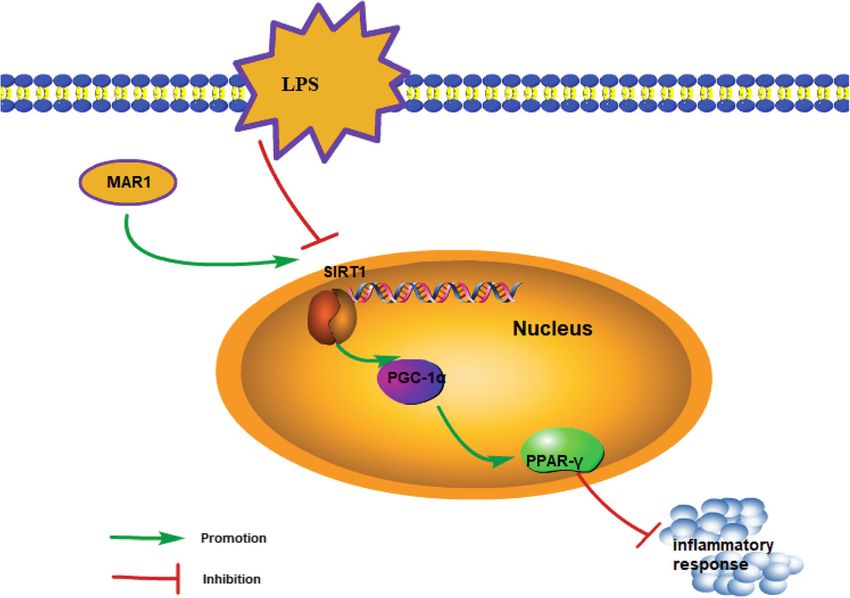
Wang et al. Journal of Inflammation      (2021) 18:8                                                                               Page 9 of 11

 (See figure on previous page.)
 Fig. 4 MAR1 suppressed inflammatory response in LPS-induced RAW 264.7 cells and hPBMCs via the SIRT1/PGC-1α/PPAR-γ pathway. Cells were
 treated with LPS (10 µg/ mL) for 24 h or treated with 10 nM of MAR1 for 12 h before LPS treatment. a, TNF-α, IL-1β and IL-6 expression were
 detected by RT-qPCR; b, TNF-α, IL-1β and IL-6 secretion were measured using ELISA. c-d, The mRNA and protein levels of SIRT1, PGC-1α and
 PPAR-γ were detected by RT-qPCR and Western blot, respectively. Data were shown as the means ± SD. Each experiment was repeated in
 triplicate. *p < 0.05, **p < 0.01, ***p < 0.001

the proinflammatory cytokines IL-6, IL-1 β and TNF-                      concluded from the results of this study that MAR1
α expression and secretion increased. Under the action                   reduces the inflammatory response by activating the
of MAR1, the elevated levels of SIRT1, PGC-1α, and                       regulatory mechanism of the SIRT1/PGC-1α/ PPAR-γ
PPAR-γ protein and inhibited the expression of pro-                      axis (Fig. 5).
inflammatory factors and exerted anti-inflammatory                         The present study also has some limitations. It is
effects were observed. In subsequent observations, we                    an in vitro study, thus the in vivo effects of MAR1 on
inhibited the expression of SIRT1 in RAW264.7 cells                      sepsis as well as its clinical effects are not clear. More
and hPBMCs by transfected with shRNA against                             researches are still necessary to confirm the in vivo
SIRT1 to explore the role of SIRT1 in the anti-                          and potential clinical application of MAR1 in sepsis
inflammatory activity of MAR1. The results showed                        treatment.
that the expression of PGC-1α and PPAR-γ protein                           In conclusion, this study demonstrates that the SIRT1/
was inhibited, the content of pro-inflammatory factors                   PGC-1α/PPAR-γ axis plays an important role in the
increased, and LPS-induced sepsis worsened. It was                       process of MAR1 in reducing LPS-induced inflammation
proved that SIRT1 was activated in RAW264.7 cells                        in in vitro sepsis model and provides clues for further
and hPBMCs under MAR1 treatment to reduce LPS-                           exploring the mechanism of MAR1 in reducing inflam-
induced inflammatory response. At the same time,                         mation in sepsis. Altogether, we propose that MAR1
effect of suppressing SIRT1 expression was reversed                      could not only be a potential biomarker but also a novel
by overexpression of PGC-1α. In summary, it can be                       therapeutic target for sepsis.

 Fig. 5 The schematic diagram consisting of identified pathway showing that MAR1 reduces the inflammatory response by activating the
 regulatory mechanism of the SIRT1/PGC-1α/PPAR-γ axis
Wang et al. Journal of Inflammation             (2021) 18:8                                                                                         Page 10 of 11

Acknowledgements                                                                    12. Serhan CN. Pro-resolving lipid mediators are leads for resolution physiology.
Not applicable.                                                                         Nature. 2014;510(7503):92–101.
                                                                                    13. Serhan CN, et al. Macrophage proresolving mediator maresin 1 stimulates
Authors’ contributions                                                                  tissue regeneration and controls pain. FASEB J. 2012;26(4):1755–65.
Guarantor of integrity of the entire study: Rui Chen; Study concepts: Wei           14. Schlegel M, et al. Inhibition of neogenin fosters resolution of inflammation
Wang, Rong-Li Xu, Rui Chen; Study design: Wei Wang, Rong-Li Xu; Definition              and tissue regeneration. J Clin Invest. 2018;128(10):4711–26.
of intellectual content: Rong-Li Xu; Literature research: Ping He; Experimental     15. Xian W, et al. Maresin 1 attenuates the inflammatory response and
studies: Rui Chen; Data acquisition: Rong-Li Xu; Data analysis: Wei Wang; Stat-         mitochondrial damage in mice with cerebral ischemia/reperfusion in a
istical analysis: Wei Wang, Rong-Li Xu; Manuscript preparation: Wei Wang,               SIRT1-dependent manner. Brain Res. 2019;1711:83–90.
Rong-Li Xu; Manuscript editing: Wei Wang, Rong-Li Xu, Rui Chen; Manuscript          16. Xian W, et al. The pro-resolving lipid mediator Maresin 1 protects against
review: Rui Chen. The author(s) read and approved the final manuscript.                 cerebral ischemia/reperfusion injury by attenuating the pro-inflammatory
                                                                                        response. Biochem Biophys Res Commun. 2016;472(1):175–81.
Funding                                                                             17. Yuk JM, et al. Orphan Nuclear Receptor ERRalpha Controls Macrophage
None.                                                                                   Metabolic Signaling and A20 Expression to Negatively Regulate TLR-Induced
                                                                                        Inflammation. Immunity. 2015;43(1):80–91.
                                                                                    18. Xu S, et al. SIRT1/3 Activation by Resveratrol Attenuates Acute Kidney Injury
Availability of data and materials
                                                                                        in a Septic Rat Model. Oxid Med Cell Longev. 2016;2016:7296092.
All data generated or analysed during this study are included in this
                                                                                    19. Zhuo Y, et al. Resolvin D1 promotes SIRT1 expression to counteract the
published article [and its supplementary information files].
                                                                                        activation of STAT3 and NF-κB in mice with septic-associated lung injury.
                                                                                        Inflammation. 2018;41(5):1762–71.
Ethics approval and consent to participate                                          20. Quan M, et al. Tanshinone IIA protects against lipopolysaccharide-induced
Not Applicable. This article does not contain any studies with human                    lung injury through targeting Sirt1. J Pharm Pharmacol. 2019;71(7):1142–51.
participants or animals performed by any of the authors.                            21. Gao R, et al. Sirt1 restrains lung inflammasome activation in a murine model
                                                                                        of sepsis. Am J Physiol Lung Cell Mol Physiol. 2015;308(8):L847-53.
Consent for publication                                                             22. Scarpulla RC. Transcriptional paradigms in mammalian mitochondrial
Not Applicable.                                                                         biogenesis and function. Physiol Rev. 2008;88(2):611–38.
                                                                                    23. Scarpulla RC. Metabolic control of mitochondrial biogenesis through the
Competing interests                                                                     PGC-1 family regulatory network. Biochim Biophys Acta. 2011;1813(7):
The authors declare that they have no conflict of interest.                             1269–78.
                                                                                    24. Higashida K, et al. Effects of resveratrol and SIRT1 on PGC-1alpha activity
Author details                                                                          and mitochondrial biogenesis: a reevaluation. PLoS Biol. 2013;11(7):
1                                                                                       e1001603.
 Department of Emergency, Hainan General Hospital, Hainan Affiliated
Hospital of Hainan Medical University, No.19, Xiuhua Road, 570311 Haikou,           25. Makela J, et al. Peroxisome proliferator-activated receptor-gamma
Hainan Province, People’s Republic of China. 2Department of Cardiology,                 (PPARgamma) agonist is neuroprotective and stimulates PGC-1alpha
Hainan General Hospital, Hainan Affiliated Hospital of Hainan Medical                   expression and CREB phosphorylation in human dopaminergic neurons.
University, No.19, Xiuhua Road, 570311 Haikou, Hainan Province, People’s                Neuropharmacology. 2016;102:266–75.
Republic of China. 3Department of Medical Intensive Care Unit, General              26. Wang L, et al. Natural product agonists of peroxisome proliferator-activated
Hospital of Southern Theater Command, PLA, No.111, Liuhua Road, 510010                  receptor gamma (PPARgamma): a review. Biochem Pharmacol. 2014;92(1):
Guangzhou, People’s Republic of China.                                                  73–89.
                                                                                    27. Peng S, et al. PPAR-gamma Activation Prevents Septic Cardiac Dysfunction
Received: 22 July 2020 Accepted: 21 January 2021                                        via Inhibition of Apoptosis and Necroptosis. Oxid Med Cell Longev. 2017;
                                                                                        2017:8326749.
                                                                                    28. Fang Y, et al. LncRNA H19 functions as an Aquaporin 1 competitive
References                                                                              endogenous RNA to regulate microRNA-874 expression in LPS sepsis.
1. Angus DC, van der Poll T. Severe sepsis and septic shock. N Engl J Med.              Biomed Pharmacother. 2018;105:1183–91.
    2013;369(9):840–51.                                                             29. Huang W, et al. Long non-coding RNA PVT1 promote LPS-induced septic
2. Rello J, et al. Sepsis: A Review of Advances in Management. Adv Ther. 2017;          acute kidney injury by regulating TNFalpha and JNK/NF-kappaB pathways in
    34(11):2393–411.                                                                    HK-2 cells. Int Immunopharmacol. 2017;47:134–40.
3. Cavaillon J-M, Adib-Conquy M. Monocytes/macrophages and sepsis. Crit             30. Zhong W, et al. Curcumin alleviates lipopolysaccharide induced sepsis
    Care Med. 2005;33(12):S506-9.                                                       and liver failure by suppression of oxidative stress-related inflammation
4. Qiu P, Liu Y, Zhang J. the role and mechanisms of macrophage autophagy               via PI3K/AKT and NF-kappaB related signaling. Biomed Pharmacother.
    in sepsis. Inflammation. 2019;42(1):6–19.                                           2016;83:302–13.
5. Hu SB, Zider A, Deng JC. When host defense goes awry: Modeling sepsis-           31. Vachharajani VT, et al. Sirtuins Link Inflammation and Metabolism. J
    induced immunosuppression. Drug Discovery Today Disease Models. 2012;               Immunol Res. 2016;2016:8167273.
    9(1):e33–8.                                                                     32. Gu J, et al. Maresin 1 attenuates mitochondrial dysfunction through the
6. Hung Y-L, et al. Corylin protects LPS-induced sepsis and attenuates LPS-             ALX/cAMP/ROS pathway in the cecal ligation and puncture mouse model
    induced inflammatory response. Sci Rep. 2017;7:46299.                               and sepsis patients. Lab Invest. 2018;98(6):715–33.
7. Napier BA, et al. Western diet regulates immune status and the response to       33. Li R, et al. Maresin 1 Mitigates Inflammatory Response and Protects Mice
    LPS-driven sepsis independent of diet-associated microbiome. Proc Natl              from Sepsis. Mediators Inflamm. 2016;2016:3798465.
    Acad Sci. 2019;116(9):3688–94.                                                  34. Serhan CN, et al. Maresins: novel macrophage mediators with potent
8. Pfeiffer D, et al. miR-146a, miR-146b, and miR-155 increase expression of IL-6       antiinflammatory and proresolving actions. J Exp Med. 2009;206(1):15–23.
    and IL-8 and support HSP10 in an in vitro sepsis model. PLoS One. 2017;         35. Gu Z, et al. Resolvin D1, resolvin D2 and maresin 1 activate the GSK3beta
    12(6):e0179850.                                                                     anti-inflammatory axis in TLR4-engaged human monocytes. Innate Immun.
9. Chen L, et al. Sonchus oleraceus Linn protects against LPS-induced sepsis            2016;22(3):186–95.
    and inhibits inflammatory responses in RAW264. 7 cells. J Ethnopharmacol.       36. Kou DQ, et al. Magnolol attenuates the inflammation and apoptosis
    2019;236:63–9.                                                                      through the activation of SIRT1 in experimental stroke rats. Pharmacol Rep.
10. Zhou W, et al. MicroRNA-205–5b inhibits HMGB1 expression in LPS-induced             2017;69(4):642–7.
    sepsis. Int J Mol Med. 2016;38(1):312–8.                                        37. Poulose N, Raju R. Sirtuin regulation in aging and injury. Biochim Biophys
11. Serhan CN, et al. Protectins and maresins: New pro-resolving families of            Acta. 2015;1852(11):2442–55.
    mediators in acute inflammation and resolution bioactive metabolome.            38. Cho RL, et al. Heme oxygenase-1 induction by rosiglitazone via PKCalpha/
    Biochim Biophys Acta. 2015;1851(4):397–413.                                         AMPKalpha/p38 MAPKalpha/SIRT1/PPARgamma pathway suppresses
Wang et al. Journal of Inflammation (2021) 18:8 Page 11 of 11
lipopolysaccharide-mediated pulmonary inflammation. Biochem Pharmacol.
2018;148:222–37.
39. Li HY, et al. Curcumin inhibits angiotensin II-induced inflammation and
proliferation of rat vascular smooth muscle cells by elevating PPAR-gamma
activity and reducing oxidative stress. Int J Mol Med. 2017;39(5):1307–16.
40. Yue L, et al. Adiponectin Protects against Glutamate-Induced Excitotoxicity
via Activating SIRT1-Dependent PGC-1alpha Expression in HT22
Hippocampal Neurons. Oxid Med Cell Longev. 2016;2016:2957354.
Publisher’s Note
Springer Nature remains neutral with regard to jurisdictional claims in
published maps and institutional affiliations.You can also read