Angiopoietin-2 regulated by progesterone induces uterine vascular remodeling during pregnancy - Spandidos Publications
←
→
Page content transcription
If your browser does not render page correctly, please read the page content below
Molecular Medicine REPORTS 22: 1235-1242, 2020

                 Angiopoietin-2 regulated by progesterone induces
                  uterine vascular remodeling during pregnancy
                                   YANG-GYU PARK, JAWUN CHOI and JAE-WON SEOL

                 Biosafety Research Institute and Laboratory of Biochemistry, College of Veterinary Medicine,
                            Jeonbuk National University, Iksan, Jeonbuk 54596, Republic of Korea

                                         Received January 9, 2020; Accepted May 4, 2020

                                                   DOI: 10.3892/mmr.2020.11185

Abstract. During pregnancy, the uterus undergoes intense             solution to prevent pregnancy failure due to a lack of vascu-
neovascularization and vascular remodeling to supply oxygen          larity in the uterus in advance.
and nutrients to the embryo. During this period, progesterone
secreted from the ovary has effects on vascular remodeling           Introduction
in the endometrium and interacts with angiogenic factors.
However, the exact mechanism of uterine vascular remod-              Each organ contains blood vessels that undergo neovascu-
eling during pregnancy is poorly understood. Therefore, the          larization and vascular remodeling processes for the organ's
aim of the present study was to investigate the association          growth. To understand the regulation of neovascularization
between angiopoietin-2 (Ang-2), one of the angiopoietins,            and vascular remodeling for each organ, it is necessary to
and intrauterine vessel remodeling during pregnancy, and to          regulate these processes (1). The uterus is a unique organ;
determine the effect of progesterone on Ang-2 levels. Changes        it undergoes extensive neovascularization and vessel degen-
in Ang-2 expression were observed according to quantitative          eration during the menstrual cycle, differing from typical
modification of progesterone using pregnant mice and human           neovascularization, which occurs mostly during premenstrual
uterine microvascular endothelial cells. As a result, Ang-2 was      and postmenopausal stages of life. During early pregnancy,
observed mainly in the mesometrial region (MR) of the uterus         the embryo rapidly develops in the uterus; the uterus supplies
during the period between implantation and placentation.             sufficient oxygen and nutrients for neovascularization and
Furthermore, a substantial amount of Ang-2 also appeared in          vascular remodeling until the placenta becomes structurally
endothelial cells, particularly of the venous sinus region (VSR).    complete and functional (2,3).
Interestingly, Ang-2 expression was increased by proges-                 Furthermore, in early pregnancy, the decidua supplies a
terone, whereas estrogen had limited effects. To confirm the         vascular network for the developing embryo before placenta-
association between Ang-2 and progesterone, the function of          tion (4). Decidualization of the uterine endometrium involves
the progesterone receptor (PR) was inhibited using RU486, a          dramatic differentiation of the uterine tissue, including
blocker of PR. Ang-2 expression and vascular remodeling of           morphological and functional transformations (5,6). However,
the VSR in the uterus were decreased when the functions of           during this process, a lack of vascularity in the decidua leads to
progesterone were inhibited. Overall, the regulation of Ang-2        early abortion and preeclampsia via nutritional deficiency (7).
by progesterone/PR was associated with vascular remodeling           It is fair to say that the uterus requires profuse vascularity
in the VSR during pregnancy. The present study proposed a            in the decidua to prevent pregnancy failure. Endometrium
                                                                     formation resulting from decidualization is termed ‘decidual
                                                                     angiogenesis’, and involves the impressive development of
                                                                     uterine neovascularization; which includes angiogenesis,
Correspondence to: Dr Jae-Won Seol, Biosafety Research Institute     vasculogenesis, arteriogenesis - and vascular remodeling
and Laboratory of Biochemistry, College of Veterinary Medicine,      stimulated by steroid hormones (8-10).
Jeonbuk National University, Gobongro 79, Iksan, Jeonbuk 54596,          The steroid hormones released from the ovaries stimulate
Republic of Korea                                                    vascular remodeling and uterine neovascularization, which are
E-mail: jwsseol@jbnu.ac.kr                                           necessary for successful pregnancy. Progesterone and estrogen
                                                                     are representative hormones which bind to the progesterone
Abbreviations: HUtMEC, human uterine microvascular endothelial
cell; Ang-2, angiopoietin-2; PR, progesterone receptor; MR,
                                                                     receptor (PR) and estrogen receptor (ER), respectively, and
mesometrial region; AMR, anti-mesometrial region; VSR; venous        cooperate to regulate decidua formation during early preg-
sinus region; VEGFs, vascular endothelial growth factors; DPC, day   nancy (11,12). In addition, progesterone promotes decidual
post coitum                                                          angiogenesis during this period via the vascular endothe-
                                                                     lial growth factor-A/vascular endothelial growth factore
Key words: angiopoietin-2, vascular remodeling, pregnant uterus,     receptore-2 (VEGF-A/VEGFR-2) system. Furthermore,
progesterone, human uterine microvascular endothelial cell           progesterone and estrogen further regulate the induction of
                                                                     angiogenesis in the uterus (1,13).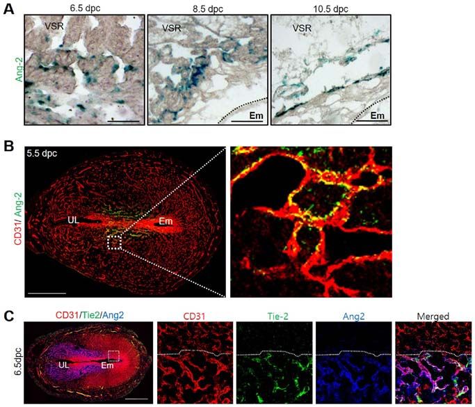
1236              park et al: UTERINE ANGIOGENESIS AND ANGIOPOIETIN-2 REGULATION DURING PREGNANCY

    Angiogenesis, one of the essential processes in blood         Identification of a vaginal plug the following morning was
vessel formation, represents the development of new branching     interpreted as successful mating, and designated 0.5 day
vessels from existing vascular networks and is operated by        post coitum (dpc). Ang-2+/LacZ mice were transferred and
endothelial cells, the main components of blood vessels (14).     bred in our pathogen-free animal facilities. The Specific
The phenomenon is essential for embryonic growth, wound           pathogen-free (SPF) C57BL/6J mice were all given ad libitum
healing in recovery of adults, and the menstrual cycle in         access to standard diet (PMI Lab diet) and water. All animal
thickening the uterus (15). Angiogenesis also contributes         experiments were performed following approval from the
to inflammatory disease and tumor growth. In some cases,          Institutional Animal Care and Use Committees (IACUC) of
inappropriate angiogenesis may result in ischemia (16). Given     Jeonbuk National University.
the variety of functions it performs, vascular remodeling,
which results from angiogenesis of the uterus, is essential for   Histological analysis. Mice were sacrificed using the
successful pregnancy. Angiogenesis and vascular remodeling        cervical dislocation method on the indicated days. Segments
are thought to be regulated by the cooperative interaction        of the uterus containing implanted embryos were fixed in
between several angiogenic factors (17). There are two major      4% paraformaldehyde (Biosesang; cat. no. PC2031) for 4 h,
angiogenic factors and their respective responsive receptors:     followed by overnight dehydration in 20% sucrose solution.
VEGF and the receptor VEGF-R, and angiopoietin and the Tie        Dehydrated samples were embedded with tissue freezing
receptor, which regulate both neovascularization, including       medium (Scigen; cat. no. 4586) and the frozen blocks cut into
vasculogenesis and angiogenesis, and vascular remodeling,         20 µm sections.
including enlargement and blood network formation, in the             Samples were blocked with 5% donkey serum (Jackson
uterus (18-20).                                                   ImmunoResearch; cat. no. 017-000-121) or goat serum (Jackson
    The VEGF, one of the major angiogenic factors of vascular     ImmunoResearch; cat. no. 005-000-121) in PBST (0.03% Triton
regulator in the endometrium, increases endothelial cells'        X-100 in PBS) and then incubated for 4 h at room tempera-
proliferation, permeability, and migration (21). Another          ture (RT) with the following primary antibodies: anti-CD31
vascular growth factor, Angiopoietin-1 (Ang-1), increases         (hamster monoclonal, Millipore; cat. no. MAB1398Z), anti-
the recruitment of endothelial cells with pericytes and           Ang-2 (rabbit polyclonal, Proteintech TM; cat. no. 24613‑1‑AP),
vascular smooth muscle cells to remodel newly formed blood        anti-PR (rabbit polyclonal, Cell signaling; cat. no. 8757), and
vessels, stimulating and stabilizing their maturation (22).       anti-Tie-2 (mouse monoclonal, Abcam; cat. no. ab24859).
Angiopoietin-2 (Ang-2), as an antagonist of Ang-1, plays an       After several washes, the samples were incubated for 2 h at
important role alongside VEGF, as a regulator of vascular         RT with the following secondary antibodies: Cy3-conjugated
remodeling, to migrate and proliferate endothelial cells. The     anti-hamster IgG (Jackson ImmunoResearch; cat. no. 127-165-
Ang-1/Ang-2 ratio is inversely associated with blood vessel       160), and Cy3- or FITC-conjugated anti-rabbit IgG (Jackson
destabilization, a prerequisite for new blood vessel forma-       ImmunoResearch; cat. no. 711-165-152 or cat. no. 111-095-003).
tion (23). During angiogenesis, Ang-2 binds to its receptor       Nuclei were stained with 4',6-diamidino‑2-phenylindole (Enzo;
named Tie-2, competitively with Ang-1 (24). Recently, many        cat. no. BML-AP402). Afterward, the samples were mounted in
studies have proved that Ang-2 holds a crucial role in female     fluorescent mounting medium (DAKO; cat. no. S3023).
reproduction (25). Overexpression of Ang-2 in mice resulted           To examine β-galactosidase activity, the cryo-sections were
in embryonic fatality in consequence to failure of angiogen-      incubated with a staining solution [2 mM magnesium chloride,
esis (26). Interestingly, Ang-2 is initially expressed in the     5 mM potassium ferricyanide, 5 mM potassium ferrocyanide and
ovaries and later, during early pregnancy, in the uterus and      1 mg/ml 4-chloro-5-bromo-3-indolyl-β-D-galactopyranoside
placenta (23).                                                    (X-gal) in PBS] at 37˚C for 24 h. Immunofluorescent images
    Previous research has shown that progesterone governs         and β -gal activity were acquired using a Zeiss LSM510
uterine angiogenesis and vascular remodeling via VEGF-A/          confocal fluorescence microscope (Carl Zeiss) and a micro-
VEGFR-2 signaling, especially in the anti-mesometrial             scope equipped with a CCD camera (Carl Zeiss).
region (AMR), where the embryo resides during pregnancy (1).
However, the functional role of spatiotemporal-localized Ang-2    Detection of Ang-2 expression by reverse transcription
expression in the pregnancy uterus is not yet fully understood.   (RT)-qPCR. Total RNA was extracted from the uterus using
In our study, we hypothesized that spatiotemporal changes are     TRIzol® Reagent (Invitrogen; cat. no. 15596018) according to
focused on the mesometrial region (MR) of the uterus because      the manufacturer's instructions. The RNA concentration was
decidual development and vascular remodeling are both devel-      measured using NanoDrop 2000 (Thermo Fisher Scientific,
oped by Ang-2 which is regulated by progesterone during           Inc.). The RNA (2 µg) was reverse transcribed into cDNA
early pregnancy. To examine the relationship between Ang-2        using SuperScript II Reverse Transcriptase (Invitrogen; cat.
and progesterone, we underwent in vitro and in vivo experi-       no. 18064071). RT-qPCR was carried out using the following
ments. Consequently, our results supported our hypothesis that    conditions: preheating for 5 min at temperature 95˚C; and
Ang-2 regulated by progesterone is a key regulator of vascular    then repeating 32 cycles in temperature 95˚C for 20 sec and
remodeling in the uterus during pregnancy.                        30 sec at 59˚C. The primer sequences were as follows: (1)
                                                                  Ang-2, Foward; 5'-GGATCTGGGGAGAGAGGAAC-3',
Materials and methods                                             Reverse; 5'- CTCTGCACCGAGTCATCGTA -3'. (2) GAPDH,
                                                                  Forward; 5'-ACCACAGTCCATGCCATCAC-3', Reverse;
Mice. C57BL/6 mice aged 8 to 10 weeks were used for this          5'-TCCACCACCCTGTTGCTGTA-3'. The PCR products
study and female mice were mated with adult male mice.            were loaded onto a 1.5% agarose gel containing Loading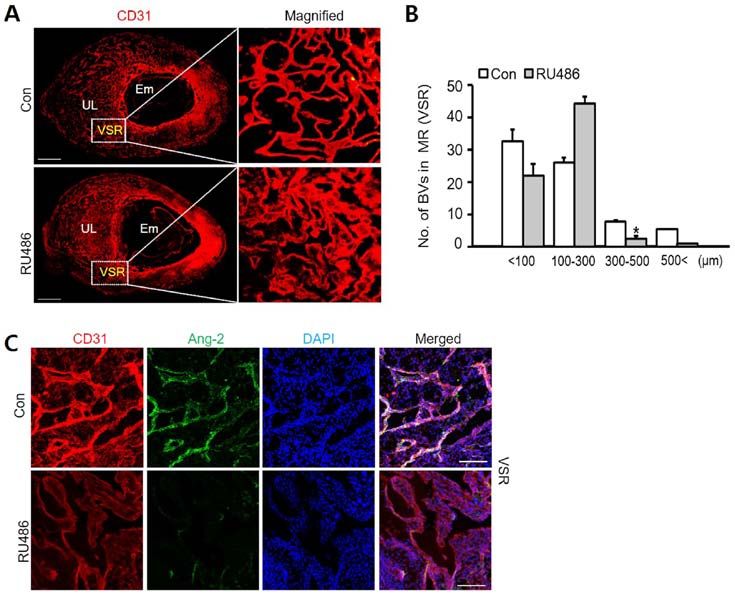
Molecular Medicine REPORTS 22: 1235-1242, 2020                                        1237

STAR nucleic acid dye (6X, Dynebio; cat. no. A750), electro-       Statistical analysis. Values are presented as mean ± standard
phoresed, and photographed using a Fusion FX7 acquisition          deviation (SD). Significant differences between means were
system (Vilbert Lourmat). The band was semi‑quantified             determined by unpaired Student's t-tests or analysis of vari-
using Quantity One software (v4.6.2; Bio‑Rad Laboratories,         ance with one-way and two-way ANOVA followed by the
Inc.) with GAPDH as the loading controls.                          Student-Newman-Keuls test or Bonferroni post hoc test. All
                                                                   statistical analysis was performed using the GraphPad Prism
Cell culture. Human uterine microvascular endothelial              software. P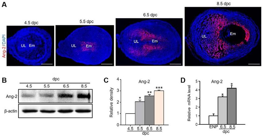
1238                  park et al: UTERINE ANGIOGENESIS AND ANGIOPOIETIN-2 REGULATION DURING PREGNANCY

Figure 1. Ang-2 expression in the uterus during pregnancy. (A) Image showing Ang-2 expression in mouse uteri during early pregnancy at 4.5, 5.5, 6.5 and
8.5 dpc. Scale bar, 500 µm. (B) Protein expression levels and (C) semi-quantitative analysis of Ang-2 in the uteri at 4.5, 5.5, 6.5 and 8.5 dpc were measured
by western blotting. Loading of similar amounts of protein for each sample was verified by a similar intensity of β-actin signal. Data are presented as the
mean ± SD from three independent experiments. *P
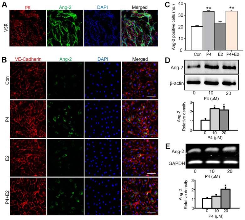
Molecular Medicine REPORTS 22: 1235-1242, 2020                                                        1239

Figure 3. Ang-2 expression is increased by progesterone treatment in HUtMECs. (A) Image showing PR+ region and Ang-2+ cells in the uterus at 6.5 days
post coitum. Scale bar, 100 µm. (B) Immunofluorescence staining of VE-Cadherin and Ang-2 in HUtMECs treated with Con, P4 (10 µM), E2 (10 µM) and
P4 + E2 (each 10 µM). Nuclei were counterstained with DAPI (blue). Scale bar, 100 µm. (C) Comparisons of Ang-2+ HUtMECs treated with Con, P4, E2,
P4 + E2 (10 µM). **P
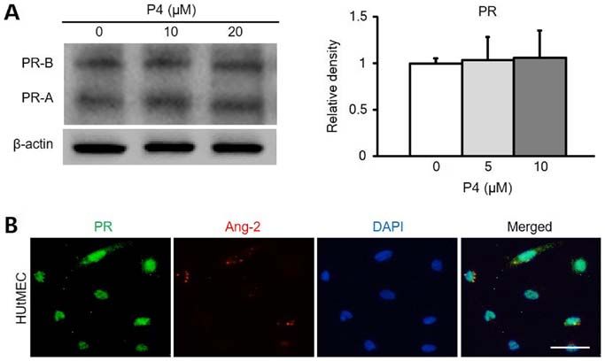
1240                  park et al: UTERINE ANGIOGENESIS AND ANGIOPOIETIN-2 REGULATION DURING PREGNANCY

Figure 4. Ang-2 expression is associated with PR in HUtMECs. (A) Comparison of PR protein levels and relative density (right graph) in HUtMECs treated
with control and P4 (5, 10 and 20 µM). Loading of similar amounts of protein for each sample was verified by a similar intensity of β-actin and relative density
was determined according to the control level. (B) Immunofluorescence staining of PR and Ang-2. Nuclei were counterstained with DAPI (blue). Scale bar,
100 µm. Ang-2, angiopoietin-2; HUtMECs, human uterine microvascular endothelial cells; P4, progesterone; PR, progesterone receptor

Figure 5. Vascular remodeling in VSR is decreased by RU486. (A) Images showing CD31+ BVs in the uterus at 8.5 dpc following treatment with Con and
RU486 (8 mg/kg). n=4. Scale bar, 500 µm. (B) Number of BVs in MR (VSR) was dependent on their size, compared with Con and treatment RU486. n=4.
*
 P
Molecular Medicine REPORTS 22: 1235-1242, 2020                                               1241

    Our results demonstrated that Ang-2 steadily increases        Funding
after implantation until placentation. Remarkably, Ang-2 was
expressed spatiotemporally in the uterus - especially in the      The present study was supported by the National Research
VSR of the MR. In this period, Ang-2 also constantly rose         Foundation of Korea funded by the Ministry of Science, ICT
in terms of the total level of protein and mRNA in uterus.        and Future Planning (grant no. 2015R1A1A1A05001546 to
Other studies indicated that the amount of mRNA of Ang-2          JWS).
increased during early pregnancy (23). From this result, we
predicted Ang-2 has an effect on vascular remodeling in           Availability of data and materials
VSR where lots of blood vessels are lined up by endothe-
lial cells. We found out the correlation between Ang-2 and        The datasets used and/or analyzed during the present study are
endothelial cell by performing an X-gal stain. Based on these     available from the corresponding author on reasonable request.
results, the treatment of HUtMEC with progesterone is very
different compared to treatment with progesterone in vitro.       Authors' contributions
Progesterone increased the expression of Ang-2. Progesterone
binds to a PR to activate its mechanism. As shown in Fig. 4,      YGP and JC designed and performed the experiments,
PR expressed in the HUtMEC and PR expression was not              analyzed the data, generated the figures and wrote the manu-
dose regulated by progesterone concentration. Expression          script. JWS designed, organized and supervised the project,
of PR is not related to progesterone level but other factors.     and wrote the manuscript. All authors read and approved the
According to another study, PR is up-regulated by estrogen at     final manuscript.
the level of gene expression (30). It seems that the concentra-
tion of progesterone has no influence on the expression of PR     Ethics approval and consent to participate
in HUtMEC.
    Progesterone is a biomarker for decidualization of            All experiments were performed following approval from the
pregnant uterus (31). Ang-2 was observed in MR during             Institutional Animal Care and Use Committees (IACUC) of
this period. Therefore, we tested the relationship between        Jeonbuk National University.
Ang-2 and PR in decidual angiogenesis using PR blockade
named RU486. We injected it intraperitoneally in pregnant         Patient consent for publication
mice to observe vascular remodeling during pregnancy.
RU486 inhibited Ang-2 mediated vascular remodeling in             Not applicable.
the MR. Our results indicated that vascular remodeling and
decidual angiogenesis are related to signaling pathways that      Competing interests
are affected differently by progesterone and Ang-2 mediated
vascular remodeling. Notably, vascular remodeling induced         The authors declare that they have no competing interests.
by Ang-2 expression was not dependent on VEGF-A-related
signaling in the MR. VEGF-A was mainly expressed in the           References
AMR and was not detected in the VSR during pregnancy (1).
Thus, vascular remodeling mediated by Ang-2 signaling              1. Kim M, Park HJ, Seol JW, Jang JY, Cho YS, Kim KR, Choi Y,
                                                                      Lydon JP, Demayo FJ, Shibuya M, et al: VEGF-A regulated by
would be different to VEGF-A/VEGFR-2 signaling in the                 progesterone governs uterine angiogenesis and vascular remod-
pregnant uterus.                                                      elling during pregnancy. EMBO Mol Med 5: 1415-1430, 2013.
    Taking all of this information into account, we speculate      2. Cha J, Sun X and Dey SK: Mechanisms of implantation: Strategies
                                                                      for successful pregnancy. Nat Med 18: 1754-1767, 2012.
that the expression of Ang-2 is stimulated by progesterone         3. Wang H and Dey SK: Roadmap to embryo implantation: Clues
which influences spatiotemporally different parts of the uterus       from mouse models. Nat Rev Genet 7: 185-199, 2006.
in early pregnancy. Progesterone most likely induces vascular      4. Smith SD, Choudhury RH, Matos P, Horn JA, Lye SJ, Dunk CE,
                                                                      Aplin JD, Jones RL and Harris LK: Changes in vascular extracellular
remodeling via VEGF-A/VEGFR-2 signaling only in the                   matrix composition during decidual spiral arteriole remodeling in
AMR. In the MR, Ang-2 expression by progesterone affects              early human pregnancy. Histol Histopathol 31: 557-571, 2016.
vascular remodeling for placentation.                              5. Bany BM and Cross JC: Post-implantation mouse conceptuses
                                                                      produce paracrine signals that regulate the uterine endometrium
    In conclusion, our results provide insight on previously          undergoing decidualization. Dev Biol 294: 445-456, 2006.
undefined features of vascular remodeling in the pregnant          6. Jones RL and Critchley HO: Morphological and functional
uterus by Ang-2-mediated signaling during early preg-                 changes in human endometrium following intrauterine levonorg-
                                                                      estrel delivery. Hum Reprod 15 (Suppl 3): 162-172, 2000.
nancy. In the post-implantation period, the pregnant uterus        7. Osol G and Moore LG: Maternal uterine vascular remodeling
undergoes profound vascular remodeling in response to                 during pregnancy. Microcirculation 21: 38-47, 2014.
progesterone for placentation, which has spatiotemporally          8. Kiepiela P, Smith AN and Rosenberg E: Retraction notice
                                                                      to ‘Laboratory markers associated with progression of HIV
different effects on the AMR and MR. Progesterone-PR-                 infection [Best Pract Res Clin Obstet Gynaecol 19: 243-254,
regulated Ang-2 is a key regulator for placentation and               2005]’. Best Pract Res Clin Obstet Gynaecol 21: 883, 2007.
prevention of pregnancy failure through spatiotemporal             9. Conneely OM, Mulac-Jericevic B and Lydon JP: Progesterone-
                                                                      dependent regulation of female reproductive activity by two distinct
vascular remodeling in the MR.                                        progesterone receptor isoforms. Steroids 68: 771-778, 2003.
                                                                  10. Carson DD, Bagchi I, Dey SK, Enders AC, Fazleabas AT, Lessey BA
Acknowledgements                                                      and Yoshinaga K: Embryo implantation. Dev Biol 223: 217-237, 2000.
                                                                  11. Schumacher A, Costa SD and Zenclussen AC: Endocrine factors
                                                                      modulating immune responses in pregnancy. Front Immunol 5:
Not applicable.                                                       196, 2014.
1242 park et al: UTERINE ANGIOGENESIS AND ANGIOPOIETIN-2 REGULATION DURING PREGNANCY
12. Wetendorf M and DeMayo FJ: The progesterone receptor 23. Matsumoto H, Ma WG, Daikoku T, Zhao X, Paria BC, Das SK,
regulates implantation, decidualization, and glandular devel- Trzaskos JM and Dey SK: Cyclooxygenase-2 differentially
opment via a complex paracrine signaling network. Mol Cell directs uterine angiogenesis during implantation in mice. J Biol
Endocrinol 357: 108-118, 2012. Chem 277: 29260-29267, 2002.
13. Groothuis PG, Dassen HH, Romano A and Punyadeera C: 24. Felcht M, Luck R, Schering A, Seidel P, Srivastava K, Hu J,
Estrogen and the endometrium: Lessons learned from gene Bartol A, Kienast Y, Vettel C, Loos EK, et al: Angiopoietin-2
expression profiling in rodents and human. Hum Reprod differentially regulates angiogenesis through TIE2 and integrin
Update 13: 405-417, 2007. signaling. J Clin Invest 122: 1991-2005, 2012.
14. Saraswati S, Kumar S and Alhaider AA: α-santalol inhibits the 25. Geva E, Ginzinger DG, Moore DH II, Ursell PC and Jaffe RB:
angiogenesis and growth of human prostate tumor growth by In utero angiopoietin-2 gene delivery remodels placental blood
targeting vascular endothelial growth factor receptor 2-mediated vessel phenotype: A murine model for studying placental angio-
AKT/mTOR/P70S6K signaling pathway. Mol Cancer 12: 147, genesis. Mol Hum Reprod 11: 253-260, 2005.
2013. 26. Tsuzuki T, Okada H, Cho H, Shimoi K, Miyashiro H, Yasuda K
15. Bauer SM, Bauer RJ and Velazquez OC: Angiogenesis, vascu- and Kanzaki H: Divergent regulation of angiopoietin-1, angio-
logenesis, and induction of healing in chronic wounds. Vasc poietin-2, and vascular endothelial growth factor by hypoxia and
Endovascular Surg 39: 293-306, 2005. female sex steroids in human endometrial stromal cells. Eur J
16. Abbas OL, Borman H, Terzi YK, Terzi A, Bayraktar N and Obstet Gynecol Reprod Biol 168: 95-101, 2013.
Yazıcı AC: The Notch pathway is a critical regulator of angio- 27. Akwii RG, Sajib MS, Zahra FT and Mikelis CM: Role of
genesis in a skin model of ischemia. Vasc Med 20: 205-211, Angiopoietin-2 in Vascular Physiology and Pathophysiology.
2015. Cells 8: 471, 2019.
17. Koga K, Osuga Y, Tsutsumi O, Yano T, Yoshino O, Takai Y, 28. Cartwright JE, Fraser R, Leslie K, Wallace AE and James JL:
Matsumi H, Hiroi H, Kugu K, Momoeda M, et al: Demonstration Remodelling at the maternal-fetal interface: Relevance to human
of angiogenin in human endometrium and its enhanced pregnancy disorders. Reproduction 140: 803-813, 2010.
expression in endometrial tissues in the secretory phase and the 29. Walter LM, Rogers PA and Girling JE: The role of progesterone
decidua. J Clin Endocrinol Metab 86: 5609-5614, 2001. in endometrial angiogenesis in pregnant and ovariectomised
18. Augustin HG, Koh GY, Thurston G and Alitalo K: Control of mice. Reproduction 129: 765-777, 2005.
vascular morphogenesis and homeostasis through the angio- 30. Ing NH and Tornesi MB: Estradiol up-regulates estrogen
poietin-Tie system. Nat Rev Mol Cell Biol 10: 165-177, 2009. receptor and progesterone receptor gene expression in specific
19. Carmeliet P: Angiogenesis in health and disease. Nat Med 9: ovine uterine cells. Biol Reprod 56: 1205-1215, 1997.
653-660, 2003. 31. Das A, Mantena SR, Kannan A, Evans DB, Bagchi MK and
20. Chung AS and Ferrara N: Developmental and pathological Bagchi IC: De novo synthesis of estrogen in pregnant uterus is
angiogenesis. Annu Rev Cell Dev Biol 27: 563-584, 2011. critical for stromal decidualization and angiogenesis. Proc Natl
21. Fan X, Krieg S, Kuo CJ, Wiegand SJ, Rabinovitch M, Druzin ML, Acad Sci USA 106: 12542-12547, 2009.
Brenner RM, Giudice LC and Nayak NR: VEGF blockade
inhibits angiogenesis and reepithelialization of endometrium.
FASEB J 22: 3571-3580, 2008. This work is licensed under a Creative Commons
22. Kwak HJ, So JN, Lee SJ, Kim I and Koh GY: Angiopoietin-1 is Attribution-NonCommercial-NoDerivatives 4.0
an apoptosis survival factor for endothelial cells. FEBS Lett 448: International (CC BY-NC-ND 4.0) License.
249-253, 1999.You can also read