Platelet-derived growth factor (PDGF) and PDGF receptors in rat corpus cavernosum: changes in expression after transient in vivo hypoxia - Journal ...
←
→
Page content transcription
If your browser does not render page correctly, please read the page content below
395

Platelet-derived growth factor (PDGF) and PDGF receptors in
rat corpus cavernosum: changes in expression after transient
in vivo hypoxia
A Aversa1,2, S Basciani3, P Visca4, M Arizzi3, L Gnessi3,
G Frajese2 and A Fabbri2
1
 AFaR-CRCCS, Ospedale Fatebenefratelli-Isola Tiberina, 00186 Rome, Italy
2
 Cattedra di Endocrinologia, Università degli Studi ‘Tor Vergata’, Ospedale Fatebenefratelli-Isola Tiberina, 00186 Rome, Italy
3
 Dipartimento Fisiopatologia Medica, Università degli Studi ‘La Sapienza’ 00161 Rome, Italy
4
 Servizio di Anatomia Patologica-IFO, Polo Oncologico ‘Istituto Regina Elena’, 00100 Rome, Italy
(Requests for offprints should be addressed to A Aversa, Cattedra di Endocrinologia, Università di Roma ‘Tor Vergata’, Via di Torvergata 135,
   00133 Roma, Italy; Email: amfaversa@yahoo.com)

Abstract
Platelet-derived growth factor (PDGF) overactivity has                              transcripts in hypoxic versus normoxic animals. The
been implicated in atherosclerosis and several fibrotic                             immunohistochemical analysis showed that the localization
conditions including lung and kidney fibrosis, liver                                of PDGF subunits and PDGFR- was confined to the
cirrhosis and myelofibrosis. Low oxygen tension (hypoxia)                           cytoplasm of the perivascular smooth muscle cells, endo-
is a known stimulus for transcriptional induction of PDGF                           thelium and trabecular fibroblasts. Our findings indicate
ligand and receptor genes in different tissues. We studied                           that transient low oxygen tension induces PDGF over-
the expression and localization of PDGF-A, PDGF-B, and                              expression in rat CC, which in the long term may lead to
PDGF receptor (PDGFR)- and - subunits in adult                                    an increase of connective tissue production. We suggest
rat isolated corpus cavernosum (CC) under generalized                               that a local impairment of the PDGF/PDGFR system may
transient hypoxia (pO2 10%) in comparison with                                      contribute to CC fibrosis, which is an established cause of
normoxic conditions. Semi-quantitative RT-PCR analysis                              erectile dysfunction in man.
of mRNA extracted from rat penis showed higher                                      Journal of Endocrinology (2001) 170, 395–402
amounts of PDGF-A, PDGF-B and PDGFR- mRNA

Introduction                                                                        in penile tunica albuginea obtained from patients with
                                                                                    veno-occlusive dysfunction and Peyronie’s disease
Platelet-derived growth factor (PDGF) is a major mitogen                            (Gentile et al. 1996), suggesting that it could be involved
for cells of mesenchymal origin, such as fibroblasts and                            in the pathogenesis of these two conditions that are
smooth muscle cells. PDGF is widely expressed in normal                             frequently associated with erectile dysfunction (ED) in
and transformed cells and is produced by monocytes and                              men.
macrophages, vascular endothelial and smooth muscle cells                              In vivo, the reduction in the environmental oxygen
(Antoniades 1991, Heldin 1992). There are three PDGF                                tension to which cells are exposed leads to physiological
isoforms (AA, AB and BB) that exert their biological                                and, eventually, pathological consequences associated with
actions via binding to cell surface receptors ( and ) that                        differential expression of specific genes which encode for
belong to the protein tyrosine-kinase family of receptors                           cytokines and growth factors thought to play key roles in
(Williams 1989). PDGF-mediated events, which include                                the regulation of synthesis and assembly of connective
chemoattraction, activation of inflammatory cells, vaso-                            tissue proteins (Gerritsen & Bloor 1993, Bunn & Poyton
constriction and influence on the synthesis or degradation                          1996). For example, in rat corpus cavernosum smooth
of matrix constituents (Heldin 1992), are most likely                               muscle cells in culture hypoxia stimulates the expression of
exerted locally in an autocrine or paracrine manner and are                         transforming growth factor 1 (TGF-1), a pleotrophic
involved in natural as well as pathological processes, such as                      cytokine that is known to induce extracellular matrix
neoplasia, atherosclerosis and fibrosis (Heldin 1992, Gnessi                        expression and inhibit growth and proliferation of vascular
et al. 1993). Recently, immunohistochemical and electron                            smooth muscle cells (Faller 1999). Indeed, in human
microscopy studies showed that PDGF is highly expressed                             corpus cavernosum smooth muscle cells, TGF-1 is a

Journal of Endocrinology (2001) 170, 395–402                                                                    Online version via http://www.endocrinology.org
0022–0795/01/0170–395  2001 Society for Endocrinology Printed in Great Britain

                                                                                                                 Downloaded from Bioscientifica.com at 06/01/2021 05:14:31AM
                                                                                                                                                               via free access
396 A AVERSA and others · Rat penile PDGF and hypoxia
mitogen and induces a two- to fourfold increase in Table 1 Primers utilized in PCR reactions
collagen synthesis (Moreland et al. 1995); also, it has been
5 –3 sequence Position
found to be overexpressed in tunica albuginea from men
suffering venocclusive dysfunction (Nehra et al. 1996) and Substance
in plaques obtained from Peyronie’s disease (El-Sakka et al. PDGF-A1 CCTGTGCCATCCGCAGGAAGAGA 215–239
1997). PDGF-A2 TTGGCCACCTTGACGCTGCGGTG 418–441
PDGF-B1 GATCCGCTCCTTTGATGATC 1203–1223
In this study, we compared for the first time the PDGF-B2 GTCTCACACTTGCATGCCAG 1617–1637
expression of PDGF subunits and PDGF receptor mRNAs PDGF-1 CGACTCCAGATGGGAGTTCCC 1881–1902
in corpora cavernosa (CC) isolated from adult rats both PDGF-2 TGACATCCACTTCACAGGCA 2704–2724
in normal conditions and after acute hypoxia (pO2 = PDGF-1 CACCATTTCGAGCACCTTTGT 28–49
10 mmHg). The immunohistochemical localization of PDGF-2 AGGGCACTCCGAAGAGGTAA 684–704
-actin 1 ATTGGCAATGAGCGGTTCCGC 2413–2437
PDGF-A, PDGF-B and PDGF receptor (PDGFR)- and -actin 2 CTCCTGCTTGCTGATCCACATC 2727–2349
- subunits was also evaluated in comparison with that of
the TGF-1.
Materials and Methods ensure amplification in the exponential phase of PCR,
reactions were temporarily halted at 20, 25, 30, 35 and
Hypoxic exposure and CC preparation 40 cycles, and 10 µl of PCR products were removed from
each tube (see Fig. 1). All products were analyzed by 1·5%
Male Sprague–Dawley rats (55–60 days) purchased from agarose gel electrophoresis and 30 cycles were chosen for
Charles River Italia (Calco, Italy), were continuously further analysis. Quantitation of the signals was performed
gassed for 6 h with a mixture of 10% O2 and 90% N2 (ten by densitometric analysis, using densitometry computer
rats) or normal air (ten rats) in a 7060100 cm software (Kodak Digital Science ID Image Analysis
gas-tight box (Bucher et al. 1996); animals were conscious Software, Eastman Kodak Company, Rochester, NY,
and had free access to food and water. At the end of the USA). Dilution of RT products was made where neces-
procedure decapitation was performed, blood was col- sary and the amplification procedure was repeated until all
lected from the left ventricle for pO2, sO2 and pCO2 samples were standardized for -actin content. After
determination by hemogasanalysis (Franchini et al. 1994), standardization, PCR was performed using appropriately
and CC were rapidly prepared according to Broderick diluted RT products in 50 µl of the reaction mix by
et al. (1994) with minor modifications. Briefly, rat penises utilizing 20 µM of each rat PDGFs and PDGFRs primers
were surgically removed and the corpus spongiosum and (Table 1). For each gene examined, all primers were
the urethra were excised. The CC tissues were carefully derived from separate exons and spanned at least one
dissected free from the surrounding tunica albuginea and intron of genomic sequence, thus excluding the possibility
made available for RT-PCR and immunohistochemical of genomic DNA contamination. No PCR product was
studies. The Animal Care Committee of the University of obtained with any of the set of primers in the absence of
Rome Medical School approved this protocol. cDNA template (negative control) (Caprio et al. 1999).
Thermocycling conditions were: initial denaturation for
3 min at 94 C, 30 cycles of amplification (since levels of
RNA extraction and RT-PCR analysis
PCR products increased in a linear fashion for up to
Tissues mRNA were extracted by using a commercial kit 35 cycles for PDGFs and PDGFRs, Fig. 1) with 1 min of
(Micro-Fast-Track Kit, Invitrogen, San Diego, CA, denaturation at 94 C, different annealing temperature for
USA). Reverse transcription was performed using an each pair of primers (Table 1), 1 min extension at 72 C,
annealing temperature of 70 C in a final volume of 25 µl followed by a final elongation of 5 min at 72 C.
containing 250 mM Tris–HCl, 375 mM KCl, 15 mM
MgCl2, 50 mM dithiothreitol (DTT), 0·5 mM dNTPs,
0·5 µg random hexamer oligonucleotide, 200 U M-MLV- Immunohistochemistry and light microscopy
RT, 26 U ribonuclease inhibitor (Promega, Madison, WI, After decapitation, the skin overlying the penis was
USA). -actin was used as a constutively expressed gene incised, and the whole penis body, including the CC crura
product for comparison of PDGFs and PDGFRs mRNA and the bulbospongiosum covered by the ischiocavernous
abundance between samples. A 0·5 µl volume of the RT and bulbospongiosus skeletal muscles, was excised in
products was amplified with 2·5 units of Taq DNA one piece, fixed in Bouin’s solution for 12 h, and prepared
polymerase (Promega) and 20 µM specific rat -actin for immunostaining (Gnessi et al. 1993, 2000). Immuno-
primer (Table 1) in 50 µl of reaction mix containing staining was carried out by incubating tissue sections
500 mM KCl, 200 mM Tris–HCl, and 1·5 mM MgCl2 as (3 µm) with TGF-1, PDGFs and PDGFRs antisera
follows: 94 C, 1 min; 58 C, 1 min; 72 C, 1 min. To (1:100) overnight at 4 C (Gnessi et al. 1993, Caprio
Journal of Endocrinology (2001) 170, 395–402 www.endocrinology.org
Downloaded from Bioscientifica.com at 06/01/2021 05:14:31AM
via free accessRat penile PDGF and hypoxia · A AVERSA and others 397
Heldin, Ludwig Institute for Cancer Research, Uppsala,
Sweden). PDGFR-7 was generated against a synthetic
peptide covering amino acids 1066–1084 of the COOH-
terminal region of human PDGFR- subunit and does not
cross-react with the PDGFR- subunit. It recognizes both
human and rat PDGFR- subunit. PDGFR-3 was raised
against a synthetic peptide corresponding to amino acids
981–994 of the mouse PDGFR-. It recognizes rat
PDGFR- subunit. PDGFR-7 and PDGFR-3 were
affinity purified on columns with immobilized syn-
thetic peptides against which the antisera were raised
(Hermanson et al. 1992, Gnessi et al. 1995). All the
antibodies react specifically with the respective antigens in
immunoprecipitation and Western blotting experiments
(Hermanson et al. 1992, Eccleston et al. 1993). For better
identification of smooth muscle cells lining the cavernosal
spaces, adjacent sections were immunostained with a
primary antismooth muscle -actin antibody diluted up to
2 µg/ml at room temperature (DAKO Corp., Trappes,
France). At the end of incubation immunopositivity was
visualized by the streptavidin–biotin immunoperoxidase
technique, using a commercial kit (Zymed Lab. Inc., San
Francisco, CA, USA). Slides were developed using
amino-ethylcarbazole (AEC) as chromogenic substrate
that is converted by the peroxidase into a red to brownish-
red precipitate at the sites of antigen localization in the
tissue. The preparations were counterstained with hema-
toxylin, dehydrated, cleared and mounted (Claesson-
Welsh et al. 1989). The immunohistochemical expression
of TGF-1 was used as a positive control of tissue hypoxia.
Results were evaluated by using a semi-quantitative
staining intensity of immunoreactive TGF-1, PDGF
peptides and receptors on three consecutive sections of rat
CC for each antisera were examined. We evaluated
positive endothelial, perivascular smooth muscle and
trabecular fibroblast cells counted on five microscopic
Figure 1 Optimization of RT-PCR conditions for semi-quantitative cellular areas (40) for all sections of hypoxic and
determination of hypoxic PDGFs and PDGFRs and -actin mRNA. normoxic rat CC. The positive staining intensity was
For amplification in the exponential phase of PCR, different
numbers of cycles were tested for each message. Quantitative scored on a four-tiered scale: negative=0; low inten-
analysis of cycle-dependency for the generated PCR signals sity=1; moderate intensity=2; and strong intensity=3.
revealed a strong linear relationship between cycles 20 and 35 for The staining distribution and intensity were determined
PDGF-A (correlation coefficient r2 =0·9889) and between cycles by two observers independently (P V and M A) with
20 and 40 for other targets (r2 =0·9778 for PDGFR-, r2 =0·9840
for PDGF-B, r2 =0·9921 for PDGFR-, and r2 =0·9442 for -actin).
subsequent reconciliation of scored values. -actin im-
Values are given as means S.D. of three independent munostaining was considered as positive or negative
determinations. A representative ethidium bromide-stained gel (Gnessi et al. 1995, Visca et al. 1999). Thereafter, we
electrophoresis of the DNA products generated for each target is calculated the overall means (n=15, resulting from evalu-
presented in the insets. OD, optical density. ation of 5 microscopic cellular areas for three sections) of
staining intensity for each cellular subtype of each hypoxic
and normoxic CC examined. The total means of 150
et al. 1999). The following antisera were used: rabbit scores (15 scores for ten hypoxic and normoxic rat CC
anti-human TGF-1 (Research Diagnostics, Inc., respectively) of the staining intensity for each cellular
Flanders, NJ, USA), affinity purified polyclonal rabbit components of hypoxic tissues were then compared with
anti-PDGF-BB and anti-PDGF-AA antibodies the total means of the 150 scores obtained from normoxic
(Genzyme, Cambridge, MA, USA); PDGFR-7 and tissues. A score of zero was considered to be negative (–);
PDGFR-3, rabbit polyclonal antisera to the PDGFR- mean scores between 0 and 1 were considered as weak
and subunit respectively (provided by Dr Carl-Henrik staining intensity (+/–); mean scores between 1·1 and 2
www.endocrinology.org Journal of Endocrinology (2001) 170, 395–402
Downloaded from Bioscientifica.com at 06/01/2021 05:14:31AM
via free access398 A AVERSA and others · Rat penile PDGF and hypoxia
Table 2 Comparative semi-quantitative staining intensity of immunoreactive TGF-1, PDGF peptides and receptors within the hypoxic and
normoxic rat corpus cavernosum. Values in parentheses are means S.E.
PDGF-A PDGF-B PDGFR- PDGFR- TGF-1
H N H N H N H N H N
Corpus
cavernosum
Endothelium ++ + ++ + + + ++ + ++ +
(2·60·1) (1·20·05*) (2·60·1) (1·20·1*) (1·70·1) (1·50·1) (2·20·1) (1·20·05*) (2·80·1) (1·60·09*)
Perivascular ++ + ++ + + ++ + ++ +/
SMCs (2·70·2) (1·20·05*) (2·80·1) (0·00·0*) (1·60·1) (1·70·1) (2·50·1) (1·20·1*) (2·70·2*) (0·90·05*)
Fibroblasts of ++ + + +/ + + ++ + ++ +
the trabeculae (2·80·1) (1·30·1*) (1·90·05) (0·50·08*) (1·30·1) (1·20·1) (2·80·1) (1·30·1*) (2·80·07) (1·40·08*)
negative; +/ weak; + moderate; ++ strong staining intensity (*PRat penile PDGF and hypoxia · A AVERSA and others 399
stimulates PDGF expression in the rat lungs and may be
involved in the pathogenesis of idiopathic pulmonary
fibrosis (Katayose et al. 1993, Betsholtz & Raines 1997).
Moreover, incubation under hypoxic conditions stimulates
the release of PDGF from human macrophages and
cultured endothelial cells (Kuwabara et al. 1995, Betsholtz
& Raines 1997), as well as strongly up-regulates the
PDGF-B chain gene expression (Kourembanas et al.
1990). In normal penile human tissues, vasal endothelial
cells constitutively express PDGF; furthermore, fibroblasts
from pathological tunica albuginea of impotent men with
Peyronie’s disease and venocclusive dysfunction show
intense immunostaining for PDGF-A and -B chains
(Gentile et al. 1996). As a consequence, a higher expres-
sion of PDGF-A and -B proteins may determine an
imbalance between trabecular smooth muscle and connec-
tive tissue ratio resulting in CC fibrosis and erectile
dysfunction. In our study, we found that PDGF and
PDGFR are constitutively expressed in the rat vascular
endothelial cells as well as in penile nerves. More import-
ant, in the corpora of hypoxic rats there was a higher
expression of PDGF-A and -B proteins than in normoxic
rats. Immunohistochemistry showed that the expression
was focal in the endothelium, diffuse in the perivascular
smooth muscle cells and perinuclear in the fibroblasts of
the trabeculae. After exposure to transient low oxygen
tension, PDGFR- expression was also increased in the
same cell components of CC expressing PDGF peptides,
suggesting that in this condition these cells become a more
sensitive target for PDGF peptides. The absence of modi-
fications in PDGFR- expression may be explained with
the concomitant overexpression of TGF-1 which is
known to down-regulate PDGFR- in human fibroblasts
(Bonner et al. 1995, Kuwabara et al. 1995). Thus, the
Figure 2 Upper panel, RT-PCR expression analysis of PDGF
PDGF overexpression in penile structures under transient
peptides and PDGF receptors in corpus cavernosum of hypoxic hypoxia may well contribute to the cascade of events
and normoxic rats, in rat testis (positive control) and in the leading to tissue fibrosis under chronic hypoxic conditions.
absence of cDNA template (negative control). All data were These events may occur in some patients with erectile
normalized for -actin (internal control) and quantified by dysfunction in which a chronic CC hypoxia has been
densitometry computer software. Lower panel, mean
densitometric analysis of the results obtained from three reported (Tarhan et al. 1997).
consecutive experiments; the S.E. was less than 10% (*P400 A AVERSA and others · Rat penile PDGF and hypoxia
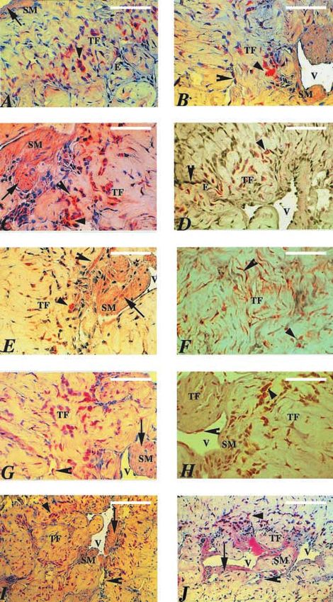
Figure 3 Immunohistochemical staining pattern of PDGF-A (A, B), -B (C, D) and PDGFR- (E, F) and - (G, H) with affinity-purified
antibody in cross-sections of hypoxic (A, C, E, G) by comparison with normoxic (B, D, F, H) rat corpus cavernosum, counterstained with
hematoxylin. The expression was intense in the cytoplasm of trabecular fibroblasts (triangles) and endothelial cells (arrowheads), and more
diffuse in smooth muscle cells (arrows). The scale bar in A represents 25 m and applies to A-H. Sections of rat corpus cavernosum
immunostained for TGF-1 in hypoxic (I) and normoxic (J) conditions are also shown. The scale bar in I represents 50 m and also applies
to J. TF=fibroblasts of the trabeculae; E=endothelium; V=vessel; SM=smooth muscle cells.
Journal of Endocrinology (2001) 170, 395–402 www.endocrinology.org
Downloaded from Bioscientifica.com at 06/01/2021 05:14:31AM
via free accessRat penile PDGF and hypoxia · A AVERSA and others 401
increased TGF-1 immunostaining in the rat penile CC, Bonner JC, Badgett A, Lindroos PM & Osornio-Vargas AR 1995
suggesting that multiple genes encoding matrix molecules Transforming growth factor beta 1 downregulates the platelet-
derived growth factor-alpha-receptor subtype on human lung
leading to fibrotic alterations inside the penis may be fibroblasts in vitro. American Journal of Respiratory Cell and Molecular
activated even during transient hypoxia, similar to that Biology 13 496–505.
described in the endothelium (Gerritsen & Bloor 1993). Border WA & Noble NA 1994 Mechanisms of disease: transforming
Low-low priapism is a frequent complication of vaso- growth factor- in tissue fibrosis. New England Journal of Medicine
active intracavernous pharmacotherapy in men affected by 331 1286–1292.
Broderick GA, Gordon D, Hypolite J & Levin RM 1994 Anoxia and
erectile dysfunction, especially when combination drugs corporeal smooth muscle dysfunction: a model for ischemic
are used (Fabbri et al. 1997). Human CC undergo major priapism. Journal of Urology 151 259–262.
ultrastructural changes, i.e. corporeal fibrosis during Bucher M, Sandner P, Wolf K & Kurtz A 1996 Cobalt but not
priapism, but pharmacological detumescence is not hypoxia stimulates PDGF gene expression in rats. American Journal of
recommended until 12 h has passed (Hauri et al. 1983). Physiology. Endocrinology and Metabolism 271 E451–E457.
However, it is known that in a rabbit model, hypoxia Bunn HF & Poyton RO 1996 Oxygen sensing and molecular
adaptation to hypoxia. Physiological Reviews 76 839–885.
induced by prolonged erection and subsequent local Caprio M, Isidori AM, Carta AR, Moretti C, Dufau ML & Fabbri A
acidosis impair contractility of trabecular smooth muscles. 1999 Expression of functional leptin receptors in rodent Leydig
This phenomenon impedes the drainage of blood, per- cells. Endocrinology 140 4939–4947.
petuates the ischemic state and may cause early ultrastruc- Claesson-Welsh L, Hammacher A, Westermark B, Heldin CH &
tural changes (Kim et al. 1996, Saenz de Tejada et al. 1997, Nister M 1989 Identification and structural analysis of the type A
receptor for platelet-derived growth factor: similarity with the type
Moon et al. 1999). Taking into account our findings, B receptor. Journal of Biological Chemistry 264 1742–1747.
the risk that fibrotic alterations may begin early after Eccleston PA, Funa K & Heldin CH 1993 Expression of
acute hypoxia has occurred inside the corpora is quite platelet-derived growth factor (PDGF) and PDGF - and
elevated. Thus, in the clinical outpatient setting prompt -receptors in the peripheral nervous system: an analysis of
detumescence of erections that exceed a duration of 2 h sciatic nerve and dorsal root ganglia. Developmental Biology 155
459–470.
should be recommended (Aversa et al. 2000).
El-Sakka AI, Hassoba HM, Pillarisetty RJ, Dahiya R & Lue TF 1997
In conclusion, transient in vivo hypoxia increases the Peyronie’s disease is associated with an increase in transforming
expression of the PDGF system in the rat penis. It is growth factor- protein expression. Journal of Urology 158
conceivable that these changes may occur also in con- 1391–1394.
ditions of chronic hypoxia in men and may lead to Fabbri A, Aversa A & Isidori A 1997 Erectile dysfunction: an
alterations in penile structures similar to those already overview. Human Reproduction Update 3 455–466.
Faller DV 1999 Endothelial cell responses to hypoxic stress. Clinical
described in other organs (Wespes et al. 1998, Okabe et al. and Experimental Pharmacology and Physiology 26 74–84.
1999). These phenomena might contribute to the patho- Franchini KG, Cestari IA & Krieger EM 1994 Restoration of arterial
genesis of erectile dysfunction that frequently complicate blood oxygen tension increases arterial pressure in sinoaotric-
atherosclerosis, diabetes mellitus, hypertension, obstruc- denervated rats. American Journal of Physiology 226 H1055–H1061.
tive pulmonary disease and intense cigarette smoking. Gentile V, Modesti A, La Pera G, Vasaturo F, Modica A, Prigiotti G,
Di Silverio F & Scarpa S 1996 Ultrastructural and
immunohistochemical characterization of the tunica albuginea in
Peyronie’s disease and veno-occlusive dysfunction. Journal of
Acknowledgements Andrology 17 96–103.
Gerritsen ME & Bloor CM 1993 Endothelial cell gene expression in
We thank Dr Massimiliano Caprio for the critical reading response to injury. FASEB Journal 7 523–532.
of the manuscript. Presented at the 81st International Gnessi L, Emidi A, Scarpa S, Palleschi S, Ragano-Caracciolo M,
Congress of the Endocrine Society, 12–15 June, San Silvestroni L, Modesti A & Spera G 1993 Platelet-derived growth
Diego, CA, 300, 1999, P2–91. factor effects on purified testicular peritubular myoid cells: binding,
cytosolic Ca2+ increase, mitogenic activity and extracellular matrix
production enhancement. Endocrinology 133 1880–1890.
Gnessi L, Emidi A, Jannini EA, Carosa E, Maroder M, Arizzi M,
References Ulisse S & Spera G 1995 Testicular development involves the
spatiotemporal control of PDGFs and PDGF receptors gene
Antoniades HN 1991 PDGF: a multifunctional growth factor. Baillieres expression and action. Journal of Cellular Biology 4 1105–1121.
Clinical Endocrinology and Metabolism 5 595–613.
Gnessi L, Basciani S, Mariani S, Arizzi M, Spera G, Wang C,
Aversa A, Bonifacio V, Moretti C, Frajese G & Fabbri A 2000
Bondjers C, Karlsson L & Betsoltz C 2000 Leydig cell loss and
Re-dosing of prostaglandin-E1 versus prostaglandin-E1 plus
spermatogenic arrest in platelet-derived growth factor (PDGF)-
phentolamine in male erectile dysfunction: a dynamic color power
A-deficient mice. Journal of Cellular Biology 149 1019–
Doppler study. International Journal of Impotence Research 12 33–40.
1025.
Battegay EJ, Raines EW, Seifert RA, Bowen-Pope DF & Ross R
1990 TGF-beta induces bimodal proliferation of connective tissue Hauri D, Spycher M & Bruhlmann W 1983 Erection and priapism: a
cells via complex control of an autocrine PDGF loop. Cell 63 new physiopathological concept. Urology International 38 138–145.
515–524. Heldin CH 1992 Structural and functional studies on platelet-derived
Betsholtz C & Raines EW 1997 Platelet-derived growth factor: a key growth factor. EMBO Journal 11 4251–4259.
regulator of connective tissue cells in embryogenesis and Hermanson M, Funa K, Hartman M, Claesson-Welsh L, Heldin CH,
pathogenesis. Kidney International 51 1361–1369. Westermark B & Nister M 1992 Platelet-derived growth factor and
www.endocrinology.org Journal of Endocrinology (2001) 170, 395–402
Downloaded from Bioscientifica.com at 06/01/2021 05:14:31AM
via free access402 A AVERSA and others · Rat penile PDGF and hypoxia
its receptors in human glioma tissue: expression of messenger RNA Moreland RB 1998 Is there a role of hypoxemia in penile fibrosis: a
and protein suggests the presence of autocrine and paracrine loop. viewpoint presented to the Society for the Study of Impotence.
Cancer Research 52 3213–3219. International Journal of Impotence Research 10 113–120.
Katayose D, Ohe M, Yamauchi K, Ogata M, Shirato K, Fujita H, Nehra A, Goldstein I, Pabby A, Nugent M, Huang YH, De las
Shibahara S & Takishima T 1993 Increased expression of PDGF Morenas A, Krane RJ, Udelson D, Saenz de Tejada I & Moreland
A- and B-chain genes in rat lungs with hypoxic pulmonary RB 1996 Mechanisms of venous leakage: a prospective
hypertension. American Journal of Physiology 264 L100–L106. clinicopathological correlation of corporeal function and structure.
Kim N, Vardi Y, Padma-Nathan H, Daley J & Saenz de Tejada I Journal of Urology 156 1320–1329.
1993 Oxygen tension regulates the nitric oxide pathway. Okabe H, Hale TM, Kumon H, Heaton JPW & Adams MA 1999
Physiological role in penile erection. Journal of Clinical Investigation The penis is not protected – in hypertension there are vascular
91 437–442. changes in the penis which are similar to those in other vascular
Kim NN, Kim JJ, Hypolite J, Garcia-Diaz JF, Broderick GA, beds. International Journal of Impotence Research 11 133–140.
Tornheim K, Daley JT, Levin R & Saenz de Tejada I 1996 Altered Saenz de Tejada I, Kim NH, Daley JT, Royai R, Hypolite J,
contractility of rabbit penile corpus cavernosum smooth muscle by Broderick GA, Garcia-Diaz F & Levin R 1997 Acidosis impairs
hypoxia. Journal of Urology 155 772–778. rabbit trabecular smooth muscle contractility. Journal of Urology 157
Kourembanas S, Hannan RL & Faller DV 1990 Oxygen regulates 722–726.
the expression of the platelet-derived growth factor-B chain gene Tarhan F, Kuyumcuoglu U, Kolsuz A, Ozgul A & Canguven O 1997
in human endothelial cells. Journal of Clinical Investigation 86 Cavernous oxygen tension in the patients with erectile dysfunction.
670–674. International Journal of Impotence Research 9 149–153.
Kourembanas S, Morita T, Liu Y & Christou H 1997 Mechanisms by Visca P, Alò PL, Del Nonno F, Botti C, Trombetta G, Marandino F,
which oxygen regulates gene expression and cell–cell interaction in Filippi S, Di Tondo U & Perrone-Donnorso R 1999
the vasculature. Kidney International 51 438–443. Immunohistochemical expression of fatty acid synthase, apoptotic-
Kuwabara K, Ogawa S, Matsumoto M, Koga S, Clauss M, Pinsky DJ, regulating genes, proliferating factors, and ras protein product in
Lyn P, Leavy J, Witte L, Joseph-Silverstein J et al. 1995 colorectal adenomas, carcinomas, and adjacent nonneoplastic
Hypoxia-mediated induction of acidic/basic fibroblast growth factor mucosa. Clinical Cancer Research 5 4111–4118.
platelet-derived growth factor in mononuclear phagocytes stimulates Wespes E, Raviv G, Vanegas JP, Decaestecker C, Petein M, Danguy
growth of hypoxic endothelial cells. PNAS 9 4606–4610. A, Schulman CC & Kiss R 1998 Corporeal veno-occlusive
Moon DG, Lee DS & Kim JJ 1999 Altered contractile response of dysfunction: a distal arterial pathology? Journal of Urology 160
penis under hypoxia with metabolic acidosis. International Journal of 2054–2057.
Impotence Research 11 265–271. Williams LT 1989 Signal transduction by the platelet-derived growth
Moreland RB, Traish A, McMillin MA, Smith B, Goldstein I & factor. Science 243 1564–1570.
Saenz de Tejada I 1995 PGE1 suppresses the induction of
collagen synthesis by transforming growth factor-1 in human
corpus cavernosum smooth muscle. Journal of Urology 153 Received 29 March 2001
826–834. Accepted 26 April 2001
Journal of Endocrinology (2001) 170, 395–402 www.endocrinology.org
Downloaded from Bioscientifica.com at 06/01/2021 05:14:31AM
via free accessYou can also read