MiR-128-3p inhibits apoptosis and inflammation in LPS-induced sepsis by targeting TGFBR2
←
→
Page content transcription
If your browser does not render page correctly, please read the page content below
Open Medicine 2021; 16: 274–283
Research Article
Peng Yang#, Jianhua Han#, Shigeng Li, Shaoning Luo, Xusheng Tu, Zhiqiang Ye*
miR-128-3p inhibits apoptosis and inflammation
in LPS-induced sepsis by targeting TGFBR2
https://doi.org/10.1515/med-2021-0222 overexpression reversed the inhibitory effects of miR-
received June 4, 2020; accepted December 4, 2020 128-3p overexpression on inflammation and apoptosis
Abstract in LPS-induced HK2 cells. Besides, overexpression of miR-
Background ‒ Sepsis is a systemic inflammatory response 128-3p downregulated TGFBR2 to suppress the activation
that can lead to the dysfunction of many organs. The of the Smad signaling pathway.
aberrant expression of miRNAs is associated with the Conclusion ‒ miR-128-3p could inhibit apoptosis and
pathogenesis of sepsis. However, the biological functions inflammation by targeting TGFBR2 in LPS-induced HK2
of miR-128-3p in sepsis remain largely unknown, and its cells, which might provide therapeutic strategy for the
mechanism should be further investigated. This study treatment of sepsis.
aimed to determine the regulatory network of miR-128- Keywords: sepsis, miR-128-3p, TGFBR2, apoptosis,
3p and TGFBR2 in lipopolysaccharide (LPS)-induced inflammation
sepsis.
Methods ‒ The expression levels of miR-128-3p and
transforming growth factor beta receptors II (TGFBR2)
were detected by quantitative polymerase chain reaction 1 Introduction
(qPCR). The protein levels of TGFBR2, Bcl-2, Bax, cleaved
caspase 3, Smad2, and Smad3 were measured by western Sepsis is a life-threatening organ dysfunction caused by a
blot. Cell apoptosis was analyzed by flow cytometry. dysregulated host response to infection [1,2]. Although
Cytokine production was detected by enzyme-linked there have been significant improvements in critical care
immunosorbent assay (ELISA). The binding sites of miR- medicine in recent years, sepsis remains one of the leading
128-3p and TGFBR2 were predicted by Targetscan online causes of mortality in the intensive care unit (ICU) [3,4].
software and confirmed by dual-luciferase reporter assay Therefore, it is critical to explain the pathogenic mechan-
and RNA immunoprecipitation (RIP) assay. isms at the molecular level, providing a potentially effec-
Results ‒ The level of miR-128-3p was decreased, and tive treatment for sepsis.
TGFBR2 expression was increased in serum samples of MicroRNAs (miRNAs) are a kind of small noncoding
sepsis patients and LPS-induced HK2 cells. Overexpres- RNAs that regulate the expression of protein through
sion of miR-128-3p or knockdown of TGFBR2 ameliorated targeting the 3′UTR of messenger RNA (mRNA), resulting
LPS-induced inflammation and apoptosis. Moreover, in mRNA degradation or suppression of translation [5].
TGFBR2 was a direct target of miR-128-3p, and its Increasing evidence has suggested that the dysregulation
of miRNAs is associated with the pathogenesis of sepsis.
For example, serum miR-146a and miR-223 were evidently
decreased in septic patients compared with healthy con-
# Contributed equally.
trols [6]. In addition, miR-27a expression was increased
and promoted inflammatory response in sepsis [7]. miR-
* Corresponding author: Zhiqiang Ye, Department of Emergency, 128-3p was recently reported to be downregulated in
The Third Affiliated Hospital, Sun Yat-sen University, No. 600 Tianhe several types of cancer, such as hepatocellular carcinoma
Road, Tianhe District, 510630, Guangzhou, China, and lung cancer [8,9]. Besides, it has been reported that
e-mail: yezhiq@mail.sysu.edu.cn, tel: +86-020-85253010 the expression of miR-128 was decreased in podocytes of a
Peng Yang: Department of Anesthesiology, The First Affiliated
sepsis patient [10]. However, the biological function of
Hospital, Sun Yat-sen University, 510080, Guangzhou, China
Jianhua Han, Shigeng Li, Shaoning Luo, Xusheng Tu: Department of
miR-128-3p in sepsis has not been well elucidated.
Emergency, The Third Affiliated Hospital, Sun Yat-sen University, Identification of miRNA-regulated targeting genes
No. 600 Tianhe Road, Tianhe District, 510630, Guangzhou, China is important for understanding their specific biological
Open Access. © 2021 Peng Yang et al., published by De Gruyter. This work is licensed under the Creative Commons Attribution 4.0
International License.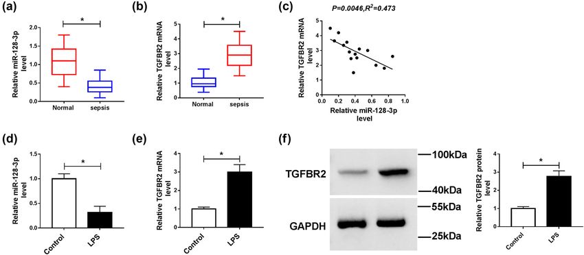
Effect of miR-128-3p in LPS-induced sepsis 275
functions [11]. Transforming growth factor-β (TGF-β) sig- 2.2 Serum total RNA isolation
naling pathway is a multifunctional cytokine and plays
important roles in regulating cell proliferation, cell cycle, In brief, special tubes containing separating gel and clot
differentiation, migration, and apoptosis in a broad spec- activator were used to collect blood samples and then
trum of tissues [12]. TGF-β signaling is initiated when the centrifuged at 3,000 rpm for 15 min at room temperature.
ligand binds to type II serine/threonine kinase receptor The supernatant was transferred to clean Eppendorf tubes
(TGFBR2), which then phosphorylates and activates type and centrifuged at 1,500 rpm for 30 min to remove cell
I serine/threonine kinase receptor (TGFBR1) [13]. The debris. The final supernatant was stored at −80°C until
activated type I receptor can activate Smad proteins RNA extraction. Serum total RNA including miRNAs was
that regulate transcription [14]. A previous study has isolated using mirVana miRNA Isolation Kit (Ambion,
shown that the blood level of TGF-β could distinguish Austin, TX, USA), and the serum total mRNA was extracted
between H1N1 virus sepsis and sepsis due to other forms by using the RNeasy Mini Kit (Qiagen, Valencia, CA, USA).
of community-acquired pneumonia [15]. In addition, levo- All procedures followed the manufacturer’s protocol for
simendan upregulated TGF-β and Smad signaling in the liquid samples.
aorta in the early stage of sepsis [16]. These studies
revealed that TGFBR2 might play important roles in sepsis.
Therefore, the underlying mechanisms of TGFBR2 in sepsis
should be clearly elucidated. Interestingly, online bioinfor- 2.3 Cell culture and transfection
matics database showed that TGFBR2 had complementary
binding sites for miR-128-3p, which prompted us to con- Human kidney-2 (HK2) cell line was purchased from
struct a miRNA-mRNA regulatory network in sepsis. American Tissue Culture Collection (ATCC; Manassas,
In this study, the levels of miR-128-3p and TGFBR2 in VA, USA). HK2 cells were cultured in Dulbecco’s modi-
serum samples of sepsis patients and LPS-induced HK2 fied eagle medium (DMEM; Hyclone, Logan, UT, USA)
cells were detected. Moreover, we investigated the effects supplemented with 10% fetal bovine serum (FBS; Gibco,
of miR-128-3p on apoptosis and inflammation and explored Carlsbad, CA, USA) in an incubator with 5% CO2 at 37°C.
the regulatory network of miR-128-3p and TGFBR2 in lipo- We used HK2 cells induced by LPS (10 μg/mL) to establish
polysaccharide (LPS)-induced HK2 cells. In conclusion, the sepsis model.
this study might provide potential therapeutic value for miR-128-3p mimic (miR-128-3p) and mimic negative
sepsis treatment. control (NC; a random sequence miRNA mimic molecule),
miR-128-3p inhibitor (anti-miR-128-3p) and inhibitor
negative control (anti-NC; a chemically modified single-
stranded nucleic acid of random sequence), small inter-
fering RNA (siRNA) against TGFBR2 (si-TGFBR2) and
2 Materials and methods siRNA negative control (Scramble, nonspecific scramble
siRNA), TGFBR2 overexpression vector (TGFBR2), and
2.1 Patients and blood samples collection vector negative control (vector, transfected empty vector)
were obtained from GenePharma (Shanghai, China) and
All blood samples were obtained from The First Affiliated transfected into HK2 cells using Lipofectamine 3000
Hospital of Sun Yat-sen University. All participants did (Invitrogen, Carlsbad, CA, USA) according to the man-
not receive chemotherapy or radiotherapy. Human blood ufacturer’s protocol.
samples were obtained from 15 patients with sepsis
(mean age: 56.27 ± 7.11, 66.67% male) following the defini-
tions of the American College of Chest Physicians/Society
of Critical Care Conference [17]. Fifteen age- and sex- 2.4 Quantitative polymerase chain reaction
matched healthy volunteers (mean age: 56.27 ± 7.11,
66.67% male) served as control subjects. This study was Total RNA was isolated from cells using Trizol reagent
granted by the ethics committee of The First Affiliated (Invitrogen) following the manufacturer’s instructions.
Hospital of Sun Yat-sen University, and written informed Complementary DNA (cDNA) was synthesized from total
consent was obtained from all participants. RNA by using TaqMan Reverse Transcription Kit or276 Peng Yang et al.
TaqMan microRNA Reverse Transcription Kit (Applied 2.6 Cell apoptosis assay
Biosystems, Foster City, CA, USA). Subsequently, qPCR
was conducted using SYBR Green PCR Kit (Toyobo, Tokyo, Cell apoptosis was measured by flow cytometry with
Japan) on ABI Prism 7500 Detection System (Applied Annexin V-fluorescein isothiocyanate (FITC)/propidium
Biosystems). U6 or GAPDH served as an internal reference iodide (PI) apoptosis detection kit (Sigma, St. Louis, MO,
for miR-128-3p or TGFBR2, respectively. The relative expres- USA). In brief, treated or transfected HK2 cells were seeded
sion of mRNAs was evaluated with the 2−ΔΔCt method. The into six-well plates. After 48 h, cells were collected and
primers were obtained from Sangon Biotech (Shanghai, stained with Annexin V-FITC and PI for 20 min in a dark
China), and primer sequences were listed below: miR-128- place. Finally, cell apoptosis was detected using the flow
3p (forward, 5′-GACTGCCGAGCGAGCG-3′; reverse, 5′-GACG cytometry (B.D. FACS Calibur) and analyzed by the flow
CCGAGGCACTCTCTCCT-3′), U6 (forward, 5′-CCATCGGAAG cytometer software.
CTCGTATACGAAATT-3′; reverse, 5′-GGCCTCTCGAACTTGC
GTGTCAG-3′), TGFBR2 (forward, 5′-GCTGATCACCGCCTT
CCA-3′; reverse, 5′-CAGGTCCTCCCAGCTGATGA-3′), and
GAPDH (forward, 5′-CCACATCGCTCAGACACCAT-3′; reverse, 2.7 Enzyme-linked immunosorbent assay
5′-GCG CCCAATACGACCAAAT-3′).
The concentrations of TNF-α and IL-2 were measured by
using commercially available enzyme-linked immunosor-
bent assay (ELISA) kits (R & D Systems Inc., Minneapolis,
2.5 Western blot assay MN, USA) following the manufacturer’s instructions.
Total protein was extracted from collected cells using radio-
immunoprecipitation assay (RIPA) lysis buffer (Thermo
Fisher Scientific, Wilmington, DE, USA) containing the pro- 2.8 Dual-luciferase reporter assay
tease inhibitors (Roche, Basel, Switzerland) and then cen-
trifuged at 12,000 rpm for 15 min to collect the supernatant. The putative binding sites of miR-128-3p and TGFBR2
Next, protein samples were quantified using bicinchoninic were predicted by online software Targetscan (http://
acid (BCA) protein assay kit (Beyotime Biotechnology, www.targetscan.org/cgi-bin/targetscan/vert_71/view_gene.
Shanghai, China) and boiled for 10 min with 2× loading cgi?rs=ENST00000359013.4&taxid=9606&members=miR-
buffer (Beyotime Biotechnology). Subsequently, an equiva- 128-3p&showcnc=0&shownc=0&subset=1). The sequences
lent protein in each sample was loaded and separated on of wild-type TGFBR2 (TGFBR2-wt) and mutant TGFBR2
10–12% sodium dodecyl sulfate-polyacrylamide gel elec- (TGFBR2-mut) with predicted binding sites to human
trophoresis (SDS-PAGE) and then transferred to polyviny- miR-128-3p were amplified and cloned into the pGL3 luci-
lidene difluoride (PVDF) membranes (Millipore, Billerica, ferase reporter vectors (Promega, Madison, WI, USA). The
MA, USA). The membranes were then blocked with 5% constructed pGL3 vectors were then co-transfected with
nonfat milk in Tris-buffer saline containing 0.1% Tween NC or miR-128-3p into HK2 cells according to the manu-
20 (TBST; 1 h, room temperature) and then probed with a facturer’s protocols. After transfection for 48 h, the luci-
specific primary antibody (4°C, overnight) against TGFBR2 ferase activities were analyzed by Dual-Luciferase Assay
(1:1,000, ab186838, Abcam, Cambridge, UK), Bcl-2 Kit (Promega) and normalized to the Renilla luciferase
(1:1,000, ab196495, Abcam), Bax (1:1,000, ab199677, activity.
Abcam), cleaved caspase 3 (1:500, ab49822, Abcam),
Smad2 (1:2,000, ab40855, Abcam), Smad3 (1:2,000,
ab40854, Abcam), or GAPDH (1:2,500, ab9485, Abcam).
Then, the membranes were washed with TBST three times 2.9 RNA Immunoprecipitation assay
and incubated with horseradish peroxidase (HRP)-conju-
gated secondary antibodies (1:4,000, Sangon Biotech, The relationship between miR-128-3p and TGFBR2 was
Shanghai, China) for 2 h at room temperature. Finally, measured by Magna RIP Kit (Millipore) following the
the protein bands were visualized by the enhanced che- manufacturer’s instructions. In brief, HK2 cells trans-
miluminescence (ECL) system (Thermo Fisher Scientific) fected with miR-128-3p or NC were collected and resus-
in the dark and quantitated using ImageJ software pended in RNA immunoprecipitation lysis buffer containing
(National Institutes of Health, Bethesda, MD, USA). magnetic beads and then incubated with anti-argonauteEffect of miR-128-3p in LPS-induced sepsis 277
2 (anti-Ago2) or IgG antibodies. Subsequently, the protein 3.2 Overexpression of miR-128-3p reversed
was digested through proteinase K buffer, followed by RNA the effects of LPS on apoptosis and
purification. Finally, the purified RNA was used for the inflammation in HK2 cells
qPCR analysis of the TGFBR2 level.
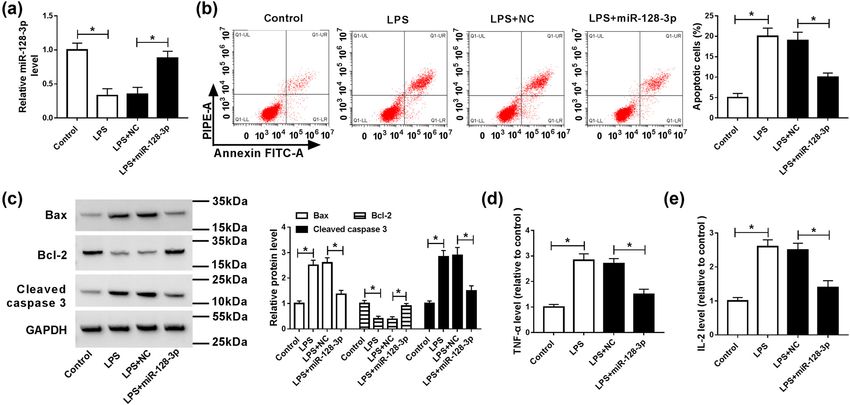
To analyze the effects of miR-128-3p on the LPS-induced
apoptosis and inflammatory response in HK2 cells, miR-
128-3p was transfected into the LPS-induced HK2 cells.
2.10 Statistical analysis The results of qPCR analysis showed that LPS exposure
significantly decreased the level of miR-128-3p, which
The data were presented as the mean ± standard devia- was abolished by the addition of miR-128-3p (Figure 2a).
tion (SD) from at least three independent experiments. Moreover, the apoptotic rate was remarkably increased in
Spearman rank correlation was performed to explore the HK2 cells treated with LPS compared with the control cells,
correlation between miR-128-3p and TGFBR2. Statistical whereas it was ablated by upregulation of miR-128-3p
analyses were performed using Graphpad Prism version (Figure 2b). Besides, apoptosis-related proteins were ana-
6.0 software (GraphPad Software, San Diego, CA, USA). lyzed by western blot. The results suggested that LPS treat-
The differences between two groups were assessed using ment prominently increased the protein levels of Bax and
the two-tailed Student’s t test. P < 0.05 was considered to cleaved caspase 3 but reduced the protein expression of
indicate a statistically significant difference. Bcl-2, which was reversed by accumulation of miR-128-3p
(Figure 2c). Furthermore, levels of pro-inflammatory cyto-
kines (TNF-α and IL-2) were evidently elevated in HK2 cells
induced with LPS, while overexpression of miR-128-3p
3 Results overturned this effect (Figure 2d and e). Altogether, these
data indicated that miR-128-3p reversed LPS-induced apop-
3.1 The expression of miR-128-3p was tosis and inflammatory response in HK2 cells.
decreased and TGFBR2 expression was
increased in serum samples with sepsis
and LPS-induced HK2 cells
3.3 Knockdown of TGFBR2 had the
To explore the roles of miR-128-3p and TGFBR2 in sepsis,
we determined their levels by qPCR in serum samples similar effects with overexpression
from patients with sepsis. Results proved that the level of miR-128-3p in LPS-induced
of miR-128-3p was markedly downregulated, and the HK2 cells
expression of TGFBR2 was notably upregulated in serum
samples of patients with sepsis compared with control To further study the function of TGFBR2 in LPS-induced
groups (Figure 1a and b). In addition, the relationship HK2 cells, si-TGFBR2 was transfected into HK2 cells
between miR-128-3p and TGFBR2 was analyzed in serum treated with LPS. Results revealed that TGFBR2 knock-
samples of patients with sepsis. We found that the level down reversed LPS-mediated promotion of TGFBR2 expres-
of TGFBR2 was negatively associated with miR-128-3p sion in HK2 cells (Figure 3a). Moreover, abrogation of
expression (Figure 1c). Next, we detected the levels of TGFBR2 abolished LPS-induced apoptosis in HK2 cells
miR-128-3p and TGFBR2 in the LPS-induced HK2 cells. (Figure 3b). Besides, downregulation of TGFBR2 alleviated
The results suggested that the abundance of miR-128-3p LPS-mediated promotion of Bax and cleaved caspase 3
was remarkably decreased in the LPS-induced HK2 cells protein levels and reduction of Bcl-2 expression (Figure 3c).
compared with control cells (Figure 1d). Besides, the Furthermore, inhibition of TGFBR2 also weakened the pro-
mRNA and protein levels of TGFBR2 were apparently ele- motion of inflammatory cytokines TNF-α and IL-2 levels
vated in the LPS-treated HK2 cells (Figure 1e and f). These caused by LPS (Figure 3d and e). Thus, these data proved
findings indicated that miR-128-3p and TGFBR2 might that inhibition of TGFBR2 attenuated LPS-induced apoptosis
play important roles in the development of sepsis. and inflammatory response in HK2 cells.278 Peng Yang et al. Figure 1: Expression levels of miR-128-3p and TGFBR2 in serum samples of sepsis patients and LPS-induced HK2 cells. (a and b) The expression levels of miR-128-3p and TGFBR2 were measured in serum samples of patients with sepsis and control groups by qPCR. (c) The association between TGFBR2 mRNA level and miR-128-3p abundance was measured in serum samples of patients with sepsis. (d and e) The expression levels of miR-128-3p and TGFBR2 were detected in LPS-induced HK2 cells by qPCR. (f) Western blot was performed to detect the protein level of TGFBR2. *P < 0.05. Figure 2: Effects of miR-128-3p on apoptosis and inflammation in LPS-induced HK2 cells. HK2 cells were treated with LPS or/and transfected with miR-128-3p. (a) The expression level of miR-128-3p was measured by qPCR. (b) Cell apoptosis was detected by flow cytometry. (c) The protein levels of Bax, Bcl-2, and cleaved caspase 3 were analyzed by western blot. (d and e) The production pro-inflammatory cytokines (TNF-α and IL-2) was detected by ELISA kit. *P < 0.05.
Effect of miR-128-3p in LPS-induced sepsis 279
Figure 3: Effects of TGFBR2 on apoptosis and inflammation in LPS-induced HK2 cells. HK2 cells were treated with LPS or/and transfected
with si-TGFBR2. (a) The protein level of TGFBR2 was measured by western blot analysis. (b) Cell apoptosis was detected by the flow
cytometry. (c) The protein levels of Bax, Bcl-2, and cleaved caspase 3 were analyzed by western blot. (d and e) ELISA kit was used to
determine the production of pro-inflammatory cytokines (TNF-α and IL-2). *P < 0.05.
3.4 TGFBR2 was a direct target of confirmed by dual-luciferase reporter assay and RNA
miR-128-3p immunoprecipitation (RIP) analysis in HK2 cells. Results
suggested that transfection of miR-128-3p obviously
To further explore the relationship between miR-128-3p decreased the luciferase activity of TGFBR2-wt, whereas
and TGFBR2 in sepsis, the potential binding sites of miR- luciferase activity of TGFBR2-mut was not evidently
128-3p and TGFBR2 were predicted by Targetscan online affected after transfection with miR-128-3p (Figure 4b).
website, indicating that TGFBR2 might be a target of RIP analysis suggested that TGFBR2 was notably enriched
miR-128-3p (Figure 4a). Subsequently, the prediction was in miR-128-3p group coated with Ago2 antibody compared280 Peng Yang et al.
Figure 4: miR-128-3p directly targeted TGFBR2 in HK2 cells. (a) The putative binding sites of miR-128-3p and TGFBR2 were provided by
TargetScan. (b) Luciferase activity was measured in HK2 cells co-transfected with TGFBR2-wt or TGFBR2-mut and miR-128-3p or NC by dual-
luciferase reporter assay. (c) RIP assay was performed to detect TGFBR2 enrichment level in HK2 cells transfected with miR-128-3p or NC.
*P < 0.05. (d) The protein level of TGFBR2 was measured in HK2 cells transfected with miR-128-3p, NC, anti-miR-128-3p, or anti-NC by
western blot analysis. *P < 0.05.
with the control group (Figure 4c). Besides, upregulation LPS-induced HK2 cells (Figure 5c). In addition, upregulation
of miR-128-3p apparently reduced the protein expression of TGFBR2 attenuated the effect of miR-128-3p overexpres-
of TGFBR2 in HK2 cells, and its knockdown presented an sion on the decreased levels of inflammatory cytokine TNF-α
opposite effect (Figure 4d). Taken together, these data and IL-2 (Figure 5d and e). Altogether, these data indicated
indicated that miR-128-3p directly targeted TGFBR2 in that upregulation of TGFBR2 reversed the effects of miR-128-
HK2 cells. 3p overexpression on apoptosis and inflammatory response
in the LPS-induced HK2 cells.
3.5 Upregulation of TGFBR2 reversed the
effects of miR-128-3p overexpression in 3.6 miR-128-3p suppressed the activation of
LPS-induced HK2 cells the Smad signaling pathway by
affecting TGFBR2 expression
To explore whether TGFBR2 was involved in miR-128-3p
overexpression-mediated inhibition of progression of Smad signaling pathway may play an essential role in the
sepsis, NC, miR-128-3p, miR-128-3p + vector, or miR-128- development and progression of sepsis. To further explore
3p + TGFBR2 was transfected into the LPS-induced HK2 the molecular mechanism by which miR-128-3p regulates
cells. As shown in Figure 5a, overexpression of miR-128-3p the biological functions in sepsis, the expression levels of
prominently reduced the protein level of TGFBR2 in the proteins related to the Smad signaling pathway (Smad2
LPS-induced HK2 cells, which could be abated by the and Smad3) were measured in controls cells and LPS-
addition of TGFBR2. Moreover, upregulation of TRAF3 induced HK2 cells transfected with NC, miR-128-3p,
abolished the inhibitory effect of miR-128-3p overexpres- miR-128-3p + vector, or miR128-3p + TGFBR2. Western
sion on apoptosis in LPS-induced HK2 cells (Figure 5b). blot indicated that overexpression of miR-128-3p reversed
Besides, transfection of TGFBR2 reversed the effects of the LPS-induced promotion of Smad2 and Smad3 protein
miR-128-3p upregulation on reduction of Bax and cleaved levels of in HK2 cells, whereas addition of TGFBR2 abo-
caspase 3 expression and promotion of Bcl-2 expression in lished the effects of miR-128-3p overexpression onEffect of miR-128-3p in LPS-induced sepsis 281 Figure 5: Upregulation of TGFBR2 weakened the effects of miR-128-3p overexpression on apoptosis and inflammation in LPS-induced HK2 cells. miR-128-3p, NC, miR-128-3p + vector, or miR-128-3p + TGFBR2 was transfected into LPS-induced HK2. (a) The protein level of TGFBR2 was detected by western blot analysis. (b) Cell apoptosis was measured by flow cytometry. (c) The protein levels of Bax, Bcl-2, and cleaved caspase 3 were analyzed by western blot. (d and e) The production of pro-inflammatory cytokines (TNF-α and IL-2) was detected by ELISA kit. *P < 0.05. Figure 6: miR-128-3p regulated Smad singling pathway by affecting TGFBR2 expression. LPS-induced HK2 cells were transfected with miR- 128-3p, NC, miR128-3p + TGFBR2, or miR128-3p + vector. (a and b) The protein levels of Smad2 and Smad3 were detected by western blot analysis. *P < 0.05.
282 Peng Yang et al.
expression of Smad2 and Smad3 (Figure 6a and b). From TGFBR2/Smad2/DNMT1/miR-145 negative regulatory loop
aforementioned outcomes, we demonstrated that miR-128- might be a potential target for the treatment of sepsis [26].
3p could regulate Smad singling pathway via affecting Zhang et al. showed that SUMO protease SENP1 acted as
TGFBR2 expression. a ceRNA for TGFBR2, which activated TGFBR2/Smad
signaling responsible for LPS-induced sepsis [27]. Cao et al.
identified that the expression TGFBR2 was obviously
increased with LPS treatment in a time-dependent manner,
4 Discussion and miR-145 ameliorated sepsis-induced lung injury by inhi-
biting TGFBR2 signaling [28]. Here, we found that knock-
Sepsis is a systemic inflammation response syndrome, down of TGFBR2 ameliorated LPS-induced inflammation
which frequently causes extensive tissue injury and and apoptosis. Besides, upregulation of TGFBR2 abrogated
multiple organ dysfunctions [18]. If sepsis is not con- miR-128-3p overexpression-mediated attenuation on LPS-
trolled timely and effectively, it can lead to multiple induced sepsis, suggesting that miR-128-3p was involved
organ dysfunction syndrome, which is the main cause in the development and progression of sepsis by regulation
of death in trauma, burn, and critical surgical patients. of TGFBR2.
Recent reports have demonstrated that aberrant expres- TGF-β1 exerts its biological functions via activating
sion of miRNAs might be used to improve diagnosis and Smad2 and Smad3 [29]. Recently, it had been reported
treatment of sepsis. Hence, it is particularly important that inhibition of the TGF-β1/Smad3 pathway might play
to understand the underlying molecular mechanisms of a protective role in sepsis-induced acute lung injury [30].
miRNAs for the treatment of sepsis. Besides, propofol could provide protection against acute
miR-128-3p has been suggested to be dysregulated in lung injury through suppressing the TGF-β1-Smad2-depen-
many conditions and participate in multiple cell beha- dent pathway [28]. In this study, we found that transfec-
viors, including proliferation, migration and invasion, tion of miR-128-3p reversed the LPS-induced promotion of
epithelial-mesenchymal transition (EMT), and angio- Smad2 and Smad3 protein levels of in HK2 cells, whereas
genesis [8,9,19,20]. Increasing evidence suggested that addition of TGFBR2 abolished the effects of overexpres-
miR128-3p has also been regarded as a prognostic marker sion of miR-128-3p on expression of Smad2 and Smad3.
in many diseases [20–22]. In addition, miR-128-3p could These results indicated that miR-128-3p could regulate
regulate inflammatory response in LPS-stimulated macro- Smad singling pathway by affecting TGFBR2 expression.
phages via the TLR4-NF-κB pathway [23]. Besides, miR- In conclusion, we demonstrated that miR-128-3p was
128 specifically inhibited the development of inflamma- decreased and TGFBR2 expression was increased in serum
tion by downregulation of MyD88 [24]. Previous study samples of sepsis patients and LPS-induced HK2 cells.
had revealed that decreased level of miR-128 synergisti- Overexpression of miR-128-3p or knockdown of TGFBR2
cally promoted cell injuries through targeting Snail and ameliorated LPS-induced inflammation and apoptosis.
PTEN [10]. As mentioned earlier, miR-128 could regulate Moreover, our study presented the first evidence that
inflammatory responses and might play important roles in TGFBR2 was a direct target of miR-128-3p, and its overex-
sepsis. Consistent with the previous results, the expres- pression reversed the effects of miR-128-3p overexpression
sion of miR-128-3p was decreased in serum samples of on inflammation and apoptosis. Besides, overexpression of
sepsis patients and LPS-induced HK2 cells. Moreover, miR-128-3p downregulated TGFBR2 to suppress the activa-
treatment of LPS promoted apoptosis and inflammation tion of the Smad signaling pathway. Collectively, miR-128-
in HK2 cells, which was abated by overexpression of 3p could inhibit apoptosis and inflammation by targeting
miR-128-3p. These findings suggested that miR-128-3p TGFBR2 in LPS-induced HK2 cells, providing viable thera-
might play vital roles in sepsis. peutic avenues for the treatment of sepsis.
Increasing evidence shows that miRNAs exert their
functions through directly binding to target mRNAs and Funding: This work was financially supported by research
inhibiting mRNA stability and translation [25]. In our fund of Guangdong Provincial Science and Technology
study, we found that TGFBR2 was a direct target of miR- Plan Project (No. 201704020049).
128-3p, and the level of TGFBR2 was negatively associated
with miR-128-3p expression. TGFBR2 has been reported to Disclosure of interest: The authors declare that they have
play essential roles in sepsis. Ma et al. demonstrated that no financial conflicts of interest.Effect of miR-128-3p in LPS-induced sepsis 283
References beta and smad signaling in the aorta in the early stage of
sepsis. Ulus Travma Acil Cerrahi Derg. 2010;16(4):293–9.
[1] Hotchkiss RS, Karl IE. The pathophysiology and treatment of [17] Bone RC, Balk RA, Cerra FB, Dellinger RP, Fein AM, Knaus WA,
sepsis. N Engl J Med. 2003;348(2):138–50. et al. Definitions for sepsis and organ failure and guidelines
[2] Cecconi M, Evans L, Levy M, Rhodes A. Sepsis and septic for the use of innovative therapies in sepsis. Crit Care Med.
1992;101(6):1644–55.
shock. Lancet. 2018;392(10141):75–87.
[18] Rhodes A, Evans LE, Alhazzani W, Levy MM, Antonelli M,
[3] Kampmeier TG, Rehberg S, Westphal M, Lange M. Vasopressin
Ferrer R, et al. Surviving sepsis campaign: international
in sepsis and septic shock. Minerva Anestesiol.
guidelines for management of sepsis and septic shock: 2016.
2010;76(10):844–50.
Intensive Care Med. 2017;43(3):304–77.
[4] Angus DC, Linde-Zwirble WT, Lidicker J, Clermont G, Carcillo J,
[19] Evangelisti C, Florian MC, Massimi I, Dominici C, Giannini G,
Pinsky MR. Epidemiology of severe sepsis in the United States:
Galardi S, et al. miR-128 up-regulation inhibits reelin and DCX
analysis of incidence, outcome, and associated costs of care.
expression and reduces neuroblastoma cell motility and
Crit Care Med. 2001;29(7):1303–10.
invasiveness. FASEB J. 2009;23(12):4276–87.
[5] Cai Y, Yu X, Hu S, Yu J. A brief review on the mechanisms of
[20] Zhao L, Li R, Xu S, Li Y, Zhao P, Dong W, et al. Tumor sup-
miRNA regulation. Genomics Proteomics Bioinf.
pressor miR-128-3p inhibits metastasis and epithelial-
2009;7(4):147–54.
mesenchymal transition by targeting ZEB1 in esophageal
[6] Wang JF, Yu ML, Yu G, Bian JJ, Deng XM, Wan XJ, et al. Serum
squamous-cell cancer. Acta Biochim Biophys Sin. 2018 Feb
miR-146a and miR-223 as potential new biomarkers for sepsis.
1;50(2):171–80.
Biochem Biophys Res Commun. 2010;394(1):184–8.
[21] Cai J, Fang L, Huang Y, Li R, Xu X, Hu Z, et al. Simultaneous
[7] Wang Z, Ruan Z, Mao Y, Dong W, Zhang Y, Yin N, et al. miR-27a
overactivation of Wnt/β-catenin and TGFβ signalling by miR-
is up regulated and promotes inflammatory response in
128-3p confers chemoresistance-associated metastasis in
sepsis. Cell Immunol. 2014;290(2):190–5.
NSCLC. Nat Commun. 2017;8:15870.
[8] Huang CY, Huang XP, Zhu JY, Chen ZG, Li XJ, Zhang XH, et al.
[22] Li B, Chen H, Wu N, Zhang WJ, Shang LX. Deregulation of miR-
miR-128-3p suppresses hepatocellular carcinoma proliferation
by regulating PIK3R1 and is correlated with the prognosis of 128 in ovarian cancer promotes cisplatin resistance. Int J
HCC patients. Oncol Rep. 2015;33(6):2889–98. Gynecol Cancer. 2014;24(8):1381–8.
[9] Hu J, Cheng Y, Li Y, Jin Z, Pan Y, Liu G, et al. microRNA-128 plays [23] Xiaokelaiti H, Zhi X, Yusufujiang Y, Aikebaier A. miR-128-3p
a critical role in human non-small cell lung cancer tumouri- regulates inflammatory response in LPS-stimulated macro-
genesis, angiogenesis and lymphangiogenesis by directly phages through the TLR4-NF-κB pathway. Int J Clin Exp Med.
targeting vascular endothelial growth factor-C. Eur J Cancer. 2016;9(5):8005–13.
2014;50(13):2336–50. [24] Ma X, Guo S, Jiang K, Wang X, Yin N, Yang Y, et al. miR-128
[10] Wang S, Wang J, Zhang Z, Miao H. Decreased miR-128 and mediates negative regulation in Staphylococcus aureus
increased miR-21 synergistically cause podocyte injury in induced inflammation by targeting MyD88. Int
sepsis. J Nephrol. 2017;30(4):543–50. Immunopharmacol. 2019;70:135–46.
[11] Yue D, Liu H, Huang Y. Survey of computational algorithms for [25] Hudder A, Novak RF. miRNAs: effectors of environmental
microRNA target prediction. Curr Genomics. influences on gene expression and disease. Toxicol Sci.
2009;10(7):478–92. 2008;103(2):228–40.
[12] Javelaud D, Mauviel A. Mammalian transforming growth factor- [26] Ma F, Li Z, Cao J, Kong X, Gong G. A TGFBR2/SMAD2/DNMT1/
betas: smad signaling and physio-pathological roles. Int J miR-145 negative regulatory loop is responsible for LPS-
Biochem Cell Biol. 2004;36(7):1161–5. induced sepsis. Biomed Pharmacother. 2019;112:108626.
[13] Massagué J. TGF-beta signal transduction. Annu Rev Biochem. [27] Zhang C, Li J, Qiu X, Chen Y, Zhang X. SUMO protease SENP1
1998;67:753–91. acts as a ceRNA for TGFBR2 and thus activates TGFBR2/Smad
[14] Weinstein M, Monga SP, Liu Y, Brodie SG, Tang Y, Li C, et al. signaling responsible for LPS-induced sepsis. Biomed
Smad proteins and hepatocyte growth factor control parallel Pharmacother. 2019;112:108620.
regulatory pathways that converge on beta1-integrin to pro- [28] Cao X, Zhang C, Zhang X, Chen Y, Zhang H. miR-145
mote normal liver development. Mol Cell Biol. negatively regulates TGFBR2 signaling responsible for sepsis-
2001;21(15):5122–31. induced acute lung injury. Biomed Pharmacother.
[15] Rendón-Ramirez EJ, Ortiz-Stern A, Martinez-Mejia C, Salinas- 2019;111:852–8.
Carmona MC, Rendon A, Mata-Tijerina VL, et al. TGF-β blood [29] Lan HY, Chung AC. TGF-β/Smad signaling in kidney disease.
levels distinguish between influenza A (H1N1)pdm09 virus Semin Nephrol. 2012;32(3):236–43.
sepsis and sepsis due to other forms of community-acquired [30] Xu F, Lin SH, Yang YZ, Guo R, Cao J, Liu Q. The effect of cur-
pneumonia. Viral Immunol. 2015;28(5):248–54. cumin on sepsis-induced acute lung injury in a rat model
[16] Erbüyün K, Tok D, Vatansever S, Ok G, Türköz E, Aydede H, through the inhibition of the TGF-β1/SMAD3 pathway. Int
et al. Levosimendan up-regulates transforming growth factor- Immunopharmacol. 2013;16(1):1–6.You can also read