Postnatal Recapitulation of Embryonic Hedgehog Pathway in Response to Skeletal Muscle Ischemia
←
→
Page content transcription
If your browser does not render page correctly, please read the page content below
Postnatal Recapitulation of Embryonic Hedgehog Pathway
             in Response to Skeletal Muscle Ischemia
    Roberto Pola, MD, PhD*; Leona E. Ling, PhD*; Tamar R. Aprahamian, BS; Elena Barban, MD;
     Marta Bosch-Marce, PhD; Cynthia Curry, BS; Michael Corbley, PhD; Marianne Kearney, BS;
                         Jeffrey M. Isner, MD†; Douglas W. Losordo, MD

Background—Hedgehog (Hh) proteins are morphogens regulating epithelial–mesenchymal signaling during several
  crucial processes of embryonic development, including muscle patterning. Sonic (Shh), Indian (Ihh), and Desert (Dhh)
  hedgehog constitute the repertoire of Hh genes in humans. The activities of all 3 are transduced via the Patched (Ptc1)
  receptor. Recent observations indicate that exogenous administration of Shh induces angiogenesis. Here, we studied
  whether the endogenous Hh pathway, in addition to its functions during embryogenesis, plays a physiological role in
  muscle regeneration after ischemia in adults.
Methods and Results—We found that skeletal muscle ischemia induces strong local upregulation of Shh mRNA and
  protein. In addition, the Ptc1 receptor is activated in interstitial mesenchymal cells within the ischemic area, indicating
  that these cells respond to Shh and that the Shh pathway is functional. We also found that Shh-responding cells produce
  vascular endothelial growth factor under ischemic conditions and that systemic treatment with a Shh-blocking antibody
  inhibits the local angiogenic response and the upregulation of vascular endothelial growth factor.
Conclusions—Our study shows that the Hh signaling may be recapitulated postnatally in adult and fully differentiated
  muscular tissues and has a regulatory role on angiogenesis during muscle regeneration after ischemia. These findings
  demonstrate a novel biological activity for the Hh pathway with both fundamental and potential therapeutic implications.
  (Circulation. 2003;108:479-485.)
             Key Words: genes, hedgehog 䡲 ischemia 䡲 muscle, skeletal 䡲 angiogenesis 䡲 tissue regeneration

I  n the past decade, there has been increasing appreciation of
   the fact that pathways studied predominantly during embry-
ogenesis and known to be relatively silent during normal adult
                                                                                Previous observations have suggested that Hh might also
                                                                             be involved in the vascularization of certain embryonic
                                                                             tissues. First, transgenic overexpression of Shh in the dorsal
life may be recruited postnatally in response to tissue injury.1             neural tube is associated with hypervascularization of neuro-
   Hedgehog (Hh) proteins are morphogens that act in a wide                  ectoderm,19 whereas a knockout of the zebra fish Shh
variety of tissues during embryonic development2–9 and                       homologue results in disorganization of the endothelial pre-
regulate epithelial–mesenchymal interactions that are crucial                cursor cells and inability to form the dorsal aorta or axial
to morphogenesis of the nervous system, somite, limb, lung,                  vein. In addition, Shh-null mice lack proper vascularization
gut, hair follicle, and bone.3–7,10 –12 There are 3 highly                   of the developing lung.4 More recently, it has been reported
conserved Hh genes in mammals: Sonic hedgehog (Shh),                         that vasculogenesis in the mouse embryo is regulated by
Indian hedgehog (Ihh), and Desert hedgehog (Dhh).13 The                      Ihh.20 Finally, very recently, we found that cells in the adult
interaction of Hh proteins with their specific receptor                      cardiovascular tissues express Ptc1 and can respond to
patched-1 (Ptc1) inactivates the repression of the transmem-                 exogenous administration of Shh by upregulating Ptc1.21 We
brane protein smoothened (Smo), leading to activation of the                 also demonstrated that Shh induces neovascularization in 2
transcription factor Gli,14 –16 the principal mediator of the Hh             different murine models of angiogenesis and upregulates 2
signaling pathway. Gli induces expression of downstream                      families of angiogenic growth factors, including vascular
target genes of the Hh pathway, including Ptc1 and Gli                       endothelial growth factor (VEGF) and angiopoietins.21
itself.17,18 Thus, Ptc1 and Gli are both components and                         The aim of this study was to investigate whether the
transcriptional targets of the Hh signaling pathway.                         endogenous Hh pathway is physiologically involved in the

  Received December 18, 2002; revision received April 10, 2003; accepted April 14, 2003.
  From the Department of Medicine (Cardiovascular Research), St Elizabeth’s Medical Center, Tufts University School of Medicine, Boston, Mass (R.P.,
T.R.A., E.B., M.B.-M., C.C., M.K., J.M.I., D.W.L.); the Department of Medicine, A. Gemelli University Hospital, Rome, Italy (R.P.); and Biogen, Inc,
Cambridge, Mass (L.E.L., M.C.).
  *The first 2 authors contributed equally to this work.
  †Deceased.
  Correspondence to Douglas W. Losordo, MD, Division of Vascular Medicine, St Elizabeth’s Medical Center, 736 Cambridge St, Boston, MA
02135-2997. E-mail douglas.losordo@tufts.edu
  © 2003 American Heart Association, Inc.
  Circulation is available at http://www.circulationaha.org                                             DOI: 10.1161/01.CIR.0000080338.60981.FA

                                                                       479
                               Downloaded from http://circ.ahajournals.org/ by guest on September 22, 2015
480 Circulation July 29, 2003
revascularization of ischemic tissue in adults. We used a anti-Shh C-terminus, goat polyclonal IgG anti-Ihh C-terminus, and
murine model of muscle regeneration by inducing ischemia goat polyclonal IgG anti-Dhh C-terminus (Santa Cruz Biotechnolo-
gy). Horse anti-goat IgG horseradish peroxidase– conjugated anti-
of the hindlimb. Then, we observed the expression pattern of
body (1:500 dilution) (Vector Laboratories) was used as secondary
different components of the Hh pathway, including Shh, Dhh, antiserum. Staining was visualized by using FITC-conjugated
Ihh, and Ptc1, and studied the relationship between Hh streptavidin (Pharmingen). For vimentin immunostaining, muscles
activation, VEGF expression, and angiogenesis. We found were fixed in 1% paraformaldehyde for 2 hours. The staining was
that Shh is activated in the regeneration after ischemia and done on frozen sections with anti-vimentin goat serum (Sigma)
compared with normal goat serum (Sigma) using horseradish per-
that interstitial cells within the ischemic area strongly express oxidase– conjugated donkey anti-goat secondary antibody (Jackson
Ptc1, indicating the postnatal activity of the Hh signaling Immunoresearch) or rhodamine-conjugated donkey anti-goat anti-
pathway. We found that Ptc1 expression was associated with body (Santa Cruz Biotechnology). Staining for VEGF was per-
VEGF production and angiogenesis. Inhibition of Shh inhib- formed with a rabbit polyclonal anti-VEGF antibody (Santa Cruz
its endogenous angiogenesis and VEGF production in the Biotechnology) with a biotinylated goat anti-rabbit Ig as secondary
antibody.
ischemic hindlimb. Our data suggest a novel and unexpected
physiological role for Shh. LacZ Immunofluorescence and Histochemistry in
nls-Ptc1-lacZ Mice
Methods For -gal immunofluorescence staining, hindlimb muscles from
Animals nls-Ptc1-lacZ mice were harvested and processed as described
previously.26
Male C57BL/6J mice (Jackson Laboratories, Bar Harbor, Me), male
or female nls-Ptc1-lacZ mice, or their wild-type littermates (Ontog-
eny, Inc) were used for the ischemic hindlimb experiments.21 All the Inhibition of the Hh Pathway and Analysis of
experiments were conducted in accordance with the St Elizabeth’s or Local VEGF Expression and Analysis of the
Biogen Institutional Animal Care and Use Committee. Angiogenic Response to Ischemia
Unilateral hindlimb ischemia was induced as described above in 8-
Murine Ischemic Hindlimb Model to 12-week-old C57BL/6J mice. Animals were treated with daily
Ischemia was induced in 8- to 12-week-old C57BL/6J mice, nls- intraperitoneal injections of 10 mg/kg 5E1 blocking antibody or the
Ptc1-lacZ mice, and their wild-type littermates as described same amount of 1E6 control antibody. 5E1 blocks the binding of Shh
previously.21,22 to Ptc1; it was obtained from Curis Inc and Dr Thomas Jessell
(Columbia University) and prepared as purified IgG1 in PBS.18
In Situ Hybridization for Hh Members Seven days after induction of ischemia, mice were killed. Hindlimb
Skeletal muscles were harvested 4 and 7 days after surgery and muscle specimens were harvested, processed, and analyzed by
immediately immersion-fixed overnight in 4% paraformaldehyde, Western blotting for VEGF expression. For analysis of the response
paraffin-embedded, and sectioned longitudinally at 7 to 8 m. Shh, to ischemia, animals were divided into 2 groups: the first group
Ihh, and Dhh in situ hybridization was performed with digoxigenin- received a total dose of 1.25 mg 5E1, delivered systemically via an
labeled sense and antisense cRNA probes. osmotic pump over a period of 21 days, and the second group
received an equal amount of 1E6. Ten animals in each group were
studied. At days 7, 14, 21, and 28 after induction of ischemia, blood
Quantitative Real-Time Reverse flow was measured with a laser Doppler perfusion imaging system as
Transcription–Polymerase Chain Reaction described previously.20,21
Mice were killed 4 and 7 days after ischemia, and reverse transcrip- Analysis of capillary density was performed as described
tion–polymerase chain reaction (RT-PCR) was performed as de- previously.21,22
scribed previously. The primer sequences were as follows: Shh
forward, GAGCAGACCGGCTGATGACT; Shh reverse, Statistical Analysis
AGAGATGGCCAAGGCATTTAAC; Dhh forward, CGCAGAC- All results are expressed as mean⫾SD, with the exception of the
CGCCTGATGAC; Dhh reverse, GCGATGGCTAGAGCGTTCAC; real-time RT-PCR results, which are presented as mean⫾SEM.
Ihh forward, CAAACCGGCTGAGAGCTTTC; Ihh reverse, AGC- Group differences were analyzed by ANOVA or Student’s t test.
CGACGCGGAGGAT. The probe sequences were as follows: Shh Differences were considered statistically significant at a value of
6FAM, AGAGGTGCAAAGACA-MGBNFQ; Dhh 6FAM, P⬍0.05.
AGCGTTGCAAAGAG-MGBNFQ; Ihh 6FAM, AGGTCATC-
GAGACTCA-MGBNFQ.
Results
ELISA Shh Signaling Pathway Is Activated in Ischemic
Mice were killed 4 and 7 days after ischemia, and hindlimb muscle Regenerating Skeletal Muscles
specimens were harvested and homogenized in lysis buffer. Shh The expression of Shh mRNA was increased at 4 and 7 days
protein levels were determined by ELISA as described previously.23
after injury in ischemic compared with nonischemic skeletal
Western Blotting for Ptc1 muscle, as detected by in situ hybridization (Figure 1a). Shh
Protein extracts from the skeletal muscles of mice killed 7 days after mRNA in ischemic muscle was strongly induced, particularly
ischemia were used for Western blotting analysis of Ptc1 expression in the interstitial regions. Expression of Ihh was also slightly
as described previously.24 Densitometric analysis was performed elevated in ischemic muscle, whereas little Dhh expression
(NIH Imaging program) to allow for quantitative comparison of was detected at 4 or 7 days after ischemic injury (Figure 1b).
protein expression. Upregulation of Shh was confirmed and quantified by
real-time RT-PCR: Shh expression increased 10⫾3.3-fold in
Immunofluorescence and Immunohistochemistry
Ischemic and contralateral muscle specimens were harvested 7 days day 4 ischemic muscles (P⫽0.01) and 16⫾3.8-fold at day 7
after induction of ischemia, and frozen sections were processed as (P⫽0.0007) (Figure 2a and data not shown). No significant
detailed previously.20,25 Primary antisera were goat polyclonal IgG increment of Dhh and Ihh expression was documented.
Downloaded from http://circ.ahajournals.org/ by guest on September 22, 2015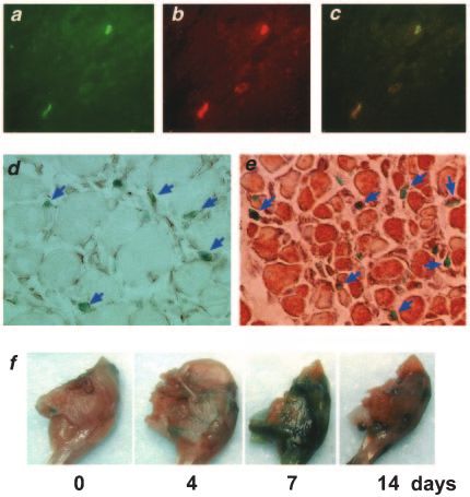
Pola et al Hedgehog in Postnatal Skeletal Muscle Ischemia 481
in response to Shh signaling, expression of Ptc1 constitutes
evidence of active Shh signaling in ischemic skeletal muscle.
In addition, these findings suggest that in this model, an
autocrine interaction occurs between the Shh ligand and its
receptor Ptc1.
Shh and Ptc1 Activation Occur in Interstitial
Mesenchymal Cells and Are Associated With
VEGF Production
To determine the identity of the Shh-producing and
-responding cells during ischemia, we performed further
immunohistochemical analyses. These interstitial cells were
not positive for the endothelial cell marker CD31 or the
smooth muscle cell marker ␣-SM-actin (data not shown). In
contrast, we found that Shh- and Ptc1-positive cells stained
positive for vimentin, consistent with mesenchyme-derived
fibroblasts (Figure 4, a– d).
We also analyzed the relationship between the activated,
endogenous Shh pathway and VEGF in ischemic skeletal
muscle. We found that Ptc1-positive interstitial cells located
within the ischemic area were strikingly immunopositive for
VEGF (Figure 4e). The colocalization in the same cells of
Ptc1 and VEGF suggests that the Shh signaling pathway may
stimulate, either directly or indirectly, VEGF expression
Figure 1. Shh mRNA is upregulated during skeletal muscle
regeneration after ischemia. In situ hybridization for Shh, Ihh, within the neovascular foci. No Ptc1 (X-gal) or VEGF
and Dhh 4 and 7 days after ischemia. Strong positive signal for staining was observed in either the contralateral or control
Shh mRNA is present in interstitium of ischemic muscles at both muscles (data not shown).
days 4 and 7 after surgery, whereas no signal is detected in
contralateral nonischemic specimens (a); Ihh mRNA seems The time course of upregulated Ptc1 expression after the onset
slightly elevated in ischemic muscle (b). Very little Dhh expres- of ischemia, inferred from whole-mount X-gal staining of
sion is detected at 4 or 7 days after ischemic injury (b) (H&E nls-Ptc1-lacZ hindlimbs, was characterized by Ptc1 upregulation
indicates hematoxylin and eosin; Shh AS, Shh Antisense probe;
beginning 4 days after ischemia, peaking at day 7, and decreas-
Shh S, Shh Sense probe; Ihh AS, Ihh Antisense probe; Dhh AS,
Dhh Antisense probe). ing significantly by day 14 after ischemia (Figure 4f).
Inhibition of Shh Signaling Pathway Impairs Local
The concentration of Shh protein in ischemic and contralat-
VEGF Upregulation
eral muscles was studied by ELISA and was shown to be To determine whether the Shh signaling pathway is necessary
increased at day 4 (P⫽0.03) and day 7 (P⫽0.02) (Figure 2b). for VEGF upregulation during ischemia, we used a Shh-
Ptc1 upregulation was verified by Western blotting (Figure neutralizing antibody (5E1). After unilateral hindlimb ische-
2c) and normalized for tubulin expression, was also statisti- mia had been induced in mice, animals were treated for 7
cally significant (P⬍0.01) (Figure 2d). days with systemic injections of 5E1 or control antibody
Immunofluorescence analysis demonstrated that several (1E6). Local VEGF expression was studied by Western
cells are immunopositive for Shh within 7 days after induc- blotting in both ischemic and contralateral muscles. As
tion of ischemia (Figure 3, a and b). No positive immuno- expected, mice treated with the 1E6 control antibody exhib-
staining in ischemic muscle was observed at this time point ited a physiological upregulation of VEGF in the ischemic
for either Ihh or Dhh (data not shown). Immunofluorescence hindlimbs (Figure 5a). In contrast, animals treated with the
staining for -gal, performed in nls-Ptc1-lacZ mice, demon- 5E1 antibody did not upregulate VEGF in the ischemic
strated the expression of Ptc1 in several cells in the ischemic hindlimb (Figure 5a). Comparison of VEGF expression in
tissue (Figure 3, c and d). Interestingly, both Shh- and ischemic muscle, normalized for tubulin expression, indi-
Ptc1-positive cells appeared to be interstitial cells widely cated a statistically significant difference between mice
distributed in the ischemic area, within and around skeletal treated with 5E1 versus 1E6 (P⬍0.01) (Figure 5b).
muscle fibers. By performing double immunofluorescence
staining for Shh and -gal in nls-Ptc1-lacZ mice, we demon- Inhibition of the Shh Signaling Pathway Decreases
strated that Shh and Ptc1 are coexpressed in the same cells the Angiogenic Response to Ischemia
(Figure 3, e– g). These data demonstrate that cells in the Hh-blocking antibody (5E1) or a control antibody (1E6) was
ischemic tissue produce Shh and express the Ptc1 gene, administered by continuous subcutaneous infusion through an
indicating that the Shh pathway is physiologically active osmotic pump for 3 weeks after induction of hindlimb
during muscle regeneration after ischemia in adults. Because ischemia. Twenty-eight days after induction of ischemia,
Ptc1 is a downstream transcriptional target of the Shh signal blood flow was significantly lower in animals treated with the
transduction pathway and Ptc1 expression is known to occur Shh-blocking antibody (P⬍0.01) (Figure 5c). Capillary den-
Downloaded from http://circ.ahajournals.org/ by guest on September 22, 2015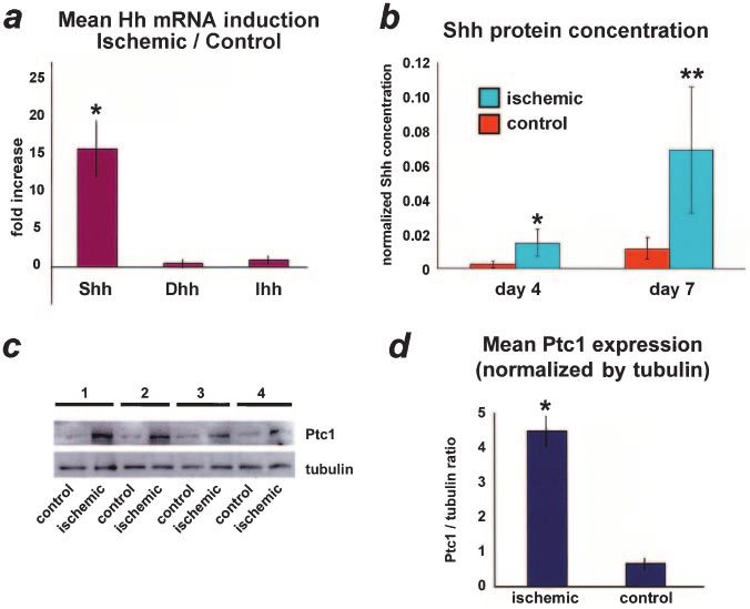
482 Circulation July 29, 2003
Figure 2. Quantification of Shh and Ptc1
upregulation during skeletal muscle
regeneration after ischemia. Real-time
RT-PCR for Shh, Ihh, and Dhh 7 days
after ischemia (a): Shh mRNA is
increased 16⫾3.8-fold (*P⫽0.0007),
whereas Dhh and Ihh gene expression
are not substantially altered. ELISA for
Shh 4 and 7 days after ischemia (b): Shh
protein concentration, normalized by
total protein concentration, is signifi-
cantly increased in ischemic skeletal
muscles at both time points (*P⫽0.03
and **P⫽0.02). Representative Western
blotting for Ptc1, 7 days after ischemia
(c): Ptc1 signal is increased in ischemic
muscles compared with controls. Quanti-
fication of Ptc1 upregulation (d): Ptc1
signal, quantified by densitometric analy-
sis and normalized for tubulin, is upregu-
lated significantly in ischemic tissues
(*P⬍0.01).
sity was assessed by CD31 immunostaining and was signif- Discussion
icantly reduced as well (P⬍0.0001) (Figure 5, d and e). These The Hh pathway has been studied and characterized exten-
results indicate that the activation of the Shh pathway is a sively during embryogenesis. The vast majority of these
prerequisite for the postnatal angiogenic response to skeletal prenatal studies have focused on the role of Hh family
muscle ischemia. members in the regulation of epithelial–mesenchymal inter-
actions crucial to limb, lung, gut, hair follicle, and bone
formation,3– 6 including a possible role during vascularization
of certain embryonic tissues.4,19,20,27–29 In contrast, a role for
the Hh family in the regulation of postnatal tissue regenera-
tion and revascularization has received limited attention.30 –32
We recently demonstrated that exogenous administration of
Shh induces neovascularization in both corneal and ischemic
hindlimb models of angiogenesis.21 Shh stimulates fibroblasts
in vitro to produce a combination of potent angiogenic
factors, including the 3 major isoforms of VEGF, Ang-1, and
Ang-2.21 Shh seems to act as an indirect angiogenic agent and
may trigger neovascularization through Shh/Ptc1 signaling
specifically in mesenchymal cells.21
Following these observations, we investigated the hypoth-
esis that the Hh pathway may be postnatally recapitulated in
response to skeletal muscle ischemia and discovered that in
adult mice, a strong upregulation of Shh and Ptc1 occurs
during regeneration of ischemic skeletal muscle. These find-
ings are consistent with previous reports in the literature
describing the association of both ischemia and tissue regen-
Figure 3. Immunostaining for Shh and Ptc1 in ischemic regener- eration with the reactivation of genes involved in fetal
ating skeletal muscle. Cross sections of day 7 ischemic and transcription programs.33–35
nonischemic hindlimbs (magnification ⫻20). Immunofluores- After ischemia, Ptc1 expression occurs in interstitial mes-
cence staining shows that Shh is strongly upregulated in several
interstitial cells in ischemic tissues (a and b). Likewise, -gal im- enchymal fibroblasts. The ability of fibroblasts to respond to
munofluorescence staining in nls-Ptc1-lacZ mice (magnification Shh stimulation has already been demonstrated: eg, fibro-
⫻20) demonstrates strong upregulation of Ptc1 during ischemia blasts respond to Shh stimulation in vitro,21 physiologically
(c and d). Double immunofluorescence in nls-Ptc1-lacZ mice
(magnification ⫻20) shows that Shh staining (e) and Ptc1 (-gal) express Ptc1 in adult perineural sheaths and dermis,6,9 and
staining (f) are coexpressed in same interstitial cells (g). upregulate Ptc1 and VEGF during Shh-induced corneal neo-
Downloaded from http://circ.ahajournals.org/ by guest on September 22, 2015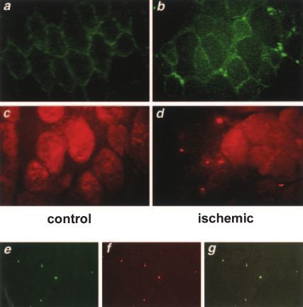
Pola et al Hedgehog in Postnatal Skeletal Muscle Ischemia 483
vascularization.21 Taken together, these data strongly suggest
that fibroblasts are central mediators of Shh activity during
muscle regeneration.
In our ischemic model, interstitial mesenchymal fibroblasts
also expressed Shh. The coexpression of Shh and Ptc1 in the
same cells indicates the presence of an autocrine mechanism
regulating Shh signaling in ischemic muscle. Such an auto-
crine mechanism has already been described in adult pan-
creas, in which Dhh and Ihh are coexpressed with Ptc1 in
pancreatic -cells and regulate insulin production.25
In muscle regeneration after ischemia, a crucial role is
played by angiogenesis.22 In this study, we show that Ptc1-
positive interstitial fibroblasts within the ischemic area pro-
duce VEGF. We also show that the inhibition of Shh
signaling is sufficient to decrease local VEGF upregulation.
Similarly, ischemia-induced angiogenesis is decreased by
inhibition of the Shh pathway. These results indicate that
although Ptc1-positive fibroblasts do not represent the major-
ity of VEGF-producing cells during ischemia, the activation
of the Shh signaling pathway is crucial for the overall
production of VEGF and the related angiogenic response.
Indeed, interstitial fibroblasts are important supporting cells,
and their function, modulated by Shh, might be fundamental
for stimulating the angiogenic activity of neighboring cells.
The mechanism by which ischemia and/or hypoxia upregu-
lates Shh expression remains uncertain. The promoter regions
Figure 5. Inhibition of Shh signaling decreases VEGF upregula-
tion and angiogenic response to ischemia. Hindlimb ischemia
was induced in young mice, after which they were treated with
a Shh-blocking antibody (Ab) (5E1) or a control antibody (1E6).
Western Blotting analysis of VEGF expression shows upregula-
tion in ischemic limbs of mice treated with 1E6 compared with
normal limbs (a). No upregulation of VEGF was detected in is-
chemic limbs of mice in which Shh signaling was blocked by
5E1 (a). Quantification of VEGF signal normalized for tubulin in
mice treated with 5E1 and 1E6 shows a statistically significant
difference between 2 groups (*P⬍0.01) (b). Blood flow was
assessed by laser Doppler perfusion imaging (LDPI) (c): no sig-
nificant increase in hindlimb perfusion was seen over time in
group treated with 5E1. In these animals, at day 28 after sur-
gery, blood flow was reduced significantly compared with con-
trols (0.414⫾0.019 vs 0.741⫾0.126, §P⬍0.01). Capillary density
assessed 28 days after surgery was reduced significantly in
mice treated with Shh-blocking antibody vs controls
(*P⬍0.0001) (d). CD31 staining of skeletal muscle sections from
ischemic hindlimbs of mice treated with 5E1 and control anti-
Figure 4. Ptc1 upregulation occurs in interstitial mesenchymal body (magnification ⫻10) (e): number of vessels is strikingly
cells, is associated with VEGF production, and is maximal 7 reduced in 5E1-treated tissues.
days after ischemia. Shh and vimentin double immunofluores-
cence in skeletal muscle 7 days after ischemia: Shh staining (a) of Hh family members are not known to include a hypoxia-
and vimentin staining (b) are colocalized in same cells (c) (mag-
nification ⫻40). Staining for X-gal and vimentin in day 7 ische- inducible factor sequence, and no data are available about the
mic hindlimb of nls-Ptc1-lacZ mouse (magnification ⫻10) (d): possible interactions between Shh- and hypoxia-inducible
Ptc1-positive cells (green nuclei) are interstitial mesenchymal factor pathways in regulating VEGF synthesis and stabiliza-
cells (brown cytoplasm) (arrows). Staining for X-gal and VEGF in
day 7 ischemic hindlimb of nls-Ptc1-lacZ mouse (magnification
tion. Interestingly, it has been reported that in mice with a
⫻10) (e): Ptc1-positive mesenchymal cells (green nuclei) are deletion of the hypoxia-response element in the VEGF
VEGF-positive (brown cytoplasm) (arrows). Time course for Ptc1 promoter, fibroblasts are still able to upregulate VEGF under
expression after ischemia (f): whole-mount X-gal staining of is- hypoxic conditions.36 This finding is apparently specific for
chemic hindlimbs from nls-Ptc1-lacZ mice shows that expres-
sion of Ptc1 increases progressively after ischemia, reaching its fibroblasts and, in association with the ability of these cells to
maximal upregulation at day 7. produce VEGF on Shh stimulation, indicates that fibroblasts
Downloaded from http://circ.ahajournals.org/ by guest on September 22, 2015
484       Circulation         July 29, 2003

                                                                        Ministry of University and Scientific and Technological Research.
                                                                        We also acknowledge Norm Allaire for performing the Taqman
                                                                        experiments, the Beth Israel–Deaconess In Situ Hybridization Core
                                                                        Facility, and Dr Urs Berger for his excellent technical assistance.

                                                                                                      References
                                                                         1. Losordo DW, Kawamoto A. Biological revascularization and the inter-
                                                                            ventional molecular cardiologist: bypass for the next generation. Circu-
                                                                            lation. 2002;106:3002–3005.
                                                                         2. Chiang C, Litingtung Y, Lee E, et al. Cyclopia and defective axial
                                                                            patterning in mice lacking sonic hedgehog gene function. Nature. 1996;
                                                                            383:407– 413.
                                                                         3. Johnson RL, Tabin CJ. Molecular models for vertebrate limb devel-
                                                                            opment. Cell. 1997;90:979 –990.
                                                                         4. Pepicelli CV, Lewis PM, McMahon AP. Sonic hedgehog regulates
                                                                            branching morphogenesis in the mammalian lung. Curr Biol. 1998;8:
                                                                            1083–1086.
                                                                         5. Ramalho-Santos M, Melton DA, McMahon AP. Hedgehog signals
Figure 6. Schematic of proposed Shh-dependent signaling                     regulate multiple aspects of gastrointestinal development. Development.
pathway responsible for upregulation of angiogenic growth fac-              2000;127:2763–2772.
tors during ischemia. Under ischemic conditions, Shh, produced           6. S-Jacques B, Dassule HR, Karavanova I, et al. Sonic hedgehog signaling
by mesenchymal fibroblasts, binds receptor Ptc1 on membrane                 is essential for hair development. Curr Biol. 1998;8:1058 –1068.
of Shh-producing cells. Interaction between Shh and Ptc1                 7. S-Jacques B, Hammerschmidt M, McMahon AP. Indian hedgehog sig-
results in activation of transmembrane protein Smo, which acti-             naling regulates proliferation and differentiation of chondrocytes and is
vates transcription factor Gli, responsible for upregulation of             essential for bone formation. Genes Dev. 1999;13:2072–2086.
ptc1 gene. Alternatively, Shh may lead to activation of nuclear          8. Bitgood MJ, Shen L, McMahon AP. Sertoli cell signaling by Desert
orphan receptor COUP-TFII through a Gli-independent pathway.                hedgehog regulates the male germline. Curr Biol. 1996;6:298 –304.
Upregulation of genes encoding for angiogenic growth factors             9. Parmantier E, Lynn B, Lawson D, et al. Schwann cell-derived Desert
might depend on either Gli or COUP-TFII activation (open                    hedgehog controls the development of peripheral nerve sheaths. Neuron.
arrows). Shh pathway may be inhibited by an Hh antibody,                    1999;23:713–724.
which blocks binding of Shh to Ptc1.                                    10. Wang M, Jin P, Bumcrot DA, et al. Induction of the dopaminergic neuron
                                                                            phenotype in the midbrain by sonic hedgehog protein. Nat Med. 1995;1:
                                                                            1184 –1188.
may have hypoxia-independent mechanisms to upregulate                   11. Goodrich L, Scott M. Hedgehog and patched in neural development and
VEGF, potentially involving direct regulation by the Hh                     disease. Neuron. 1998;21:1243–1257.
pathway transcriptional factor Gli. However, no Gli response            12. Roelink H, Augsburger A, Heemskerk J, et al. Floor plate and motor
elements are present in the VEGF promoter region. Hh can,                   neuron induction by Vhh-1, a vertebrate homologue of hedgehog
                                                                            expressed by the notochord. Cell. 1994;76:761–775.
however, also induce a Gli-independent pathway, which                   13. Zardoya R, Abouheif E, Meyer A. Evolution and orthology of hedgehog
activates the orphan nuclear receptor COUPTF-II.37 Interest-                genes. Trends Genet. 1996;12:496 – 497.
ingly, COUPTF-II–null embryos are defective in maturation               14. Kogerman P, Grimm T, Kogerman L, et al. Mammalian suppressor-
of the primary vascular plexus.38 Thus, it is possible that the             of-fused modulates nuclear-cytoplasmic shuttling of Gli-1. Nature Cell
                                                                            Biology. 1999;1:312–319.
induction of angiogenic growth factors by Hh occurs via                 15. Sisson JC, Ho KS, Suyama K, et al. Costal2, a novel kinesin-related
COUPTF-II activation in mesenchymal cells (Figure 6).                       protein in the Hedgehog signaling pathway. Cell. 1997;90:235–245.
   The development of functional neovasculature in regener-             16. Robbins DJ, Nybakken KE, Kobayashi R, et al. Hedgehog elicits signal
                                                                            transduction by means of a large complex containing the kinesis-related
ating tissues requires precise spatial–temporal regulation of
                                                                            protein costal2. Cell. 1997;90:225–234.
cell proliferation, migration, interaction, and differentiation.        17. Marigo V, Johnson RL, Vortkamp A, et al. Sonic hedgehog differentially
The role of Shh as a morphogen may be relevant to its                       regulates expression of GLI and GLI3 during limb development. Dev
potential activity to orchestrate appropriate postnatal angio-              Biol. 1996;180:273–283.
                                                                        18. Marigo V, Tabin C. Regulation of patched by sonic hedgehog in the
genesis after tissue injury. The activation of components of                developing neural tube. Proc Natl Acad Sci U S A. 1996;93:9346 –9351.
the Hh pathway during ischemia and the reduced angiogen-                19. Rowitch DH, S-Jacques B, Lee SM, et al. Sonic hedgehog regulates
esis observed after inhibition of Hh suggest a crucial role for             proliferation and inhibits differentiation of CNS precursor cells.
these morphogens in the pathophysiology of muscle regener-                  J Neurosci. 1999;19:8954 – 8965.
                                                                        20. Dyer MA, Farrington SM, Mohn D, et al. Indian hedgehog activates
ation. In addition, these results open the possibility that                 hematopoiesis and vasculogenesis and can respectify prospective neuro-
members of the Hh family might play a role in the develop-                  ectodermal cell fate in the mouse embryo. Development. 2001;128:
ment of angiogenesis-related diseases, such as diabetic reti-               1717–1730.
nopathy or tumor angiogenesis. Finally, influencing angio-              21. Pola R, Ling L, Silver M, et al. The morphogen sonic hedgehog is an
                                                                            indirect angiogenic agent upregulating two families of angiogenic growth
genesis by modulating the Hh pathway might have important                   factor. Nat Med. 2001;7:706 –711.
implications for both proangiogenic and antiangiogenic ther-            22. Couffinhal T, Silver M, Zheng LP, et al. A mouse model of angiogenesis.
apeutic strategies.                                                         Am J Pathol. 1998;152:1667–1679.
                                                                        23. Wang LC, Liu ZY, Gambardella L, et al. Regular articles: conditional
                                                                            disruption of hedgehog signaling pathway defines its critical role in hair
                     Acknowledgments                                        development and regeneration. J Invest Dermatol. 2000;114:901–908.
This project was supported in part by National Institutes of Health     24. Karpen HE, Bukowski JT, Hughes T, et al. The sonic hedgehog receptor
grants HL-53354, HL-57516, HL-60911, HL-63414, HL-63695,                    patched associates with caveolin-1 in cholesterol-rich microdomains of
AG-16332, and HL-66957 and the Shaughnessy Center for Clinical              the plasma membrane. J Biol Chem. 2001;276:19503–19511.
Genetics. Dr Pola is a recipient of a grant for Young Investigators     25. Thomas MK, Rastalsky N, Lee JH, et al. Hedgehog signaling regulation
from the A. Gemelli University Hospital (Rome, Italy) and the Italian       of insulin production by pancreatic-cells. Diabetes. 2000;49:2039 –2047.

                              Downloaded from http://circ.ahajournals.org/ by guest on September 22, 2015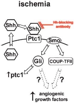
Pola et al          Hedgehog in Postnatal Skeletal Muscle Ischemia                             485

26. Shin D, Garcia-Cardena G, Hayashi S, et al. Expression of ephrinB2              32. Murakami S, Noda M. Expression of Indian hedgehog during fracture
    identifies a stable genetic difference between arterial and venous vascular         healing in adult rat femora. Calcif Tissue Int. 2000;66:272–276.
    smooth muscle as well as endothelial cells, and marks subsets of                33. Meng X, Shames BD, Pulido EJ, et al. Adrenergic induction of bimodal
    microvessels at sites of adult neovascularization. Dev Biol. 2001;230:              myocardial protection: signal transduction and cardiac gene repro-
    139 –150.                                                                           gramming. Am J Physiol. 1999;276:R1525–R1533.
27. Vu TH, Shipley JM, Bergers G, et al. MMP-9/gelatinase B is a key                34. Sharma M, Kambadur R, Matthews KG, et al. Myostatin, a transforming
    regulator of growth plate angiogenesis and apoptosis of hypertrophic                growth factor-beta superfamily member, is expressed in heart muscle and is
    chondrocytes. Cell. 1998;93:411– 422.                                               upregulated in cardiomyocytes after infarct. J Cell Physiol. 1999;180:1–9.
                                                                                    35. Dufourcq P, Couffinhal T, Ezan J, et al. FrzA, a secreted frizzled related
28. Mecklenburg L, Tobin DJ, Muller-Rover S, et al. Active hair growth
                                                                                        protein, displays a novel angiogenic pathway. Circulation. 2002;106:
    (anagen) is associated with angiogenesis. J Invest Dermatol. 2000;114:
                                                                                        3097–3103.
    909 –916.
                                                                                    36. Oosthuyse B, Moons L, Storkebaum E, et al. Deletion of the hypoxia-
29. Wang LC, Liu ZY, Gambardella L, et al. Conditional disruption of                    response element in the vascular endothelial growth factor promoter
    hedgehog signaling pathway defines its critical role in hair development            causes motor neuron degeneration. Nat Genet. 2001;28:131–138.
    and regeneration. J Invest Dermatol. 2000;114:901–908.                          37. Krishnan V, Pereira FA, Qiu Y, et al. Mediation of Sonic hedgehog-
30. Torok MA, Gardiner DM, Izpisua-Belmonte JC, et al. Sonic hedgehog                   induced expression of COUP-TFII by a protein phosphatase. Science.
    (shh) expression in developing and regenerating axolotl limbs. J Exp                1997;278:1947–1950.
    Zool. 1999;284:197–206.                                                         38. Pereira FA, Qiu Y, Zhou G, et al. The orphan nuclear receptor
31. Poss KD, Shen J, Nechiporuk A, et al. Roles of Fgf signaling during                 COUP-TFII is required for angiogenesis and heart development. Genes
    zebrafish fin regeneration. Dev Biol. 2000;222:347–358.                             Dev. 1999;13:1037–1049.

                                   Downloaded from http://circ.ahajournals.org/ by guest on September 22, 2015
Postnatal Recapitulation of Embryonic Hedgehog Pathway in Response to Skeletal Muscle
Ischemia
Roberto Pola, Leona E. Ling, Tamar R. Aprahamian, Elena Barban, Marta Bosch-Marce, Cynthia
Curry, Michael Corbley, Marianne Kearney, Jeffrey M. Isner and Douglas W. Losordo
Circulation. 2003;108:479-485; originally published online July 14, 2003;
doi: 10.1161/01.CIR.0000080338.60981.FA
Circulation is published by the American Heart Association, 7272 Greenville Avenue, Dallas, TX 75231
Copyright © 2003 American Heart Association, Inc. All rights reserved.
Print ISSN: 0009-7322. Online ISSN: 1524-4539
The online version of this article, along with updated information and services, is located on the
World Wide Web at:
http://circ.ahajournals.org/content/108/4/479
Permissions: Requests for permissions to reproduce figures, tables, or portions of articles originally published in
Circulation can be obtained via RightsLink, a service of the Copyright Clearance Center, not the Editorial Office.
Once the online version of the published article for which permission is being requested is located, click Request
Permissions in the middle column of the Web page under Services. Further information about this process is
available in the Permissions and Rights Question and Answer document.
Reprints: Information about reprints can be found online at:
http://www.lww.com/reprints
Subscriptions: Information about subscribing to Circulation is online at:
http://circ.ahajournals.org//subscriptions/
Downloaded from http://circ.ahajournals.org/ by guest on September 22, 2015You can also read