Estradiol Alters the Virulence Traits of Uropathogenic Escherichia coli - Frontiers
←
→
Page content transcription
If your browser does not render page correctly, please read the page content below
ORIGINAL RESEARCH
                                                                                                                                              published: 20 July 2021
                                                                                                                                    doi: 10.3389/fmicb.2021.682626

                                             Estradiol Alters the Virulence Traits
                                             of Uropathogenic Escherichia coli
                                             Ulrik Engelsöy 1 , Maria A. Svensson 1,2 and Isak Demirel 1,3*
                                             1
                                              School of Medical Sciences, Örebro University, Örebro, Sweden, 2 Department of Research and Education, Faculty
                                             of Medicine and Health, Örebro University, Örebro, Sweden, 3 Faculty of Medicine and Health, iRiSC–Inflammatory Response
                                             and Infection Susceptibility Centre, Örebro University, Örebro, Sweden

                                             Uropathogenic Escherichia coli (UPEC) is the most common bacteria to cause urinary
                                             tract infection (UTI). Postmenopausal women have an increased risk of recurrent UTI.
                                             This is partly explained by estrogenic effects on host defenses against UTI. Current
                                             research is mostly focused on how UPEC affects host factors, but not so much is known
                                             about how host factors like hormones affect UPEC virulence. The aim of the present
                                             study was to investigate the impact of estradiol exposure on the virulence of UPEC. We
                                             found that a postmenopausal concentration of estradiol increased CFT073 growth and
                                             biofilm formation, but not the premenopausal concentrations. Real-time qPCR showed
                                             that estradiol altered the expression of genes associated with the iron acquisition system
                          Edited by:         and metabolic pathways in CFT073. We also found that estradiol in a dose-dependent
                          Yuji Morita,
Meiji Pharmaceutical University, Japan
                                             manner increased the expression of fimH and papC adhesins and increased colonization
                        Reviewed by:
                                             and invasion of bladder epithelial cells. The premenopausal concentration of estradiol
                        Defne Gümüş,        also suppressed cytokine release from bladder epithelial cells. Additionally, we also
          Yeni Yüzyıl University, Turkey     showed using a Caenorhabditis elegans killing assay that estradiol increased the survival
                Kristin M. Burkholder,
           University of New England,        of CFT073-infected C. elegans worms. Taken together, our findings show that estradiol
                         United States       has the ability to alter the virulence traits of UPEC.
               Krishnendu Mukherjee,
University Hospital Münster, Germany         Keywords: uropathogenic Escherichia coli, estradiol, growth, virulence, cross-kingdom interaction

                  *Correspondence:
                         Isak Demirel
                  isak.demirel@oru.se       INTRODUCTION
                    Specialty section:      Urinary tract infection (UTI), the majority caused by uropathogenic Escherichia coli (E. coli), is
         This article was submitted to      one of the most widespread infections in humans. Approximately 50% of all women will suffer
                   Infectious Diseases,     from one UTI during their lifetime. Furthermore, women with a history of UTI have an increased
               a section of the journal     risk of recurrency within 3–4 months (Flores-Mireles et al., 2015). We know today that estrogen
             Frontiers in Microbiology
                                            is very important for the fight of the host against UTI (Lüthje et al., 2013, 2014). The female
           Received: 18 March 2021          body becomes more prone to UTI after menopause because lower estrogen levels lead to changes
            Accepted: 21 June 2021
                                            all along the urinary tract. Reduced levels of estrogen in postmenopausal women are associated
            Published: 20 July 2021
                                            with decreased proliferation of bladder epithelium (Blakeman et al., 2001) and lower levels of
                              Citation:     antimicrobial peptides (AMPs) (Lüthje et al., 2014). In addition, lower estrogen levels induce
       Engelsöy U, Svensson MA and
                                            vaginal mucosa atrophy, leading to a decreased number of lactobacilli and, hence, a higher pH. The
       Demirel I (2021) Estradiol Alters
 the Virulence Traits of Uropathogenic
                                            reduced number of lactobacilli and the higher pH are associated with an increased risk of urinary
                       Escherichia coli.    tract colonization by UPEC (Gupta et al., 1998). In the 1990s, several studies were conducted
          Front. Microbiol. 12:682626.      focusing on how to reduce recurrent UTI in menopausal women (Kjaergaard et al., 1990; Cardozo
     doi: 10.3389/fmicb.2021.682626         et al., 1998; Eriksen, 1999). One study investigated the use of topical estrogen cream vs. prophylactic

Frontiers in Microbiology | www.frontiersin.org                                    1                                           July 2021 | Volume 12 | Article 682626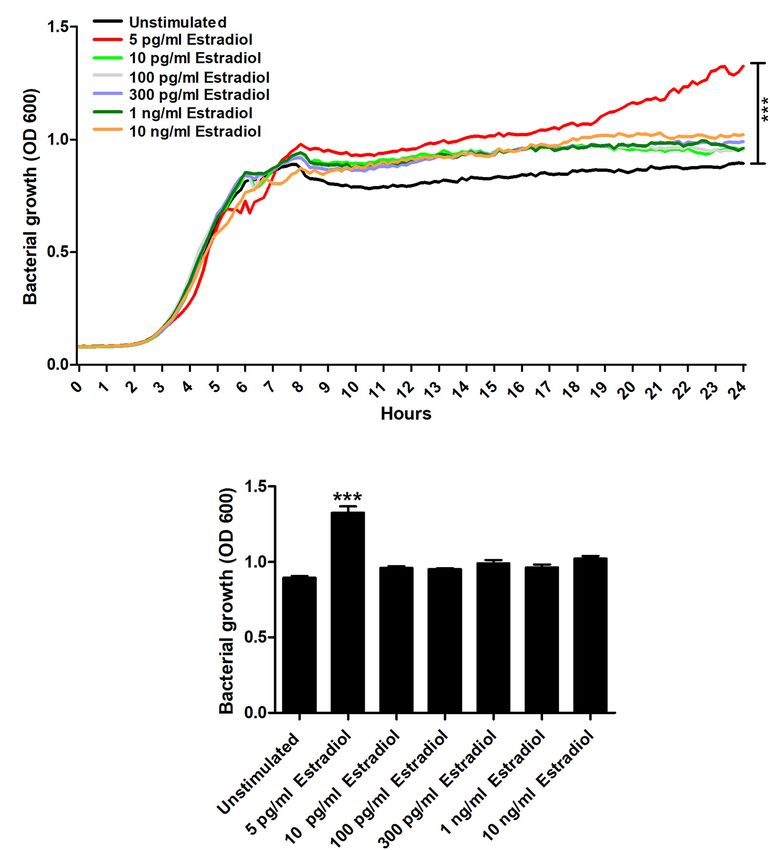
Engelsöy et al.                                                                                                       Estradiol Alters UPEC Virulence

antibiotics in postmenopausal females as a treatment against               (eGFP) expressing pLMB449 plasmid (Karunakaran et al.,
recurrency. The study found that the control group without any             2005) (kind gift from Professor Philip Poole at the University
treatment experienced an average of 5.9 UTI episodes per year.             of Oxford, Oxford, United Kingdom) was used for the
With estrogen treatment, the average dropped to 0.5 episodes               colonization experiments.
per year compared with 0.8 episodes per year with an antibiotic                The bladder epithelial cell line 5,637 is a commercial cell line
(Raz and Stamm, 1993). Hence, vaginal estrogen therapy was                 acquired from the American Type Culture Collection (Manassas,
shown to be able to reduce the number of episodes of UTI in                VA, United States). The cells were grown in Dulbecco’s modified
menopausal women.                                                          Eagle’s medium (DMEM) (Lonza, Basel, Switzerland) with 10%
   Most of the research conducted today in the field of host–              fetal bovine serum (FBS), 2 mM L-glutamine, 1 mM non-
pathogen interaction is focused on elucidating how pathogens,              essential amino acids (Thermo Fisher Scientific, Waltham, MA,
with their respective virulence factors, successfully modulate             United States) and incubated at 37◦ C and 5% CO2 atmosphere.
or evade the immune responses to cause infections. However,                The culture medium was changed during the experiments to
less is known about how host immune factors like cytokines                 DMEM with 2% FBS, 1 mM non-essential amino acids, and 2 mM
and hormones are affecting the virulence of UPEC by cross-                 L -glutamine.
kingdom interaction. The majority of the present research
conducted on UTI and estrogen has focused on its effects                   Growth Assessment
on the host and analysis of risk factors. Wang et al. (2013)               The UPEC strain CFT073 was grown in Lysogeny broth (Becton
have shown that mice with low estrogen levels have higher                  Dickinson) overnight on a shaker at 37◦ C prior to the growth
levels of bacteriuria compared with controls in a UTI model.               assay. CFT073 (1 × 10⁶ CFU/ml) was then grown in minimal
The effect of host factors (e.g., hormones and proinflammatory             salt medium [MSM, 0.3% KH2 PO4 , 1.3% (wt/vol) Na2 HPO4 ,
cytokines) on bacteria is a relatively unexplored field. What has          0.05% NaCl, and 0.1% NH4 Cl supplemented with 20 mM glucose,
been shown is that cytokines can bind to bacterial DNA and                 2 mM MgSO4 , 100 mM CaCl2 , and 0.25% casamino acids] with
alter gene expression in Neisseria meningitidis (Mahdavi et al.,           or without the presence of 17β-estradiol [5 pg/ml, 10 pg/ml,
2013). We have also shown that proinflammatory cytokines alter             100 pg/ml, 300 pg/ml, 1 ng/ml, and 10 ng/ml (E2758, Sigma-
UPEC virulence, leading to significantly decreased survival of             Aldrich, St. Louis, MO, United States)] in a 96-well plate. The
C. elegans worms (Engelsöy et al., 2019). Estrogen has been                96-well plate was then incubated at 37◦ C and the optical density
shown to enhance the growth and survival of several Gram-                  (600 nm) was measured every 10 min using a spectrophotometer
negative bacteria. The interaction of estradiol with Pseudomonas           (Cytation 3, Biotek Inc., Winooski, VT, United States).
aeruginosa results in an enhancement of its virulent mucoid
biofilm phenotype (Chotirmall et al., 2012). The mechanism
for this shift appears related to a P. aeruginosa constitutive
                                                                           Biofilm and Endotoxin Measurement
                                                                           After the growth assay, the same 96-well plate was used for
cytosolic estrogen-binding protein (Rowland et al., 1992). In
                                                                           evaluating biofilm formation after 24 h. The wells were washed
Chlamydia trachomatis, estradiol downregulates a significant
                                                                           with sterile RO water three times. Crystal violet (0.1%, Thermo
portion of genes involved in nucleotide metabolism and fatty
                                                                           Fisher Scientific) was added to the wells to stain the biofilm.
acid biosynthesis and upregulates genes associated with the
                                                                           Excess crystal violet was washed away with RO water and the
chlamydial stress response (Amirshahi et al., 2011). These studies
                                                                           plate was left to dry overnight. Ethanol (95%) was then used
strengthen the notion that estrogen has a direct effect on bacterial
                                                                           to dissolve the crystal violet and the solution was transferred to
virulence. A recent in vitro study investigated the effects of
                                                                           a new plate and the absorbance (540 nm) was measured by a
estrogen on E. coli growth and gene expression. They showed
                                                                           spectrophotometer (Cytation 3).
that estrogen increased the growth of E. coli (Gümüş et al., 2019).
                                                                              CFT073 was grown in MSM in the presence or absence
Hence, we know that there is a strong clinical association between
                                                                           of estradiol (5 and 300 pg/ml) statically at 37◦ C for 24 h.
estradiol levels and the development of a UPEC-mediated UTI.
                                                                           After 24 h of stimulation, the bacterial supernatants were
However, we do not know if the direct effects of estradiol on
                                                                           centrifuged for 5 min at 5,000×g and the endotoxin levels were
UPEC virulence could partially explain the association between
                                                                           analyzed using Pierce LAL chromogenic endotoxin quantitation
estrogen levels and UPEC-mediated UTI. The aim of this study
                                                                           kit (Thermo Fisher Scientific) according to the instructions of
was to investigate the impact of estrogen exposure on the
                                                                           the manufacturer.
virulence of UPEC.

                                                                           RNA Isolation, cDNA Generation, and
MATERIALS AND METHODS                                                      Quantitative Polymerase Chain Reaction
                                                                           CFT073 (1 × 10⁶ CFU/ml) was grown in MSM with or
Cell and Bacterial Culture                                                 without estradiol (5 and 300 pg/ml) statically at 37◦ C for
CFT073 is a UPEC strain isolated from a patient with                       6 or 24 h. RNAlater (Sigma-Aldrich) was used before RNA
pyelonephritis, which is fully genome sequenced (Welch                     isolation to stabilize the mRNA in CFT073. The E.Z.N.A Total     R

et al., 2002). The bacteria were kept on a tryptic soy agar                RNA Kit I (Omega Bio-tek, Inc., Norcross, GA, United States)
plate (Becton Dickinson, Franklin Lakes, NJ, United States).               was used according to the instructions of the manufacturer
CFT073 containing an enhanced green fluorescent protein                    for the isolation of total RNA. DNase digestion (TURBO

Frontiers in Microbiology | www.frontiersin.org                        2                                       July 2021 | Volume 12 | Article 682626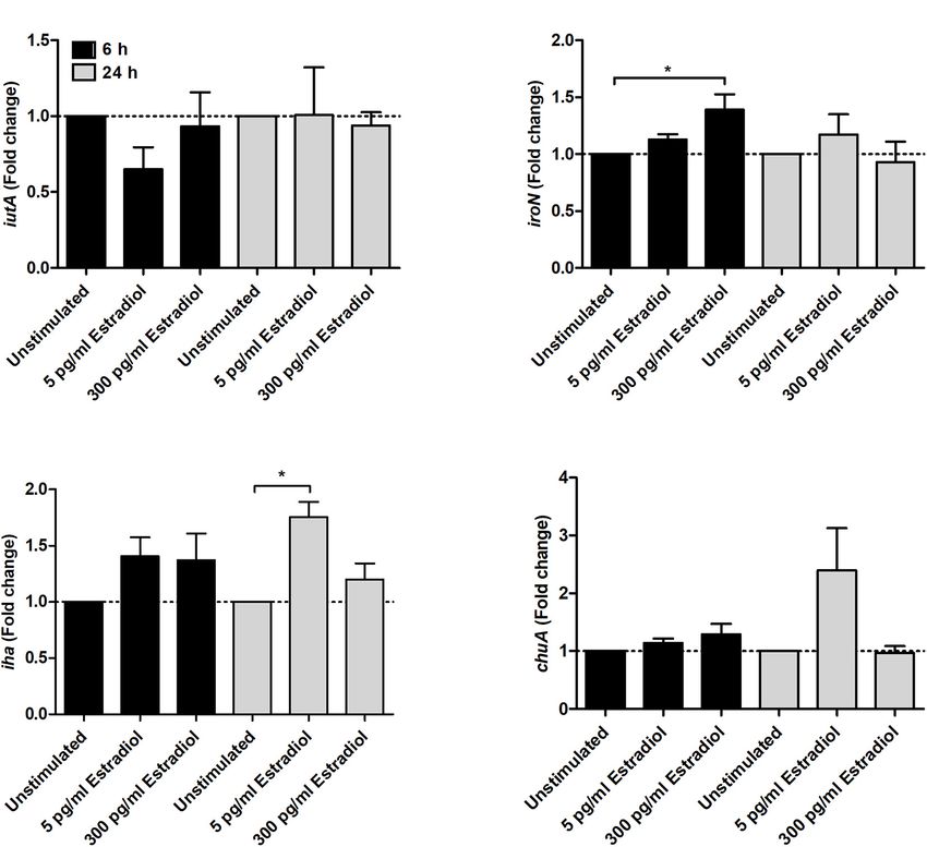
Engelsöy et al.                                                                                                                    Estradiol Alters UPEC Virulence

DNase, Life Technologies, Waltham, MA, United States) was                               plate reader. Colonization is presented as % mean fluorescence
conducted according to the instructions of the manufacturer to                          intensity (MFI) of CFT073.
minimize DNA contamination. RNA concentration and purity
were measured with a spectrophotometer (Nano Drop 2000,                                 Invasion Assay
Wilmington, NC, United States) before cDNA synthesis. The                               CFT073 was grown in MSM in the presence or absence of
cDNA synthesis was performed with 100 ng total RNA using                                estradiol (5 and 300 pg/ml) statically at 37◦ C for 24 h. Estradiol
the High-Capacity cDNA Reverse Transcription Kit (Applied                               was washed away from the bacteria with PBS and the bladder
Biosystems, Foster City, CA, United States). For the RT-                                epithelial cell line 5,637 (250,000 cells) was infected with the
qPCR, 5 ng cDNA and 250 nM of primer (Table 1) (Eurofins                                respective treatment at MOI of 100 for 2 h at 37◦ C and 5%
MWG Synthesis GmbH, Ebersberg, Munich, Germany) were                                    CO2 . Then, the cells were washed with PBS 10 times. DMEM
used with Maxima SYBR Green qPCR Master Mix (Thermo                                     supplemented with 2% FBS and 100 µg/ml gentamicin was then
Fisher Scientific). A CFX96 TouchTM Real-Time PCR Detection                             added to the cells to kill remaining extracellular bacteria during
System (Bio-Rad, Hercules, CA, United States) was used for                              2 h. The plate was then washed again three times and the cells
the amplification using the following protocol for 40 cycles:                           were lysed with 0.1% Triton X-100 in PBS (with calcium chloride
denaturation at 95◦ C for 15 s, annealing at 60◦ C for 30 s, and                        100 mg/L and magnesium chloride 100 mg/L) for 10 min under
extension at 72◦ C for 30 s. A dissociation curve between 60 and                        gentle rotation. Finally, the bacteria were plated on TSA plates
95◦ C was also done after the qPCR. CT values were obtained                             and incubated overnight at 37◦ C, and the colonies were counted
and the 11Ct method (2−11Ct ) was used to calculate the fold                            the next morning.
difference between groups. The results were normalized to the
endogenous control glyceraldehyde 3-phosphate dehydrogenase                             Cytokine Release and Viability Assay
A (gapA).                                                                               CFT073 was grown in MSM in the presence of absence of
                                                                                        estradiol (5 and 300 pg/ml) statically at 37◦ C for 24 h. Estradiol
Colonization Assay                                                                      was washed away from the bacteria with PBS and the bladder
                                                                                        epithelial cell line 5,637 (50,000 cells) was infected with the
CFT073 (harboring an enhanced GFP-expressing plasmid, eGFP)
                                                                                        respective treatment at MOI of 10 for 6 h at 37◦ C and 5% CO2 .
was grown in MSM (with gentamicin) in the presence of absence
                                                                                        Supernatants were collected after the infection and centrifuged
of estradiol (5 and 300 pg/ml) statically at 37◦ C for 24 h.
                                                                                        for 5 min at 5,000×g and stored at –80◦ C. An enzyme-linked
Estradiol was washed away from the bacteria with phosphate-
                                                                                        immunosorbent assay (ELISA) was performed to measure IL-
buffered saline (PBS) and the bladder epithelial cell line 5,637
                                                                                        1β and IL-8 release from the 5,637 cells. The cytokine was
(50,000 cells) was infected with the respective treatment at
                                                                                        measured with the IL-1β and IL-8 kits (ELISA MAX Deluxe
multiplicity of infection (MOI) of 10 for 4 h at 37◦ C and 5% CO2
                                                                                        Sets, BioLegend, San Diego, CA, United States) according to the
to measure bacterial colonization (adherent and intracellular
                                                                                        instructions of the kit. Cell viability after infection was assessed
bacteria). Then, the cells were washed with PBS 10 times and
                                                                                        by Pierce LDH cytotoxicity assay (Thermo Fisher Scientific)
the eGFP-expressing CFT073 was quantified with the Cytation 3
                                                                                        according to the instructions of the kit.

                                                                                        Caenorhabditis elegans Killing Assay
TABLE 1 | Primers used in the real-time qPCR.                                           The C. elegans wild-type Bristol strain N2 (Caenorhabditis
Gene symbol            Description          Oligonucleotide sequences   (50 –30 )
                                                                                        Genetics Center, University of Minnesota, United States)
                                                                                        (Brenner, 1974) was maintained on nematode growth medium
iutA               Siderophore receptor     F: AAAGAGCTGAAAGACGCACTGG                   plates seeded with E. coli OP50 (Brenner, 1974) (Caenorhabditis
                                            R: TGTCGGAACGTGAAGAGTTGAG
                                                                                        Genetics Center) at 21◦ C. Prior to the experiments, C. elegans
iroN               Siderophore receptor     F: ATTACCAAACGTCCCACCAACG
                                                                                        were synchronized (0.25 M NaOH, 1% HOCl) and maintained
                                            R: AAACGCGTGGTAAGAGCATCAC
                                                                                        on nematode growth medium plates for 48 h at 21◦ C to
iha                Siderophore receptor     F: TGCGAATAACCACTCTGGCTTC
                                            R: TAATCACAGAAACACTGGCGGC                   reach the L4 stage. CFT073 was grown in MSM in the
chuA                  Heme receptor         F: AAGGCGTTGCCCAATACCAGAGTA                 presence or absence of estradiol (5 and 300 pg/ml) statically at
                                            R: TATTCCGATCGCTCACAGTGGCTT                 37◦ C for 24 h. Estradiol was washed away from the bacteria
FimH                Adhesin subunit of      F: GTGCCAATTCCTCTTACCGTT                    and CFT073 (5 × 10⁸ CFU/ml) was transferred to a 96-
                     type 1 fimbriae        R: TGGAATAATCGTACCGTTGCG                    well plate. The L4 worms were washed with M9 buffer and
papC                Enables P-fimbriae      F: GTGGCAGTATGAGTAATGACCGTTA                10 worms were then transferred to the respective well of
                        assembly            R: ATATCCTTTCTGCAGGGATGCAATA                bacteria that had been grown in the presence or absence
pgi                 Glycolytic enzyme       F: CTCTGGCGAGAAGATCAACC                     of estradiol. After the addition of the worms to the wells,
                                            R: TCACCGGAAATAATCGCTTC
                                                                                        C. elegans and the bacteria were incubated together for 10 h
ppsA              Gluconeogenetic enzyme F: GCAAAACAGGCCGTACAAAT
                                         R: CAGCGTATAACGCTCCATGA
                                                                                        at 21◦ C. The viability of the worms was evaluated every hour.
frdA               Anaerobic respiratory    F: CAACACCGACCTGCTCTACA
                                                                                        A worm was considered dead when it failed to respond to
                         enzyme             R: GCGGCAGCGTAGTAATCTTC                     touch. Dead worms were also visualized with 1 µM SYTOX
gapA               Endogenous control       F: AAGTTGGTGTTGACGTTG                       Green (Thermo Fisher Scientific) using a spectrophotometer
                    (glycolytic enzyme)     R: AGCGCCTTTAACGAACATCG                     (Cytation 3) (Gill et al., 2003).

Frontiers in Microbiology | www.frontiersin.org                                     3                                       July 2021 | Volume 12 | Article 682626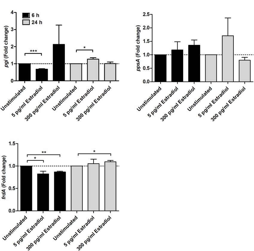
Engelsöy et al.                                                                                                                      Estradiol Alters UPEC Virulence

Statistical Methods                                                                  significant growth increase compared with unstimulated CFT073
Student’s unpaired t-test was used to analyze the difference                         (Figures 1A,B).
between stimulated CFT073 and unstimulated CFT073. Data are
expressed as mean ± SEM. Results were considered statistically                       Biofilm Formation and Endotoxin
significant at p < 0.05. n is equal to the number of independent                     Release
biological experiments.                                                              We continued by investigating the effects of estradiol on biofilm
                                                                                     formation and endotoxin release. The biofilm formation was
                                                                                     significantly increased by 5 pg/ml estradiol compared with
                                                                                     unstimulated CFT073 after 24 h. The higher concentrations
RESULTS
                                                                                     of estradiol did not differ from unstimulated CFT073 in their
                                                                                     ability to induce biofilm formation (Figure 2A). When looking
Estradiol Induces Increased UPEC                                                     at endotoxin levels, we found no significant changes induced by
Growth                                                                               estradiol compared with unstimulated CFT073 (Figure 2B).
We began by evaluating the effects estradiol had on the growth
of CFT073. We found that 5 pg/ml of estradiol significantly                          Estradiol Alters the Expression Levels of
increased the growth of CFT073 compared with unstimulated
CFT073 (Figure 1A). Statistical significance (p < 0.05) was
                                                                                     Genes Encoding Different Metabolic
reached from 8 h onward for 5 pg/ml estradiol. The biggest                           Pathways, Fimbriae, and Iron Acquisition
growth difference was observed between 5 pg/ml estradiol                             Systems
and unstimulated CFT073 after 24 h (Figure 1B). However,                             We investigated the effects of estradiol on virulence-associated
the higher concentrations of estradiol did not induce a                              genes in UPEC. We found that the gene expression related to

  FIGURE 1 | CFT073 growth with or without the presence of estradiol (5 pg/ml, 10 pg/ml, 100 pg/ml, 300 pg/ml, 1 ng/ml, and 10 ng/ml) during 24 h (A) and at 24 h
  (B). Data are presented as mean (A) and mean ± SEM (B) of n = 3 independent experiments. The asterisk distinguishes statistical significance: ***p < 0.001 vs.
  unstimulated CFT073.

Frontiers in Microbiology | www.frontiersin.org                                  4                                            July 2021 | Volume 12 | Article 682626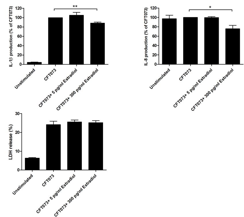
Engelsöy et al.                                                                                                                       Estradiol Alters UPEC Virulence

  FIGURE 2 | Biofilm formation (A) and endotoxin release (B) in the presence or absence of estradiol after 24 h. Data are presented as mean ± SEM of n = 3
  independent experiments. The asterisk distinguishes statistical significance: *p < 0.05 vs. unstimulated CFT073.

  FIGURE 3 | Real-time qPCR analysis of pgi (A), ppsA (B), and frdA (C) gene expression in the presence or absence of estradiol (5 and 300 pg/ml) at 6 and 24 h.
  Data are presented as mean ± SEM of n = 3 independent experiments. The asterisks distinguish statistical significance: *p < 0.05; **p < 0.01; ***p < 0.001 vs.
  unstimulated CFT073.

energy metabolism was altered. Estradiol at 5 pg/ml significantly                    trend toward an increased expression was found compared with
changed the expression of pgi at both 6 and 24 h compared                            unstimulated CFT073 (Figure 3B). As for frdA, the expression
with unstimulated CFT073 (Figure 3A). At 6 h, the expression                         was significantly decreased at 6 h by 5 and 300 pg/ml estradiol. At
was decreased, and at 24 h, an increase was observed. The                            24 h, a slight increased expression induced by 300 pg/ml estradiol
expression of ppsA showed no significant changes, but a possible                     was seen (Figure 3C).

Frontiers in Microbiology | www.frontiersin.org                                  5                                             July 2021 | Volume 12 | Article 682626
Engelsöy et al. Estradiol Alters UPEC Virulence
Regarding iron acquisition-associated genes, we found that seems to increase with estradiol-stimulated CFT073 in a dose-
300 pg/ml estradiol increased the expression of iroN at 6 h response manner; 300 pg/ml estradiol mediated a significantly
(Figure 4B) and 5 pg/ml estradiol increased the expression of increased colonization compared with unstimulated CFT073
iha at 24 h (Figure 4C) compared with unstimulated CFT073. (Figures 6A,C). We also found that estradiol increased the
However, estradiol did not induce any significant changes in the invasion capability of CFT073, at MOI 100, in a dose-dependent
expression of the iutA (Figure 4A) or ChuA genes (Figure 4D). manner; 300 pg/ml estradiol mediated a significantly increased
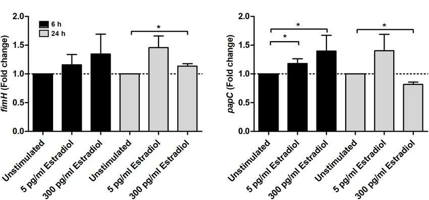
For adhesion-associated genes, the expression of papC was invasion of bladder epithelial cells compared with unstimulated
significantly increased at 6 h by both 5 and 300 pg/ml CFT073 (Figure 6B).
estradiol compared with unstimulated CFT073. After 24 h, papC
expression was significantly decreased by 300 pg/ml estradiol Altered Cytokine Release Induced by
(Figure 5B). The fimH expression was increased by estradiol at Estradiol
6 h but significance was not reached. However, the expression of We continued with evaluating if estradiol-stimulated CFT073,
fimH was significantly increased by 300 pg/ml estradiol at 24 h at MOI 10, could alter the release of IL-1β and IL-8 from
compared with unstimulated CFT073 (Figure 5A). bladder epithelial cells. We found that the cytokine release was
significantly lowered by CFT073 stimulated with 300 pg/ml
Colonization and Invasion of Bladder estradiol for both IL-1β and IL-8 compared with unstimulated
Epithelial Cells CFT073 (Figures 7A,B). No difference in IL-1β and IL-8
We proceeded with evaluating the effects of estradiol on the release was observed for CFT073 stimulated with 5 pg/ml
ability of UPEC to colonize (adhere and invade) and invade estradiol (Figures 7A,B). Furthermore, we did not find any
human bladder epithelial cells. Bacterial colonization, at MOI 10, differences in LDH release from bladder epithelial infected
FIGURE 4 | Real-time qPCR analysis of iutA (A), iroN (B), iha (C), and chuA (D) gene expression in the presence or absence of estradiol (5 and 300 pg/ml) at 6 and
24 h. Data are presented as mean ± SEM of n = 3 independent experiments. The asterisks distinguish statistical significance: *p < 0.05 vs. unstimulated CFT073.
Frontiers in Microbiology | www.frontiersin.org 6 July 2021 | Volume 12 | Article 682626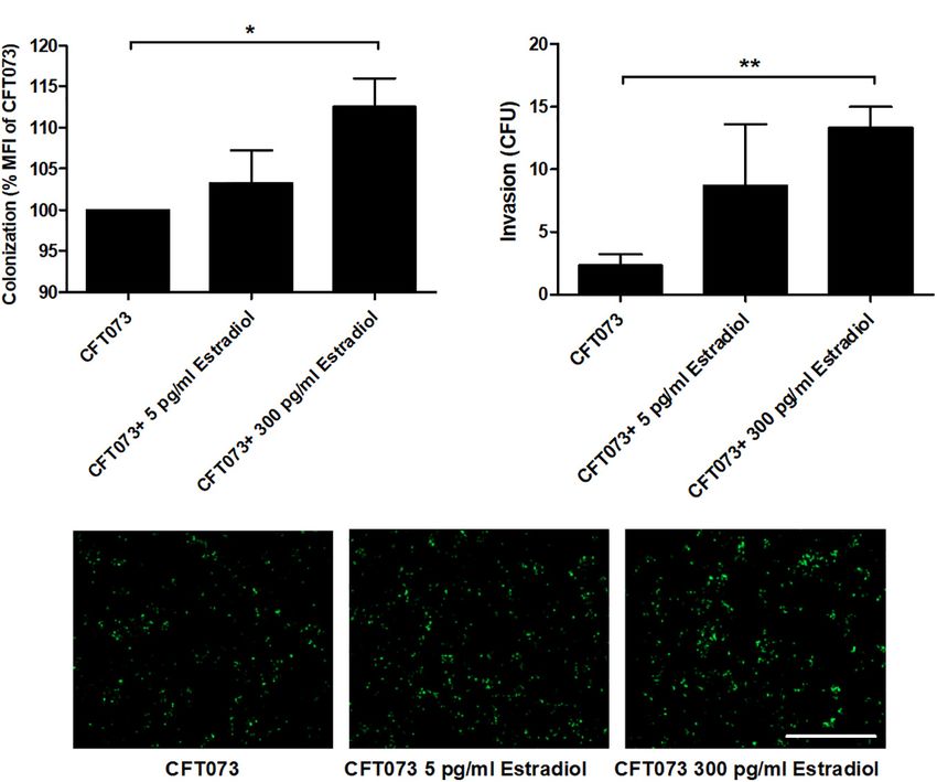
Engelsöy et al.                                                                                                                           Estradiol Alters UPEC Virulence

  FIGURE 5 | Real-time qPCR analysis of fimH (A) and papC (B) gene expression in the presence or absence of estradiol (5 and 300 pg/ml) at 6 and 24 h. Data are
  presented as mean ± SEM of n = 3 independent experiments. The asterisks distinguish statistical significance: *p < 0.05 vs. unstimulated CFT073.

  FIGURE 6 | CFT073 colonization (A,C, 4 h) or invasion (B, 2 h) of bladder epithelial cells after CFT073 pretreated with or without estradiol (5 and 300 pg/ml). CFT073
  (harboring a GFP-expressing plasmid) colonization was quantified as mean fluorescence intensity (MFI) (A) and imaged (C). Data are presented as mean ± SEM of
  n = 3 independent experiments. The asterisk distinguishes statistical significance: *p < 0.05, **p < 0.01 vs. unstimulated CFT073. Scale bar: 200 µm.

with estradiol-stimulated CFT073 compared with unstimulated                             model. This was done to evaluate the combined significance
CFT073 (Figure 7C).                                                                     our findings had on the virulence of CFT073. We found that
                                                                                        CFT073 in the presence of 5 and 300 pg/ml estradiol significantly
                                                                                        increased the survival of C. elegans worms compared with
Reduced CFT073 Toxicity in the                                                          unstimulated CFT073 (Figure 8A). The dead worms were also
Presence of Estradiol                                                                   visualized by the uptake of SYTOX Green (Figures 8B–D).
We proceeded with evaluating the virulence of CFT073 in                                 Estradiol per se did not induce any C. elegans toxicity
the presence of estradiol with an in vivo C. elegans infection                          (data not shown).

Frontiers in Microbiology | www.frontiersin.org                                     7                                              July 2021 | Volume 12 | Article 682626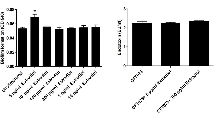
Engelsöy et al.                                                                                                                          Estradiol Alters UPEC Virulence

  FIGURE 7 | IL-1β (A), IL-8 (B), and LDH (C) release from bladder epithelial cells infected with CFT073 pretreated with or without estradiol (5 and 300 pg/ml) at 6 h.
  Data are presented as mean ± SEM of n = 4 independent experiments. The asterisks distinguish statistical significance: *p < 0.05 and **p < 0.01 vs. unstimulated
  CFT073.

DISCUSSION                                                                             we focused on investigating the cross-kingdom effects of estradiol
                                                                                       on the virulence of UPEC.
Understanding how UPEC interacts and modulates the human                                  Our findings showed that 5 pg/ml of estradiol
host in order to colonize the urinary tract is becoming more                           (postmenopausal concentration) (Stanczyk and Clarke, 2014)
important in an age where antibiotic resistance is increasing                          significantly increased the growth of the UPEC strain CFT073.
worldwide. Evidence exists showing that human factors (e.g.,                           However, the premenopausal concentrations of estradiol
cytokines and hormones) synthesized and released by our cells                          (100–300 pg/ml) (Stanczyk and Clarke, 2014) did not induce
can activate the virulence of different bacteria through bacterial                     a growth increase. Previous studies have shown that estradiol
sensors (Mahdavi et al., 2013). The bacterial sensors have a                           can increase the growth of several Gram-negative bacteria
dual function: recognition of the complex host environment and                         (Mahdavi et al., 2013; Wang et al., 2013), but we found that
transduction of the message to initiate bacterial adaptation. It is                    this growth increase was associated with postmenopausal, and
now evident that bacterial virulence is regulated by the detection                     not premenopausal, concentrations of estradiol. These findings
of host factors released in the microenvironment like hormones                         could be a contributing factor explaining why women are more
and cytokines (Mahdavi et al., 2013; Engelsöy et al., 2019; Gümüş                     prone to UTI after menopause, as bacterial growth is strongly
et al., 2019). We know today that estrogen is very important for                       associated with UTI (Anderson et al., 2004).
the protection of the urinary tract against UPEC (Eriksen, 1999;                          We also found that biofilm formation was significantly
Lüthje et al., 2014). The female body becomes more prone to                            increased by the postmenopausal concentration of estradiol
UTI after menopause because lower estrogen levels lead to lower                        (5 pg/ml), but not by the premenopausal. Others have also shown
levels of antimicrobial peptides, vaginal mucosa atrophy, and a                        that estradiol has the ability to increase the biofilm formation of
higher pH (Gupta et al., 1998; Lüthje et al., 2014). In this study,                    Gram-negative bacteria (Wang et al., 2013; Fteita et al., 2014).

Frontiers in Microbiology | www.frontiersin.org                                    8                                              July 2021 | Volume 12 | Article 682626
Engelsöy et al. Estradiol Alters UPEC Virulence FIGURE 8 | CFT073 mediated C. elegans killing in the presence or absence of estradiol (5 and 300 pg/ml) (A). Data are presented as mean ± SEM of n = 3 independent experiments. Statistical significance is denoted with asterisks: ***p < 0.001 vs. unstimulated CFT073. Immunofluorescence staining of dead worm with 1 µM SYTOX Green (B–D). Scale bar: 1,000 µm. Biofilm formation is associated with protection of UPEC from show that estradiol has the ability to modify the expression of antimicrobial agents, environmental conditions, and the host various metabolic pathways in UPEC. However, further studies immune response (Mah and O’Toole, 2001; Hatt and Rather, are needed to understand the link between estradiol and the 2008). We also investigated if estradiol could alter the expression metabolic activity of UPEC. of UPEC lipopolysaccharide (LPS), which is known to activate Iron is a critical nutrient for UPEC survival and pathogenicity and modulate the host immune response during a UTI. However, in the urinary tract (Subashchandrabose and Mobley, 2015). no differences in the expression of LPS were found in the As soluble iron levels are low in the urinary tract, acquisition presence of estradiol. Taken together, we have shown that only of iron is essential for the pathogenicity of UPEC (Roos and the postmenopausal concentration of estradiol induces increased Klemm, 2006). Acquisition of iron is facilitated by numerous bacterial growth and biofilm formation, which may promote mechanisms such as outer membrane receptors for heme, ferrous UPEC persistence in the urinary tract of postmenopausal women. iron transporters, and via ferric iron chelators called siderophores We continued with investigating the effects of estradiol on (Robinson et al., 2018). We observed that estradiol increased the metabolic pathways of UPEC. We showed that the gene the expression of the salmochelin uptake receptor IroN and the expression of pgi (encoding for glucose-6-phosphate isomerase), enterobactin uptake receptor Iha (Yep et al., 2014). Upregulation a glycolytic enzyme, was decreased by 5 pg/ml estradiol of the uptake receptors may indicate that estradiol alters the after 6 h and increased after 24 h. We also found that iron acquisition systems in UPEC, which may have an effect on ppsA (encoding for phosphoenolpyruvate synthase, an enzyme UPEC pathogenicity. involved in gluconeogenesis) showed a possible trend toward an The capacity of UPEC to adhere and invade bladder increased expression induced by estradiol. Studies have found epithelial cells is essential for the colonization of the urinary that tricarboxylic acid cycle (TCA) and gluconeogenesis, but not tract. We found that the gene expression of fimH (type-1 glycolysis, are essential for the fitness of UPEC in mice (Alteri fimbriae) and papC (P-fimbriae) was upregulated by both et al., 2009; Subashchandrabose and Mobley, 2015). We also 5 and 300 pg/ml estradiol. In addition, we also observed know that UPEC uses predominantly amino acids and peptides a dose-dependent increased colonization and invasion of in urine as carbon sources instead of glucose (Alteri et al., 2009). bladder epithelial cells mediated by estradiol exposure. If Furthermore, we also found that frdA (encoding for the catalytic we factor in estrogenic effects on bladder epithelium, it subunit of fumarate reductase), an enzyme used for anaerobic makes these findings even stronger. Premenopausal levels of respiration, was altered and downregulated by estradiol after estrogen are associated with several differences in bladder 6 h. This indicated that aerobic respiration was preferred in the epithelium compared with lower postmenopausal levels. presence of estradiol at an early timepoint. Alteri and Mobley Higher antimicrobial peptide production, stronger tight (2015) have previously shown that fumarate reductase is not junctions between the epithelial cells, and seemingly increased essential for the fitness of UPEC. Taken together, these data expression of uroplakins and β1-integrins are linked to the Frontiers in Microbiology | www.frontiersin.org 9 July 2021 | Volume 12 | Article 682626
Engelsöy et al.                                                                                                                               Estradiol Alters UPEC Virulence

higher, premenopausal levels of estrogen (Lüthje et al., 2013).                            to be generalizable to UPEC in general, an evaluation of other
β1-Integrins and uroplakins interact with the type-1 fimbriae                              strains than CFT073 is needed. Another limitation of the study is
adhesin fimH and are important for CFT073 invasion into                                    that additional concentrations lower than 5 pg/ml estradiol would
bladder epithelial cells (Eto et al., 2007). Hence, an initial stronger                    be interesting to investigate. A general limitation of the study is
adhesion would lead to increased invasion. In addition, as higher                          that we focus on the direct effects of estradiol on UPEC virulence
extracellular antimicrobial peptide levels are associated with                             in vitro, without considering the in vivo state of multiple host
premenopausal levels of estrogen, the increased colonization and                           factors and cells interacting with UPEC.
invasion of UPEC at 300 pg/ml could be an antimicrobial peptide                                Understanding how the human host affects the virulence of
evasion strategy.                                                                          UPEC may be a new frontier in the fight against infection. If we
   We continued to investigate if CFT073 exposed to estrogen                               can elucidate how UPEC senses its environment and mobilizes
could alter the cytokine release from bladder epithelial cells.                            its virulence, we may inhibit this activation and dampen or
We found that 300 pg/ml estradiol but not 5 pg/ml reduced                                  completely prevent the infection. By focusing of inhibiting
the ability of CFT073 to induce IL-1β and IL-8 from bladder                                virulence, we reduce antibiotic selection pressure (Brannon and
epithelial cells. This is not an effect of decreased cell viability as no                  Hadjifrangiskou, 2016), which will lead to reduced antibiotic
increased LDH release was observed. This very interesting finding                          resistance. We have seen that a postmenopausal concentration
supports the hypothesis of host defense avoidance. IL-1β and IL-                           of estradiol both increased the growth and biofilm formation,
8 are very important cytokines during a UTI. Both are important                            which may be a contributing factor as to why women are more
for neutrophil recruitment, which is the primary immune cell                               prone to UTI after menopause. However, the premenopausal
that clears the infection (Hedges and Svanborg, 1994; Godaly                               concentration of estradiol mediated the increased bacterial
et al., 1997). Taken together, we have shown that 300 pg/ml                                colonization and suppression of proinflammatory cytokines. Our
estradiol increases the colonization of bladder epithelial cells and                       study suggests that the evasion mechanisms induced by UPEC
strengthens the evasion strategies of CFT073.                                              are an adaptation to the primed immune responses associated
   An in vivo C. elegans infection model was used to comprehend                            with high estradiol levels. Although we have found that estradiol
how the individual virulence alteration induced by estradiol                               can change the virulence of UPEC, further research is needed
contributes to the total cytotoxicity of CFT073. We and others                             to understand the mechanism behind these findings and what
have used C. elegans previously to evaluate the virulence of                               clinical significance they may have.
UPEC in vivo. It was shown that there is a significant correlation
between the virulence of UPEC in a murine model and in
C. elegans (Diard et al., 2007). We showed that CFT073 in
the presence of pre- and postmenopausal concentrations of
                                                                                           DATA AVAILABILITY STATEMENT
estradiol significantly increased the survival of C. elegans. As                           The raw data supporting the conclusions of this article will be
the estradiol is removed prior to infecting the worms, no                                  made available by the authors, without undue reservation.
alteration in bacterial growth is observed to explain the reduced
cytotoxicity. In addition, we did not observe any difference in
α-hemolysins activity from CFT073 in the presence of estradiol
(data not shown) that could explain our data. However, there                               AUTHOR CONTRIBUTIONS
are a number of CFT073 virulence factors that we have not
explored like the vacuolating autotransporter toxin (Vat) and                              UE, MS, and ID design the study, analyzed the data, and drafted
secreted autotransporter toxin (Sat). Both Vat and Sat have been                           the article. UE and ID conducted the experiments. All authors
shown to induce tissue damage (Wiles et al., 2008; Engelsöy et al.,                        read and approved the final manuscript.
2019). Exploring the effects of estradiol on these toxins may give
us a better understanding of why the cytotoxicity of CFT073 is
reduced in the presence of estradiol. Taken together, we have                              FUNDING
shown that estradiol at pre- and postmenopausal concentrations
decreases the total cytotoxicity of CFT073.                                                This project was financially supported by the Faculty of Medicine
   The limitations of this study include the number of UPEC                                and Health at Örebro University and by the Research Committee
strains used and the in vitro experimental setup. For the results                          of Örebro County Council.

REFERENCES                                                                                    the sex hormones estradiol and progesterone. BMC Microbiol. 11:150. doi:
                                                                                              10.1186/1471-2180-11-150
Alteri, C. J., and Mobley, H. L. T. (2015). Metabolism and fitness of urinary tract        Anderson, G. G., Dodson, K. W., Hooton, T. M., and Hultgren, S. J. (2004).
   pathogens. Microbiol. Spectr. 3. doi: 10.1128/microbiolspec.MBP-0016-2015                  Intracellular bacterial communities of uropathogenic Escherichia coli in urinary
Alteri, C. J., Smith, S. N., and Mobley, H. L. (2009). Fitness of Escherichia coli            tract pathogenesis. Trends Microbiol. 12, 424–430. doi: 10.1016/j.tim.2004.07.
   during urinary tract infection requires gluconeogenesis and the TCA cycle.                 005
   PLoS Pathog. 5:e1000448. doi: 10.1371/journal.ppat.1000448                              Blakeman, P. J., Hilton, P., and Bulmer, J. N. (2001). Cellular proliferation in
Amirshahi, A., Wan, C., Beagley, K., Latter, J., Symonds, I., and Timms, P. (2011).           the female lower urinary tract with reference to oestrogen status. BJOG 108,
   Modulation of the Chlamydia trachomatis in vitro transcriptome response by                 813–816. doi: 10.1016/s0306-5456(00)00210-2

Frontiers in Microbiology | www.frontiersin.org                                       10                                               July 2021 | Volume 12 | Article 682626
Engelsöy et al. Estradiol Alters UPEC Virulence Brannon, J. R., and Hadjifrangiskou, M. (2016). The arsenal of pathogens and reporter proteins for studying gene expression in gram-negative bacteria. antivirulence therapeutic strategies for disarming them. Drug Des. Devel. Ther. Microbiology (Reading) 151, 3249–3256. doi: 10.1099/mic.0.28311-0 10, 1795–1806. doi: 10.2147/dddt.s98939 Kjaergaard, B., Walter, S., Knudsen, A., Johansen, B., and Barlebo, H. (1990). Brenner, S. (1974). The genetics of Caenorhabditis elegans. Genetics 77, 71–94. [Treatment with low-dose vaginal estradiol in post-menopausal women. doi: 10.1093/genetics/77.1.71 A double-blind controlled trial]. Ugeskr. Laege 152, 658–659. Cardozo, L., Benness, C., and Abbott, D. (1998). Low dose oestrogen prophylaxis Lüthje, P., Brauner, H., Ramos, N. L., Ovregaard, A., Gläser, R., Hirschberg, A. L., for recurrent urinary tract infections in elderly women. BJOG 105, 403–407. et al. (2013). Estrogen supports urothelial defense mechanisms. Sci. Transl. Med. doi: 10.1111/j.1471-0528.1998.tb10124.x 5:190ra180. Chotirmall, S. H., Smith, S. G., Gunaratnam, C., Cosgrove, S., Dimitrov, B. D., Lüthje, P., Hirschberg, A. L., and Brauner, A. (2014). Estrogenic action on innate O’neill, S. J., et al. (2012). Effect of estrogen on Pseudomonas mucoidy and defense mechanisms in the urinary tract. Maturitas 77, 32–36. doi: 10.1016/j. exacerbations in cystic fibrosis. N. Engl. J. Med. 366, 1978–1986. doi: 10.1056/ maturitas.2013.10.018 nejmoa1106126 Mah, T. F., and O’Toole, G. A. (2001). Mechanisms of biofilm resistance to Diard, M., Baeriswyl, S., Clermont, O., Gouriou, S., Picard, B., Taddei, antimicrobial agents. Trends Microbiol. 9, 34–39. doi: 10.1016/s0966-842x(00) F., et al. (2007). Caenorhabditis elegans as a simple model to study 01913-2 phenotypic and genetic virulence determinants of extraintestinal pathogenic Mahdavi, J., Royer, P.-J., Sjölinder, H. S., Azimi, S., Self, T., Stoof, J., et al. (2013). Escherichia coli. Microbes Infect. 9, 214–223. doi: 10.1016/j.micinf.2006. Pro-inflammatory cytokines can act as intracellular modulators of commensal 11.009 bacterial virulence. Open Biol. 3:130048. doi: 10.1098/rsob.130048 Engelsöy, U., Rangel, I., and Demirel, I. J. F. I. M. (2019). Impact of Raz, R., and Stamm, W. E. (1993). A controlled trial of intravaginal estriol in proinflammatory cytokines on the virulence of uropathogenic Escherichia coli. postmenopausal women with recurrent urinary tract infections. N. Engl. J. Med. Front. Microbiol. 10:1051. doi: 10.3389/fmicb.2019.01051 329, 753–756. doi: 10.1056/nejm199309093291102 Eriksen, B. C. (1999). A randomized, open, parallel-group study on the preventive Robinson, A. E., Heffernan, J. R., and Henderson, J. P. (2018). The iron hand of effect of an estradiol-releasing vaginal ring (Estring) on recurrent urinary tract uropathogenic Escherichia coli: the role of transition metal control in virulence. infections in postmenopausal women. Am. J. Obstet. Gynecol. 180, 1072–1079. Future Microbiol. 13, 745–756. doi: 10.2217/fmb-2017-0295 doi: 10.1016/s0002-9378(99)70597-1 Roos, V., and Klemm, P. (2006). Global gene expression profiling of the Eto, D. S., Jones, T. A., Sundsbak, J. L., and Mulvey, M. A. (2007). Integrin- asymptomatic bacteriuria Escherichia coli strain 83972 in the human urinary mediated host cell invasion by type 1–piliated uropathogenic Escherichia coli. tract. Infect. Immun. 74, 3565–3575. doi: 10.1128/iai.01959-05 PLoS Pathog. 3:e100. doi: 10.1371/journal.ppat.0030100 Rowland, S. S., Falkler, W. A., and Bashirelahi, N. (1992). Identification of an Flores-Mireles, A. L., Walker, J. N., Caparon, M., and Hultgren, S. J. (2015). Urinary estrogen-binding protein in Pseudomonas aeruginosa. J. Steroid Biochem. Mol. tract infections: epidemiology, mechanisms of infection and treatment options. Biol. 42, 721–727. Nat. Rev. Microbiol. 13, 269–284. doi: 10.1038/nrmicro3432 Stanczyk, F. Z., and Clarke, N. J. (2014). Measurement of estradiol–challenges Fteita, D., Könönen, E., Söderling, E., and Gürsoy, U. K. (2014). Effect of estradiol ahead. J. Clin. Endocrinol. Metab. 99, 56–58. doi: 10.1210/jc.2013-2905 on planktonic growth, coaggregation, and biofilm formation of the Prevotella Subashchandrabose, S., and Mobley, H. L. (2015). Virulence and fitness intermedia group bacteria. Anaerobe 27, 7–13. doi: 10.1016/j.anaerobe.2014.02. determinants of uropathogenic Escherichia coli. Microbiol. Spectr 3. doi: 10. 003 1128/microbiolspec.UTI-0015-2012 Gill, M. S., Olsen, A., Sampayo, J. N., and Lithgow, G. J. (2003). An Wang, C., Symington, J. W., Ma, E., Cao, B., and Mysorekar, I. U. (2013). automated high-throughput assay for survival of the nematode Caenorhabditis Estrogenic modulation of uropathogenic Escherichia coli infection pathogenesis elegans. Free Radic. Biol Med. 35, 558–565. doi: 10.1016/s0891-5849(03)003 in a murine menopause model. Infect. Immun. 81, 733–739. doi: 10.1128/iai. 28-9 01234-12 Godaly, G., Proudfoot, A. E., Offord, R. E., Svanborg, C., and Agace, Welch, R. A., Burland, V., Plunkett, G., Redford, P., Roesch, P., Rasko, D., et al. W. W. (1997). Role of epithelial interleukin-8 (IL-8) and neutrophil (2002). Extensive mosaic structure revealed by the complete genome sequence IL-8 receptor A in Escherichia coli-induced transuroepithelial neutrophil of uropathogenic Escherichia coli. Proc. Natl. Acad. Sci. U.S.A. 99, 17020–17024. migration. Infect. Immun. 65, 3451–3456. doi: 10.1128/iai.65.8.3451-3456. Wiles, T. J., Kulesus, R. R., and Mulvey, M. A. (2008). Origins and virulence 1997 mechanisms of uropathogenic Escherichia coli. Exp. Mol. Pathol. 85, 11–19. Gümüş, D., Kalaycı Yüksek, F., Sefer, Ö, Yörük, E., Uz, G., and Anğ Küçüker, M. doi: 10.1016/j.yexmp.2008.03.007 (2019). The roles of hormones in the modulation of growth and virulence genes’ Yep, A., Mcquade, T., Kirchhoff, P., Larsen, M., and Mobley, H. L. T. (2014). expressions in UPEC strains. Microb. Pathog. 132, 319–324. doi: 10.1016/j. Inhibitors of TonB function identified by a high-throughput screen for micpath.2019.05.019 inhibitors of iron acquisition in uropathogenic Escherichia coli CFT073. Mbio Gupta, K., Stapleton, A. E., Hooton, T. M., Roberts, P. L., Fennell, C. L., and Stamm, 5:e01089-13. W. E. (1998). Inverse association of H2O2-producing lactobacilli and vaginal Escherichia coli colonization in women with recurrent urinary tract infections. Conflict of Interest: The authors declare that the research was conducted in the J. Infect. Dis. 178, 446–450. doi: 10.1086/515635 absence of any commercial or financial relationships that could be construed as a Hatt, J. K., and Rather, P. N. (2008). Role of bacterial biofilms in urinary tract potential conflict of interest. infections. Curr. Top Microbiol. Immunol. 322, 163–192. doi: 10.1007/978-3- 540-75418-3_8 Copyright © 2021 Engelsöy, Svensson and Demirel. This is an open-access article Hedges, S., and Svanborg, C. (1994). The mucosal cytokine response to urinary distributed under the terms of the Creative Commons Attribution License (CC BY). tract infections. Int. J. Antimicrob. Agents. 4, 89–93. doi: 10.1016/0924- The use, distribution or reproduction in other forums is permitted, provided the 8579(94)90039-6 original author(s) and the copyright owner(s) are credited and that the original Karunakaran, R., Mauchline, T. H., Hosie, A. H. F., and Poole, P. S. (2005). A family publication in this journal is cited, in accordance with accepted academic practice. No of promoter probe vectors incorporating autofluorescent and chromogenic use, distribution or reproduction is permitted which does not comply with these terms. Frontiers in Microbiology | www.frontiersin.org 11 July 2021 | Volume 12 | Article 682626
You can also read