Assessing nucleic acid binding activity of four dinoflagellate cold shock domain proteins from Symbiodinium kawagutii and Lingulodinium polyedra
←
→
Page content transcription
If your browser does not render page correctly, please read the page content below
Zaheri and Morse BMC Molecular and Cell Biology                (2021) 22:27
https://doi.org/10.1186/s12860-021-00368-4
                                                                                                                                      BMC Molecular and
                                                                                                                                      Cell Biology

 RESEARCH                                                                                                                                           Open Access

Assessing nucleic acid binding activity of
four dinoflagellate cold shock domain
proteins from Symbiodinium kawagutii and
Lingulodinium polyedra
Bahareh Zaheri and David Morse*

  Abstract
  Background: Dinoflagellates have a generally large number of genes but only a small percentage of these are
  annotated as transcription factors. Cold shock domain (CSD) containing proteins (CSPs) account for roughly 60% of
  these. CSDs are not prevalent in other eukaryotic lineages, perhaps suggesting a lineage-specific expansion of this
  type of transcription factors in dinoflagellates, but there is little experimental data to support a role for dinoflagellate
  CSPs as transcription factors. Here we evaluate the hypothesis that dinoflagellate CSPs can act as transcription factors
  by binding double-stranded DNA in a sequence dependent manner.
  Results: We find that both electrophoretic mobility shift assay (EMSA) competition experiments and selection and
  amplification binding (SAAB) assays indicate binding is not sequence specific for four different CSPs from two
  dinoflagellate species. Competition experiments indicate all four CSPs bind to RNA better than double-stranded DNA.
  Conclusion: Dinoflagellate CSPs do not share the nucleic acid binding properties expected for them to function as
  bone fide transcription factors. We conclude the transcription factor complement of dinoflagellates is even smaller than
  previously thought suggesting that dinoflagellates have a reduced dependance on transcriptional control compared to
  other eukaryotes.
  Keywords: Transcription factors, Cold shock domain proteins, Dinoflagellates, RNA binding domain, DNA binding
  domain, Transcription

Background                                                                             annotated as transcription factors (TF). This is in sharp
Dinoflagellates are an important group of unicellular                                  contrast to the roughly 6% of genes annotated as TF in
eukaryotes perhaps best known for their large genomes                                  plants [1] or animals [2]. In addition, a high proportion
and permanently condensed chromosomes. Surprisingly,                                   (~ 60%) of the annotated dinoflagellate TF in transcrip-
little is known how gene expression is regulated in these                              tomes are cold shock domain (CSD) containing proteins
organisms. Transcriptome analyses in several species,                                  (CSPs) [3, 4] yet this class is typically less than 1% of the
including Lingulodinium and Symbiodinium, have re-                                     TF in other eucaryotes. CSDs are small (roughly 70
vealed a general paucity (typically 0.15%) of sequences                                amino acid) nucleic acid binding domains containing
                                                                                       two conserved RNA recognition motifs, KGFGFI and
                                                                                       VFVHF, that are known to bind both DNA and RNA.
* Correspondence: david.morse@umontreal.ca
                                                                                       All dinoflagellate CSPs contain the two RNA binding
Institut de Recherche en Biologie Végétale, Département de Sciences
Biologiques, 4101 Sherbrooke Est, Université de Montréal, Montréal H1X 2B2,            motifs characteristic of the CSD. Four divergent domain
Canada

                                        © The Author(s). 2021 Open Access This article is licensed under a Creative Commons Attribution 4.0 International License,
                                        which permits use, sharing, adaptation, distribution and reproduction in any medium or format, as long as you give
                                        appropriate credit to the original author(s) and the source, provide a link to the Creative Commons licence, and indicate if
                                        changes were made. The images or other third party material in this article are included in the article's Creative Commons
                                        licence, unless indicated otherwise in a credit line to the material. If material is not included in the article's Creative Commons
                                        licence and your intended use is not permitted by statutory regulation or exceeds the permitted use, you will need to obtain
                                        permission directly from the copyright holder. To view a copy of this licence, visit http://creativecommons.org/licenses/by/4.0/.
                                        The Creative Commons Public Domain Dedication waiver (http://creativecommons.org/publicdomain/zero/1.0/) applies to the
                                        data made available in this article, unless otherwise stated in a credit line to the data.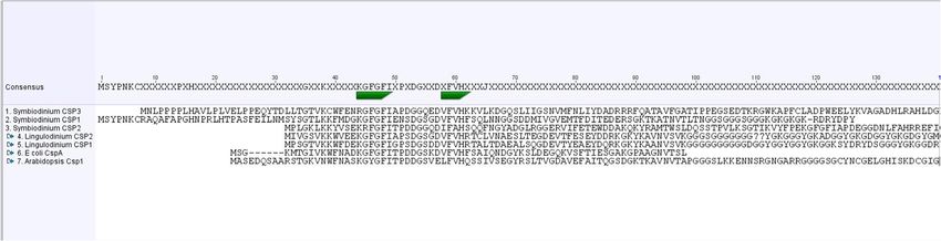
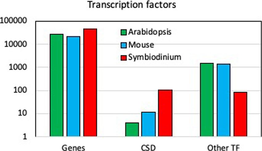
Zaheri and Morse BMC Molecular and Cell Biology      (2021) 22:27                                                               Page 2 of 9

structures have been found in Lingulodinium and Sym-                   measure if they were active in binding nucleic acids. In a
biodinium proteins, the most prevalent ones containing                 second approach, selection and amplification binding as-
a CSD either alone or with a C-terminal G-rich domain.                 says (SAAB) was used to determine if these proteins could
Less frequently observed are some structures containing                bind a specific sequence on DNA. All these CSPs were
a Zn-finger domain following the G-rich domain, and                    able to bind to DNA and RNA, and no sequence specific
also examples of sequences with multiple CSDs and one                  binding activity toward DNA was observed.
or more RNA recognition motifs (RRM). Thus, many of
the dinoflagellate CSPs are similar to what are found in               Results
bacteria as these typically contain only a CSD [5].                    SkCSP1, SkCSP2 and SkCSP3 belong to a Symbiodinium
  In E. coli, CSPs have a wide range of functions, including           unique clade
binding DNA as transcription factors, binding to RNA,                  The number of annotated DNA binding proteins in the
regulating transcription, splicing, and translation, and af-           genome of the S. kawagutii genome [10] belonging either
fecting mRNA stability as RNA chaperones [6, 7]. Bacter-               to CSD family or other TF (Fig. 1) shows the relative im-
ial CSPs have a non-specific RNA binding function during               portance of CSDs in dinoflagellates compared to plants
cold stress, which is correlated to their chaperone activity,          and animals. All CSDs contain the two RNA recognition
and this helps transcription by acting as an antiterminator            motifs (KGFGFI and VFVHF) shared with bacteria and
[7, 8]. However, the dinoflagellate proteins may be differ-            plants [3, 4]. Phylogenetic analysis of CSDs from 12 pre-
ent from their bacterial counterparts as two Lingulodi-                dicted Symbiodinium kawagutii protein sequences was
nium CSPs, both containing a single CSD followed by a                  performed using RaxML, and all were found to cluster to-
glycine-rich C-terminal region, were both unable to com-               gether within a single well defined clade together with
plement the growth of an E. coli strain lacking four differ-           some bacterial sequences (Fig. 2, Table S2). This is slightly
ent CSP genes at low temperature [5]. Furthermore, cold                different from the situation in Lingulodinium where se-
temperatures did not induce the CSP transcripts in L.                  quences are distributed among two different clades. The
polyedra [9]. Previous work on L. polyedra CSPs showed                 phylogenetic positions of the four CSPs examined here:
binding to both single- and double-stranded DNA as well                LpCSP1 (JO732587), SkCSP1 (Skav223430), SkCSP2
as to RNA, but it was unclear if binding would show any                (Skav207008) and SkCSP3 (Skav233957) are boxed.
sequence specificity that would be likely if they were to                LpCSP1 with a size of 113 amino acids has been previ-
function as transcription factors [5]. Here we performed               ously cloned [5]. For this study, SkCSP1, SkCSP2 and
two experimental approaches to assess the specific nucleic             SkCSP3 were also cloned and have sizes of 128, 120 and
acid binding activity of L. polyedra CSP1 (LpCSP1) and                 182 residues, respectively. All four CSPs were expressed
three S. kawagutii CSPs (SkCSP1, SkCSP2 and SkCSP3).                   as GST-tagged proteins and used for EMSA after re-
Initially, these four CSPs were expressed, purified and                moval of the GST tag (Fig. S1). Two of the S. kawagutii
used in electrophoretic mobility assays (EMSAs) to                     proteins contain an N-terminal extension (Fig. 3).

 Fig. 1 The abundance of DNA-binding domain families detected in S. kawagutii compared with plants and animals. The number of genes
 annotated as CSD and as other TF are shown for the most recent S. kawagutii genome. Note the log scale at left
Zaheri and Morse BMC Molecular and Cell Biology         (2021) 22:27                                                                     Page 3 of 9

 Fig. 2 Phylogenetic reconstruction of a variety of dinoflagellate CSP. Sequences were aligned and the phylogeny reconstructed with RaxML.
 LpCSP1, SkCSP1, SkCSP2 and SkCSP3 sequences are boxed

Lingulodinium and Symbiodinium CSPs bind to DNA and                         increase with increasing concentrations of the CSPs, al-
RNA                                                                         though not precisely proportional to the amount of pro-
EMSA experiments were conducted on LpCSP1, SkCSP1,                          tein. We conclude that all four CSPs were able to bind
SkCSP2 and SkCSP3 to analyze their binding to radiola-                      to all three types of nucleic acids tested.
beled double-stranded (dsDNA), single-stranded (ssDNA)
and RNA probes (Fig. 4). Fusion proteins still containing                   Symbiodinium CSP1 shows preferential binding to single-
the glutathione S-transferase (GST) tags also bind nucleic                  stranded nucleic acids
acids but migrating slower on the gel, and all EMSA experi-                 To assess the specificity of Symbiodinium CSPs interactions
ments used proteins after removal of the tag by thrombin.                   with different nucleic acid substrates, binding to dsDNA
  All proteins were able to bind dsDNA, ssDNA and                           and ssDNA probes was evaluated using SkCSP1 and un-
RNA as seen by the presence of a radioactive band of                        labeled (cold) competitors (Fig. 5). When dsDNA was used
lower mobility. The mobility of probe sequence was re-                      as a probe, the intensity of the slowly migrating bands de-
duced to roughly the same extent with all proteins with                     creased dramatically when the amount of competing cold
the exception of LpCSP1 binding to ssDNA or RNA.                            ssDNA was increased. In contrast, band intensity using
The amount of the reduced mobility band seemed to                           ssDNA probes was mostly stable using increasing amounts

 Fig. 3 Alignment of CSD domains from the dinoflagellates L. polyedra, and S. kawagutii, the bacterium E. coli and the higher plant Arabidopsis
 thaliana. The two RNA recognition motifs are marked in green
Zaheri and Morse BMC Molecular and Cell Biology (2021) 22:27 Page 4 of 9
Fig. 4 Nucleic acid binding activity of L. polyedra and S. kawagutii CSPs in EMSA. ssDNA (a, b), dsDNA (c, d) and RNA (e, f) probes were used. The
black triangle shows the different concentrations of the CSPs (0.5, 1 and 3 μg in all the assays); position of the shifts are shown by arrows
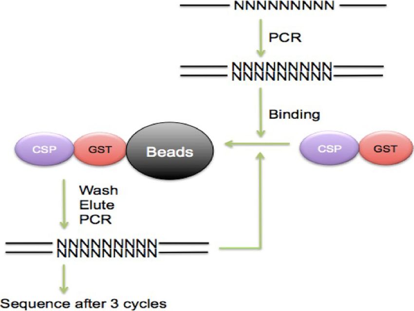
of cold dsDNA. Furthermore, RNA appears to compete ef- performed a selection and amplification binding enrich-
ficiently with both dsDNA and ssDNA. These results indi- ment (SAAB) with DNA containing 9 random nucleotides
cate that SkCSP1 has a preference for single-stranded (N9) flanked by PCR primers. These experiments used the
nucleic acids, with RNA preferred over DNA. This is con- fusion proteins directly to facilitate purification of bound
sistent with a previous report for Lingulodinium CSP1 [5]. DNA sequences, as the presence of the GST tag did not
While the potential tendency to bind to ssDNA may sup- affect DNA binding on EMSA assays. After 3 rounds of
port a role for these proteins in uncoiling the DNA struc- SAAB, samples containing double-stranded N9 enriched
ture during transcription, preferential binding to RNA by binding to LpCSP1, SkCSP1, SkCSP2 and SkCSP3 were
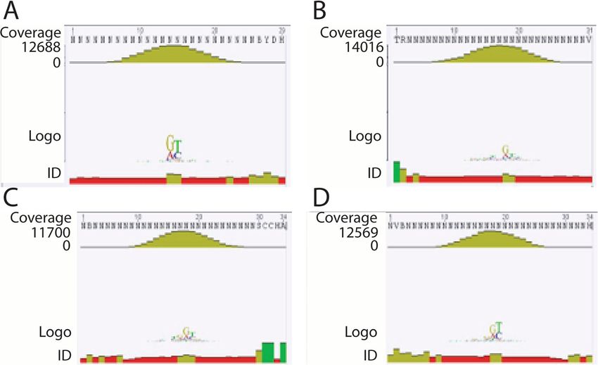
suggests this may not be their primary role. sequenced (Fig. 6). Over 12,000 sequences were been ob-
tained for each CSP, but sequence alignments after bind-
L. polyedra and S. kawagutii CSPs bind non-specifically to ing to all four shows no evidence for a consensus motif
DNA and RNA for any of the CSPs (Fig. 7). We conclude that there is no
To assess the possibility of sequence specific binding of specific dsDNA which can be enriched by binding to Lin-
Lingulodinium and Symbiodinium CSPs to dsDNA, we gulodinium or Symbiodinium CSPs.Zaheri and Morse BMC Molecular and Cell Biology (2021) 22:27 Page 5 of 9 Fig. 5 Competition assays of SkCSP1 with ssDNA, dsDNA and RNA. Symbiodinium CSP1 binds to ssDNA better than dsDNA. Cold oligos have a different sequence than the ssDNA. Concentration of SkCSP1 is 0.5 μg in all the assays; position of the shifts are shown by arrows. RNA competes efficiently with both dsDNA and ssDNA. The black triangle shows the different concentrations of the unlabeled nucleic acids (1x, 10x, 30x, and 80x the probe concentration) Discussion regulation of transcription by binding to a Y-box specific Cold shock domain (CSD) proteins were recognized in sequence, and is also involved in regulation of transla- Escherichia coli during cold shock stress [7, 11, 12]. The tion and RNA processing [20–24] and DNA repair [7, conservation of CSD in these proteins was discovered in 12, 25]. YB1 prefers to bind to ssDNA rather than bacteria, archaea, plants, and animals. In prokaryotes, dsDNA, thus disentangling the double helix structure of CSPs containing only a CSD act mainly as RNA chaper- DNA has been proposed for the activation of transcrip- ones. Some E. coli CSPs are cold-inducible and act as tion [7, 26]. YB1 also prefers RNA over ssDNA [7] with RNA chaperons disrupting RNA secondary structures [7, the consensus CA(U/C) C sequence as the RNA-binding 13]. They are also involved in the transcription regula- site [27, 28]. In dinoflagellates, CSPs are mostly in the tion by binding specifically to gyrA promoter (CspA) form of one conserved CSD either alone or with a C- [12, 14]. In eukaryotes, CSPs are composed of CSD and terminal G-rich domain [5]. Previously, a Y-box se- additional domains and aid in responding to cold stress, quence (CTGATTGGCT) was used to study the binding nutrient limitation and growth [7, 13, 15–17]. Plants specify of L. polyedra CSPs [5]. Here we used different CSPs are engaged in regulation of translation during random C-rich sequences to test the possibility of se- cold stress and also complicated physiological processes quence privileged targeting. For the SAAB assay, we syn- such as seed and flower germination [7, 18, 19]. In A. thesized a DNA sequence with 9 random nucleotides (N9) thaliana, CSP3 interacts with other proteins involved in nestled between flanking PCR primers. The goal of this mRNA processing path [19]. A vertebrate CSP called experiment was to see if several cycles of binding, elution YB1 (Y-box binding protein) is responsible for the and amplification would enrich for a particular sequence
Zaheri and Morse BMC Molecular and Cell Biology        (2021) 22:27                                                                    Page 6 of 9

 Fig. 6 Schematic model for analyzing the specificity of DNA sequence binding by selection and amplification binding assays (SAAB)

 Fig. 7 Consensus nucleotide binding activity of 4 different dinoflagellates CSPs. Over 12,000 different N9 sequences bound by LpCSP1 (a), SkCSP1
 (b), SkCSP2 (c), and SkCSP3 (d) were aligned and used to prepare a sequence logo showing the frequency of each nucleotide at each position
Zaheri and Morse BMC Molecular and Cell Biology   (2021) 22:27                                                   Page 7 of 9

motif that could constitute a potential promoter element.        Methods
However, no sequence motifs were enriched by binding to          Cell cultures
any of the four CSPs indicating that these proteins are un-      Cultures of Symbiodinium kawagutii (strain CCMP2468)
likely to function as conventional sequence-specific tran-       and Lingulodinium polyedra (strain CCMP1936) were
scription factors. It is not possible to rule out a role in      obtained from the National Center for Marine Algae
DNA unwinding similar to what has been proposed for              (Boothbay Harbor, Maine). Cells were grown in f/2 sea
YB1, in which non-specific binding of CSPs to ssDNA was          water medium prepared from Instant Ocean under 12 h
thought to help stabilize the structure, but it must be          cool white fluorescent light and 12 h darkness as de-
noted CSPs have no known helicase activity.                      scribed [30] except that the temperature was 25 ± 1 °C
   The importance of examining the nucleic acid binding          for S. kawagutii.
properties of CSPs is due to the finding that the majority
of the proteins annotated as transcription factors in the        Phylogenetic reconstruction and primer design
transcriptome of Lingulodinium [3], Symbiodinium [4]             The CSP sequences for Lingulodinium and Symbiodinium
and the genome of Symbiodinium [10, 29] (Fig. 1) are             were obtained from the dinoflagellate transcriptomes de-
CSDs. Our hypothesis was that to act as transcription            posited at NCBI and from the Symbiodinium kawagutii
factors, dinoflagellates CSPs should bind to dsDNA in a          genome at the Symbiodiniaceae and Algal Genomic Re-
sequence specific manner. We assessed nucleic acid               source (SAGER) database [29]. Phylogenetic analysis of
binding activity of LpCSP1, SkCSP1, SkCSP2 and                   CSDs from the predicted protein sequences (Table
SkCSP3 using two different approaches. In one ap-                S2) was performed using a webserver for alignments
proach, electrophoretic mobility shifts assays (EMSA)            (http://www.phylo.org/sub_sections/portal/) [31]. The ser-
were used to show that all four CSPs could bind both             ver performs sequence alignments using MUSCLE, and
double- and single- stranded DNA as well as RNA (Fig.            curation using GBlocks. Phylogenetic reconstructions
4). When tested in competition EMSA experiments,                 were built with RaxML using the CIPRES portal (http://
RNA was found to compete with binding to DNA                     www.phylo.org/sub_sections/portal/). Trees were visual-
probes better than DNA competed with binding to RNA              ized by TreeDyn. Primers were designed using Geneious
probes (Fig. 5). These characteristics are not what would        software [32] or BLAST integrated into Galaxy [33] for
be predicted for a transcription factor. In a second ap-         amplification and subsequent cloning of the CSPs. Gen-
proach, selection and amplification binding (SAAB) ex-           eious software [32] was also used for sequence alignments.
periments showed none of the four CSPs tested enriched
a specific motif after three cycles of binding and PCR           Cloning, expression and purifying of CSPs
amplification, again inconsistent with a role as a se-           Symbiodinium cultures were harvested by centrifugation
quence specific transcription factor.                            and the pellets frozen in liquid nitrogen. Frozen pellets
   Our results indicate that LpCSP1, SkCSP1, SkCSP2              were crushed into a fine powder using a pre-chilled
and SkCSP3 binding to nucleic acids does not depend              mortar and pestle, and the powder was added to Trizol
on sequence. We infer that the dinoflagellate CSPs in            (Invitrogen). Primer pairs based on sequences from the
general are unlikely to act as sequence-specific transcrip-      Symbiodinium transcriptome or genome were used to
tion factors. Although only one S. kawagutii CSP                 amplify CSPs from a first strand cDNA reaction product
(SkCSP1) was extensively analyzed by competition                 using the total RNA extracted from Symbiodinium cells
EMSA, the similarity to the Lingulodinium CSP1 sug-              as described [5]. For cDNA amplification, the reverse
gests the nucleic acid binding properties found may be a         transcription reaction was performed with ProtoScript II
consistent lineage-specific feature. The balance of the          first strand cDNA synthesis kit (New England BioLabs).
evidence thus suggests that CSPs do bind nucleic acids,          The sequences were cloned into the pGEM-T vector
thus explaining why they were annotated as transcrip-            (Promega) and sequenced. A second PCR was performed
tion factors. However the details of the binding suggest         on the insert in the pGEM-T plasmid using primers con-
they are unlikely to play this role in vivo. Additional          taining restriction sites required for directional cloning
characterization studies of dinoflagellate CSPs would be         into the bacterial expression vectors pGEX-4 T2 (GE
essential to recognize more about their function and             Healthcare) [34] (Table. S1 in the supplementary data).
possible interaction with other partners.                        The reading frame of all clones were confirmed by se-
                                                                 quencing and the size of the CSP fusion protein verified
Conclusions                                                      by SDS PAGE (Fig. S1). The pGEX4T2 vectors contain-
The four CSPs examined here do not bind DNA in a se-             ing CSP sequences were used to transform the chem-
quence specific manner. Furthermore, SkCSP1 prefers              ically competent cells of BL21. Liquid Luria Bertani (LB)
binding to single-stranded RNA. CSPs are unlikely to             medium was used to grow one colony of transformed E.
function as transcription factors in dinoflagellates.            coli overnight at 37 °C with vigorous shaking in the
Zaheri and Morse BMC Molecular and Cell Biology (2021) 22:27 Page 8 of 9
presence of ampicillin to maintain selection for the plas- Selection and amplification binding assays
mid. Protein expression were induced using Isopropyl β- Symbiodinium and Lingulodinium CSPs were cloned
D-1-thiogalactopyranoside (IPTG). Cells were collected and expressed as a fusion protein with a C-terminal
by centrifugation, resuspended in PBS buffer and broken GST tag as described above. The BL21 cell lysates were
in a French pressure cell (Fisher Scientific). The cell ly- centrifuged and the supernatants containing GST tagged
sates were then centrifuged and the supernatants were CSPs were incubated with Glutathione Sepharose 4B
incubated with Glutathione Sepharose 4B beads (Pro- beads (Promega) for 45 min at room temperature with
mega) for 45 min at room temperature with end-over- end-over-end agitation. Beads were washed 4 times in
end agitation. Beads were washed 4 times in PBS and re- PBS. Immobilized LpCSP1, SkCSP1, SkCSP2 and SkCSP3
suspended in PBS supplemented with thrombin to cleave were tested for sequence-specific DNA binding activities
the GST tag. The size, and purity of the single CSPs against a set of degenerate oligonucleotides using a se-
were then analyzed by SDS-PAGE on acrylamide gel lection and amplification binding assay (SAAB) [35, 36].
(Fig. S1) and the Bradford assay (BioRad) was used to as- A set of single-stranded oligonucleotides with PCR pri-
sess the protein concentration. mer sequences flanking nine random nucleotides (N9)
were synthesized and used to produce double-stranded
CSP electrophoretic mobility shift assays DNA by a single PCR cycle using the reverse primer. Fif-
[γ-32P] ATP (PerkinElmer) was used to 5′-end-label 32 teen μg of double-stranded DNA (N9) was allowed to
nt ssDNA 5′-TCCGCCCTCCCTCCCCCCGCCCTCCC bind to 10 μL of immobilized CSPs in a 100 μL total vol-
TCCCCA-3′ and 25 bp dsDNA 5′-GGCGCCCTCC ume solution containing 75 mM NaCl, 1 mM DTT, 1
CTCCGCCCTCCCTCA-3′ C-rich sequences using a T4 mM phenylmethylsulfonyl fluoride, 0.1% Triton X-100,
polynucleotide kinase kit (NEB). A QIAquick nucleotide 10 ng of poly (dI-dC) per μL, 10 mM Tris-HCl (pH 7),
removal kit (Qiagen) was used for removing the unin- 6% glycerol and 1% BSA. After 1 h of agitation at 4 °C,
corporated nucleotides and purifying the probes. Either the supernatant containing unbound oligonucleotides
dsDNA or ssDNA 32P-labelled probes (1 ng) and in- were removed. Following 3 times of washing with bind-
creasing concentrations of CSPs (0.5–3 μg) were incu- ing buffer, DNA was released from the protein by boil-
bated in 20 μL of 2x binding buffer (20 mM Tris-Cl [pH ing in water [35]. DNA was amplified in a PCR reaction
7.0], 20 mM MgCl2, 50 mM KCl, 10% glycerol and 1 to repeat the protein binding step. Three rounds of
mM DTT) for 30 min at room temperature. The CSP/ SAAB were performed before sending out the PCR
DNA complexes were run through a 5% native poly- products for sequencing (Fig. 6).
acrylamide gels for 45 mins at 80 V in 1× Tris-borate-
EDTA (TBE) buffer at room temperature. The gels were
Supplementary Information
dried immediately and exposed overnight at − 80 °C with The online version contains supplementary material available at https://doi.
a phosphorimager screen (Amersham). The images were org/10.1186/s12860-021-00368-4.
analyzed with a Typhoon Trio+ (Amersham) using Ima-
geQuant 5.2. Competition reactions were prepared by Additional file 1: Supplemental Figure S1. Purification of LpCSP1,
SkCSP1, SkCSP2 and SkCSP3. A shows recombinant LpCSP1-GST, SkCSP1-
incubation of the CSPs and increasing amounts of cold GST, SkCSP2-GST and SkCSP3-GST analyzed on an 18% acrylamide SDS-
unlabeled ssDNA, dsDNA or RNA probes (described PAGE gel after affinity purification. B shows LpCSP1, SkCSP1, SkCSP2 and
below) for specific binding and a 40x excess of random SkCSP3 after removal of the GST tag by thrombin digestion and binding
to glutathione-Sepharose 4B beads. The sizes of the molecular weight
22 nt single-stranded oligonucleotide (TTATTGGGGC markers (left) are shown in kilodaltons.
ACACCGCATGCT) for non-specific competition in the Additional file 2: Supplemental Table S1. List of primers used for
binding buffer for 15 mins before adding the radiola- PCR amplification and cloning of LpCSP1, SkCSP1, SkCSP2 and SkCSP3
beled probes. sequences in pGEX4T2 plasmid.
Forty nt RNAs were synthesized by T7 RiboMAX Additional file 3: Supplementary Table S2. List of proteins selected
for phylogenetic reconstruction.
RNA production kit (Promega) using dsDNA templates
containing the N9 and T7 promoter sequences. There-
after, RQI RNase-free DNase (Promega) was used for Authors’ contributions
degradation of the dsDNA templates. The in vitro tran- BZ designed and performed experiments, analysed data, and participated in
writing the manuscript. DM analysed data, participated in writing the
scribed RNAs were quantitated using spectrophotometry manuscript and obtained funding. The author(s) read and approved the final
(1.2 μg/μL), end-labeled using [γ-32P] ATP (PerkinEl- manuscript.
mer) (see above) and purified using filtration chromatog-
raphy on a Bio-Gel P10 column (Bio-Rad). One ng Funding
labelled probe was incubated with increasing concentra- Funding was obtained from the Canadian National science and Engineering
Research Council (grant number 171382–03 to D.M.). The funding body
tions of CSPs in the binding reactions as described played no role in the design of the study and collection, analysis, and
above. interpretation of data, or in writing the manuscript and decision to publish.Zaheri and Morse BMC Molecular and Cell Biology (2021) 22:27 Page 9 of 9
Availability of data and materials 17. Wistow G. Cold shock and DNA binding. Nature. 1990;344(6269):823–4.
All data generated or analysed during this study are included in this https://doi.org/10.1038/344823c0.
published article. 18. Fusaro AF, Bocca SN, Ramos RL, Barroco RM, Magioli C, Jorge VC, et al.
AtGRP2, a cold-induced nucleo-cytoplasmic RNA-binding protein, has a role
Declarations in flower and seed development. Planta. 2007;225(6):1339–51. https://doi.
org/10.1007/s00425-006-0444-4.
Ethics approval and consent to participate 19. Kim MH, Sonoda Y, Sasaki K, Kaminaka H, Imai R. Interactome analysis
Not applicable. reveals versatile functions of Arabidopsis COLD SHOCK DOMAIN PROTEIN 3
in RNA processing within the nucleus and cytoplasm. Cell Stress
Chaperones. 2013;18(4):517–25. https://doi.org/10.1007/s12192-012-0398-3.
Consent for publication 20. Izumi H, Imamura T, Nagatani G, Ise T, Murakami T, Uramoto H, et al. Y box-
Not applicable. binding protein-1 binds preferentially to single-stranded nucleic acids and
exhibits 3′-->5′ exonuclease activity. Nucleic Acids Res. 2001;29(5):1200–7.
Competing interests https://doi.org/10.1093/nar/29.5.1200.
The authors declare that they have no competing interests. 21. Lasham A, Moloney S, Hale T, Homer C, Zhang YF, Murison JG, et al. The Y-box-
binding protein, YB1, is a potential negative regulator of the p53 tumor suppressor.
Received: 23 March 2021 Accepted: 21 April 2021 J Biol Chem. 2003;278(37):35516–23. https://doi.org/10.1074/jbc.M303920200.
22. Sommerville J. Activities of cold-shock domain proteins in translation
control. Bioessays. 1999;21(4):319–25. https://doi.org/10.1002/(SICI)1521-1
878(199904)21:43.0.CO;2-3.
References 23. Kleene KC. Y-box proteins combine versatile cold shock domains and
1. Riechmann JL, Heard J, Martin G, Reuber L, Jiang C, Keddie J, et al. arginine-rich motifs (ARMs) for pleiotropic functions in RNA biology.
Arabidopsis transcription factors: genome-wide comparative analysis among Biochem J. 2018;475(17):2769–84. https://doi.org/10.1042/BCJ20170956.
eukaryotes. Science. 2000;290(5499):2105–10. https://doi.org/10.1126/ 24. Mordovkina D, Lyabin DN, Smolin EA, Sogorina EM, Ovchinnikov LP, Eliseeva
science.290.5499.2105. I. Y-box binding proteins in mrnp assembly, translation, and stability control.
2. Zhang HM, Chen H, Liu W, Liu H, Gong J, Wang H, et al. AnimalTFDB: a Biomolecules. 2020;10(4):591. https://doi.org/10.3390/biom10040591.
comprehensive animal transcription factor database. Nucleic Acids Res. 25. Sangermano F, Delicato A, Calabro V. Y box binding protein 1 (YB-1)
2012;40(Database issue):D144–9. https://doi.org/10.1093/nar/gkr965. oncoprotein at the hub of DNA proliferation, damage and cancer progression.
3. Beauchemin M, Roy S, Daoust P, Dagenais-Bellefeuille S, Bertomeu T, Biochimie. 2020;179:205–16. https://doi.org/10.1016/j.biochi.2020.10.004.
Letourneau L, et al. Dinoflagellate tandem array gene transcripts are highly 26. MacDonald GH, Itoh-Lindstrom Y, Ting JP. The transcriptional regulatory
conserved and not polycistronic. Proc Natl Acad Sci U S A. 2012;109(39): protein, YB-1, promotes single-stranded regions in the DRA promoter. J Biol
15793–8. https://doi.org/10.1073/pnas.1206683109. Chem. 1995;270(8):3527–33. https://doi.org/10.1074/jbc.270.8.3527.
4. Bayer T, Aranda M, Sunagawa S, Yum LK, Desalvo MK, Lindquist E, et al. 27. Wei WJ, Mu SR, Heiner M, Fu X, Cao LJ, Gong XF, et al. YB-1 binds to CAUC
Symbiodinium transcriptomes: genome insights into the dinoflagellate motifs and stimulates exon inclusion by enhancing the recruitment of U2AF
symbionts of reef-building corals. PLoS One. 2012;7(4):e35269. https://doi. to weak polypyrimidine tracts. Nucleic Acids Res. 2012;40(17):8622–36.
org/10.1371/journal.pone.0035269. https://doi.org/10.1093/nar/gks579.
5. Beauchemin M, Roy S, Pelletier S, Averback A, Morse D. Characterization of 28. Yang XJ, Zhu H, Mu SR, Wei WJ, Yuan X, Wang M, et al. Crystal structure of
two dinoflagellate cold shock domain proteins. mSphere. 2016;1:e00034–15. a Y-box binding protein 1 (YB-1)-RNA complex reveals key features and
6. Mihailovich M, Militti C, Gabaldon T, Gebauer F. Eukaryotic cold shock residues interacting with RNA. J Biol Chem. 2019;294(28):10998–1010.
domain proteins: highly versatile regulators of gene expression. Bioessays. https://doi.org/10.1074/jbc.RA119.007545.
2010;32(2):109–18. https://doi.org/10.1002/bies.200900122. 29. Yu L, Li T, Li L, Lin X, Li H, Liu C, et al. SAGER: a database of
7. Budkina KS, Zlobin NE, Kononova SV, Ovchinnikov LP, Babakov AV. Cold Symbiodiniaceae and algal genomic resource. Database (Oxford). 2020;2020.
shock domain proteins: structure and interaction with nucleic acids. https://doi.org/10.1093/database/baaa051.
Biochemistry (Mosc). 2020;85(Suppl 1):S1–S19. https://doi.org/10.1134/ 30. Wang Y, Jensen L, Hojrup P, Morse D. Synthesis and degradation of
S0006297920140011. dinoflagellate plastid-encoded psbA proteins are light-regulated, not
8. Bae W, Xia B, Inouye M, Severinov K. Escherichia coli CspA-family RNA circadian-regulated. Proc Natl Acad Sci U S A. 2005;102(8):2844–9. https://
chaperones are transcription antiterminators. Proc Natl Acad Sci U S A. doi.org/10.1073/pnas.0406522102.
2000;97(14):7784–9. https://doi.org/10.1073/pnas.97.14.7784. 31. Dereeper A, Guignon V, Blanc G, Audic S, Buffet S, Chevenet F, et al.
9. Roy S, Letourneau L, Morse D. Cold-induced cysts of the photosynthetic Phylogeny.fr: robust phylogenetic analysis for the non-specialist. Nucleic
dinoflagellate Lingulodinium polyedrum have an arrested circadian Acids Res. 2008;36(Web Server issue):W465–9.
bioluminescence rhythm and lower levels of protein phosphorylation. Plant 32. Kearse M, Moir R, Wilson A, Stones-Havas S, Cheung M, Sturrock S, et al.
Physiol. 2014;164(2):966–77. https://doi.org/10.1104/pp.113.229856. Geneious basic: an integrated and extendable desktop software platform for
10. Li T, Yu L, Song B, Song Y, Li L, Lin X, et al. Genome improvement and core the organization and analysis of sequence data. Bioinformatics. 2012;28(12):
gene set refinement of fugacium kawagutii. Microorganisms. 2020;8(1):102. 1647–9. https://doi.org/10.1093/bioinformatics/bts199.
https://doi.org/10.3390/microorganisms8010102. 33. Cock PJ, Chilton JM, Gruning B, Johnson JE, Soranzo N. NCBI BLAST+ integrated
11. Jones PG, VanBogelen RA, Neidhardt FC. Induction of proteins in response into galaxy. Gigascience. 2015;4(1):39. https://doi.org/10.1186/s13742-015-0080-7.
to low temperature in Escherichia coli. J Bacteriol. 1987;169(5):2092–5. 34. Xia B, Ke H, Inouye M. Acquirement of cold sensitivity by quadruple
https://doi.org/10.1128/JB.169.5.2092-2095.1987. deletion of the cspA family and its suppression by PNPase S1 domain in
12. Heinemann U, Roske Y. Cold-shock domains-abundance, structure, Escherichia coli. Mol Microbiol. 2001;40(1):179–88. https://doi.org/10.1046/
properties, and nucleic-acid binding. Cancers. 2021;13(2):190. https://doi. j.1365-2958.2001.02372.x.
org/10.3390/cancers13020190. 35. Chang C, Jacobs Y, Nakamura T, Jenkins NA, Copeland NG, Cleary ML. Meis
13. Graumann PL, Marahiel MA. A superfamily of proteins that contain the cold- proteins are major in vivo DNA binding partners for wild-type but not
shock domain. Trends Biochem Sci. 1998;23(8):286–90. https://doi.org/10.101 chimeric pbx proteins. Mol Cell Biol. 1997;17(10):56795687.
6/S0968-0004(98)01255-9. 36. Magnani E, Sjolander K, Hake S. From endonucleases to transcription factors:
14. Jones PG, Krah R, Tafuri SR, Wolffe AP. DNA gyrase, CS7.4, and the cold evolution of the AP2 DNA binding domain in plants. Plant Cell. 2004;16(9):
shock response in Escherichia coli. J Bacteriol. 1992;174(18):5798–802. 2265–77. https://doi.org/10.1105/tpc.104.023135.
https://doi.org/10.1128/JB.174.18.5798-5802.1992.
15. Karlson D, Imai R. Conservation of the cold shock domain protein family in
plants. Plant Physiol. 2003;131(1):12–5. https://doi.org/10.1104/pp.014472. Publisher’s Note
16. Nakaminami K, Karlson DT, Imai R. Functional conservation of cold shock Springer Nature remains neutral with regard to jurisdictional claims in
domains in bacteria and higher plants. Proc Natl Acad Sci U S A. 2006; published maps and institutional affiliations.
103(26):10122–7. https://doi.org/10.1073/pnas.0603168103.You can also read