Unresolved questions in human copper pump mechanisms
←
→
Page content transcription
If your browser does not render page correctly, please read the page content below
PERSPECTIVE

                      Unresolved questions in human copper
                      pump mechanisms
                      Pernilla Wittung-Stafshede

                      Chemistry Department, Umeå University, 90187 Umeå, Sweden

                      Quarterly Reviews of Biophysics (2015), 48(4), pages 471–478   doi:10.1017/S0033583515000128

                      Abstract. Copper (Cu) is an essential transition metal providing activity to key enzymes in the human body. To regulate the levels and avoid
                      toxicity, cells have developed elaborate systems for loading these enzymes with Cu. Most Cu-dependent enzymes obtain the metal from the mem-
                      brane-bound Cu pumps ATP7A/B in the Golgi network. ATP7A/B receives Cu from the cytoplasmic Cu chaperone Atox1 that acts as the cyto-
                      plasmic shuttle between the cell membrane Cu importer, Ctr1 and ATP7A/B. Biological, genetic and structural efforts have provided a
                      tremendous amount of information for how the proteins in this pathway work. Nonetheless, basic mechanistic-biophysical questions (such
                      as how and where ATP7A/B receives Cu, how ATP7A/B conformational changes and domain–domain interactions facilitate Cu movement
                      through the membrane, and, finally, how target polypeptides are loaded with Cu in the Golgi) remain elusive. In this perspective, unresolved
                      inquiries regarding ATP7A/B mechanism will be highlighted. The answers are important from a fundamental view, since mechanistic aspects
                      may be common to other metal transport systems, and for medical purposes, since many diseases appear related to Cu transport dysregulation.

                      Key words: copper transport, copperchaperone, Wilson disease protein, Menke’s disease protein, ceruloplasmin, biophysical methods.

                      Copper (Cu) pumps of the human secretory pathway
                      Cu is found in the active sites of essential proteins that partici-              cytoplasmic metal-binding domains in ATP7A and
                      pate in cellular reactions such as respiration, antioxidant                      ATP7B (also called Menke’s and Wilson disease proteins,
                      defense, neurotransmitter biosynthesis, connective-tissue                        respectively), two homologous multi-domain P1B-type
                      biosynthesis and pigment formation (Harris, 2003; Huffman                         ATPases located in the trans-Golgi network (Fig. 1). Most
                      & O’Halloran, 2001; Puig & Thiele, 2002). The ability of Cu                      Cu-dependent enzymes acquire Cu from ATP7A/B in the
                      to oxidize/reduce (switch between Cu+ and Cu2+) allows                           Golgi before reaching their final destination (e.g. blood clot-
                      Cu-containing proteins to play important roles as electron car-                  ting factors, tyrosinase, lysyl oxidase and ceruloplasmin)
                      riers and redox catalysts in living systems. To avoid Cu toxicity,               (Festa & Thiele, 2011; Koch et al. 1997; O’Halloran &
                      the intracellular concentration of Cu is regulated via dedicated                 Culotta, 2000; Robinson & Winge, 2010). Once transferred
                      proteins that facilitate uptake, efflux as well as distribution to                 to ATP7A/B, the Cu ion is channeled to the lumen of the
                      target Cu-dependent proteins and enzymes (Festa & Thiele,                        Golgi and loaded onto target Cu-dependent proteins. We re-
                      2011; O’Halloran & Culotta, 2000; Robinson & Winge,                              cently reported that at least in vitro, in addition to ATP7A/B
                      2010). In the human cytoplasm, after Cu has entered the cell                     interactions, Atox1 can cross-react and exchange Cu with
                      via the Cu importer Ctr1 (Ohrvik & Thiele, 2014), there are                      another cytoplasmic Cu chaperone, the Cu chaperone for
                      at least three independent Cu transport pathways.                                superoxide dismutase, CCS, which delivers Cu specifically
                                                                                                       to SOD1 (Petzoldt et al. 2015). Thus, cross talk between cyto-
                      In the general pathway, conserved in most organisms, the
                                                                                                       plasmic chaperones may be an unexplored mechanism that
                      68-residue Cu chaperone Atox1 transports the metal to
                                                                                                       allows for efficient usage of cytoplasmic Cu ions.

                      Authors for Correspondence: P. Wittung-Stafshede, Chemistry Department,
                                                                                                       ATP7A/B are elaborate multi-domain Cu pumps with eight
                      Umeå University, 90187 Umeå, Sweden. Tel.: +467865347; Fax: +467867655;          membrane-spanning helices, an actuator (A) domain, as
                      E-mail: Pernilla.wittung@chem.umu.se                                             well as nucleotide-(N) and phosphorylation-(P) domains,

                      © Cambridge University Press 2015. This is an Open Access article, distributed under the terms of the Creative Commons Attribution licence (http://
                       creativecommons.org/licenses/by/3.0/),
Downloaded from https://www.cambridge.org/core.               which permits
                                                 IP address: 46.4.80.155,    unrestricted
                                                                          on 13 Nov 2021 atre-use, distribution,
                                                                                             18:06:47, subject to and  reproduction
                                                                                                                  the Cambridge     interms
                                                                                                                                 Core  any medium,    provided
                                                                                                                                            of use, available    the original work is
                                                                                                                                                              at https://www.cambridge.org/core/terms.
https://doi.org/10.1017/S0033583515000128
                       properly cited.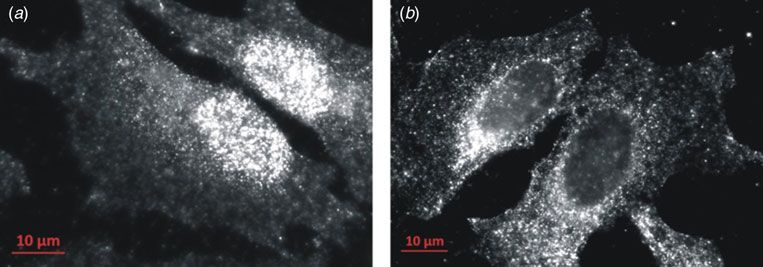
Fig. 1. Left: Illustration of the Cu transport pathway to the Golgi for Cu loading of proteins in the secretory path. Uptake of Cu takes
                      place via Ctr1, then cytoplasmic transport is facilitated by the Cu chaperone Atox1 to membrane-bound ATP7A/B for loading of
                      Cu-dependent enzymes. If there is too much Cu in the cell, ATP7A/B can move to vesicles and facilitate Cu export out of the cell. Right:
                      Schematic structure of the domain arrangement of ATP7A/B (six metal-binding domains, an actuator (A) domain, N- and P-domains
                      that bind ATP and become phosphorylated, respectively, and membrane-spanning helices (gray).

                      with nucleotide-binding site and an invariant Asp (that is                       Cu-bridged hetero-dimer complexes where the metal is
                      transiently phosphorylated during the catalytic cycle), re-                      shared between the two metal-binding sites (Fig. 2). Cu is
                      spectively, protruding into the cytoplasm (Fig. 1). In                           thought to move from one protein to the other via
                      addition, ATP7A and ATP7B both have six cytoplasmic                              ligand-exchange reactions involving tri-coordinated Cu–sul-
                      metal-binding domains in the N-terminus connected by pep-                        fur intermediates (Pufahl et al. 1997). All six domains of
                      tide linkers of various lengths (Lutsenko et al. 2007). Notably,                 ATP7A/B can be loaded with Cu by Atox1 but only in
                      much of our current knowledge of ATP7A/B comes from stu-                         some cases, have Cu-dependent protein–protein complexes
                      dies of individual domains (as it is difficult to prepare the full                 been detected by NMR via slower tumbling times (Achila
                      length proteins) and from work on yeast and bacterial homo-                      et al. 2006; Banci et al. 2005, 2008, 2009a, b). Based on af-
                      logs (Culotta et al. 2005; Gourdon et al. 2011).                                 finity and NMR studies, Cu binding to an ATP7B metal-
                                                                                                       binding domain is favored over binding to Atox1 by a factor
                      Each metal-binding domain in ATP7A/B, as well as Atox1,
                                                                                                       of 3–5 providing a shallow directional thermodynamic driv-
                      has a ferredoxin-like αβ-fold and a surface-exposed in-
                                                                                                       ing force. We found that upon mixing of Cu–Atox1 and the
                      variant MXCXXC motif (X = any residue) in which a single
                                                                                                       fourth metal-binding domain of ATP7B (WD4), a stable
                      Cu can bind via the two cysteine sulfurs. In contrast to
                                                                                                       ternary complex assembled that was in equilibrium with
                      humans, bacterial and yeast P1B-type ATPases have only
                                                                                                       substrates and products (Niemiec et al. 2012). In contrast,
                      one or two metal-binding domains. The purpose for as
                                                                                                       when mixing a two-domain construct of domains 5 and 6
                      many as six metal-binding domains in ATP7A/B is unknown,
                                                                                                       in ATP7B (WD56) and Cu–Atox1, the protein–protein in-
                      albeit regulation has been proposed. The MXCXXC motif
                                                                                                       teraction was transient such that it did not survive size ex-
                      does not confer intrinsic specificity to Cu ions, although
                                                                                                       clusion chromatography (SEC) but Cu transfer still took
                      soft metals are favored by sulfur ligands, as both Atox1 and
                                                                                                       place (Nilsson et al. 2013).
                      individual ATP7B metal-binding domains can bind other
                      metals, such as Zn, strongly in vitro (Niemiec et al. 2014).                     For the Atox1 and WD4 pair, SEC in combination with ti-
                      At normal cell conditions, however, metal-binding degener-                       tration calorimetry made possible thermodynamic analysis of
                      acy is not a problem since metal transport is strictly governed                  the reaction in Fig. 2 and we identified that Atox1–Cu–WD4
                      by protein–protein interactions (Tottey et al. 2005, 2008).                      hetero-protein complex formation is driven by favorable en-
                                                                                                       thalpy and entropy changes, whereas the overall reaction,
                                                                                                       from Atox1 to WD4, relies on only enthalpy (Niemiec et al.
                      Moving Cu from chaperone to ATP7A/B                                              2012). In additional studies, involving protein engineering,
                      It was originally assumed that Atox1 delivers Cu to one of                       we revealed that the first cysteine in each protein’s Cu
                      the metal-binding domains of ATP7A/B and the metal                               binding motif was essential for hetero-protein complex forma-
                      then is shuttled within the protein to Cu-binding sites in                       tion but one of the second cysteines was not required.
                      the membrane channel. In vitro (Achila et al. 2006; Banci,                       Thermodynamic analysis disclosed that the wild-type Cu site
                      2006; Banci et al. 2008, 2009a, b; Pufahl et al. 1997;                           in the hetero-protein complex was dynamic (in agreement
                      Wernimont et al. 2000) and in silico (Rodriguez-Granillo                         with positive entropy change, see above), involving entropy–
                      et al. 2010) work has shown that Cu transfer from Atox1                          enthalpy compensation (Niemiec et al. 2015). It remains un-
                      to metal-binding domains of ATP7A/B proceeds via                                 known if the same mechanism and thermodynamic principles

                                                                                                 472
Downloaded from https://www.cambridge.org/core. IP address: 46.4.80.155, on 13 Nov 2021 at 18:06:47, subject to the Cambridge Core terms of use, available at https://www.cambridge.org/core/terms.
https://doi.org/10.1017/S0033583515000128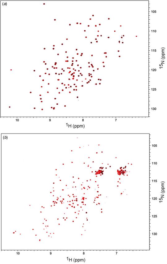
Fig. 2. Top: Scheme of Cu transfer mechanism from Atox1 to the 4th metal-binding domain of ATP7B (WD4) indicating an intermedi-
                      ate hetero-protein complex in which the Cu ion is coordinated by cysteines in both proteins’ metal-binding loops. Bottom: Structural
                      model of the Cu-dependent Atox1–WD4 hetero-protein complex.

                      apply to Atox1 interactions with the other five domains                           are found in the metal-binding domains of ATP7B (Hamza
                      in ATP7B and when the target domain is surrounded by its                         et al. 1999).
                      natural domains within the full-length protein. Information
                      on these (apparent) straightforward issues has been hampered
                      by the difficulty to prepare large membrane proteins for
                                                                                                       Internal interactions that modulate Cu
                      biophysical studies.
                                                                                                       movement
                      The finding that the Cu chaperone in bacteria (that is
                                                                                                       During the catalytic cycle, that requires ATP hydrolysis and
                      homologous to Atox1) could bypass the single metal-binding
                                                                                                       ultimately results in Cu transfer to the lumen side of the mem-
                      domain of the bacterial P1B-type ATPase and instead deliver
                                                                                                       brane, ATP7A/B are likely to undergo significant conforma-
                      Cu directly to the membrane entry site (Gonzalez-Guerrero &
                                                                                                       tional changes driven by domain–domain interactions
                      Arguello, 2008) reinforced the idea that the human metal-
                                                                                                       (Lutsenko et al. 2007). Available predictions for how
                      binding domains played regulatory roles. Nonetheless, yeast
                                                                                                       ATP7A/B works catalytically come from analogy with the cal-
                      complementation studies have shown that the presence of
                                                                                                       cium pump SERCA, for which structures of different enzy-
                      the human and yeast metal-binding domains, at least some
                                                                                                       matic stages have been resolved (Bublitz et al. 2013). Since
                      domains in the case of the human protein, is essential for
                                                                                                       there are no structures of the arrangement of the six metal-
                      Cu transfer activity (Forbes et al. 1999; Morin et al. 2009).
                                                                                                       binding domains within full-length ATP7A/B, it is unclear
                      In 2011, the crystal structure of the bacterial ATP7A/B homo-
                                                                                                       how these domains participate in the catalytic cycle.
                      log Legionella pneumophila CopA was reported (Gourdon
                                                                                                       Interactions among the metal-binding domains of ATP7A/B
                      et al. 2011). Although the CopA structure was a breakthrough,
                                                                                                       are proposed to transmit signals long-range (Gourdon et al.
                      there was no electron density resolved for its metal-binding
                                                                                                       2012). In support, we found that even in a small construct
                      domain (Gourdon et al. 2011). The CopA structure revealed
                                                                                                       of only domains 5 and 6 of ATP7B (WD56), minute variations
                      a putative docking site for a chaperone, or an internal metal-
                                                                                                       in salt and pH conditions perturbed domain–domain relative
                      binding domain, at the membrane entry site for Cu in the
                                                                                                       fluctuations such that the efficiency of Atox1-mediated Cu de-
                      form of a kinked helix. Subsequent modeling studies indi-
                                                                                                       livery to these domains was modulated (Nilsson et al. 2013).
                      cated that this kinked helix could be a docking site for
                                                                                                       This implies that local (temporal and spatial) fluctuations in
                      Atox1 (Gourdon et al. 2012) as well as for the 6th metal-
                                                                                                       the cellular environment may tune overall Cu pump activity
                      binding domain (Arumugam & Crouzy, 2012) making the
                                                                                                       via changes in domain–domain interactions.
                      question of where Atox1 delivers the Cu ion still unresolved.
                      Regardless, the importance of the metal-binding domains in                       For ATP7B, an interaction between the ATP-binding do-
                      vivo is clear: at least three disease-causing point mutations                    main (N-domain) and a construct of the six metal-binding
                                                                                                 473
Downloaded from https://www.cambridge.org/core. IP address: 46.4.80.155, on 13 Nov 2021 at 18:06:47, subject to the Cambridge Core terms of use, available at https://www.cambridge.org/core/terms.
https://doi.org/10.1017/S0033583515000128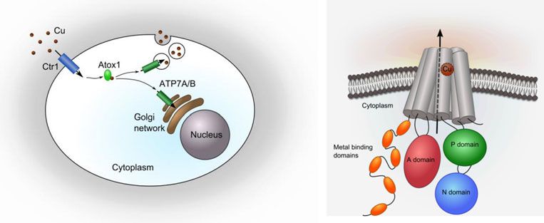
domains of ATP7B was reported based on pull-down data                            Interactions with other proteins and
                      (Tsivkovskii et al. 2001); this interaction was found to de-                     new functions
                      pend on the metal-loading status as well as on phosphoryla-
                      tion events and it was suggested to be an auto-inhibitory                        In addition to internal domain–domain interactions, other
                      interaction (Hasan et al. 2012). A similar intra-protein in-                     proteins have also been proposed to regulate ATP7A/B ac-
                      teraction was reported for domains of a homologous bac-                          tivity. For example, the protein COMMD1 (Copper
                      terial Cu pump, also using pull down experiments with                            Metabolism Murr1 Domain) was recently shown to interact
                      tagged proteins (Gonzalez-Guerrero et al. 2009). Since the                       with the metal-binding domains of ATP7B and this ap-
                      bacterial homolog has only one metal-binding domain,                             peared to regulate overall ATP7B stability via the ER degra-
                      one may speculate that the 6th metal-binding domain in                           dation pathway (de Bie et al. 2007; Materia et al. 2012). The
                      ATP7B plays a key role in the interaction within the                             binding sites on neither protein nor the physical mechanism
                      human protein, perhaps with the additional five metal-                            resulting in ER degradation (interaction causing protein
                      binding domains and the unstructured loop, unique to the                         destabilization or triggering of a cellular signal) are
                      human N-domain, fine-tuning the binding. However, our                             known. Surprisingly, COMMD1 was found to specifically
                      biophysical studies using 15N-labeled purified domains                            bind Cu2+ in vitro and, therefore, it was speculated that
                      (N-domain mixed with four-domain construct, WD1-4 or                             the COMMD1–ATP7B interaction may be a way to elimin-
                      with WD56) did not reveal any interaction for any protein                        ate oxidized (toxic) Cu from cells (Sarkar & Roberts, 2011).
                      pair (Åden and Wittung-Stafshede, unpublished), although
                                                                                                       The general idea has been that mammalian Cu transport protein
                      we used high μM protein concentrations (Fig. 3). This sug-
                                                                                                       levels are primarily regulated post-transcriptionally, such as via
                      gests that the interaction identified by pull-down experi-
                                                                                                       degradation pathways (Hasan & Lutsenko, 2012). To control for
                      ments depends sensitively on the environment, such as
                                                                                                       elevated Cu levels, ATP7A/B can redistribute reversibly from the
                      pH, salt and inter-domain interactions, or possibly on ad-
                                                                                                       Golgi to the plasma membrane to expel Cu and thereby protect
                      ditional components present in the lysate. In a general
                                                                                                       against Cu toxicity (La Fontaine & Mercer, 2007). However, in
                      sense, this result highlights the elusive nature of regulatory
                                                                                                       2008 it was reported that Atox1 had dual functionality and
                      interactions; one must clearly test a range of conditions
                                                                                                       also acted as a Cu-dependent transcription factor (TF) that
                      and constructs before making conclusions.
                                                                                                       drives the expression of Ccd1 (cyclin D1), a protein involved
                                                                                                       in cell proliferation. A direct Cu-dependent interaction of
                                                                                                       GST-tagged Atox1 with a GAAAGA sequence in the promotor
                      Fate of Cu after reaching the lumen                                              region of Ccnd1 was demonstrated by an electrophoretic mo-
                      After Cu has passed the ATP7A/B channel to the lumen it is                       bility shift assay (EMSA) (Itoh et al. 2008). Subsequently, this
                      unclear how it is added onto target polypeptides. In ATP7A,                      motif was found in the promotor regions of several other
                      a luminal loop has been identified that has Cu-binding ca-                        genes (Muller & Klomp, 2009). In support of playing a role
                      pacity (Barry et al. 2011). In ATP7B, this loop is shorter                       in the nucleus, the sequence of Atox1 contains an apparent
                      but still has Cu-binding residues. Thus, this loop may be a                      nuclear localization signal KKTGK within its C-terminal
                      transient site for the Cu ion before it is loaded on a target                    part and, although not discussed, in the initial discovery
                      polypeptide. The mechanism of Cu loading onto target                             paper of Atox1 from 1999 (Hamza et al. 1999), immunofluor-
                      polypeptides is unknown; one possibility is that Cu becomes                      escence of HeLa cells indicated that Atox1 was distributed
                      free in solution after leaving ATP7A/B and binding is sim-                       throughout the cell, including the nucleus. In our work, we
                      ply driven by Cu–protein affinity.                                                 have also detected Atox1 in the nuclei of HeLa cells (Fig. 4)
                                                                                                       although we find no binding to DNA duplexes harboring
                      Ceruloplasmin is a large six-domain ferroxidase, requiring
                                                                                                       the target sequence in vitro (Kahra et al. 2015). The answer
                      six Cu ions for activity, which is Cu-loaded by ATP7B in
                                                                                                       to this apparent paradox may be that Atox1 is an indirect
                      the secretory pathway. Our in vitro studies of ceruloplasmin
                                                                                                       TF working via interaction with another protein that contains
                      unfolding imply that metal binding must take place early on
                                                                                                       the DNA-binding ability.
                      during protein folding in order for the polypeptide to fold
                      into its functional form. If the polypeptide is allowed to                       To search for new Atox1 partners, we performed a yeast two-
                      fold without metals, then it misfolds into a dead end species                    hybrid screen (Hybrigenics; similar to (Rain et al. 2001))
                      that cannot bind Cu (Sedlak & Wittung-Stafsheden, 2007;                          using Atox1 as a LexA fusion bait toward a human placenta
                      Sedlak et al. 2008). Thus, appropriate timing of folding                         library of protein fragments (Wittung-Stafshede, unpub-
                      and binding events in the final step of the Cu transport cas-                     lished); among 98 million possible interactions, we found
                      cade appears crucial. In the case of ceruloplasmin, this                         25 confident target proteins that interacted with Atox1
                      becomes important in the treatment of aceruloplasminemia                         (Table 1). Of these interactions partners, at least six are
                      (a condition where mutated ceruloplasmin it not loaded                           known DNA/RNA-binding proteins. The results of this
                      with Cu) since Cu supplementation will not rescue already                        screen demonstrate that the Cu transport network is greater
                      misfolded apo-forms of ceruloplasmin.                                            than what is currently known and, moreover, suggest that

                                                                                                 474
Downloaded from https://www.cambridge.org/core. IP address: 46.4.80.155, on 13 Nov 2021 at 18:06:47, subject to the Cambridge Core terms of use, available at https://www.cambridge.org/core/terms.
https://doi.org/10.1017/S0033583515000128
Fig. 3. 1H–15N–HSQC spectra recorded at 850 MHz in 50 mM Tris, 50 mM NaCl, 2 mM DTT, 6% D2O (v/v) at pH 8·0 and 25°C. (a)
150 μM 15N-labeled N-domain (blue), and together with 150 μM unlabeled WD56 (red). (b) 200 μM 15N-labeled WD1-4 (blue), and
together with 200 μM unlabeled N-domain (red).
Cu transport proteins (i.e. Atox1) likely have additional (yet Outlook for Cu pump biophysics
unknown) partners and functions. Although only a screen
that will require extensive follow up, these types of large-scale Many mutations in the genes that code for Cu transport pro-
experiments that are available today are important in that teins have been linked to human diseases. Mutations in
they may identify new directions for future biophysical work. ATP7A/B constitute the basis of Wilson and Menke’s
475
Downloaded from https://www.cambridge.org/core. IP address: 46.4.80.155, on 13 Nov 2021 at 18:06:47, subject to the Cambridge Core terms of use, available at https://www.cambridge.org/core/terms.
https://doi.org/10.1017/S0033583515000128Fig. 4. Localization of Atox1 in HeLa cells detected by wide-field fluorescence microscopy using monoclonal Alexa Fluor488 tagged anti-
mouse antibodies specific for Atox1. In most cells Atox1 appears in perinuclear areas and as punctuate structures in close contact with
the plasma membrane (b), indicative of Golgi compartment and transport vesicle localization, respectively. However, there are also cells
with increased Atox1 levels within the nucleus (a).
Table 1. Results from a cDNA yeast two hybrid screen using Atox1 as bind specifically to the amyloidogenic proteins that are
bait (LexA fusion) and a human placenta RP6 library as prey involved in Huntington’s, Parkinson’s and Alzheimer’s dis-
(Hybrigenics). 98·2 million interactions were analyzed and 310 posi- eases; upon binding, amyloid formation is promoted in
tive clones were fully processed. Detected interactions with the highest
predicted biological scores (PBS) are listed below divided in four cat-
vitro (Faller et al. 2013), suggesting that Cu may be a causative
egories from A (the highest confidence rank) to D. There were two ad- agent of these diseases in vivo.
ditional unknown proteins in B, and 12 additional proteins in the D
Another aspect is drug treatment and side effects. For the can-
category (three ubiquitin specific peptidases and nine unknown pro-
teins) not listed here. No C scores were found. Several of the detected cer drug cisplatin it has been reported that the drug hijacks Cu
interaction partners have DNA/RNA binding capacity (bold name) transport proteins as a possible mechanism to become
exported out of cells. We showed that Atox1 can bind cispla-
PBS Target Comment on target protein tin together with Cu creating a di-metal site. Cisplatin binding
causes Atox1 unfolding in vitro but prior to this, if a partner
A PTPRF Protein tyrosine phosphatase, receptor-like; cell such as ATP7B is present, the cancer drug can be transferred
signaling, cancer further (Palm et al. 2011; Palm-Espling & Wittung-Stafshede,
A KIAA0947 Nuclear protein involved in RNA binding and
2012; Palm-Espling et al. 2013, 2014). This may result in drug
regulation
A ATP7B Known Atox1 partner (positive control) resistance but, considering the possibility of Atox1 entering
B ATP7A Known Atox1 partner (positive control) the cell nucleus, Atox1 may in fact mediate the delivery of
B DNMT1 C-5 cytosine methyl transferase the drug to DNA.
B CRELD2 Cysteine-rich with EFG-like Ca-binding
domains Clearly, biophysical knowledge of mechanisms and regu-
B ZFHX3 TF, with zinc fingers lation of Cu transport proteins may aid the development of
D CPEB4 RNA-binding protein new approaches to target disorders involving Cu transport
D LMCD1 Zinc-finger protein with LIM and cysteine-rich proteins and imbalance in Cu metabolism. Biophysical stu-
domains, regulator of transcription
D PPM1A Phosphatase; regulator of stress response, cell
dies of the mechanisms and proteins that facilitate human
cycle control Cu transport may also provide predictions for how other
D TRIM26 Protein with tripartite motif (includes zinc- metal transport systems work. Like in Cu transport proteins,
binding domains), DNA binding the ferredoxin-like structural fold appears commonly used by
zinc transport proteins (Lu & Fu, 2007; Lu et al. 2009), which
is a group of metal transporters for which mechanistic and
biophysical knowledge is severely lacking. Taken together,
diseases; missense mutations in almost all domains of ATP7B the unresolved questions described above emphasize the
have been linked to Wilson disease with the most common need for more careful biophysical studies and invite young
mutation being H1069Q in the N-domain. Using a range of biophysical scientists to enter this wide-open field.
biophysical methods we could explain the underlying mech-
anism: the mutation did not affect domain stability or
ATP-binding affinity; however, it made ATP bind in such a
way that hydrolysis was hindered (Rodriguez-Granillo et al.
Acknowledgements
2008). In addition to direct genetic defects, Cu accumulation I thank my group members Jörgen Åden, Moritz Niemiec,
(either due to, or causing, Cu transport dysregulation) is often Dana Kahra and Tanumoy Mondol for providing unpub-
found in cancer tumors and upon neurodegeneration. Cu can lished data, Åden for preparing Fig. 3, and Dana Kahra for
476
Downloaded from https://www.cambridge.org/core. IP address: 46.4.80.155, on 13 Nov 2021 at 18:06:47, subject to the Cambridge Core terms of use, available at https://www.cambridge.org/core/terms.
https://doi.org/10.1017/S0033583515000128Fig. 4. Former graduate student Maria Palm-Espling made associated with enhanced binding to COMMD1 and reduced
Fig. 1. The Swedish Research Council, the Wallenberg foun- stability of ATP7B. Gastroenterology 133(4), 1316–1326.
dation, and Umeå University are acknowledged for financial FALLER, P., HUREAU, C. & BERTHOUMIEU, O. (2013). Role of metal ions
support. This text was written during a sabbatical period at in the self-assembly of the Alzheimer’s amyloid-beta peptide.
Inorganic Chemistry 52(21), 12193–12206.
California Institute of Technology.
FESTA, R. A. & THIELE, D. J. (2011). Copper: an essential metal in bi-
ology. Current Biology 21(21), R877–R883.
FORBES, J. R., HIS, G. & COX, D. W. (1999). Role of the copper-
Conflict of interest binding domain in the copper transport function of ATP7B,
None. the P-type ATPase defective in Wilson disease. Journal of
Biological Chemistry 274(18), 12408–12413.
GONZALEZ-GUERRERO, M. & ARGUELLO, J. M. (2008). Mechanism of
Cu+-transporting ATPases: soluble Cu+ chaperones directly
References transfer Cu+ to transmembrane transport sites. Proceedings of
ACHILA, D., BANCI, L., BERTINI, I., BUNCE, J., CIOFI-BAFFONI, S. & the National Academy of Sciences of the United States of
HUFFMAN, D. L. (2006). Structure of human Wilson protein America 105(16), 5992–5997.
domains 5 and 6 and their interplay with domain 4 and the cop- GONZALEZ-GUERRERO, M., HONG, D. & ARGUELLO, J. M. (2009).
per chaperone HAH1 in copper uptake. Proceedings of the Chaperone-mediated Cu+ delivery to Cu+ transport ATPases: re-
National Academy of Sciences of the United States of America quirement of nucleotide binding. Journal of Biological Chemistry
103(15), 5729–5734. 284(31), 20804–20811.
ARUMUGAM, K. & CROUZY, S. (2012). Dynamics and stability of the GOURDON, P., LIU, X. Y., SKJORRINGE, T., MORTH, J. P., MOLLER, L. B.,
metal binding domains of the Menkes ATPase and their interac- PEDERSEN, B. P. & NISSEN, P. (2011). Crystal structure of a copper-
tion with metallochaperone HAH1. Biochemistry 51(44), transporting PIB-type ATPase. Nature 475(7354), 59–64.
8885–8906. GOURDON, P., SITSEL, O., LYKKEGAARD KARLSEN, J., BIRK MOLLER, L. &
BANCI, L. (2006). The Atx1-Ccc2 complex is a metal-mediated pro- NISSEN, P. (2012). Structural models of the human copper P-type
tein–protein interaction. Nature Chemical Biology 2, 367–368. ATPases ATP7A and ATP7B. Biological Chemistry 393(4),
BANCI, L., BERTINI, I., CALDERONE, V., DELLA-MALVA, N., FELLI, I. C., 205–216.
NERI, S., PAVELKOVA, A. & ROSATO, A. (2009a). Copper HAMZA, I., SCHAEFER, M., KLOMP, L. W. & GITLIN, J. D. (1999).
(I)-mediated protein–protein interactions result from suboptimal Interaction of the copper chaperone HAH1 with the Wilson dis-
interaction surfaces. Biochemical Journal 422(1), 37–42. ease protein is essential for copper homeostasis. Proceedings of
BANCI, L., BERTINI, I., CANTINI, F., MASSAGNI, C., MIGLIARDI, M. & the National Academy of Sciences of the United States of
ROSATO, A. (2009b). An NMR study of the interaction of the America 96(23), 13363–13368.
N-terminal cytoplasmic tail of the Wilson disease protein with HARRIS, E. D. (2003). Basic and clinical aspects of copper. Critical
copper(I)-HAH1. Journal of Biological Chemistry 284(14), Reviews in Clinical Laboratory Sciences 40(5), 547–586.
9354–9360. HASAN, N. M., GUPTA, A., POLISHCHUK, E., YU, C. H., POLISHCHUK, R.,
BANCI, L., BERTINI, I., CANTINI, F., ROSENZWEIG, A. C. & YATSUNYK, L. DMITRIEV, O. Y. & LUTSENKO, S. (2012). Molecular events initiat-
A. (2008). Metal binding domains 3 and 4 of the Wilson disease ing exit of a copper-transporting ATPase ATP7B from the
protein: solution structure and interaction with the copper(I) trans-Golgi network. Journal of Biological Chemistry 287(43),
chaperone HAH1. Biochemistry 47(28), 7423–7429. 36041–36050.
BANCI, L., BERTINI, I., CIOFI-BAFFONI, S., CHASAPIS, C. T., HADJILIADIS, HASAN, N. M. & LUTSENKO, S. (2012). Regulation of copper transpor-
N. & ROSATO, A. (2005). An NMR study of the interaction be- ters in human cells. Current Topics in Membrane 69, 137–161.
tween the human copper(I) chaperone and the second and fifth HUFFMAN, D. L. & O’HALLORAN, T. V. (2001). Function, structure,
metal-binding domains of the Menkes protein. FEBS Journal and mechanism of intracellular copper trafficking proteins.
272(3), 865–871. Annual Reviews of Biochemistry 70, 677–701.
BARRY, A. N., OTOIKHIAN, A., BHATT, S., SHINDE, U., TSIVKOVSKII, R., ITOH, S., KIM, H. W., NAKAGAWA, O., OZUMI, K., LESSNER, S. M., AOKI,
BLACKBURN, N. J. & LUTSENKO, S. (2011). The lumenal loop H., AKRAM, K., MCKINNEY, R. D., USHIO-FUKAI, M. & FUKAI, T.
Met672-Pro707 of copper-transporting ATPase ATP7A binds (2008). Novel role of antioxidant-1 (Atox1) as a copper-
metals and facilitates copper release from the intramembrane dependent transcription factor involved in cell proliferation.
sites. Journal of Biological Chemistry 286(30), 26585–26594. Journal of Biological Chemistry 283(14), 9157–9167.
BUBLITZ, M., MUSGAARD, M., POULSEN, H., THOGERSEN, L., OLESEN, C., KAHRA, D., MONDOL, T., NIEMIEC, M. & WITTUNG-STAFSHEDE, P.
SCHIOTT, B., MORTH, J. P., MOLLER, J. V. & NISSEN, P. (2013). Ion (2015). Human copper chaperone Atox1 translocates to the
pathways in the sarcoplasmic reticulum Ca2+-ATPase. Journal nucleus but does not bind DNA in vitro. Protein & Peptide
of Biological Chemistry 288(15), 10759–10765. Letters 22(6), 532–538.
CULOTTA, V. C., YANG, M. & HALL, M. D. (2005). Manganese trans- KOCH, K. A., PENA, M. M. & THIELE, D. J. (1997). Copper-binding
port and trafficking: lessons learned from Saccharomyces cerevi- motifs in catalysis, transport, detoxification and signaling.
siae. Eukaryotic Cell 4(7), 1159–1165. Chemistry and Biology 4(8), 549–560.
DE BIE, P., VAN DE SLUIS, B., BURSTEIN, E., VAN DE BERGHE, P. V., LA FONTAINE, S. & MERCER, J. F. (2007). Trafficking of the
MULLER, P., BERGER, R., GITLIN, J. D., WIJMENGA, C. & KLOMP, L. copper-ATPases, ATP7A and ATP7B: role in copper homeosta-
W. (2007). Distinct Wilson’s disease mutations in ATP7B are sis. Archives of Biochemistry and Biophysics 463(2), 149–167.
477
Downloaded from https://www.cambridge.org/core. IP address: 46.4.80.155, on 13 Nov 2021 at 18:06:47, subject to the Cambridge Core terms of use, available at https://www.cambridge.org/core/terms.
https://doi.org/10.1017/S0033583515000128LU, M., CHAI, J. & FU, D. (2009). Structural basis for autoregulation unfolding in vitro. Proceedings of the National Academy of
of the zinc transporter YiiP. Nature Structural and Molecular Sciences of the United States of America 108(17), 6951–6956.
Biology 16(10), 1063–1067. PETZOLDT, S., KAHRA, D., KOVERMANN, M., DINGELDEIN, A. P., NIEMIEC,
LU, M. & FU, D. (2007). Structure of the zinc transporter YiiP. M. S., ADEN, J. & WITTUNG-STAFSHEDE, P. (2015). Human cytoplas-
Science 317(5845), 1746–1748. mic copper chaperones Atox1 and CCS exchange copper ions in
LUTSENKO, S., BARNES, N. L., BARTEE, M. Y. & DMITRIEV, O. Y. (2007). vitro. Biometals 28, 577–585.
Function and regulation of human copper-transporting ATPases. PUFAHL, R. A., SINGER, C. P., PEARISO, K. L., LIN, S. J., SCHMIDT, P. J.,
Physiological Reviews 87(3), 1011–1046. FAHRNI, C. J., CULOTTA, V. C., PENNER-HAHN, J. E. & O’HALLORAN,
MATERIA, S., CATER, M. A., KLOMP, L. W., MERCER, J. F. & LA T. V. (1997). Metal ion chaperone function of the soluble Cu(I)
FONTAINE, S. (2012). Clusterin and COMMD1 independently receptor Atx1. Science 278(5339), 853–856.
regulate degradation of the mammalian copper ATPases ATP7A PUIG, S. & THIELE, D. J. (2002). Molecular mechanisms of copper
and ATP7B. Journal of Biological Chemistry 287(4), 2485–2499. uptake and distribution. Current Opinion in Chemical Biology 6
MORIN, I., GUDIN, S., MINTZ, E. & CUILLEL, M. (2009). Dissecting the (2), 171–180.
role of the N-terminal metal-binding domains in activating the RAIN, J. C., SELIG, L., DE REUSE, H., BATTAGLIA, V., REVERDY, C., SIMON,
yeast copper ATPase in vivo. FEBS Journal 276(16), 4483–4495. S., LENZEN, G., PETEL, F., WOJCIK, J., SCHACHTER, V., CHEMAMA, Y.,
MULLER, P. A. & KLOMP, L. W. (2009). ATOX1: a novel copper- LABIGNE, A. & LEGRAIN, P. (2001). The protein–protein interaction
responsive transcription factor in mammals? International map of Helicobacter pylori. Nature 409(6817), 211–215.
Journal of Biochemistry and Cell Biology 41(6), 1233–1236. ROBINSON, N. J. & WINGE, D. R. (2010). Copper metallochaperones.
NIEMIEC, M. S., DINGELDEIN, A. P. & WITTUNG-STAFSHEDE, P. (2014). T Annual Reviews of Biochemistry 79, 537–562.
versus D in the MTCXXC motif of copper transport proteins RODRIGUEZ-GRANILLO, A., CRESPO, A., ESTRIN, D. A. &
plays a role in directional metal transport. Journal of Biology WITTUNG-STAFSHEDE, P. (2010). Copper-transfer mechanism from
and Inorganic Chemistry 19, 1037–1047. the human chaperone Atox1 to a metal-binding domain of Wilson
NIEMIEC, M.S., DINGELDEIN, A.P. & WITTUNG-STAFSHEDE, P. (2015). disease protein. Journal of Physical Chemistry B 114(10), 3698–3706.
Enthalpy-entropy compensation at play in human copper ion RODRIGUEZ-GRANILLO, A., SEDLAK, E. & WITTUNG-STAFSHEDE, P.
transfer. International Journal of Scientific Reports 5, 10518. (2008). Stability and ATP binding of the nucleotide-binding
NIEMIEC, M. S., WEISE, C. F. & WITTUNG-STAFSHEDE, P. (2012). In vitro domain of the Wilson disease protein: effect of the common
thermodynamic dissection of human copper transfer from cha- H1069Q mutation. Journal of Molecular Biology 383(5),
perone to target protein. PLoS ONE 7(5): e36102. 1097–1111.
NILSSON, L., ADEN, J., NIEMIEC, M. S., NAM, K. & WITTUNG-STAFSHEDE, SARKAR, B. & ROBERTS, E. A. (2011). The puzzle posed by COMMD1, a
P. (2013). Small pH and salt variations radically alter the thermal newly discovered protein binding Cu(II). Metallomics 3(1), 20–27.
stability of metal-binding domains in the copper transporter, SEDLAK, E. & WITTUNG-STAFSHEDE, P. (2007). Discrete roles of copper
Wilson disease protein. Journal of Physical Chemistry B 117 ions in chemical unfolding of human ceruloplasmin.
(42), 13038–13050. Biochemistry 46(33), 9638–9644.
O’HALLORAN, T. V. & CULOTTA, V. C. (2000). Metallochaperones, an SEDLAK, E., ZOLDAK, G. & WITTUNG-STAFSHEDE, P. (2008). Role of
intracellular shuttle service for metal ions. Journal of Biological copper in thermal stability of human ceruloplasmin. Biophysical
Chemistry 275(33), 25057–25060. Journal 94(4), 1384–1391.
OHRVIK, H. & THIELE, D. J. (2014). How copper traverses cellular TOTTEY, S., HARVIE, D. R. & ROBINSON, N. J. (2005). Understanding
membranes through the mammalian copper transporter 1, how cells allocate metals using metal sensors and metallochaper-
Ctr1. Annals of the New York Academy of Sciences 1314, 32–41. ones. Accounts of Chemical Research 38(10), 775–783.
PALM-ESPLING, M. E., ANDERSSON, C. D., BJORN, E., LINUSSON, A. & TOTTEY, S., WALDRON, K. J., FIRBANK, S. J., REALE, B., BESSANT, C., SATO,
WITTUNG-STAFSHEDE, P. (2013). Determinants for simultaneous K., CHEEK, T. R., GRAY, J., BANFIELD, M. J., DENNISON, C. &
binding of copper and platinum to human chaperone Atox1: ROBINSON, N. J. (2008). Protein-folding location can regulate
hitchhiking not hijacking. PLoS ONE 8(7), e70473. manganese-binding versus copper- or zinc-binding. Nature 455
PALM-ESPLING, M. E., LUNDIN, C., BJORN, E., NAREDI, P. & (7216), 1138–1142.
WITTUNG-STAFSHEDE, P. (2014). Interaction between the anticancer TSIVKOVSKII, R., MACARTHUR, B. C. & LUTSENKO, S. (2001). The
drug Cisplatin and the copper chaperone Atox1 in human Lys1010-Lys1325 fragment of the Wilson’s disease protein
melanoma cells. Protein and Peptide Letters 21(1), 63–68. binds nucleotides and interacts with the N-terminal domain of
PALM-ESPLING, M. E. & WITTUNG-STAFSHEDE, P. (2012). Reaction of plati- this protein in a copper-dependent manner. Journal of
num anticancer drugs and drug derivatives with a copper transport- Biological Chemistry 276(3), 2234–2242.
ing protein, Atox1. Biochemical Pharmacology 83(7), 874–881. WERNIMONT, A. K., HUFFMAN, D. L., LAMB, A. L., O’HALLORAN, T. V. &
PALM, M. E., WEISE, C. F., LUNDIN, C., WINGSLE, G., NYGREN, Y., BJORN, ROSENZWEIG, A. C. (2000). Structural basis for copper transfer by
E., NAREDI, P., WOLF-WATZ, M. & WITTUNG-STAFSHEDE, P. (2011). the metallochaperone for the Menkes/Wilson disease proteins.
Cisplatin binds human copper chaperone Atox1 and promotes Nature Structural Biology 7(9), 766–771.
478
Downloaded from https://www.cambridge.org/core. IP address: 46.4.80.155, on 13 Nov 2021 at 18:06:47, subject to the Cambridge Core terms of use, available at https://www.cambridge.org/core/terms.
https://doi.org/10.1017/S0033583515000128You can also read