A cardiac myosin binding protein C mutation in the Maine Coon cat with familial hypertrophic cardiomyopathy
←
→
Page content transcription
If your browser does not render page correctly, please read the page content below
Human Molecular Genetics, 2005, Vol. 14, No. 23              3587–3593
                                                                  doi:10.1093/hmg/ddi386
                                                                  Advance Access published on October 19, 2005

A cardiac myosin binding protein C mutation
in the Maine Coon cat with familial hypertrophic
cardiomyopathy
Kathryn M. Meurs1,*, Ximena Sanchez2, Ryan M. David2, Neil E. Bowles2, Jeffrey A. Towbin2,
Peter J. Reiser3, Judith A. Kittleson4, Marcia J. Munro4, Keith Dryburgh1, Kristin A. MacDonald4
and Mark D. Kittleson4

                                                                                                                                               Downloaded from http://hmg.oxfordjournals.org/ at University of California, Davis - Library on September 17, 2012
1
 Department of Veterinary Clinical Sciences, College of Veterinary Medicine, The Ohio State University, 601 Vernon
Tharp Street, Columbus, OH 43210, USA, 2Department of Pediatric Cardiology, 6621 Fannin, Baylor College of
Medicine, Houston, TX 77030, USA, 3Department of Oral Biology, College of Dentistry, The Ohio State University,
4195 12th Street, Columbus, OH 43210, USA and 4Department of Medicine and Epidemiology, School of Veterinary
Medicine, University of California, Davis, One Shields Avenue, Davis, CA 95616, USA

Received August 13, 2005; Revised and Accepted October 11, 2005

Hypertrophic cardiomyopathy (HCM) is one of the most common causes of sudden cardiac death in young
adults and is a familial disease in at least 60% of cases. Causative mutations have been identified in several
sarcomeric genes, including the myosin binding protein C (MYBPC3) gene. Although numerous causative
mutations have been identified, the pathogenetic process is still poorly understood. A large animal model
of familial HCM in the cat has been identified and may be used for additional study. As the first spontaneous
large animal model of this familial disease, feline familial HCM provides a valuable model for investigators to
evaluate pathophysiologic processes and therapeutic (pharmacologic or genetic) manipulations. The
MYBPC3 gene was chosen as a candidate gene in this model after identifying a reduction in the protein in
myocardium from affected cats in comparison to control cats (P < 0.001). DNA sequencing was performed
and sequence alterations were evaluated for evidence that they changed the amino acid produced, that
the amino acid was conserved and that the protein structure was altered. We identified a single base pair
change (G to C) in the feline MYBPC3 gene in affected cats that computationally alters the protein confor-
mation of this gene and results in sarcomeric disorganization. We have identified a causative mutation in
the feline MYBPC3 gene that results in the development of familial HCM. This is the first report of a spon-
taneous mutation causing HCM in a non-human species. It should provide a valuable model for evaluating
pathophysiologic processes and therapeutic manipulations.

INTRODUCTION                                                           it is a familial disease. Spontaneous causative mutations
                                                                       have been identified in several genes that encode sarcomeric
Hypertrophic cardiomyopathy (HCM) is a clinically hetero-              proteins including the alpha and beta myosin heavy chains,
geneous myocardial disease characterized by increased left             cardiac myosin binding protein C (MYBPC3), cardiac tropo-
ventricular (LV) mass due to an increase in wall thickness             nins T, I and C, alpha tropomyosin, the essential and regulatory
in the absence of apparent pressure overload or metabolic              light chains, actin and, most recently, titin (1 –9). Mutations
stimuli and histologically by myofibrillar and myocyte dis-            within the genes that encode for the sarcomeric proteins
array (1,2). It has an estimated prevalence of one in 500              may lead to the development of the HCM phenotype by
humans and is one of the most common causes of sudden                  affecting either protein function or protein structure or both
cardiac death in young adults (1). In at least 60% of cases,           (1,9 –12).

*To whom correspondence should be addressed at: Department of Veterinary Clinical Sciences, College of Veterinary Medicine, Washington State
University, Pullman, WA 99164-6610, USA. Tel: þ1 5093350738; Fax: þ1 5093350880; Email: meurs@vetmed.wsu.edu

# The Author 2005. Published by Oxford University Press. All rights reserved.
For Permissions, please email: journals.permissions@oxfordjournals.org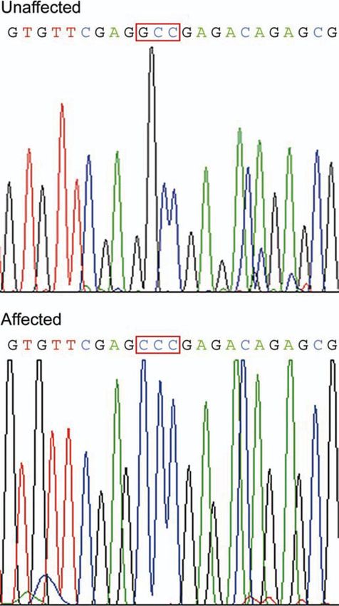
3588 Human Molecular Genetics, 2005, Vol. 14, No. 23
HCM is the most common cardiac disease identified in
domestic cats (13). A colony of a feline model of familial
HCM has been produced in the Maine Coon cat (13).
Because of the identification of over 240 mutations in genes
that encode for sarcomeric proteins in humans, we hypo-
thesized that a mutation in one of these genes would be
responsible for familial HCM in this animal model (1). After
identifying a reduction in the cMyBP-C protein in affected
cats, we identified a mutation in the feline gene that is pre-
dicted to alter the protein conformation of this gene and
results in sarcomeric disruption. This is the first report of a sarco-
meric gene mutation in a species other than human being. As
the first spontaneous large animal model of this familial
Downloaded from http://hmg.oxfordjournals.org/ at University of California, Davis - Library on September 17, 2012
disease, feline familial HCM provides an extremely valuable
model for investigators to evaluate pathophysiologic processes
and therapeutic (pharmacologic or genetic) manipulations.
RESULTS Figure 1. SDS–PAGE analysis of LV (free wall) myocardial samples from
Clinical description normal (lanes 1 and 2) and affected (lanes 3–6) cats. The cMyBP-C and myo-
mesin proteins are reduced and the anomalously migrating beta myosin
Twenty-three (16 affected, seven unaffected) Maine Coon cats appears to be increased in comparison to the normal cats. The genotypes of
from a colony with familial HCM, as previously described, the cats are shown below the lanes as G/G (normal cat), C/G (affected hetero-
zygote) and C/C (affected homozygote).
and 100 unaffected control cats were evaluated. The pedigree
of the cats in the colony studied has been published previously
(13). Disease status of adult cats was identified by repeated
echocardiographic examinations and the median of LV wall
thickness and interventricular wall thickness was 7 mm
(range: 6– 9 mm; normal ¼ 3– 5 mm) in affected cats. Most
affected cats also had systolic anterior motion of the mitral
valve and left atrial enlargement. Papillary muscle hypert-
rophy was frequently noted.
Sarcomeric protein concentrations are altered
Myocardial samples were obtained from the LV free wall of
eight affected cats at the time of death due to euthanasia for
refractory heart failure, or as soon after death as possible
from cats that died suddenly, as well as from three apparently Figure 2. Immunoblot performed to confirm the identification of the abnormal
healthy unrelated cats. Myocardial proteins were evaluated by proteins. Lanes 1 and 3 contain myocardial samples from normal cats, lane 2
sodium dodecyl sulphate –polyacrylamide gel electrophoresis contains a myocardial sample from an affected (heterozygote) cat. The geno-
(SDS– PAGE) analysis. Two proteins were noted to be types of the cats are shown below the lanes as G/G (normal cat) and C/G
(affected heterozygote).
absent or greatly reduced, and one protein was noted to be
increased in the affected cats in comparison to the normal
cats. The identification of the proteins that were reduced or
increased in the affected cats was tentatively determined by Identification of a MYBPC3 mutation
analysis with MALDI mass spectrometry and by entering the
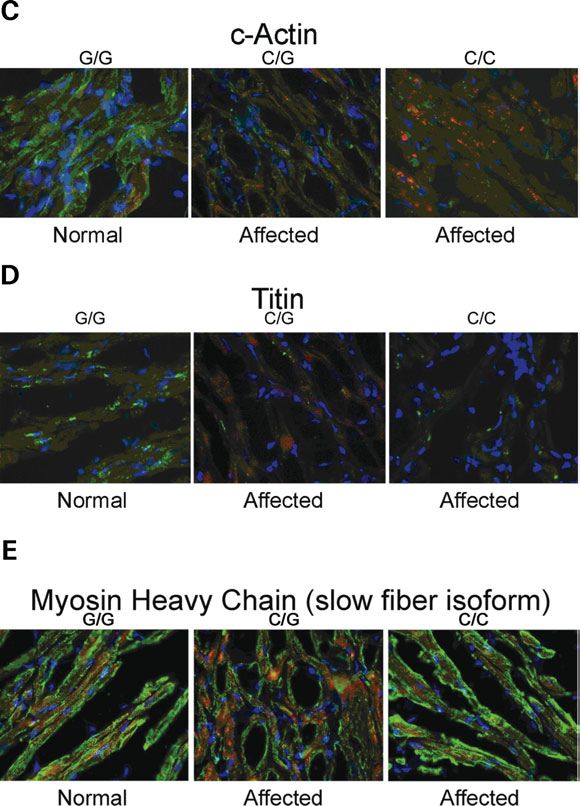
peptide mass information into the NCBI database. The pro- Because of the marked and consistent reduction in myocardial
teins reduced were identified as cMyBP-C and myomesin, MYBPC3 concentration in affected cats, the MYBPC3 gene
an M-band protein. The protein that was increased in the was targeted for analysis. DNA sequencing revealed a single
affected cats was identified as anomalously migrating beta base pair change (G to C) in codon 31 (exon 3) in affected
myosin (Fig. 1). Western blot analysis confirmed the identifi- cats (Fig. 4). This changed a conserved amino acid from
cation of the reduced proteins (Fig. 2). When the proteins from alanine (A) to proline (P) (A31P) in each of the Maine Coon
the SDS –PAGE were evaluated quantitatively by densito- cats with HCM, but none of the unaffected Maine Coon or
metry, both the cMyBP-C and myomesin proteins were sig- control cats. Affected cats were either heterozygous (n ¼ 10)
nificantly reduced in the affected cats in comparison to the or homozygous (n ¼ 6) for the mutation based on direct
control cats (cMyBP-C, P , 0.001 and myomesin, P ¼ 0.011) DNA sequence analysis. Computer protein structure analysis
(Fig. 3). A weak inverse correlation (r ¼ 20.341) was observed predicted a reduction in the alpha helix and an increase in
between the amount of anomalously migrating myosin and the random coils in this region of the molecule in the affected
cMyBP-C for affected cats. cats (Fig. 5).
Human Molecular Genetics, 2005, Vol. 14, No. 23                         3589

                                                                                                                                                                     Downloaded from http://hmg.oxfordjournals.org/ at University of California, Davis - Library on September 17, 2012
Figure 3. Quantitative (arbitrary densitometry units) SDS–PAGE analysis of
myocardial proteins in normal and affected cats. Both the cMyBP-C and myo-
mesin proteins were significantly reduced in the affected cats in comparison to
the control cats (cMyBP-C, P , 0.001 and myomesin, P ¼ 0.011). A weak
inverse correlation (r ¼ 20.341) was observed between the amount of ano-          Figure 5. Computer protein structure analysis predicted a reduction in the
malously migrating myosin and the cMyBP-C for affected cats. The genotypes        alpha helix (blue) and an increase in random coils (purple) in the amino
of the cats are shown as G/G (three normal cats), C/G (seven affected hetero-     acid region from 10 to 20 (altered amino acid is 16) of the molecule in affected
zygotes) and C/C (one affected homozygote).                                       cats compared to normal cats.

                                                                                  Sarcomeric protein organization is altered
                                                                                  Immunofluorescence analysis of sarcomeric proteins in LV
                                                                                  sections from affected and unaffected cats revealed significant
                                                                                  disruption in several sarcomeric proteins in affected cats, with
                                                                                  reductions in staining intensity of cMyBP-C, myomesin, titin
                                                                                  and cardiac actin (Fig. 6A – D). Staining for myosin heavy
                                                                                  chain (Fig. 6E) and connexin 43 (data not shown) were
                                                                                  normal.

                                                                                  MYBPC3 mRNA is increased
                                                                                  Although the cMyBP-C protein was noted to be reduced by
                                                                                  SDS –PAGE, western blot analysis and immunofluorescence,
                                                                                  quantification of MYBPC3 mRNA by reverse transcription,
                                                                                  real-time PCR in three affected (two heterozygous and one
                                                                                  homozygous) cats and three unaffected cats determined that
                                                                                  the amount of mRNA in affected cats was increased 1.25 –
                                                                                  3-fold (HPRT and actin as housekeeping genes) (Fig. 7).

                                                                                  Disease outcome relates to genotype
                                                                                  The phenotype of the affected cats evaluated in this study
                                                                                  varied from moderate to severe HCM in cats whether they
                                                                                  had one or two affected alleles. Most of the cats had echocar-
                                                                                  diographic evidence of HCM by 2– 3 years of age, but one
                                                                                  female (heterozygote) did not have echocardiographically
                                                                                  identifiable disease until 7 years of age. The clinical
                                                                                  outcome of the disease did vary with the genotype with a
                                                                                  larger number of cats with a homozygous mutation developing
                                                                                  moderate to severe disease and dying of their disease at
                                                                                  4 years of age or less, four of them suddenly (Table 1).
                                                                                  One of these cats appeared echocardiographically normal
                                                                                  but died unexpectedly under anesthesia at 4 years of age. Of
                                                                                  the 10 cats with a heterozygous mutation, three are still
                                                                                  alive at 8 –12 years of age with moderate disease and only
Figure 4. DNA sequencing identified a single base pair change (G to C) in
codon 31 of the MYBPC3 gene in the affected cats (n ¼ 16) but not in any          one died suddenly, a larger number of these cats developed
of the unaffected family members (n ¼ 7) or control cats (n ¼ 100). A homo-       severe HCM and died of heart failure. One died of an un-
zygous affected cat is displayed.                                                 related cause.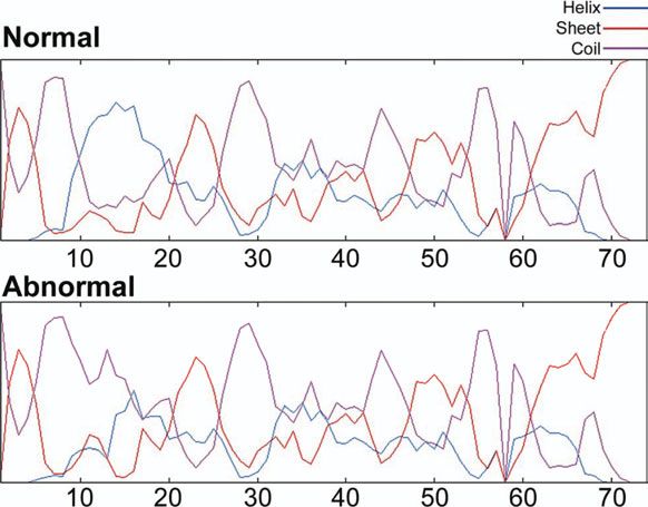
3590        Human Molecular Genetics, 2005, Vol. 14, No. 23

                                                                                                                                                                Downloaded from http://hmg.oxfordjournals.org/ at University of California, Davis - Library on September 17, 2012
                                                                                   Figure 7. Histogram of the ratio of MYPBPC3 message RNA for three
                                                                                   affected cats (two heterozygous and one homozygous) to three unaffected
                                                                                   cats demonstrating that the amount of message RNA was increased 1.25–3-
                                                                                   fold in affected cats. The HPRT and feline actin genes were used as house-
                                                                                   keeping genes.

                                                                                   Table 1. The clinical outcome of the disease appeared to vary with the
                                                                                   genotype although the number of cats in each group was small and should be
                                                                                   cautiously interpreted

                                                                                   Phenotype       Sudden death        Congestive         Died of
                                                                                                                       heart failure      non-cardiac disease

                                                                                   G/C             1/10                5/10               1/10
                                                                                   C/C             4/6                 0/6                1/6

                                                                                   HCM identified in a species other than Homo sapiens.
                                                                                   Although mutations in exon 3 of the MYBPC3 have been
                                                                                   reported as causative for familial HCM previously in human
                                                                                   beings, this particular mutation has never been reported (14).
                                                                                   The amino acid affected is located in the linker region
                                                                                   between domains C0 and C1 of the protein. The functional
                                                                                   aspects of this area are not well understood, however, there
                                                                                   is evidence that domain C0 and the C0 – C1 linker region
                                                                                   may bind to myosin and/or actin (15 –18). The observations
                                                                                   that the mutation identified in this model changes the com-
                                                                                   puted structure of this protein in this region, and was associ-
                                                                                   ated with disruption of several sarcomeric proteins may
                                                                                   suggest a change in the interaction of the abnormal cMyBP-
                                                                                   C protein with corresponding cardiac proteins.
                                                                                      Both the cMyBP-C and myomesin proteins were decreased
Figure 6 (A –E). Immunofluorescent staining of left ventricular free wall sec-     in the myocardium of affected cats in this study. In previous
tions from affected and unaffected cats. Analysis of sarcomeric proteins in left   studies that evaluated MYBPC3 mutations and familial HCM,
ventricular sections from affected and unaffected cats revealed significant dis-
ruption to several sarcomeric proteins, with reductions in staining intensity of
                                                                                   the MYBPC3 mutations frequently resulted in a frameshift that
cMyBP-C (A), myomesin (B), cardiac actin (C) and titin (D) in affected cats.       was predicted to produce a truncated protein, however, mea-
However, staining for myosin heavy chain (E) and connexin 43 (data not             surable quantities of the truncated protein were not detectable
shown) were normal. Proteins of interest are stained green, while phalloidin       (9,12). A recent study demonstrated that truncated cMyBP-C
and DAPI staining are red and blue, respectively.                                  proteins appear to be rapidly degraded by the ubiquitin –
                                                                                   proteasome system as opposed to incorporation into the sar-
                                                                                   comere of the abnormal protein (19). In the study presented
DISCUSSION
                                                                                   here, we hypothesize that changes in the protein structure of
In this study, we have identified a previously unreported                          cMyBP-C may alter the ability of the protein to be properly
MYBPC3 mutation that changes a conserved amino acid in a                           integrated into the sarcomere and that a similar degradation
purebred domestic cat model of familial HCM. To our knowl-                         system may be involved in the reduction of the abnormal
edge, this is the first known spontaneous cause of familial                        protein. This is supported by the finding that the affected
Human Molecular Genetics, 2005, Vol. 14, No. 23 3591
cats actually had a 1.25– 3 increase in MYBPC3 message pro- MATERIALS AND METHODS
duced in conjunction with the observed decrease in protein.
This study was conducted in accordance with the ‘Position of
Myomesin, a smaller (185 kDa) anchoring protein in the
the American Heart Association on Research and Animal Use’
M-band that interacts with both titin and myosin in the assem-
and under the guidelines of the Animal Care and Use Commit-
bly and stabilization of myofibrils, was also found to be
tee of the University of California at Davis.
reduced in the affected cats. Additionally, a proportion of
cardiac myosin migrated anomalously. The abnormal behavior
of the myomesin and the myosin is likely due to the significant Animal procurement and determination of phenotypic
interactions observed between these proteins and the cMyBP- expression
C protein (20). Both myomesin and the cMyBP-C are built
into the cytoskeletal lattice with titin before myosin, even Feline echocardiographic studies were performed using an
though the sarcomeric myosin heavy chain is one of the first Acuson 128XP/10 ultrasound machine (Siemens, Malvern,
myofibrillar proteins expressed (21). It could be hypothesized PA, USA) and a 7 MHz transducer using standard views
Downloaded from http://hmg.oxfordjournals.org/ at University of California, Davis - Library on September 17, 2012
that myomesin was partially degraded in these cats due (27). The cats were unsedated and restrained in right and
to failure to be properly incorporated into the sarcomeric left lateral recumbency on a Plexiglas table. Standard right
complex. The correct assembly of this cytoskeletal scaffold parasternal long-axis and short-axis views plus left apical
appears to be an important prerequisite for correct thick fila- and left cranial views were examined (27). Measurements of
ment assembly and the integration of the contractile apparatus diastolicLV wall thickness were made from the two-dimen-
into the myofibril (21). The immunohistochemical analyses sional image.Cats were definitively diagnosed with HCM
suggest that this mutation leads to disruption of the scaffold, when severe papillary muscle hypertrophy was present and/or
as indicated by the aberrant staining of myomesin and titin a region of the LV wall or the entire wall ofthe LV was
in addition to cMyBP-C. However, the immunohistochemical 6 mm thick (27).
analysis of myosin did not demonstrate significant disruption
of this protein. Although cMyBP-C protein is not needed
for formation of myosin filaments, it has been previously SDS –PAGE electrophoresis analysis and
suggested that it is probably needed for them to form immunoblotting
normally, as without the normal content of cMyBP-C The preparation of protein samples and methods for preparing
protein, synthetic myosin filaments were observed to be gels and the running conditions were as described previously
thicker and longer and to have a more heterogeneous thickness by Reiser and Kline (28) and Blough et al. (29). Samples
(22). This could be an explanation for the anomalous were weighed and homogenized in sample buffer, consisting
migration of the myosin detected by SDS–PAGE. Such aberrant of 8 M urea, 2 M thiourea, 0.05 M Trizma base, 0.075 M dithio-
electrophoretic mobility of a protein on a SDS – PAGE threitol, 3% (w/v) SDS, pH 6.8 and 0.004% (w/v) Bromo-
has been observed for proteins that undergo post-translational phenol blue. Homogenization of the samples in this buffer,
modifications (23 – 25). The mechanism for the anomalous with the high concentrations of urea and thiourea, coupled
migration of myosin in this study is unclear, but it might be with homogenization, virtually ensured complete extraction
speculated that the reduced or abnormal cMyBP-C protein of protein from the samples. Stacking gels consisted of 4%
prevented normal formation and integration of a proportion total acrylamide (acrylamide:bis ¼ 50:1) and 5% (v/v) gly-
of the myosin into the thick filaments and that myosin that cerol (pH 6.8). Separating gels consisted of 7% total acryl-
is not integrated normally may migrate anomalously (26). amide (acrylamide:bis ¼ 50:1) and 5% (v/v) glycerol (pH
However, sufficient unaffected myosin remained to be 8.8). Protein loads were 12 mg per gel lane. The gels were
detected by immunohistochemistry. run in a Hoefer SE600 unit at 250 constant volts for 15 h
Affected cats in this study had some variability of pheno- at 88C. A set of molecular weight standards was loaded in
type from mildly affected to severe hypertrophy. Some cats one lane to verify the identification of the bands. After electro-
developed congestive heart failure and some died suddenly. phoresis, the gels were silver-stained and evaluated by densi-
Although it is tempting to suggest that these variations may tometry for quantitation of the specific proteins (28). Protein
be based on gene dose, the number of affected cats in this bands of interest were evaluated by loading gels with 20
study is too small to suggest that disease outcome is related more total protein, staining the gel with Coomassie blue and
to the homozygosity or heterozygosity of the mutation. analyzing the excised bands by MALDI mass spectrometry
The identification of the first sarcomeric gene mutation in a (Ohio State Mass Spectrometry and Proteomics, Columbus,
non-human species is highly significant and completes the OH, USA).
development of this animal model of familial HCM. Our A preliminary gel (12% acrylamide) was run, stained and
findings should increase the ability of investigators to use scanned to test uniformity of protein loads. The actin band
this model to address some of the remaining questions regard- of this gel was scanned and quantitated. The coefficient of
ing HCM, such as the mechanism by which this specific variation of the actin band was 12.1%, indicating that
mutation leads to the development of hypertrophy, the effect protein loads were reasonably uniform.
of modifiers on clinical phenotype and prognosis and the Immunoblotting was performed to confirm the identity of
optimal effects of therapy on these variables. Additionally, the proteins (cMyBP-C, myomesin, myosin heavy chain) of
evaluation of this model with a unique mutation within the interest in the stained gels. Proteins were separated by
domain 0– 1 linker may aid in providing information about SDS –PAGE (as described earlier) and transferred to nitrocel-
the structure and function of this domain. lulose. Blots were incubated with an anti-myosin heavy chain3592 Human Molecular Genetics, 2005, Vol. 14, No. 23
antibody (MF 20, Developmental Studied Hybridoma Bank, Real-time PCR
University of Iowa, Iowa City, IA, USA) diluted at 1:50,
Messenger RNA was purified and quantitated from LV myo-
a rabbit polyclonal anti-rat myosin binding protein C antibody
cardial samples of three affected (one homozygous and two
(gift from Dr Samantha Harris, University of Wisconsin,
heterozygous) and three unaffected cats with a Quickprep
Madison, WI, USA) at 1:500 dilution or a mouse monoclonal
Micro mRNA purification kit (Amersham Bioscience, Piscat-
anti-chicken myomesin antibody B4 (1:500 dilution) (gift
away, NJ, USA).
from Dr H.M. Eppenberger, Institut für Zellbiologie, ETH-
Single-step reverse transcription, real-time PCR was
Zürich, Switzerland). The blots were washed with TBST
performed on purified mRNA. Probes were designed to be
three times, incubated with an anti-mouse alkaline phosphatase-
complementary to a segment located in exon 22 of MYBPC3
conjugated secondary antibody (1:6667 dilution, Promega, (F-AACCTCCCAAGATCCACCTGG, R-CTGCGTGATAG
Madison, WI, USA) and washed again three times with
CCTTCTGCC) and two housekeeping genes, the feline actin
TBST. Color development was performed with NBT and
gene (GenBank accession no. AB005557) and a HPRT gene
BCIP (Promega) as substrates.
Downloaded from http://hmg.oxfordjournals.org/ at University of California, Davis - Library on September 17, 2012
(GenBank accession nos L77488, L77489) as previously
described (32).In brief, a mixture of all reagents required
for RT –PCR was prepared to include: 12.5 ml SYBR green
Mutation analysis reaction buffer (Qiagen, Valencia, CA), 10 ml RNase-free
DNA was extracted from peripheral lymphocytes from all cats water, 0.65 ml 20 mM forward primer, 0.65 ml 20 mM reverse
as previously described (30). Oligonucleotides were designed primer, 2.0 ml purified mRNA (250 ng/reaction) and 0.25 ml
for amplification of the 38 exons of the feline MYBPC3 gene, reverse transcriptase. Samples were run in triplicate on a
using known human sequences (GenBank accession no. Stratagene Mx3000P (Stratagene, La Jolla, CA, USA) in
U91629) and Primer3 software (31). Annealing temperatures 96-well MicroAmp optical plates (Applied Biosystems,
were optimized for each exon and individual exons were Foster City, CA, USA). Reverse transcription was performed
amplified at 958C (5 min) followed by 40 cycles of 948C at 508C for 30 min, followed by inactivation of the reverse
(20 s), optimized annealing temperature (20 s) and 748C transcriptase at 958C for 15 min, and 40 cycles of 948C
(39 s). Amplified samples were sequenced using an ABI377 (15 s), 578C (30 s), 728C (30 s). Relative quantities were cal-
(Applied Biosystems, Foster City, CA, USA) sequencer and culated using the Stratagene instrument software.
compared for base pair changes. Sequences were analyzed
for species conservation with mouse, cow, dog and human Statistical analysis
being by comparison to their respective GenBank accession
nos (NM008653, XM583653, XM540744, U91629). The Student’s t-test was used to evaluate differences in protein
quantity between affected and unaffected cats. A Pearson
correlation was used to determine a correlation between quantity
of cMyBP-C and anomalously migrating myosin. Significance
Structural analysis was defined as an alpha of ,0.05.
Protein structure predictions were performed using the GOR4
(PBIL, France) and the Protein Structure Analysis software Conflict of Interest statement. The authors have no conflict of
programs (BMERC, Boston, MA, USA). interest to disclose.
REFERENCES
Immunohistochemistry
1. Marian, A.J. and Roberts, R. (2001) The molecular genetic basis for
Frozen myocardial sections (7 mm) were cut from the left ven- hypertrophic cardiomyopathy. J. Mol. Cell. Cardiol., 33, 655 –670.
tricle. Unfixed sections were stained using cMyBP-C (gift 2. Chung, M.W., Tsoutsman, T. and Semsarian, C. (2003) Hypertrophic
cardiomyopathy: from gene defect to clinical disease. Cell Res., 13, 9– 29.
from Dr Samantha Harris, University of Wisconsin,
3. Marian, A.J. (2002) Modifier genes for hypertrophic cardiomyopathy.
Madison, WI, USA), myomesin (gift from Dr H.M. Eppen- Curr. Opin. Cardiol., 17, 242– 252.
berger, Institut für Zellbiologie, ETH-Zürich, Switzerland), 4. Richard, P., Charron, P., Carrier, L., Ledeuil, C., Cheav, T., Pichereau, C.,
myosin (clone NOQ7.5.4D, Sigma M8421), connexin-43 Benaiche, A., Isnard, R., Dubourg, O., Burban, M. et al. (2003)
(clone CXN-6, Sigma C8093), actin (clone AC-40, Sigma Hypertrophic cardiomyopathy: distribution of disease genes, spectrum
of mutations and implications for a molecular diagnosis strategy.
A4700) and titin (clone T11, Sigma T9030) antibodies. Each Circulation, 107, 2227– 2232.
primary antibody was diluted 1:500 in PBS, pH 7.2 containing 5. Carrier, L., Bonne, G., Bahrend, E., Yu, B., Richard, P., Neil, F.,
5% BSA and then added to the sections. The sections were Hainque, B., Cruaud, C., Gray, F., Labeit, S. et al. (1997) Organization
incubated for 1 h at room temperature. The slides were and sequence of human cardiac myosin binding protein C gene
washed for 10 min three times in 1 PBS pH 7.2 at room (MYBPC3) and identification of mutations predicted to produce truncated
proteins in familial hypertrophic cardiomyopathy. Circ. Res., 80,
temperature. The sections were then incubated with secondary 427–434.
antibody (Alexa-488-anti-mouse conjugated secondary anti- 6. Kimura, A., Harada, H., Park, J.E., Nishi, H., Satoh, M., Takahashi, M.,
body (Invitrogen, Carlsbad, CA) diluted 1:1000 in PBS pH Hiroi, S., Sasaoka, T., Ohbuchi, N., Nakamura, T. et al. (1997) Mutations
7.2 containing 5% BSA for 1 h at room temperature. The in the cardiac troponin I gene associated with hypertrophic
cardiomyopathy. Nat. Genet., 16, 379 –382.
slides were washed three times in 0.1 PBS pH 7.2 and 7. Poetter, K., Jiang, H., Hassanzadeh, S., Master, S.R., Chang, A.,
mounted with Cytoseal 280 mounting medium (Stephens Dalakas, M.C., Rayment, I., Sellers, J.R., Fananapazir, L. and
Scientific, Riverdale, NJ, USA) prior to observation. Epstein, N.D. (1997) Mutations in either the essential or regulatoryHuman Molecular Genetics, 2005, Vol. 14, No. 23 3593
light chains of myosin are associated with a rare myopathy in human expression and/or incorporation in fetal rat cardiomyocytes. J. Mol. Biol.,
heart and skeletal muscle. Nat. Genet., 13, 63–69. 294, 443–456.
8. Thierfelder, L., Watkins, H., MacRae, C., Lamas, R., McKenna, W., 19. Sarikas, A., Carrier, L., Schenke, C., Doll, D., Flavigny, J.,
Vosberg, H.V., Seidman, J.G. and Seidman, C.E. (1994) Alpha- Lindenberg, K.S., Eschenhagen, T. and Zolk, O. (2005) Impairment of the
tropomyosin and cardiac troponin T mutations cause familial hypertrophic ubiquitin–proteasome system by truncated cardiac myosin binding
cardiomyopathy: a disease of the sarcomere. Cell, 77, 701– 712. protein C mutants. Cardiovasc. Res., 66, 33 –44.
9. Moolman, J.A., Reith, S., Uhl, K., Bailey, S., Gautel, M., Jeschke, B., 20. Speel, E.J., van der Ven, P.F., Albrecht, J.C., Ramaekers, F.C., Furst, D.O.
Fischer, C., Ochs, J., McKenna, W.J., Klues, H. et al. (2000) A newly and Hopman, A.H. (1998) Assignment of the human gene for the
created splice donor site in exon 25 of the MyBPC gene is responsible for sarcomeric M-band protein myomesin (MYOM1) to 18p11.31–p11.32.
inherited hypertrophic cardiomyopathy with incomplete disease Genomics, 54, 184 –186.
penetrance. Circulation, 101, 1396–1402. 21. van der Ven, P.F., Ehler, E., Perriard, J.C. and Furst, D.O. (1999) Thick
10. Bonne, G., Carrier, L., Bercovici, J., Cruaud, C., Richard, P., Hainque, B., filament assembly occurs after the formation of a cytoskeletal scaffold.
Gautel, M., Labeit, S., James, M., Beckmann, J. et al. (1995) Cardiac J. Muscle Res. Cell Motil., 20, 569– 579.
myosin binding protein-C gene splice acceptor site mutation is associated 22. Winegrad, S. (2004) Myosin-binding protein C (MyBP-C) in cardiac
with familial hypertrophic cardiomyopathy. Nat. Genet., 11, 438 –440. muscle and contractility. Circ. Res., 84, 1117–1126.
Downloaded from http://hmg.oxfordjournals.org/ at University of California, Davis - Library on September 17, 2012
11. Watkins, H., Seidman, C.E., Feng, H.S., Seidman, J.G. and Sweeney, H.L. 23. Iakoucheva, L.M., Kimsey, A.L., Masselon, C.D., Smith, R.D.,
(1996) Expression and functional assessment of a truncated cardiac Dunker, A.K. and Ackerman, E.J. (2001) Aberrant mobility phenomena of
troponin T that causes hypertrophic cardiomyopathy. Evidence for a the DNA repair protein XPA. Protein Sci., 10, 1353–1362.
dominant negative action. J. Clin. Invest., 98, 2456–2461. 24. Hu, C.C. and Ghabrial, S.A. (1995) The conserved, hydrophilic and
12. Rottbauer, W., Gautel, M., Zehelein, J., Labeit, S., Franz, W.M., arginine-rich N-terminal domain of cucumovirus coat proteins contributes
Fischer, C.B., Vollrath, G., Mall, G., Dietz, R., Kubler, W. et al. (1997) to their anomalous electrophoretic mobilities in sodium dodecylsulfate –
Novel splice donor site mutation in the cardiac myosin binding protein C polyacrylamide gels. J. Virol. Methods, 55, 367–379.
gene in familial hypertrophic cardiomyopathy. J. Clin. Invest., 7, 25. Billings, P.C., Orf, J.W., Palmer, D.K., Talmage, D.A., Pan, C.G. and
475 –482. Blumenfeld, M. (1979) Anomalous electrophoretic mobility of
13. Kittleson, M.D., Meurs, K.M., Munro, M.J., Kittleson, J.A., Liu, S.K., Drosophila phosphorylated H1 histone: is it related to the compaction of
Pion, P.D. and Towbin J.A. (1999) Identification of a hereditary form of satellite DNA into heterochromatin? Nucleic Acids Res., 6, 2151–2164.
hypertrophic cardiomyopathy in Maine Coon Cats: an animal model of 26. Winegrad, S. (2000) Myosin binding protein C, a potential regulator of
human disease. Circulation,99, 3172– 3180. cardiac contractility. Circ. Res., 86, 6–7.
14. Genomics of cardiovascular development, adaptation, and remodeling. 27. Kittleson, M.D. (2000) Echocardiography. In Kittleson, M.D. and
NHLBI Program for Genomic Applications, Harvard Medical School. Kienle, R.D.(eds), Small Animal Cardiovascular Medicine. Mosby,
http://www.cardiogenomics.org (July 21, 2005). St Louis, MO, pp. 95–117.
15. Oakley, C.E., Hambley, B.D., Curmi, P.M.G. and Brown, L.G. (2004) 28. Reiser, P.J. and Kline, W.O. (1998) Electrophoretic separation and
Myosin binding protein C: structural abnormalities in familial quantitation of cardiac myosin heavy chain isoforms in eight mammalian
hypertrophic cardiomyopathy. Cell Res., 14, 95 –100. species. Am. J. Physiol. Heart Cir. Physiol., 274, H1048–H1053.
16. Witt, C., Gerull, B., Davies, M.S., Centner, T., Linke, W.A. and 29. Blough, E.R., Rennie, E.R., Zhang, F. and Reiser, P.J. (1996) Enhanced
Thierfelder, L. (2001) Hypercontractile properties of cardiac muscle fibers electrophoretic separation and resolution of myosin heavy chains in
in a knock-in mouse model of cardiac myosin-binding protein-C. J. Biol. mammalian and avian skeletal muscles. Anal. Biochem., 233, 31– 35.
Chem., 276, 5353–5359. 30. Meurs, K.M., Kittleson, M.D., Ware, W.A., Miller, M.W., Womack, J.E.
17. Squire, J.M., Luther, P.K. and Knupp, C. (2003) Structural evidence for and Towbin, J.A. (2000) Nine polymorphisms within the head and hinge
the interaction of C-protein (MyBP-C) with actin and sequence region of the feline cardiac beta-myosin heavy chain gene. Anim. Genet.,
identification of a possible actin-binding domain. J. Mol. Biol., 331, 31, 231.
713 –724. 31. Rozen, S. and Skaletsky, H. (2000) Bioinformatics methods and protocols.
18. Flavigny, J., Souchet, M., Sebillon, P., Berrebi-Bertrand, I., Hainque, B., In Krawetz, S. and Misener, S. (eds), Methods in Molecular
Mallet, A., Bril, A., Schwartz, K. and Carrier, L. (1999) COOH-terminal Biology.Humana Press, Totowa, pp. 365–386.
truncated cardiac myosin binding protein C mutants resulting from 32. Gomes, A.V. and Potter, J.D. (2004) Molecular and cellular aspects of
familial hypertrophic cardiomyopathy mutations exhibit altered troponin cardiomyopathies. Ann. NY Acad. Sci., 1015, 214–224.You can also read