Histomorphological Comparisons and Expression Patterns of BOLL Gene in Sheep Testes at Different Development Stages
←
→
Page content transcription
If your browser does not render page correctly, please read the page content below
animals
Article
Histomorphological Comparisons and Expression
Patterns of BOLL Gene in Sheep Testes at Different
Development Stages
Taotao Li 1 , Xia Wang 1 , Hongyu Zhang 1 , Zhili Chen 1 , Xingxu Zhao 2 and Youji Ma 1, *
 1   College of Animal Science and Technology, Gansu Agricultural University, Lanzhou 730070, China;
     ttli2018@163.com (T.L.); wangxiaandzisu@163.com (X.W.); 18894312250@163.com (H.Z.);
     ai1781417730@163.com (Z.C.)
 2   College of Veterinary Medicine, Gansu Agricultural University, Lanzhou 730070, China;
     zhaoxx@gsau.edu.cn
 *   Correspondence: myj1124@163.com; Tel.: +86-931-7631225

 Received: 27 January 2019; Accepted: 18 March 2019; Published: 21 March 2019

 Simple Summary: Boule homolog, RNA binding protein (BOLL), an ancestral member of the
 DAZ (deleted in azoospermia) gene family, is required for testicular function, maintenance, and
 spermatogenesis in males. However, in sheep, little is known about the expression profiles and
 molecular function of BOLL. In In this study, obvious seminiferous tubule lumens and various
 spermatogenic cells, including spermatogonia, primary and secondary spermatocytes, round
 spermatids, elongated spermatids, and spermatozoa, were observed in the testes of sheep aged
 1 year and older. Our results showed that BOLL was expressed exclusively in sheep testes. Moreover,
 significant BOLL expression at the transcript and protein levels were discovered in 1- and 2-year-old
 sheep testes, in comparison with testes from 0-day-, 2-month-, and 5-month-old sheep. BOLL protein
 was located in spermatogenic cells, ranging from primary spermatocytes to round spermatids, as well
 as in spermatozoa with intensive immunoexpression. A preliminary study demonstrated that the
 sheep BOLL gene is critical for meiosis and sperm maturity. This study contributes to further
 understanding the regulatory mechanisms of the BOLL gene during spermatogenesis.

 Abstract: BOLL is implicated in mammalian testicular function maintenance and spermatogenesis.
 To understand the expression patterns and biological functions of sheep BOLL, we examined the
 expression and immunolocalization of BOLL in the developing testes of Small-Tail Han sheep aged
 0 days (D0), 2 months (2M), 5 months (5M), 1 year (1Y), and 2 years (2Y), by qPCR, Western blot,
 and immunohistochemistry methods. Firstly, morphological studies revealed that, in addition to
 spermatogonia, ordered and clear spermatocytes, as well as round and elongated spermatids and
 sperm, were found in the 1Y and 2Y testicular seminiferous tubules of the sheep testes, compared
 with the D0, 2M, and 5M testes, as analyzed by hematoxylin and eosin (H&E) staining. The diameter
 and area of the seminiferous tubules, epithelial thickness, and the area and perimeter of the tubule
 lumens gradually increased with age. BOLL was specifically expressed in testes and upregulation of
 BOLL transcript expression was higher in the testes of the 1Y and 2Y groups than in those of the D0,
 2M, and 5M groups. Similarly, BOLL protein was expressed mainly in the 1Y and 2Y testes, ranging
 from primary spermatocytes to round spermatids, as well as in the spermatozoa. This study is the
 first demonstration that sheep BOLL might serve as a key regulator of the spermiogenesis involved in
 sperm maturity, in addition to its role as a crucial meiotic regulator.

 Keywords: sheep; BOLL gene; testis; spermatogenesis; histomorphology

Animals 2019, 9, 105; doi:10.3390/ani9030105                                    www.mdpi.com/journal/animals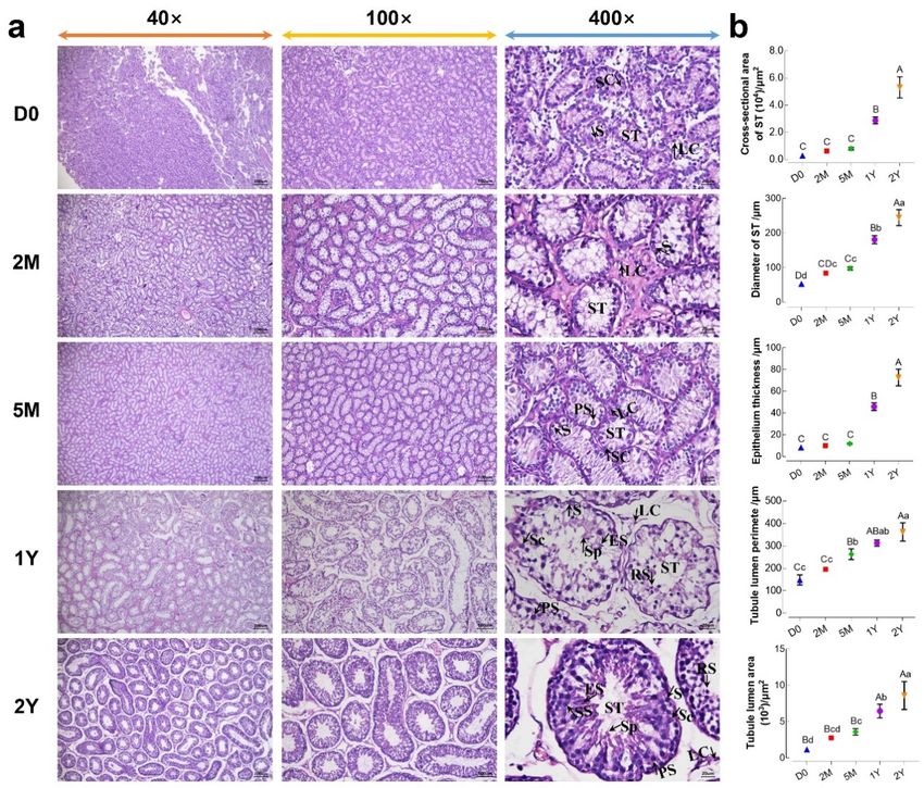
Animals 2019, 9, 105                                                                                  2 of 11

1. Introduction
      Spermatogenesis in mammals is a complex, continuous, and tightly coordinated physiological
process that is largely regulated by many genes whose expression varies at the levels of transcription
and translation during different stages [1–3]. The DAZ family genes are germ cell-specific
RNA-binding proteins (RBPs) which are implicated in male spermatogenesis [4,5]. During mammalian
spermatogenesis, there are many RBPs in mammalian testis that play important roles in regulating
gene expression at the post-transcriptional level, in order to control differentiation of the spermatogenic
cells, especially of the round spermatids [6,7]. The DAZ family consists of three known members,
including two autosomal genes, BOLL and DAZL(DAZ-like), and one Y chromosome gene, DAZ [8–11].
      BOLL (Boule), an ancestral gene of the DAZ family, is evolutionarily conserved in all metazoans [5,12].
Numerous studies have shown that the BOLL gene plays an important role in testicular function,
maintenance, and spermatogenesis and that loss of the gene may cause male dyszoospermia and
infertility [9,13–16]. In females, BOLL may regulate human embryonic stem cells to enter into meiotic
phase and produce meiotic germ cells which are further induced to form ovarian follicle-like cells by
expressing DAZL and BOLL with recombinant GDF9 and BMP15 [17]. Expression of BOLL mRNA is
significantly decreased and BOLL protein is completely lacking in the testes of infertile men, when
compared to healthy men [14,15,18]. Of note, BOLL expression is progressively reduced with the
increasing severity of testicular failure [18]. The downregulation of BOLL expression in male gonads is
related to its methylation degree and, consequently, with meiotic arrest and infertility. A long CpG
island in the BOLL promoter was hypermethylated in the testes of cattle–yak hybrids (male sterility)
with extremely low BOLL expression, compared with cattle testes [19]. For males with complete
spermatogenesis, the methylation degree of the BOLL promoter is also inversely associated with its
expression level. For example, the methylation level of the BOLL promoter in normal human testes,
with high BOLL expression, is significantly lower than that in somatic tissues with lacking or extremely
low BOLL expression [9]. The same results are reported for other mammals, such as goats [20], pigs [9],
and mice [9].
      The transcription of the BOLL gene during adult mice spermatogenesis is regulated by insulin-like
growth factor 1 (IGF1), through extracellular signal-regulated kinases 1 and 2 (ERK1/2) signaling
and T-independent pathways [10]. In males, deletion or mutation of BOLL may block production of
functional sperm, thereby leading to infertility [5,14,21]. However, blocked stages of spermatogenesis
are different among various species. In Drosophila melanogaster, for example, the mutation of Boule
results in meiotic arrest during spermatogenesis [22]. Similar findings have been published in studies
of infertility in mammalian testes from human [14,15,18] and cattle–yak [23]. However, in Boule
null mice testes, meiotic divisions proceed normally, but spermatogenic cells undergo arrest at the
subsequent stage of round spermatid, prior to deformation, and do not further differentiate into mature
spermatozoa [24]. By contrast, the overexpression of BOLL is able to promote the development and
differentiation of male germ cells. Li et al. [25] reported that the overexpression of BOLL in male dairy
goat germline stem cells can elevate the expression of meiosis-associated genes, such as STRA8, SCP3,
CDC25A, CDC2, and VASA, promoting meiosis and spermatogenesis. In vitro, overexpression of BOLL
may effectively promote the transdifferentiation of goat bone marrow mesenchymal stem cells into
early germ cells through spermatogenesis [26]. Additionally, experiments in male mice have proven
that Boll is involved in regulating the formation of stress granules (SGs) in germ cells under heat stress,
to protect those cells from heat-induced apoptosis [27]. SGs are an assembly of untranslated messenger
ribonucleoproteins that form from cytoplasmic mRNAs stalled in translation initiation under stress
conditions, such as heat, hypoxia, and oxidative stress, etc. (reviewed in [28,29]), which can inhibit
pro-apoptotic factors and thereby prevent cell damage [29].
      Until now, most reports on the BOLL gene in male mammals have focused on human, cattle and
mouse, but the regulatory mechanisms are not exactly the same in different species, considering
the entire range and wide variety. Sheep are an economically important livestock animal with
considerable agricultural significance, however, little is known about the expression profiles and
Animals 2019, 9, 105 3 of 11
biological functions of BOLL in sheep testes. Accordingly, it is important to elucidate the mechanisms
of sheep spermatogenesis by investigating the expression patterns and regulatory roles of BOLL in
developing sheep testes.
2. Materials and Methods
2.1. Experimental Animals and Design
All of the animals in this investigation were managed according to the animal care and
experimental procedure guidelines established by the Ministry of Science and Technology of the
People’s Republic of China (Approval No. 2006-398) and the study was approved by the Animal
Care Committee of Gansu Agricultural University. A total of fifteen purebred Small-Tail Han sheep,
from five developmental stages—0 days old (D0; n = 3), 2 months old (2M; n = 3), 5 months old (5M;
n = 3), 1 year old (1Y; n = 3), and 2 years old (2Y; n = 3)—were provided by Sanyang Sheep Breeding
Farm (Jingtai, Gansu, China). Samples from the same section of the right testis, heart, liver, spleen,
lung, kidney, and longissimus dorsi muscle were obtained from each sheep. Duplicated samples were
collected for all tissues: one sample was immediately placed in liquid nitrogen and stored at −80 ◦ C for
the preparation of total RNA and protein, and the other was fixed with 4% paraformaldehyde for up to
48 h, then embedded in paraffin for hematoxylin and eosin (H&E) staining and immunohistochemistry.
2.2. H&E Staining
Sections from testis tissues at different developmental stages were initially stained using the H&E
method, then dehydrated and dewaxed using conventional histological methods, as described by
Hara et al. [30] with some modifications. Sections were observed under a microscope (Sunny Optical
Technology Co. Ltd., Ningbo, China).
2.3. Total RNA Extraction and cDNA Synthesis
Total RNA from the selected tissue samples was extracted using TRIzol Reagent (TransGen
Biotech, Beijing, China), as previously described [31]. The purity, concentration, and integrity of
RNA samples were determined using a spectrophotometer (Nanodrop ND-2000, Thermo Scientific,
Niederelbert, Germany) and 1% agarose gel electrophoresis, respectively. Using random and anchored
oligo(dT)18 primers (1:5 ratio), first-strand cDNA was synthesized for each sample using 500 ng RNA,
according to the manufacturer0 s instructions (TransGen Biotech, Beijing, China).
2.4. qPCR
The BOLL gene was amplified (LightCycler 96 Real-Time System, Roche, Switzerland) using
optimized qPCR assay conditions, specifically, 1 cycle of 94 ◦ C for 30 s; 40 cycles of 94 ◦ C for 5 s,
40 cycles of 60 ◦ C for 30 s. β-actin was used as an internal control for the normalization of BOLL mRNA
expression. cDNA (1 µL) was added to 20 µL of the amplification reaction system used for qPCR,
comprised of 0.4 µL of forward primer, 0.4 µL of reverse primer, 10 µL of 2 × TransStart® Tip Green
qPCR SuperMix (TransGen Biotech, Beijing, China), and 8.2 µL of ddH2 O. The relative expression level
of BOLL mRNA was calculated according to the 2−∆∆Ct method [32]. Each experiment was biologically
replicated three times, with four technical replicates each. The BOLL mRNA expression results, taken
from an average across tissue samples, were presented in the form of bar charts. The qPCR primers,
used in the present study, are shown in Table 1.Animals 2019, 9, 105 4 of 11
Table 1. List of the primers used in qPCR.
Gene Accession No. Primer Sequence (50 –30 ) Product Length
F: AGCAGAGAGGAAGATGGAGACC
BOLL XM_004004798.3 122 bp
R: GGGCACTCGTTGGGTTATTC
F: CTTCCAGCCTTCCTTCCTGG
β-actin NM_001009784.1 180 bp
R: GCCAGGGCAGTGATCTCTTT
2.5. Western Blot
Testicular tissues at different development stages were homogenized and lysed using a radio
immunoprecipitation assay (RIPA) protein extraction kit (Solarbio, Beijing, China), according to
the operating instructions. Protein concentrations within the testis samples were determined using
a commercial bicinchoninic acid (BCA) Protein Assay kit (Beyotime, Shanghai, China). Twenty
micrograms of the denatured protein samples were separated by 12% sodium dodecyl sulfate
polyacrylamide gel electrophoresis (SDS-PAGE) and then transferred onto polyvinylidene difluoride
(PVDF) blotting membranes (Beyotime, Shanghai, China). After blocking in phosphate buffered
saline tween-20 (PBST) containing 5% non-fat milk, the membranes were incubated overnight at
4 ◦ C with either rabbit anti-BOLL polyclonal antibody (1:500, Bioss, Beijing, China) or anti-beta-actin
polyclonal antibody (1:1500, Bioss, Beijing, China). After washing, the membranes were incubated
with goat anti-rabbit IgG/HRP antibody (1:5000, Bioss, Beijing, China). Enhanced chemiluminescence
signals were visualized in an X-ray room. This experiment was biologically repeated three times.
Band intensities were quantified using AlphaEaseFC software (Protein Simple, Santa Clara, CA, USA).
The BOLL protein expression results, from an average across tissues, were presented as bar charts.
2.6. Immunohistochemistry
Immunoreactivity for the BOLL protein was visualized in 5 µm sections from testicular tissues
at postnatal development stages using a Histostain™-Plus kit (Bioss, Beijing, China), as previous
described [31]. In brief, the endogenous peroxidase activity from testicular sections was eliminated
with 3% H2 O2 and then incubated with rabbit polyclonal anti-BOLL antibody (1:120, Bioss, Beijing,
China) in a wet box, overnight, at 4 ◦ C. The negative controls were generated by replacing the primary
antibody with PBS. The positive signals (brown) of the BOLL protein in sections were visualized using
a DAB kit (Bioss, Beijing, China) and then observed using a Sunny EX31 biological microscope (Sunny,
Ningbo, China). This experiment was biologically replicated three times.
2.7. Image Analysis and Data Statistics
H&E staining and immunohistochemistry images for testicular cross sections were captured using
ImageView software (Sunny, Ningbo, China). The morphological parameters of 35 randomly selected
seminiferous tubules from the 200× magnification of H&E sections were measured using MvImage
software (Sunny, Ningbo, China). The integral optical density of immunostaining for the BOLL protein
was calculated by analyzing four random 400× microscope magnification levels in independently
replicated sections using Image-Pro Plus 6.0 software (Media Cybernetics, Rockville, MD, USA).
The data were statistically analyzed using one-way analysis of variance, with p < 0.05 considered as
statistically significant. All displayed values, in the form of bar charts, were the mean ± SD.
3. Results
3.1. Comparison of Morphological Differences between Sheep Testes at Different Ages
The morphological observation of sheep testes at different ages, using H&E staining, were
presented in Figure 1a. As the results show, testicular tissues were mainly composed of Sertoli cells
and various kinds of germ cells within seminiferous tubules, as well as Leydig cells localized in the
seminiferous tubule interspaces. In the D0 and 2M groups, spermatogonias were observed only inAnimals 2019, 9, 105 5 of 11
the seminiferous tubules near basement membranes. In the 5M group, a small number of primary
spermatocytes
Animals were observed in the seminiferous tubules, in addition to spermatogonia. In the 1Y5 of
2019, 9, x and
11
2Y groups, the spermatogenic cell layer was noticeably increased in seminiferous tubules, and various
spermatocytes, round spermatids,
spermatogenic cells—including elongated spermatids,
spermatogonia, primary and sperm—were
spermatocytes, observedspermatocytes,
secondary in an orderly
and
rounddistinct arrangement.
spermatids, Furthermore,
elongated theand
spermatids, morphological
sperm—were variables in the
observed seminiferous
in an orderly and tubules of
distinct
the testes, at different stages of development, were summarized in Figure 1b. The cross-sectional
arrangement. Furthermore, the morphological variables in the seminiferous tubules of the testes, at area
and diameter
different stagesofofthe seminiferous
development, tubules,
were epithelial
summarized thickness,
in Figure andcross-sectional
1b. The the area andarea
perimeter of the
and diameter
tubule lumen gradually increased with age. Of note, the histomorphological parameters
of the seminiferous tubules, epithelial thickness, and the area and perimeter of the tubule lumen in the above
1Y
gradually increased with age. Of note, the histomorphological parameters in the above 1Y and(p2Y
and 2Y groups were significantly increased compared to those in the D0, 2M and 5M groups <
0.05).
groups were significantly increased compared to those in the D0, 2M and 5M groups (p < 0.05).
Figure 1.
Figure Morphological comparisons
1. Morphological comparisons of of testicular
testicular tissues
tissues atat different
different development
development stages. (a) The
stages. (a) The
representative images from D0, 2M, 5M, 1Y, and 2Y testicular cross sections at 40
representative images from D0, 2M, 5M, 1Y, and 2Y testicular cross sections at 40×, 100×, and 400× × , 100 × , and 400 ×
magnification, respectively; (b) Values of the morphological parameters obtained
magnification, respectively; (b) Values of the morphological parameters obtained from the from the seminiferous
tubules at different
seminiferous tubulesages: LC, Leydig
at different ages:cell;
LC,SC, Sertolicell;
Leydig cell;SC,
ST, Sertoli
seminiferous
cell; ST,tubule; S, spermatogonia;
seminiferous tubule; S,
PS, primary spermatocyte; SS, secondary spermatocyte; RS, round spermatid;
spermatogonia; PS, primary spermatocyte; SS, secondary spermatocyte; RS, round spermatid; ES, elongated spermatid;
ES,
Sp, spermatozoa.
elongated DataSp,
spermatid; were presented as
spermatozoa. Datamean
were± presented
SD in the graphs.
as meanDifferent
± SD in thecapital letters
graphs. denote
Different
an extremely
capital letters significant difference significant
denote an extremely (p < 0.01), difference
while different lowercase
(p < 0.01), whileletters denote
different a significant
lowercase letters
difference
denote between groups
a significant (p < 0.05).
difference D0: 0groups
between days old;
(pAnimals 2019, 9, 105 6 of 11
groups (Figure 2a). Similarly, BOLL protein expression steadily increased in the testes from the D0 to
Animals 2019, 9, x 6 of 11
2Y groups,
Animals 2019, with
the highest expression observed in the 2Y group (Figure 3).
9, x 6 of 11
Figure 2. Relative expression of BOLL mRNA in selected sheep tissues detected by qPCR. (a) BOLL
Figure
Figure 2. 2. Relative
Relative expression
expression of BOLL mRNA in
in selected sheep
sheep tissues detected by qPCR. (a) BOLL
(a)
mRNA expression in sheep testis BOLL
tissuesmRNA
at differentselected
development tissues
stages; detected
(b) BOLLby qPCR.
mRNA BOLL
expression
mRNA
mRNA expression
expression in
in sheep
sheep testis
testis tissues
tissues at different
different development
development stages;
stages; (b)
(b) BOLL mRNA expression
expression
in testis and somatic tissues from 1Y sheep. β-actin was used as a reference gene. All experiments were
in
in testis
testis and somatic
somatic tissues
and replicated tissues from 1Y sheep. β-actin was used as a reference gene. All experiments were
biologically threefrom
times,1Yeach
sheep.
with four technical replicates. The bars represent the mean were
biologically
biologically replicated three times,
times, each
each with
with four
four technical
technical replicates.
replicates. The bars represent
represent the
the mean
mean
values ± SD of 12 replicate samples obtained from three sheep per group. Different capital letters
values ±
values
denote an
SD of
± SD of 12
12 replicate
extremely replicate samples
samples
significant
obtained
obtained
difference
from three
three sheep
from groups
between sheep per
per group.
(p < 0.01).group. Different
Different
D0: 0 days
capital
capital
old; 2M:
letters
letters
2 months
denote
denote an
an extremely
extremely significant
significant difference
difference between
between groups (p
groups <
(p0.01).
< D0:
0.01). 0
D0:days
0 old;
days 2M:
old; 2
2M:months
2 monthsold;
old; 5M: 5 months old; 1Y: 1 year old; 2Y: 2 years old.
5M:
old; 55M:
months old; 1Y:
5 months old;11Y:
year1 old;
year2Y:old;2 2Y:
years old. old.
2 years
Figure
Figure 3. Relative
3. Relative BOLL BOLL protein
protein expression
expression in sheep
in sheep testestestes at different
at different ages.ages.
(a) A (a) A representative
representative Western
Figure 3.
Western Relative
blot BOLL
detection protein
result; expression
(b) The in sheep
integrated testes
density at different
values (IDVs)ages.
of (a)
BOLL A representative
protein
blot detection result; (b) The integrated density values (IDVs) of BOLL protein from β-actin was used from Western
β-actin
as an
blot detection
was used asresult;
an (b)
internal The integrated
reference density
protein. Thevalues (IDVs)
experiment of
was BOLL protein
biologically from β-actin
repeated
internal reference protein. The experiment was biologically repeated three times. The bars represent threewas used
times. asthe
The an
internal
bars reference
represent protein.
the mean The experiment
values ± SD of was
three biologically
replicate repeated
samples three
obtained times.
from The
three bars
sheep
mean values ± SD of three replicate samples obtained from three sheep per group. Different capital letters represent
per group. the
mean
denote values
Different ± SD ofletters
capital
an extremely three replicate
denote
significant samples
an obtained
extremely
difference between from(pthree
significant
groups sheep
difference
< 0.01): per
D0:betweengroup.
0 days Different
groups
old; < capital
2M: 2(pmonths letters
0.01):old;
D0:5M:
denote
0 an
days extremely
old; 2M: 2 significant
months old;difference
5M: 5
5 months old; 1Y: 1 year old; 2Y: 2 years old. between
months old; groups
1Y: 1 (p
year < 0.01):
old; 2Y: D0:
2 0
yearsdays old;
old. 2M: 2 months old; 5M:
5 months old; 1Y: 1 year old; 2Y: 2 years old.
3.3. Expression Patterns of BOLL in Various Tissues of 1-Year-Old Sheep Testes
3.3. Expression Patterns of BOLL in Various Tissues of 1-Year-Old Sheep Testes
3.3. Expression Patterns
To understand of BOLL
whether in Various
sheep BOLLTissues of 1-Year-Old
was expressed Sheep tissues
in other Testes in addition to testis, we
To understand whether sheep BOLL was expressed in other tissues in addition to testis, we
subsequently used qPCR
To understand to characterize
whether sheep BOLL the expression patterns of BOLL transcript in multiple tissues
subsequently used qPCR to characterize the was expressed
expression in other
patterns of BOLLtissues in addition
transcript to testis,
in multiple we
tissues
of 1-year-old
subsequently sheep,
used qPCRincluding the
to characterizetestis, heart,
theheart, liver,
expression spleen, lung,
patternslung,of BOLLkidney, and
transcript longissimus dorsi
in multiple tissues
of 1-year-old sheep, including the testis, liver, spleen, kidney, and longissimus dorsi
muscle.
of As expected,
1-year-old sheep, BOLL mRNA
including the was only
testis, expressed
heart, liver, exclusively
spleen, lung, inkidney,
the testis,
andwhile no expression
longissimus dorsi
muscle. As expected, BOLL mRNA was only expressed exclusively in the testis, while no expression
was detected
muscle. As in somatic
expected, tissues
BOLL mRNA suchwas as the
only heart, liver, etc.
expressed (Figure 2b).
exclusively in the testis, while no expression
was detected in somatic tissues such as the heart, liver, etc. (Figure 2b).
was detected in somatic tissues such as the heart, liver, etc. (Figure 2b).
3.4. Immunolocalization of BOLL Protein in Postnatal Developmental Sheep Testes
3.4. Immunolocalization of BOLL Protein in Postnatal Developmental Sheep Testes
3.4. Immunolocalization of BOLL Protein
Immunostaining patterns for the in Postnatal
BOLL proteinDevelopmental Sheeptissue
in all testicular Testes sections, from sheep at
Immunostaining
different developmentpatterns
stages, forwasthe BOLL protein
analyzed in all testicular tissue
by immunohistochemistry sections,
using from antibody
a primary sheep at
Immunostaining
different development patterns
stages, for analyzed
was the BOLLby protein in all testicular tissue
immunohistochemistry using sections,
a primary from sheep to
antibody at
to BOLL. The representative results are provided in Figure 4. The strength of positive reactions of
different
BOLL. development
Theprotein
representative stages, was
results analyzed
are provided by immunohistochemistry
in Figure 4. intense,
The strength using a primary
of positive antibody
reactions to
of the
the BOLL in testicular tissues ranged from weak to increasing with age. Specifically,
BOLL. The
BOLL protein representative
in patterns results
testicularcorresponding are provided
tissues rangedtofrom in Figure 4. The
weak toofintense, strength
increasingof positive
with reactions of the
positive staining localization the BOLL protein in theage. Specifically,
D0, 2M, and 5M
BOLL
positiveprotein
stainingin testicular
patterns tissues
corresponding ranged from weak
to localization to intense,
of theofBOLL increasing with
protein tubules, age.
in the D0, Specifically,
2M,relatively
and 5M
groups were similar and showed its presence in the epithelia seminiferous with
positivewere
groups staining patterns
similar and corresponding
showed its to localization
presence in the of the
epithelia of BOLL protein tubules,
seminiferous in the D0, 2M,
with and 5M
relatively
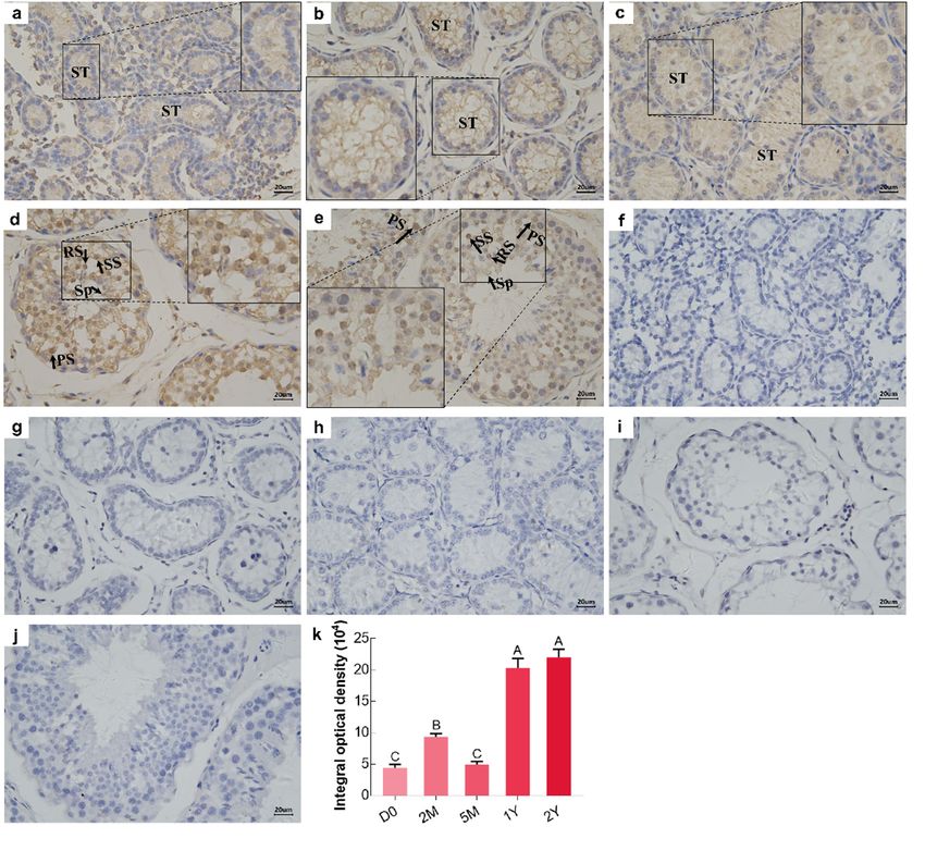
low expression (Figure 4a–c,k), whereas intense BOLL protein staining was mainly observed in the
groups
low were similar
expression and4a–c,k),
(Figure showedwhereasits presence
intensein the
BOLLepithelia
protein of staining
seminiferous tubules,observed
was mainly with relatively
in the
low expression (Figure 4a–c,k), whereas intense BOLL protein staining
primary spermatocytes, secondary spermatocytes, round spermatids, and spermatozoa in the 1Y was mainly observed inand
the
primary
2Y groupsspermatocytes,
(Figure 4d–e,k). secondary spermatocytes, round spermatids, and spermatozoa in the 1Y and
2Y groups (Figure 4d–e,k).Animals 2019, 9, 105 7 of 11
primary spermatocytes, secondary spermatocytes, round spermatids, and spermatozoa in the 1Y and
2Y groups
Animals 2019,(Figure
9, x 4d,e,k). 7 of 11
Figure 4. Immunohistochemical staining (brown) of BOLL protein in sheep testes at different
Figure 4. Immunohistochemical
developmental staining (brown)
stages. (a–e) Immunostaining patternsof of BOLL proteinininthesheep
BOLL protein D0, 2M,testes
5M, at
1Y,different
and 2Y
developmental stages. (a–e) Immunostaining patterns of BOLL protein in the D0, 2M, 5M, 1Y,
sheep testes, respectively; (f–j) Substitution of PBS for the primary antibody served as a negative control; and 2Y
sheep testes, respectively; (f–j) Substitution of PBS for the primary antibody served
(k) Integral optical density of BOLL protein: ST, seminiferous tubule; S, spermatogonia; PS, primary as a negative
control; (k) Integral
spermatocyte; opticalspermatocyte;
SS, secondary density of BOLL protein:
RS, round ST, seminiferous
spermatid; tubule; S,The
Sp, spermatozoa. spermatogonia;
experiment was PS,
primary spermatocyte;
biologically SS, times.
replicated three secondary spermatocyte;
Different RS,denote
capital letters roundan spermatid;
extremelySp, spermatozoa.
significant The
difference
experiment
between was(pbiologically
groups < 0.01). D0:replicated
0 days old;three
2M: times.
2 monthsDifferent
old; 5M:capital letters
5 months denote
old; 1Y: 1 an
yearextremely
old; 2Y:
significant difference between
2 years old. Scale bars, 20 µm. groups (p < 0.01). D0: 0 days old; 2M: 2 months old; 5M: 5 months old;
1Y: 1 year old; 2Y: 2 years old. Scale bars, 20 μm.
4. Discussion
4. Discussion
Small-Tail Han sheep are an excellent genetic germplasm resource and economic breed in China.
Therefore, studying
Small-Tail sheep are
Han sheep testicular histomorphology,
an excellent as well
genetic germplasm as geneand
resource expression
economicand regulation
breed in China.
during testis development, has an important significance for understanding sheep
Therefore, studying sheep testicular histomorphology, as well as gene expression and regulation fertility. In the
present
during study, Small-Tail Hanhas
testis development, sheep testes weresignificance
an important obtained from
for five postnatal developmental
understanding sheep fertility.stages:
In the
D0 (new birth), 2M (weaning), 5M (pre-puberty), 1Y (sexual maturity), and 2Y (adult).
present study, Small-Tail Han sheep testes were obtained from five postnatal developmental When compared stages:
toD0the(new
D0 and 2M testes,
birth), histomorphological
2M (weaning), observation1Y
5M (pre-puberty), showed
(sexualthat spermatocytes
maturity), and 2Yinitially appeared
(adult). When
in the seminiferous tubules beside the spermatogonia in 5M testes, indicating
compared to the D0 and 2M testes, histomorphological observation showed that spermatocytes that Small-Tail Han
sheep have an earlier age of sexual maturity. All levels of spermatogenic cells, from spermatogonia
initially appeared in the seminiferous tubules beside the spermatogonia in 5M testes, indicating that to
spermatozoa,
Small-Tail Han could be seen
sheep haveinan
the seminiferous
earlier tubules,
age of sexual most obviously
maturity. All levelsinof
thespermatogenic
tubules from the 1Y from
cells, and
2Y testes. The measurement of the histomorphological parameters in the cross sections
spermatogonia to spermatozoa, could be seen in the seminiferous tubules, most obviously in the of testicular
tubules from the 1Y and 2Y testes. The measurement of the histomorphological parameters in the
cross sections of testicular seminiferous tubules, at different development stages, indicated that the
area and diameter of the seminiferous tubules, epithelial thickness, and the area and perimeter of the
tubule lumen became gradually larger with testicular development. A significant increase was found
in the 1Y and 2Y testes.Animals 2019, 9, 105 8 of 11
seminiferous tubules, at different development stages, indicated that the area and diameter of the
seminiferous tubules, epithelial thickness, and the area and perimeter of the tubule lumen became
gradually larger with testicular development. A significant increase was found in the 1Y and 2Y testes.
BOLL is a marker of germ cell development and meiosis, and previous studies in some
male mammals have indicated that BOLL expression is restricted to the gonads and implicated in
spermatogenesis through regulating spermatocyte meiosis and the male gamete formation required for
fertility [5,14,33]. BOLL expression was examined in adult testes from cattle and yaks—low expression
was detectable in testes of cattle–yaks, but there was no expression in other tissues, including the
epididymis, kidney, spleen, stomach, hypothalamus, and pituitary tissues from cattle, yak, and
cattle–yak [34]. In addition, two alternative splice variants of Boule, namely Boule1 and Boule2, were also
found to be exclusively expressed in yak testes, while no expression was observed in other examined
tissues, including ovary, muscle, kidney, and spleen [23]. Similarly, testis-specific expression for BOLL
is also well-documented in male humans [9], dairy goats [25], pigs [9], mice [9] and chickens [9,33].
In addition, BOLL expression was only present in male and female gonads for fish such as Asian
seabass (Lates calcarifer) [35], Chinese sturgeon (Acipenser sinensis) [36], and medaka (Oryzias latipes) [37].
In this study, BOLL was specifically expressed in the testes of 1-year-old sheep, but was completely
lacking in other male somatic tissues, such as heart, liver, spleen, lung, kidney, and muscle, as analyzed
by qPCR, which is consistent with previous studies on cattle [34], goats [25], and mice [9]. The results
suggest a function for BOLL in sheep testes which might be related to its previously reported role in
testicular development or spermatogenesis.
The expression patterns and regulation mechanisms of BOLL are not the same in the testes of
different species nor in the same species at different development stages. In mice, Zhang et al. [9]
report that Boll expression is barely detectable from postnatal 3- to 14-day-old testes, but its expression
was significantly upregulated at 12-days-old and later. In goats, the expression of BOLL in the testes
was observed to be lacking, or low, during the embryonic and pre-pubertal stages, but gradually
increased during postnatal progression with a significant expression in mature testes [25,26]. Moreover,
BOLL expression in adult dairy goat testes with complete spermatogenesis was significantly higher
than that in testes with azoospermia or male intersex [25], which demonstrates that BOLL is crucial
for male fertility. Herein, we first examined the expression patterns of the BOLL gene at the mRNA
and protein level during postnatal sheep testis development, as analyzed using qPCR and Western
blot. Consistent with previous studies [9,26], BOLL mRNA and protein expression were observed to
be at extremely low levels in sheep testes from the D0, 2M, and 5M groups, but dramatically increased
expression was detectable in the 1Y and 2Y groups. One can speculate that the BOLL gene is crucial for
the testicular development of postnatal sheep and, particularly, for post-pubertal sheep.
BOLL is a testis-specific gene that regulates spermatogenesis in males, but its distribution in
the testis varies between different species and between different development stages. Positive BOLL
protein is restricted to spermatocytes in the testes of normal adult men, but it is completely lacking
in the testes of infertile men, which is reported by Luetjens et al. [14]. For primates, positive BOLL
protein is originally observed in zygotene spermatocytes, reaching its maximal expression in pachytene
spermatocytes. It is also found in the secondary spermatocytes and earlier round spermatids of pygmy
chimpanzee and gray mouse lemur testes, but it is distributed only in pachytene spermatocytes in
common marmoset testis [38]. In adult mice, immunostaining of the BOLL protein has been observed
in spermatocytes and round spermatids, as reported by two previous papers [10,27]. Also, the same
results are reported in adult goat testes [26]. Moreover, positive BOLL cells were mainly located in
primary spermatocytes, with a relatively low immunoexpression in secondary spermatocytes in the
adult testes of Asian seabass [35] and medaka [37]. To understand the patterns of localization of
BOLL-positive cells in developing sheep testes, BOLL protein immunoreactions were further assessed
using immunohistochemistry. As a result, BOLL protein was detectable in sheep testes throughout
different stages of development, with weak expression in the epithelia of seminiferous tubules from new
birth to the pre-pubertal stages of development, but with intense expression in primary spermatocytes,Animals 2019, 9, 105 9 of 11
secondary spermatocytes, round spermatids, and sperm from the post-pubertal developmental stage
of sheep testes. High immunoexpression patterns in testicular spermatocytes and round spermatids of
post-pubertal sheep were basically in agreement with previous studies that examined fertile men [14],
primates (except the common marmoset) [38], goats [26], and mice [10,27]. In contrast to previously
published studies, we also observed the presence of BOLL protein in sperm from the seminiferous
tubules of post-pubertal testes, which may be due to the different species as well as developmental
stages investigated in this study. Taken together, these results suggest that BOLL might play several
important roles in the meiotic phase and spermiogenesis of mature sheep.
5. Conclusions
In conclusion, our study investigated the expression patterns and cellular localization of BOLL,
an ancestral gene of the DAZ family, in sheep testes at different ages. Our data showed that BOLL
demonstrates an extremely low level of expression and is confined to the seminiferous epithelium in
testes, from birth through to the pre-pubertal stages. However, its expression demonstrates a significant
upregulation in post-pubertal testes, with strong positive signals observed in the spermatocytes,
round spermatids, and spermatozoa. These results indicate that BOLL plays a key role in sheep
spermatogenesis, especially during meiosis and spermiogenesis. Future studies will be implemented to
investigate the specific molecular mechanisms of BOLL gene expression during sheep spermatogenesis.
Author Contributions: This work was conceived and designed by T.L. and Y.M.; T.L., X.W., H.Z., and Z.C.
collected samples; T.L. and X.W. performed the experiments and analyzed the data; T.L. wrote the paper; Y.M. and
X.Z. contributed to revisions of the manuscript. All authors read and approved the final manuscript.
Funding: This research was funded by the National Key R&D Program of China (2018YFD0502103) and Fostering
Foundation for the Excellent Ph.D. Dissertation of Gansu Agricultural University (YB2018001).
Acknowledgments: We thank Sanyang Sheep Breeding Farm (Jingtai, Gansu, China) for providing the
experimental animals.
Conflicts of Interest: The authors declare no conflict of interest.
References
1. Chalmel, F.; Rolland, A.D. Linking transcriptomics and proteomics in spermatogenesis. Reproduction 2015,
150, R149–R157. [CrossRef]
2. Kotaja, N. MicroRNAs and spermatogenesis. Fertil. Steril. 2014, 101, 1552–1562. [CrossRef] [PubMed]
3. De Mateo, S.; Sassone-Corsi, P. Regulation of spermatogenesis by small non-coding RNAs: Role of the germ
granule. Semin. Cell Dev. Biol. 2014, 29, 84–92. [CrossRef] [PubMed]
4. Smorag, L.; Xu, X.; Engel, W.; Pantakani, D. The roles of DAZL in RNA biology and development.
Wiley Interdiscip. Rev. RNA 2014, 5, 527–535. [CrossRef] [PubMed]
5. Fu, X.F.; Cheng, S.F.; Wang, L.Q.; Yin, S.; Felici, M.D.; Shen, W. DAZ family proteins, key players for germ
cell development. Int. J. Biol. Sci. 2015, 11, 1226–1235. [CrossRef] [PubMed]
6. Idler, R.K.; Yan, W. Control of messenger RNA fate by RNA-binding proteins: An emphasis on mammalian
spermatogenesis. J. Androl. 2012, 33, 309–337. [CrossRef]
7. Suzuki, A.; Niimi, Y.; Shinmyozu, K.; Zhou, Z.; Kiso, M.; Saga, Y. Dead end1 is an essential partner of
NANOS2 for selective binding of target RNAs in male germ cell development. EMBO Rep. 2016, 17, 37–46.
[CrossRef]
8. Rosario, R.; Adams, I.R.; Anderson, R.A. Is there a role for DAZL in human female fertility? Mol. Hum. Reprod.
2016, 22, 377–383. [CrossRef]
9. Zhang, C.; Xue, P.; Gao, L.; Chen, X.; Lin, K.; Yang, X.; Dai, Y.; Xu, E.Y. Highly conserved epigenetic regulation
of BOULE and DAZL is associated with human fertility. FASEB J. 2016, 30, 3424–3440. [CrossRef]
10. Gonzalez, C.R.; Dorfman, V.B.; Vitullo, A.D. IGF1 regulation of BOULE and CDC25A transcripts via a
testosterone-independent pathway in spermatogenesis of adult mice. Reprod. Biol. 2015, 15, 48–55. [CrossRef]
[PubMed]Animals 2019, 9, 105 10 of 11
11. González, C.R.; Moverer, L.; Calandra, R.S.; Gonzálezcalvar, S.I.; Vitullo, A.D. Age-related and photoperiodic
variation of the DAZ gene family in the testis of the Syrian hamster (Mesocricetus auratus). Zygote 2018, 26,
127–134. [CrossRef]
12. Xu, E.Y.; Moore, F.L.; Pera, R.A. A gene family required for human germ cell development evolved from an
ancient meiotic gene conserved in metazoans. Proc. Natl. Acad. Sci. USA 2001, 98, 7414–7419. [CrossRef]
13. Ahmadivand, S.; Farahmand, H.; Teimoori-Toolabi, L.; Mirvaghefi, A.; Eagderi, S.; Geerinckx, T.;
Shokrpoor, S.; Rahmati-Holasoo, H. Boule gene expression underpins the meiotic arrest in spermatogenesis
in male rainbow trout (Oncorhynchus mykiss) exposed to DEHP and butachlor. Gen. Comp. Endocrinol. 2016,
225, 235–241. [CrossRef]
14. Luetjens, C.M.; Xu, E.Y.; Rejo Pera, R.A.; Kamischke, A.; Nieschlag, E.; Gromoll, J. Association of meiotic
arrest with lack of BOULE protein expression in infertile men. J. Clin. Endocrinol. Metab. 2004, 89, 1926–1933.
[CrossRef] [PubMed]
15. Kostova, E.; Yeung, C.H.; Luetjens, C.M.; Brune, M.; Nieschlag, E.; Gromoll, J. Association of three isoforms
of the meiotic BOULE gene with spermatogenic failure in infertile men. Mol. Hum. Reprod. 2007, 13, 85–93.
[CrossRef]
16. Kee, K.; Angeles, V.T.; Flores, M.; Nguyen, H.N.; Reijo Pera, R.A. Human DAZL, DAZ and BOULE genes
modulate primordial germ-cell and haploid gamete formation. Nature 2009, 462, 222–225. [CrossRef]
[PubMed]
17. Jung, D.; Xiong, J.; Ye, M.; Qin, X.; Li, L.; Cheng, S.; Luo, M.; Peng, J.; Dong, J.; Tang, F. In vitro differentiation
of human embryonic stem cells into ovarian follicle-like cells. Nat. Commun. 2017, 8, 15680. [CrossRef]
[PubMed]
18. Lin, Y.M.; Kuo, P.L.; Lin, Y.H.; Teng, Y.N.; Nan Lin, J.S. Messenger RNA transcripts of the meiotic regulator
BOULE in the testis of azoospermic men and their application in predicting the success of sperm retrieval.
Hum. Reprod. 2005, 20, 782–788. [CrossRef]
19. Yao, W.; Li, Y.; Li, B.; Luo, H.; Xu, H.; Pan, Z.; Xie, Z.; Li, Q. Epigenetic regulation of bovine spermatogenic
cell-specific gene boule. PLoS ONE 2015, 10, e0128250. [CrossRef] [PubMed]
20. Zhang, X.; Yu, S.; Yang, Q.; Wang, K.; Zhang, S.; Pan, C.; Yan, H.; Dang, R.; Lei, C.; Chen, H. Goat Boule:
Isoforms identification, mRNA expression in testis and functional study and promoter methylation profiles.
Theriogenology 2018, 116, 53–63. [CrossRef] [PubMed]
21. Reynolds, N.; Cooke, H.J. Role of the DAZ genes in male fertility. Reprod. Biomed. Online 2005, 10, 72–80.
[CrossRef]
22. Castrillon, D.H.; Gonczy, P.; Alexander, S.; Rawson, R.; Eberhart, C.G.; Viswanathan, S.; Dinardo, S.;
Wasserman, S.A. Toward a molecular genetic analysis of spermatogenesis in Drosophila melanogaster:
Characterization of male-sterile mutants generated by single P element mutagenesis. Genetics 1993, 135,
489–505. [CrossRef]
23. Li, B.; Ngo, S.; Wu, W.; Xu, H.; Xie, Z.; Li, Q.; Pan, Z. Identification and characterization of yak (Bos grunniens)
b-Boule gene and its alternative splice variants. Gene 2014, 550, 193–199. [CrossRef] [PubMed]
24. Vangompel, M.J.; Xu, E.Y. A novel requirement in mammalian spermatid differentiation for the DAZ-family
protein Boule. Hum. Mol. Genet. 2010, 19, 2360–2369. [CrossRef] [PubMed]
25. Li, M.; Liu, C.; Zhu, H.; Sun, J.; Yu, M.; Niu, Z.; Liu, W.; Peng, S.; Hua, J. Expression pattern of Boule in dairy
goat testis and its function in promoting the meiosis in male germline stem cells (mGSCs). J. Cell Biochem.
2013, 114, 294–302. [CrossRef] [PubMed]
26. Li, P.Z.; Yan, G.Y.; Han, L.; Pang, J.; Zhong, B.S.; Zhang, G.M.; Wang, F.; Zhang, Y.L. Overexpression of
STRA8, BOULE, and DAZL genes promotes goat bone marrow-derived mesenchymal stem cells in vitro
transdifferentiation toward putative male germ cells. Reprod. Sci. 2017, 24, 300–312. [CrossRef]
27. Kim, B.; Rhee, K. BOULE, a deleted in azoospermia homolog, is recruited to stress granules in the mouse
male germ cells. PLoS ONE 2016, 11, e0163015. [CrossRef] [PubMed]
28. Protter, D.S.W.; Parker, R. Principles and properties of stress granules. Trends Cell Biol. 2016, 26, 668–679.
[CrossRef]
29. Buchan, J.R.; Parker, R. Eukaryotic stress granules: The ins and outs of translation. Mol. Cell 2009, 36, 932–941.
[CrossRef]Animals 2019, 9, 105 11 of 11
30. Hara, A.; Abe, T.; Hirao, A.; Sanbe, K.; Ayakawa, H.; Sarantonglaga, B.; Yamaguchi, M.; Sato, A.;
Khurchabilig, A.; Ogata, K.; et al. Histochemical properties of bovine and ovine mammary glands during
fetal development. J. Vet. Med. Sci. 2018, 80, 263–271. [CrossRef]
31. Li, T.; Lu, Z.; Luo, R.; Gao, J.; Zhao, X.; Ma, Y. Expression and cellular localization of double sex and mab-3
related transcription factor 1 in testes of postnatal Small-Tail Han sheep at different developmental stages.
Gene 2018, 642, 467–473. [CrossRef] [PubMed]
32. Livak, K.J.; Schmittgen, T.D. Analysis of relative gene expression data using real-time quantitative PCR and
the 2(-Delta Delta C(T)) method. Methods 2001, 25, 402–408. [CrossRef] [PubMed]
33. Shah, C.; Vangompel, M.J.; Naeem, V.; Chen, Y.; Lee, T.; Angeloni, N.; Wang, Y.; Xu, E.Y. Widespread presence
of human BOULE homologs among animals and conservation of their ancient reproductive function.
PLoS Genet. 2010, 6, e1001022. [CrossRef] [PubMed]
34. Zhang, Q.; Li, J.; Li, Q.; Li, X.; Liu, Z.; Song, D.; Xie, Z. Cloning and characterization of the gene encoding the
bovine BOULE protein. Mol. Genet. Genomics 2009, 281, 67–75. [CrossRef]
35. Dwarakanath, M.; Lim, M.; Xu, H.; Hong, Y. Differential expression of boule and dazl in adult germ cells of
the Asian seabass. Gene 2014, 549, 237–242. [CrossRef] [PubMed]
36. Ye, H.; Li, C.J.; Yue, H.M.; Yang, X.G.; Wei, Q.W. Differential expression of fertility genes boule and dazl in
Chinese sturgeon (Acipenser sinensis), a basal fish. Cell Tissue Res. 2015, 360, 413–425. [CrossRef] [PubMed]
37. Xu, H.; Li, Z.; Li, M.; Wang, L.; Hong, Y. Boule is present in fish and bisexually expressed in adult and
embryonic germ cells of medaka. PLoS ONE 2009, 4, e6097. [CrossRef] [PubMed]
38. Tung, J.Y.; Luetjens, C.M.; Wistuba, J.; Xu, E.Y.; Reijo Pera, R.A.; Gromoll, J. Evolutionary comparison of
the reproductive genes, DAZL and BOULE, in primates with and without DAZ. Dev. Genes Evol. 2006, 216,
158–168. [CrossRef] [PubMed]
© 2019 by the authors. Licensee MDPI, Basel, Switzerland. This article is an open access
article distributed under the terms and conditions of the Creative Commons Attribution
(CC BY) license (http://creativecommons.org/licenses/by/4.0/).You can also read