Spontaneous Hinge-Bending Motions of Angiotensin I Converting Enzyme: Role in Activation and Inhibition - MDPI
←
→
Page content transcription
If your browser does not render page correctly, please read the page content below
molecules
Article
Spontaneous Hinge-Bending Motions of Angiotensin
I Converting Enzyme: Role in Activation
and Inhibition
Thi Tuong Vy 1 , Seong-Yeong Heo 1,2 , Won-Kyo Jung 1,2,3             and Myunggi Yi 1,2, *
 1   Interdisciplinary Program of Biomedical, Electrical & Mechanical Engineering, Pukyong National University,
     Busan 48513, Korea; phanvy120690@gmail.com (T.T.V.); hsyadsl@naver.com (S.-Y.H.);
     wkjung@pknu.ac.kr (W.-K.J.)
 2   Department of Biomedical Engineering, Pukyong National University, Busan 48513, Korea
 3   Marine Integrated Bionics Research Center, Pukyong National University, Busan 48513, Korea
 *   Correspondence: myunggi@pknu.ac.kr; Tel.: +82-51-629-5773

 Received: 31 January 2020; Accepted: 9 March 2020; Published: 12 March 2020

 Abstract: The inhibition of human angiotensin I converting enzyme (ACE) has been regarded as a
 promising approach for the treatment of hypertension. Despite research attempts over many years,
 our understanding the mechanisms of activation and inhibition of ACE is still far from complete.
 Here, we present results of all atom molecular dynamics simulations of ACE with and without ligands.
 Two types of inhibitors, competitive and mixed non-competitive, were used to model the ligand
 bound forms. In the absence of a ligand the simulation showed spontaneous large hinge-bending
 motions of multiple conversions between the closed and open states of ACE, while the ligand bound
 forms were stable in the closed state. Our simulation results imply that the equilibrium between
 pre-existing backbone conformations shifts in the presence of a ligand. The hinge-bending motion of
 ACE is considered as an essential to the enzyme function. A mechanistic model of activation and the
 inhibition may provide valuable information for novel inhibitors of ACE.

 Keywords: angiotensin converting enzyme; spontaneous conformational change; activation and
 inhibition mechanism; MD simulation; hinge-bending motion

1. Introduction
     Hypertension, a long-term medical condition known as high blood pressure, is a major risk
factor for vision loss, stroke, heart failure and chronic kidney disease, and more than a quarter of the
world’s adult population in 2000 had hypertension [1]. Therapeutic approaches have focused on the
renin-angiotensin-aldosterone system (RAAS) which regulates blood pressure and electrolyte balance
in humans [2]. In the RAAS, renin stimulates the generation of angiotensin I (AngI), which is converted
to vasoconstrictor angiotensin II (AngII) by angiotensin I converting enzyme (ACE). Two isoforms
(somatic and testicular) of ACE are transcribed by ACE gene in a tissue-specific manner. The somatic
form (sACE) is a zinc dependent dicarboxypeptidase, which includes two homologous domains
(N domain and C domain) with ~60% sequence identity and the same zinc motif HEXXH (X = any
amino acid residue) [3,4]. For the past decades, it has been reported that the C domain of human
sACE has the main AngI converting site in controlling blood pressure and cardiovascular functions [5].
ACE hydrolyzes decapeptide (Asp-Arg-Val-Tyr-Ile-His-Pro-Phe-His-Leu) AngI by cleaving a dipeptide
from the C-terminus to produce octapeptide (Asp-Arg-Val-Tyr-Ile-His-Pro-Phe) AngII, which causes
blood vessels to narrow and stimulates the secretion of the hormone aldosterone, resulting in increased
blood pressure [6,7]. ACE also affects blood pressure by inactivating the vasodilators bradykinin and

Molecules 2020, 25, 1288; doi:10.3390/molecules25061288                         www.mdpi.com/journal/molecules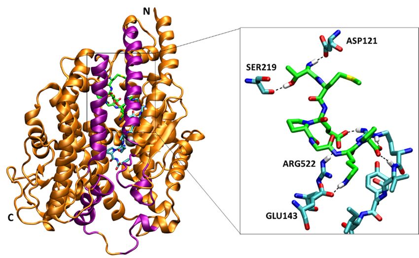
Molecules 2020, 25, 1288 2 of 12
kallidin [8]. Therefore, ACE has long been a major target for the treatment of hypertension and other
cardiovascular ailments by the use of ACE inhibitors [9].
Common synthetic ACE inhibitors such as captopril, enalapril, alacepril and lisinopril have been
on the market for decades [10]. However, it has been reported for them to have diverse side effects
including hyperkalemia and skin rashes [11], impairment of renal function [12], and
Molecules 2020, 25, x FOR PEER REVIEW
development
2 of 12
of angioedema [13]. Therefore, peptides from natural sources were considered as alternative ACE
AngII, which causes blood vessels to narrow and stimulates the secretion of the hormone aldosterone,
inhibitors and attracted researchers’ interest [14–18]. The structures of ACE complexes with various
resulting in increased blood pressure [6,7]. ACE also affects blood pressure by inactivating the
ligands including
vasodilatorssynthetic
bradykinin inhibitors and[8].
and kallidin peptides
Therefore,areACEavailable
has longinbeen
the aProtein Datafor
major target Bank
the (PDB).
The crystal structures
treatment of ACE
of hypertension andhave
otherbeen determined
cardiovascular with
ailments byhigh resolution,
the use which
of ACE inhibitors [9].give us the overall
Common synthetic
insight of the structural molecular ACEarrangement
inhibitors such as captopril,
and their enalapril, alacepril
active sites and lisinopril
[19–22]. A crystal havestructure,
been which
on the market for decades [10]. However, it has been reported for them to have diverse side effects
is a time and ensemble
including averaged
hyperkalemia andsnapshot, do not
skin rashes [11], complete
impairment ourfunction
of renal understanding. Proteinsofare not rigid.
[12], and development
They are flexible
angioedemaand [13].
dynamicTherefore,in solution,
peptides from fluctuating
natural sourcesamong weremany conformational
considered as alternative ACE sub-states [23].
inhibitors
In addition to and attracted
its structure, researchers’
knowledge interest
of the [14–18]. The
dynamics of anstructures
enzyme of and
ACE understanding
complexes with various the mechanisms
ligands including synthetic inhibitors and peptides are available in the Protein Data Bank (PDB).
of activation and inhibition is crucial for the design of better drugs [24]. Molecular dynamics (MD)
The crystal structures of ACE have been determined with high resolution, which give us the
simulationsoverall
are known
insight ofasthe a powerful tool forarrangement
structural molecular molecularand modeling
their activeandsitesinvestigating
[19–22]. A crystal dynamics of
structure, which is a time and ensemble averaged snapshot, do
proteins and have been applied to various systems successfully [25]. Recently, MD simulations of not complete our understanding.
Proteins are not rigid. They are flexible and dynamic in solution, fluctuating among many
ACE with various ligands were reported for drug discovery and molecular modeling for interactions
conformational sub-states [23]. In addition to its structure, knowledge of the dynamics of an enzyme
between theand enzyme and potential
understanding the mechanisms inhibitors [14–16,18,26].
of activation and inhibition is crucial for the design of better
In order to explore
drugs the ligand
[24]. Molecular binding
dynamics (MD)effects on the
simulations aredynamics
known as and the mechanisms
a powerful tool for molecularof activation and
modeling MD
inhibition, all-atom and simulations
investigating dynamics
of sACE of wereproteins
carried andouthave been
with andapplied
withoutto various
ligands. systems
The simulations
successfully [25]. Recently, MD simulations of ACE with various ligands were reported for drug
focus on the C domain
discovery in which
and molecular the AngI
modeling is mainly
for interactions betweenconverted
the enzyme[5]. In the ligand-free
and potential inhibitors [14– simulation
(Apo), we observed
16,18,26]. the mouth opening and closing motions of sACE like the Pac-Man of an arcade
game in 1980’s In orderThis
[27]. to explore
largethe ligandbending
hinge binding effects on theimplies
motion dynamicsthe andexistence
the mechanisms of activation backbone
of pre-existing
and inhibition, all-atom MD simulations of sACE were carried out with and without ligands. The
conformations. Figure 1 shows the overall structure of the enzyme. The secondary structure of the
simulations focus on the C domain in which the AngI is mainly converted [5]. In the ligand-free
sACE is predominantly
simulation (Apo), alpha helicalthe
we observed (Figure 1). Theand
mouth opening structure has anofelliptical
closing motions sACE like the shape
Pac-Manwith of dimensions
approximatelyan arcade
73 × game
59 × in Å3 , and
521980’s [27]. This large hinge
the cleft around bending motion site
the active implies the existence
expands about of 31
pre-existing
Å (PDB ID: 4APH).
backbone conformations. Figure 1 shows the overall structure of the enzyme. The secondary structure
The cleft divides the sACE into two subdomains, and the active site is located on the both of their
of the sACE is predominantly alpha helical (Figure 1). The structure has an elliptical shape with
inner surfaces.
dimensionssubdomains
Two approximately 73 of ×sACE
59 × 52were
Å3, anddefined
the cleftas subdomain
around the active Isite
including
expands about the N-terminus
31 Å and
the zinc ion (residues Asp40-Leu122, Pro297-Gly437, Asp551-Gly583) and subdomain II including
(PDB ID: 4APH). The cleft divides the sACE into two subdomains, and the active site is located on
the both
the C-terminus of their inner
(residues surfaces. Two subdomains
Glu123-Ala296, of sACE were
Gly438-Cys550, defined as subdomain
Gln584-Ser625). TwoI kindsincluding of inhibitors,
the N-terminus and the zinc ion (residues Asp40-Leu122, Pro297-Gly437, Asp551-Gly583) and
BPPb (bradykinin potentiating peptide b, Glu-Gly-Leu-Pro-Pro-Arg-Pro-Lys-Ile-Pro-Pro)
subdomain II including the C-terminus (residues Glu123-Ala296, Gly438-Cys550, Gln584-Ser625). [17] and
SPI (Spirulina
Twoderived heptapeptide,
kinds of inhibitors, Thr-Met-Glu-Pro-Gly-Lys-Pro)
BPPb (bradykinin [18] as competitive and mixed
potentiating peptide b, Glu-Gly-Leu-Pro-Pro-Arg-Pro-
Lys-Ile-Pro-Pro)
non-competitive, [17] and SPI
respectively, were (Spirulina
used derived heptapeptide,
to investigate theThr-Met-Glu-Pro-Gly-Lys-Pro)
difference in the inhibition [18] as
mechanisms.
competitive and mixed non-competitive, respectively, were used to investigate the difference in the
Mixed typeinhibition
non-competitive inhibition mode of SPI was determined
mechanisms. Mixed type non-competitive inhibition mode of SPI was determined by
by Lineweaver–Burk plot, and a
model of inhibition
Lineweaver–Burkmechanismplot, andwas studied
a model by the
of inhibition previous
mechanism wasstudy
studied[18].
by the previous study [18].
Figure
Figure 1. The 1. The of
overview overview of the structure
the structure of C domain
of C domain of (PDB
of sACE sACE (PDB ID: 4APH).
ID: 4APH). The The ribbon
ribbon representation
representation of sACE shows the secondary structure and the two lips (purple colored) of
of sACE shows the secondary structure and the two lips (purple colored) of the mouth. N and C indicate
the mouth. N and C indicate the N- and C-terminus of the enzyme, respectively. Zinc ion is
the N- and C-terminus of the enzyme, respectively. Zinc ion is shown as a gray sphere. The rightmost
panel shows two subdomains that form two sides of the active site in the cleft, and the subdomain I
(residues 40–122, 297–437, 551–583) and II (residues 123–296, 438–550, 584–625) are colored by blue
and red, respectively. The arrow indicates the active site near the zinc ion and the putative binding
pathway of ligands. The first lip (residues 73–100, 297–304, 348–354, 370–379) belongs to subdomain I,
and the second (109–131, 143–156, 267–276) belongs to subdomain II.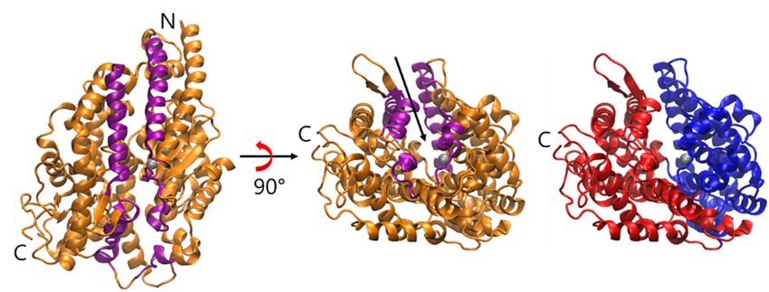
shown as a gray sphere. The rightmost panel shows two subdomains that form two sides
of the active site in the cleft, and the subdomain I (residues 40–122, 297–437, 551–583) and
II (residues 123–296, 438–550, 584–625) are colored by blue and red, respectively. The arrow
indicates the active site near the zinc ion and the putative binding pathway of ligands. The
first lip (residues 73–100, 297–304, 348–354, 370–379) belongs to subdomain I, and the
Molecules 2020, 25, 1288 3 of 12
second (109–131, 143–156, 267–276) belongs to subdomain II.
2.
2. Results
Results
2.1. Spontaneous
2.1. Spontaneous Conformational
Conformational Changes
Changes
A simulation
A simulation ofof ligand-free sACE (Apo)
ligand-free sACE was initiated
(Apo) was from the
initiated from the coordinates
coordinates after
after removing
removing the the
bound AngII from the sACE-AngII complex (PDB ID: 4APH) [19]. Like all others,
bound AngII from the sACE-AngII complex (PDB ID: 4APH) [19]. Like all others, the structure of the the structure of the
complex was also in the closed state defined by the distance between two lips (Figure
complex was also in the closed state defined by the distance between two lips (Figure 1) shorter than 1) shorter than
15
15 Å
Å (13.64
(13.64 Å).
Å). As
As simulation
simulation time
time went
went byby the
the enzyme
enzyme spontaneously
spontaneously openedopened itsits mouth,
mouth, and
and thethe
mouth gradually reclosed from the open state before returning back to the semi-open
mouth gradually reclosed from the open state before returning back to the semi-open and open states. and open states.
We
We defined
defined the
the open
open state
state with
with aa distance
distance longer
longer than
than 2020 Å
Å and
and the
the semi-open
semi-open statestate with
with distances
distances
longer than 15 Å and shorter than 20 Å. We observed multiple conversion between the open and
longer than 15 Å and shorter than 20 Å. We observed multiple conversion between the open and
closed states
closed states during
during 400
400 nsns simulation
simulation (Figure
(Figure 2). We believe
2). We believe that
that this is the
this is the first
first work
work that
that shows
shows the the
spontaneous opening
spontaneous opening and
and closing
closing motions
motions ofof ACE
ACE by by MD
MD simulation
simulation (Video
(Video S1).S1). In
In 2019,
2019, Yu
Yu et
et al. ran
al. ran
an MD simulation with ligand-free ACE only for 10 ns, but they did not report
an MD simulation with ligand-free ACE only for 10 ns, but they did not report the opening and the opening and closing
motionsmotions
closing [14]. [14].
Figure
Figure 2.2.Distance
Distance between
between twotwo
lips lips of AngII
of AngII boundbound sACE complex
sACE complex (green) and(green) andform
the Apo the(blue)
Apo
form (blue) along the simulation time after discarding the equilibration
along the simulation time after discarding the equilibration stage. A conformation with a distance stage. A
conformation
between two lips with a distance
longer than 20between two as
Å is defined lips
thelonger than 20
open state. Å isa defined
With distance as the open
shorter thanstate.
15 Å,
With a distance shorter
the conformation is definedthan 15 closed
as the Å, the state.
conformation is defined
If the distance as the15closed
is between and 20state.
Å, thenIf the
the
distance is between
conformation 15 and
is considered as 20
theÅ, then thestate.
semi-open conformation
The snapshotsis considered as thepurple
of sACE (orange, semi-open state.
for lips) are
shownsnapshots
The by superimposing
of sACE the subdomain II to the for
(orange, purple crystal structure
lips) (cyan). by superimposing the
are shown
subdomain II to the crystal structure (cyan).
In order to analyze the mouth opening and closing motion, we defined two lips and calculated
the distance
In orderbetween
to analyzethe the
centers
mouthof each lip Cα
opening andatoms throughout
closing motion, we production
defined two stagelips
of the
andsimulations
calculated
(Figure 2). Two lips of the mouth were defined as lip I in the subdomain
the distance between the centers of each lip Cα atoms throughout production stage of the simulationsI composed of residues
Ile73-Arg100,
(Figure 2). Two Pro297-Ala304,
lips of the mouth Arg348-Ala354,
were definedCys370-Val379, and lip II inI composed
as lip I in the subdomain the subdomain II composed
of residues Ile73-
of residues Pro128-Thr150, Gln160-Arg173, Ser284-Phe293. AngII bound
Arg100, Pro297-Ala304, Arg348-Ala354, Cys370-Val379, and lip II in the subdomain II composed sACE was quite stable over
of
the 400 ns simulation time, and no large backbone conformational change was observed unlike the
Apo form (in the absence of a ligand). The enzyme mainly stayed in the closed and the semi-open
states throughout the entire simulation (Figure S1).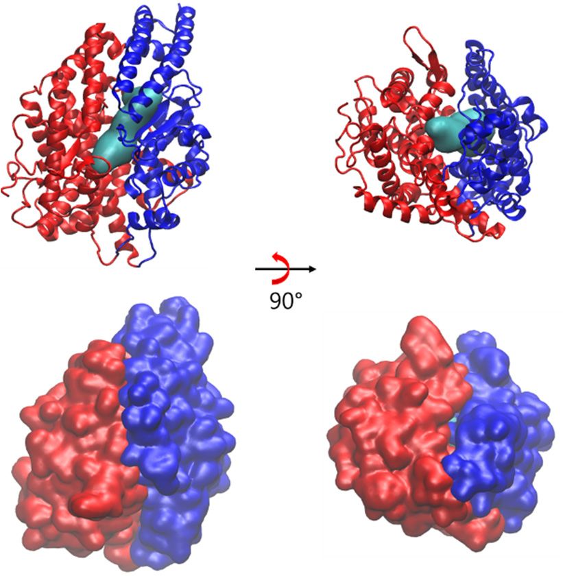
Molecules 2020, 25, x FOR PEER REVIEW 4 of 12
residues Pro128-Thr150, Gln160-Arg173, Ser284-Phe293. AngII bound sACE was quite stable over the
400 ns simulation time, and no large backbone conformational change was observed unlike the Apo
form (in the absence of a ligand). The enzyme mainly stayed in the closed and the semi-open states
Molecules 2020, 25, 1288 4 of 12
throughout the entire simulation (Figure S1).
2.2. Flexibility of sACE
In order to investigate the flexibility of the
the enzyme,
enzyme, root-mean-square deviation (RMSD) of the
Apo form
Apo form and the AngII bound
bound form were computed (Figure
form were computed (Figure 3).
3). As we expected the RMSD’s showed
the strong correlation
correlation with
withthe
thedistances
distancesbetween
betweentwotwolips.
lips.
DueDue to the
to the mouth
mouth opening
opening motions,
motions, the
the conformation
conformation of the
of the ApoApo form
form deviated
deviated farfar away
away fromthe
from theinitial
initialstructure,
structure,which
whichisis in
in the closed
conformation, reaching nearly 5 Å of the RMSD RMSD value.
value. However, the RMSD values of AngII AngII bound
form were fluctuated less than 3 Å. As compared
compared to thethe unbound
unbound form, the ligand
ligand bound form was
relatively stable.
stable.
Figure3.3.Root-mean-square
Figure Root-mean-square deviation (RMSD)
deviation and root-mean-square
(RMSD) fluctuation
and root-mean-square (RMSF) calculated
fluctuation (RMSF)
using Cα atoms
calculated usingfromCαsimulations
atoms from ofsimulations
AngII boundof sACE
AngII(green)
boundandsACE
Apo (blue).
(green)TheandredApo
bars(blue).
on the
horizontal
The red barsaxis on
of the
theRMSF graph indicate
horizontal the RMSF
axis of the residuesgraph
of subdomain
indicateI,the
showing its flexibility.
residues RMSF
of subdomain
was calculated after discarding the equilibration stage of the beginning of the MD simulations.
I, showing its flexibility. RMSF was calculated after discarding the equilibration stage of the
beginning of the MD simulations.
Cα root-mean-square fluctuation (RMSF) of each form was calculated using the production stage
of the MD simulation trajectories (Figure 3). The analysis indicates that the subdomain I is more flexible
Cα root-mean-square fluctuation (RMSF) of each form was calculated using the production stage
than subdomain II for both simulations. Interestingly, the overall flexibility of the subdomain II didn’t
of the MD simulation trajectories (Figure 3). The analysis indicates that the subdomain I is more
change much regardless of the presence of AngII. The most significant difference between the two
flexible than subdomain II for both simulations. Interestingly, the overall flexibility of the subdomain
simulations was the large increment of flexibility on the subdomain I for the Apo form. Notice that the
II didn’t change much regardless of the presence of AngII. The most significant difference between
subdomain closure movement in proteins is regarded as a common mechanism for the rearrangement
the two simulations was the large increment of flexibility on the subdomain I for the Apo form. Notice
of critical groups around substrates and inhibitors [28].
that the subdomain closure movement in proteins is regarded as a common mechanism for the
rearrangement
2.3. of critical
Open Conformation groups around substrates and inhibitors [28].
of ACE2
ThereConformation
2.3. Open is no structural report for the open conformation of human ACE yet. The angiotensin
of ACE2
converting enzyme-related carboxypeptidase (ACE2) is a homologue of the human sACE. ACE2
There
was also is no structural
identified reportreceptor
as the cellular for the open
for the conformation
SARS coronavirusof human
and ACE
novelyet. The angiotensin
coronavirus in 2019
converting enzyme-related carboxypeptidase (ACE2) is a homologue of the human
(COVID-19) [29,30]. The catalytic domains of ACE2 and sACE share 42% sequence identity [31]. sACE. ACE2 was
also identified as the cellular receptor for the SARS coronavirus and novel coronavirus
The crystal structures of ACE2 with and without its inhibitor were reported first in 2004 (PDB ID: in 2019
(COVID-19)
1R4L, 1R42). [29,30].
The open The catalytic domains
conformation of ACE2in
was observed and
thesACE share
absence 42%inhibitor,
of the sequencewhile
identity [31]. The
the inhibitor
bound one showed the closed conformation. Based on the two structures, the authors proposed a1R4L,
crystal structures of ACE2 with and without its inhibitor were reported first in 2004 (PDB ID: large
1R42). The openmotion
hinge-bending conformation was observed
is important in the
for catalytic absence
activity andofinhibitor
the inhibitor,
bindingwhile the inhibitor
of ACE too [32].bound
one showed the closed the
We superimposed conformation.
closed and Based
the openon conformations
the two structures,of ACEthe and
authors proposed
compared witha those
large
hinge-bending motion is important for catalytic activity and inhibitor binding
of ACE2, and the comparison revealed tremendous similarity between two systems (Figure S2). of ACE too [32].
We superimposed
In addition, the distancethe closedtwo
between andlips
thewas
open conformations
calculated for ACE2.of ACE
Basedandoncompared
the sequence with those of
alignment
ACE2, and the comparison revealed tremendous similarity between two systems
of ACE2 and sACE, two lips of ACE2 were identified as lip I composed of the residues 54–81, 289–296, (Figure S2). In
340–346, 361–370 of subdomain I and lip II composed of the residues 109–131, 143–156, 267–276 of
subdomain II. The distances between lip I and lip II were computed as 13.32 Å and 20.64 Å for the
closed and open states, respectively. This result is consistent with the distance analysis of sACE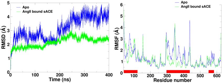
addition, the distance between two lips was calculated for ACE2. Based on the sequence alignment
 of ACE2 and sACE, two lips of ACE2 were identified as lip I composed of the residues 54–81, 289–
 296, 340–346, 361–370 of subdomain I and lip II composed of the residues 109–131, 143–156, 267–276
 of subdomain II. The distances between lip I and lip II were computed as 13.32 Å and 20.64 Å for the
Molecules 2020, 25, 1288                                                                                     5 of 12
 closed and open states, respectively. This result is consistent with the distance analysis of sACE
 (Figure 2). In order to test robustness of definition of backbone conformational states we also
(Figure  2). In
 calculated   theorder
                    mouthto test
                             openrobustness of definition
                                   angles, and  the resultsofshowed
                                                              backbone
                                                                    theconformational
                                                                        similar patternstates we also(Figure
                                                                                        as distances    calculated
                                                                                                                S3).
the mouth
 Based       open
         on our       angles, andthe
                  calculations      the results showed
                                      maximal           the similar
                                                hinge-bending       patternwas
                                                                 movement        ~18° which
                                                                             as distances (Figure  S3).toBased
                                                                                             is close            on
                                                                                                          the value
our  calculations
 (~16°)  measuredthe       maximal
                       in ACE2        hinge-bending
                                  structures  [32].    movement was ~18 which is close to the value (~16◦ )
                                                                           ◦

measured in ACE2 structures [32].
 2.4. Competitive Inhibitor Binding
2.4. Competitive Inhibitor Binding
       BPPb is a competitive inhibitor isolated from snake venom [17]. A crystal structure shows the
      BPPb is a competitive inhibitor isolated from snake venom [17]. A crystal structure shows the
 active site of sACE occupied by the octapeptide BPPb and the closed conformational state of the
active site of sACE occupied by the octapeptide BPPb and the closed conformational state of the
 complex (PDB ID: 4APJ) [19]. We set up another system with BPPb bound sACE and carried out a
complex (PDB ID: 4APJ) [19]. We set up another system with BPPb bound sACE and carried out a
 400 ns MD simulation. AngII is not only the product of catalytic function of ACE but also considered
400 ns MD simulation. AngII is not only the product of catalytic function of ACE but also considered
 as competitive inhibitor. Before leaving the active site, another substrate AngI cannot bind to the site
as competitive inhibitor. Before leaving the active site, another substrate AngI cannot bind to the site
 for the next reaction.
for the next reaction.
       The BPPb bound sACE simulation showed the similar behavior with the AngII bound one
      The BPPb bound sACE simulation showed the similar behavior with the AngII bound one (Figure
 (Figure S1). We analyzed the distance between two lips of the mouth, showing conformational
S1). We analyzed the distance between two lips of the mouth, showing conformational changes from
 changes from the closed to the semi-open states and back to the closed state again (Figure 4a). BPPb
the closed to the semi-open states and back to the closed state again (Figure 4a). BPPb bound ACE
 bound ACE was more flexible, and the AngII bound one made the mouth closed more tightly. The
was more flexible, and the AngII bound one made the mouth closed more tightly. The number of
 number of hydrogen bonds between the ligands and the enzyme was calculated over the simulation
hydrogen bonds between the ligands and the enzyme was calculated over the simulation time (Figure
 time (Figure S4). The average numbers were 6.52 ± 2.15 and 6.05 ± 1.64 for AngII and BPPb,
S4). The average numbers were 6.52 ± 2.15 and 6.05 ± 1.64 for AngII and BPPb, respectively (Table 1).
 respectively (Table 1). This indicated that the opening and closing motions were affected by the
This indicated that the opening and closing motions were affected by the interactions between the
 interactions between the ligand with the enzyme.
ligand with the enzyme.

                           (a)                                                      (b)
      Figure4.4.Comparison
     Figure      Comparison     of the
                            of the      distances
                                    distances     between
                                              between       two
                                                      two lips  lips among
                                                               among  the Apothe
                                                                              andApo  and (a) competitive
                                                                                  (a) competitive inhibitor
      inhibitor
     bound  formsbound   formsand
                   with AngII    with   AngII
                                     BPPb.  (b) and BPPb.
                                                Mixed      (b) Mixed inhibitor
                                                      non-competitive  non-competitive   inhibitor
                                                                                bound forms         bound
                                                                                             with SPI  and
      forms with SPI and SPI-AngII complex.
     SPI-AngII  complex.

     Table 1. The average number of hydrogen bonds between the ligand and the enzyme calculated over
      Table 1. The average number of hydrogen bonds between the ligand and the enzyme
     the production stage of molecular dynamics (MD) simulations.
      calculated over the production stage of molecular dynamics (MD) simulations.
                              Complex               Average Number of Hydrogen Bond
                                  Complex          Average Number of Hydrogen Bond
                        AngII  bound
                            AngII     sACE
                                   bound sACE                  6.52±±2.15
                                                              6.52    2.15
                         BPPb  bound
                            BPPb      sACE
                                   bound sACE                  6.05±±1.64
                                                              6.05    1.64
                          SPI SPI
                              bound  sACE
                                  bound sACE                   5.86±±1.42
                                                              5.86    1.42
                  SPI bound sACE-AngII complex                 6.73 ± 1.57
                      SPI bound sACE-AngII complex            6.73 ± 1.57

2.5. Mixed Non-Competitive Inhibitor Binding
    In addition to competitive inhibition, non-competitive inhibition is also a common inhibition
mechanism. Non-competitive inhibition includes pure and mixed noncompetitive inhibition.
Surprisingly for us, mechanism of mixed type non-competitive inhibition for ACE has not been studied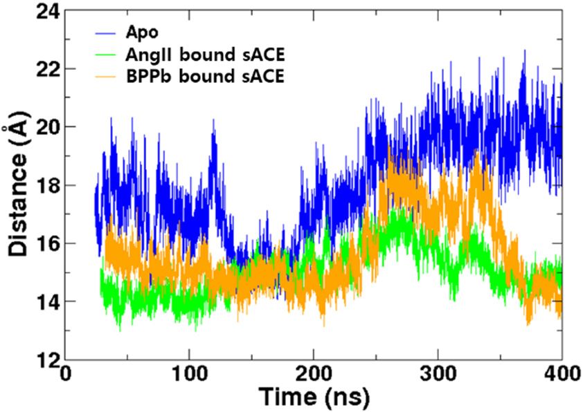
Molecules 2020, 25, x FOR PEER REVIEW 6 of 12
2.5. Mixed Non-competitive Inhibitor Binding
In addition to competitive inhibition, non-competitive inhibition is also a common inhibition
Molecules 2020, 25, 1288 6 of 12
mechanism. Non-competitive inhibition includes pure and mixed noncompetitive inhibition.
Surprisingly for us, mechanism of mixed type non-competitive inhibition for ACE has not been
studied
intensivelyintensively
so far. In so the
far. previous
In the previous
study, astudy, a heptapeptide
heptapeptide (Thr-Met-Glu-Pro-Gly-Lys-Pro)
(Thr-Met-Glu-Pro-Gly-Lys-Pro) derived
derived from a marine
from a marine microalgae
microalgae SpirulinaSpirulina was identified
was identified as a mixed
as a mixed non-competitive
non-competitive inhibitor
inhibitor (SPI)(SPI)
[18].
[18]. We investigated the interactions and dynamics of ACE bound with
We investigated the interactions and dynamics of ACE bound with the mixed non-competitive inhibitor the mixed non-competitive
inhibitor
further. MD further. MD simulations
simulations were conductedwere conducted
for the SPIfor boundthe SPI
to thebound to theand
Apo form Apotheform and thetoSPI
SPI bound the
bound to the AngII-sACE
AngII-sACE complex form. complex form.
The
The simulation
simulation of of SPI
SPIbound
boundtotosACE sACEwas was started
started with
with thethe structure
structure after
after the removing
the removing AngII AngII
from
from SPI bound
SPI bound to AngII-sACE
to AngII-sACE complex, complex,
which which is theof
is the result result
the SPIof the SPI docking
docking simulation simulation of the
of the previous
previous
study [18]. study
The[18]. The analysis
analysis of distance of distance
between between
two lips two of the lips
SPIofbound
the SPI bound
sACE sACEbackbone
showed showed
backbone
conformational fluctuation between the closed and semi-open states, but we were not able to seeable
conformational fluctuation between the closed and semi-open states, but we were not the
to see conformational
open the open conformational
state (Figure state
4b).(Figure
In other4b). In other
words, words, the spontaneous
the spontaneous conformational conformational
changes were
changes
limited, were limited,
and only the and
closedonlyand thesemi-open
closed and semi-open
states states were
were stabilized bystabilized
SPI (VideobyS2). SPI The
(Video S2).
peptide
The
SPI peptide
formed 5.86 SPI formed 5.86 ± 1.42bonds
± 1.42 hydrogen hydrogen bondsaveraged
with sACE with sACE averaged
over over thestage
the production production stage
of the whole
of the whole(Table
simulation simulation
1). (Table 1).
In
Inthe
thesimulation
simulationof ofSPI
SPIbound
boundto tothe
theAngII-sACE
AngII-sACEcomplex,complex,the thepeptide
peptidebound
boundin inthe
theN-terminal
N-terminal
side
sideofofthe
themouth
mouthand andnext
nextto tothe
the AngII
AngII (Figure
(Figure 5).5). Binding
Bindingof ofSPI
SPIwas
wasstabilized
stabilizedby byhydrogen
hydrogenbond bond
interactions not only with sACE but also with AngII [18]. We investigated
interactions not only with sACE but also with AngII [18]. We investigated the presence of hydrogen the presence of hydrogen
bonds
bonds during
during the the MD
MD simulations.
simulations. In Inaverage,
average,the theSPISPI formed
formed 3.22 3.22 ±±1.08
1.08andand 3.47
3.47 ±±0.75
0.75pairs
pairs of of
hydrogen
hydrogen bonds bonds with
withsACE
sACEand and AngII,
AngII, respectively,
respectively, andand together
together 6.73 ± 6.73
1.57± pairs
1.57 (Table
pairs (Table
1). SPI 1). SPI
formed
formed
hydrogen hydrogen bondsfrequently
bonds most most frequently
with sACE with(Thr1-Asp121,
sACE (Thr1-Asp121, Thr1-Ser219,
Thr1-Ser219, Glu3-Arg522,
Glu3-Arg522, Lys6-
Lys6-Glu143,
Glu143,
SPI-sACE SPI-sACE
residuesresidues
in order)inand order)
withandAngIIwith AngII (Glu3-Arg2,
(Glu3-Arg2, Pro7-Arg2,Pro7-Arg2,
Pro2-Val3,Pro2-Val3, SPI-AngII
SPI-AngII residues in
residues in order) (Figure 5, Table S1). In addition to hydrogen bonds,
order) (Figure 5, Table S1). In addition to hydrogen bonds, binding of the SPI was stabilized by van der binding of the SPI was
stabilized by van too.
Waals interactions der The
Waals interactions
sidechain of Pro4too. TheSPI
of the sidechain
bound inofthePro4 of the SPI
hydrophobic bound
pocket in the
formed by
hydrophobic
Ile204, Leu139 pocket formedofbysACE
and Trp220 Ile204,
(notLeu139
shown andin Trp220
Figure 5offor sACE (not shown in Figure 5 for clarity).
clarity).
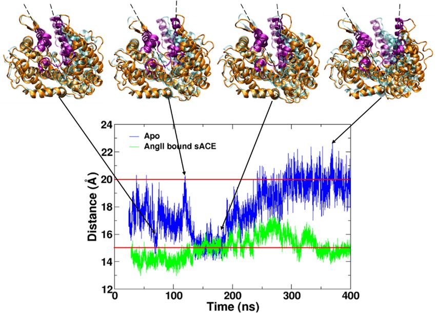
Figure
Figure 5.5. The
Thebinding
bindingsite siteofof
thethe
SPISPI (green
(green carbon)
carbon) next next to AngII
to AngII (cyan (cyan
carbon) carbon) in the
in the cleft cleft
of sACE.
sACE
of sACE.is represented by orange ribbons
sACE is represented (purple
by orange lips), and
ribbons the zinc
(purple ion in
lips), andthethe
active site
zinc is represented
ion in the activeby
a gray
site sphere. AngIIby
is represented and ACE sphere.
a gray binding AngII
site areand
represented by sticks,
ACE binding siteand
arecarbon, nitrogen,
represented by oxygen,
sticks,
sulfur
and and hydrogen
carbon, nitrogen, atoms
oxygen,are colored by cyan,
sulfur and blue, red,
hydrogen yellow
atoms areand white,
colored byrespectively.
cyan, blue, SPIred,is
represented by sticks, and carbon atoms are colored by green. Only residue
yellow and white, respectively. SPI is represented by sticks, and carbon atoms are colored numbers of sACE are
shown in the inset figure for clarity. The snapshot was taken at 200.38
by green. Only residue numbers of sACE are shown in the inset figure for clarity. The ns.
snapshot
Distance was takenofatSPI
analysis 200.38boundns. sACE-AngII showed similar behavior to other ligand bound
simulations (Figure 4b). Interestingly, the SPI bound sACE-AngII complex was stayed in the closed
state most of the simulation time. This is understandable since both SPI and AngII are acting as
inhibitors, and the number of hydrogen bonds between the ligand and the enzyme is largest among all
complexes (Table 1). The closed state was stabilized not only by the interactions between AngI and
sACE but also by the interactions between SPI and sACE. In addition, interactions between SPI and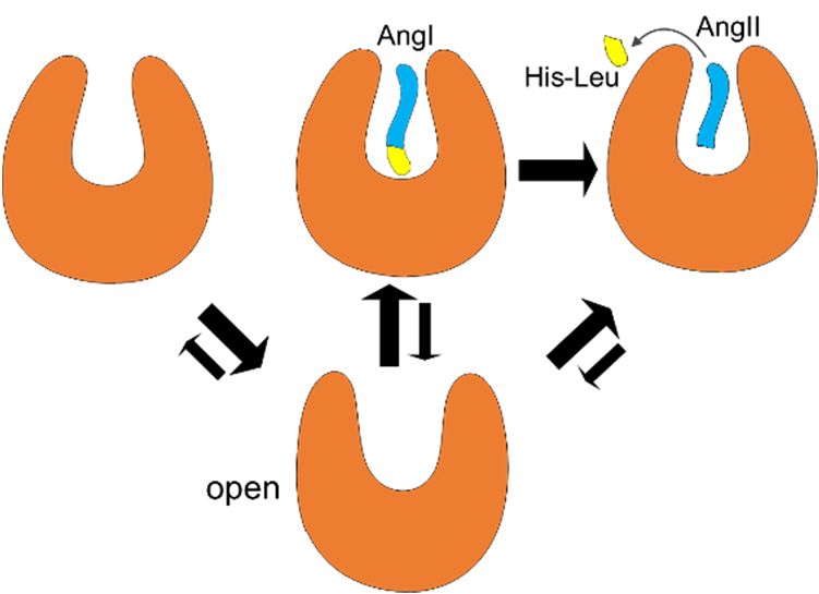
Distance analysis of SPI bound sACE-AngII showed similar behavior to other ligand bound
simulations (Figure 4b). Interestingly, the SPI bound sACE-AngII complex was stayed in the closed
state most of the simulation time. This is understandable since both SPI and AngII are acting as
inhibitors, and the number of hydrogen bonds between the ligand and the enzyme is largest among
Molecules 2020, 25, 1288 7 of 12
all complexes (Table 1). The closed state was stabilized not only by the interactions between AngI
and sACE but also by the interactions between SPI and sACE. In addition, interactions between SPI
AngII stabilized
and AngII the complex
stabilized further
the complex in theinclosed
further state.state.
the closed Analysis of alloffive
Analysis RMSF’s
all five indicated
RMSF’s that that
indicated the
most stable system was SPI bound to sACE-AngII complex (Figure S5). We recognized
the most stable system was SPI bound to sACE-AngII complex (Figure S5). We recognized that the that the binding
of SPI to of
binding sACE-AngII complex complex
SPI to sACE-AngII slightly twisted
slightlythe subdomain
twisted I (Figure IS6).
the subdomain (Figure S6).
3.
3. Discussion
Discussion
The
The active
active sites
sites and
and binding
binding interactions
interactions havehave been
been well
well known
known for for ACE
ACE with various inhibitors,
with various inhibitors,
while
while the
the conformational
conformational dynamicsdynamics and and its
its role
role inin activation
activation mechanism
mechanism have have not
not been
been studied
studied
intensively.
intensively. Drug design might benefit from backbone conformations observed by MD simulations,
Drug design might benefit from backbone conformations observed by MD simulations,
which
which have
have never
never reached
reached by bythe
theX-ray
X-raycrystallography
crystallographywith withligand
ligandbound
boundforms.
forms. Unlike
Unlike thethe closed
closed
state, the open state of sACE has not been detected in both experiments
state, the open state of sACE has not been detected in both experiments and simulations so and simulations so far.
far.
Surprisingly, over more than a decade since the first proposal [32], no further
Surprisingly, over more than a decade since the first proposal [32], no further study has been reportedstudy has been reported
about
about the
the role
role of
of large
largehinge-bending
hinge-bendingmotion motionfor forthe
theactivation
activationof ofsACE.
sACE.
In
In contrast to the lock-and-key model, many proteins can take
contrast to the lock-and-key model, many proteins can take aa ligand
ligand onlyonly through
through the the
conformational changes. The putative binding pathway of sACE
conformational changes. The putative binding pathway of sACE might be too narrow to might be too narrow to accommodate
the substrate AngI
accommodate in the closed
the substrate AngI in state
the(Figure 6), thus
closed state the enzyme
(Figure 6), thus the needs to change
enzyme needs its backbone
to change its
conformation
backbone conformation to take the substrate for its catalytic function. At least in the semi-open state,
to take the substrate for its catalytic function. At least in the semi-open state,
rearrangement
rearrangement of of sidechain
sidechain may may allow
allow thethe substrate
substrate to to enter
enter the
the active
active site
site (Figure
(Figure S7).
S7). Two
Two models
models
of
of enzyme mechanisms, induce-fit [33] and pre-existing equilibrium dynamics [34] represent the
enzyme mechanisms, induce-fit [33] and pre-existing equilibrium dynamics [34] represent the
conformational
conformational changes
changes and and dynamics
dynamics upon upon ligand
ligand binding.
binding. In In the
the former,
former, binding
binding of of aa ligand
ligand
induces
inducesconformational
conformationalchanges changes of of
a protein,
a protein,while in the
while in latter, a ligand
the latter, binds to
a ligand a certain
binds to a pre-existing
certain pre-
conformation already present and accessible by equilibrium
existing conformation already present and accessible by equilibrium dynamics. dynamics.
Figure6.6.Surface
Figure Surfacerepresentations of AngII
representations (cyan colored)
of AngII and ribbonand
(cyan colored) representation of a closed state
ribbon representation ofofa
sACE
closed(subdomain I in blue
state of sACE and subdomain
(subdomain II in and
I in blue red) subdomain
at the top panel and
II in its surface
red) representations
at the top panel and itsof
sACE at bottom panel.
surface representations of sACE at bottom panel.
In
In the
the ligand-free
ligand-free (Apo)
(Apo) MD
MD simulation,
simulation, we
we were
were able
able to
toobserve
observespontaneous
spontaneous conformational
conformational
changes
changes between the open and closed states of sACE, implying that the open
between the open and closed states of sACE, implying that the open and
and closed
closed backbone
backbone
conformational states are already present and accessible regardless of the presence
conformational states are already present and accessible regardless of the presence of a ligand. of a ligand.
In
In addition, several times of conversions between the states for the 400-nanoseconds simulation
addition, several times of conversions between the states for the 400-nanoseconds simulation indicate
indicate that the enzyme is very flexible and dynamic. Indeed, all available ACE structures are resolved
in the closed conformation either in the presence ligands or with mutations. Introducing mutations,
ligands or chimera are common methods to enhance thermal stability and to crystalize a protein.
In addition, the absence of the open conformation in a ligand-free ACE imply the intrinsic flexibility
of ACE. Again, only one available structure in the open conformation is resolved with ACE2 [32].Molecules 2020, 25, x FOR PEER REVIEW 8 of 12
that the enzyme is very flexible and dynamic. Indeed, all available ACE structures are resolved in the
closed conformation either in the presence ligands or with mutations. Introducing mutations, ligands
Molecules 2020, 25, 1288 8 of 12
or chimera are common methods to enhance thermal stability and to crystalize a protein. In addition,
the absence of the open conformation in a ligand-free ACE imply the intrinsic flexibility of ACE.
In addition,
Again, only aone previous study
available using normal
structure in the openmode analysis (NMA)
conformation with testicular
is resolved with ACE2ACE[32].
(tACE, another
In addition,
isoform of ACE) and ACE2 showed the intrinsic flexibility and open and closed
a previous study using normal mode analysis (NMA) with testicular ACE (tACE, another isoform conformational models of
of ACE
ACE) [35].
and ACE2Theshowed
authorsthealso proposed
intrinsic the hinge-bending
flexibility and open and mechanism for substratemodels
closed conformational entry into
of ACEthe
active site.
[35]. The authors also proposed the hinge-bending mechanism for substrate entry into the active site.
Based on the results of of our
our MD
MD simulations
simulations and and available
available X-ray structures, we propose the
following
following activation
activationmechanism
mechanismofofsACE sACE(Figure
(Figure7).7).Regardless
Regardless ofof
presence
presenceof of
a ligand, sACE
a ligand, sACE could
couldbe
in
be any of the
in any open,
of the semi-open
open, semi-open andand
closed
closedstates in terms
states in termsof backbone
of backboneconformation.
conformation. In the absence
In the absence of
aofsubstrate,
a substrate,or or
AngI,
AngI,the
theopen
open state
statemight
mightbebemoremorefavored
favoredthan
thanthe
theclosed
closedstate
state(Figure
(Figure 2),
2), and
and the
enzyme becomes ready ready toto accept
accept aasubstrate
substratefor forits
itscatalytic
catalyticfunction.
function.This
This equilibrium,
equilibrium, however,
however, is
is changed in the presence of AngI, and the closed state becomes more
changed in the presence of AngI, and the closed state becomes more stable than the stable than the open state due to
the binding of the substrate.
substrate. Then the C-terminal dipeptide (His-Leu) of AngI is hydrolyzed. AngII,
the product of the enzyme activity,activity, might
might be be released
released when
when sACE
sACE is in the semi-open state (Figure 2
and Figure S7). After the release of AngII from sACE, the equilibrium between conformational states
goes back to to the
thefirst
firststage
stageagain.
again.Notice
Notice that
that thethe semi-open
semi-open conformation
conformation cancan be accessed
be accessed under under
any
any condition.
condition.
Figure7. 7.
Figure Activation
Activation mechanism
mechanism of sACEofandsACE and shifts
equilibrium equilibrium shifts
between the openbetween the (lower
conformation open
conformation
panel) (lower
and closed panel) and
conformations closed
(upper conformations
panel) (upper
upon the presence panel) upon
of ligands: theleft
from the presence
to right of
in
ligands:
the figure,from
in thethe left to
absence of right in the(AngI),
a substrate figure,
in in
thethe absence
presence of a substrate
of substrate, and in (AngI), in the
the presence of
product
presence (AngII), respectively.
of substrate, and Equilibrium shifts between
in the presence of producttwo (AngII),
states are respectively.
represented qualitatively
Equilibrium by
the sizebetween
shifts of arrows.
two states are represented qualitatively by the size of arrows.
Similarly,
Similarly,we wecan derive
can a mechanism
derive a mechanism for a competitive
for a competitiveinhibitor. Now, if the
inhibitor. ligand
Now, is a competitive
if the ligand is a
inhibitor, then the bound complex becomes stable in the closed state. Qualitatively,
competitive inhibitor, then the bound complex becomes stable in the closed state. Qualitatively, the equilibrium the
shift is the same way as the case of substrate binding. After release of the inhibitor,
equilibrium shift is the same way as the case of substrate binding. After release of the inhibitor, the the equilibrium
goes back to the
equilibrium goesfirst stage
back of the
to the Apo
first stageform. Since
of the Apo AngII
form.occupies the same
Since AngII active the
occupies site,same
until active
the release
site,
of AngII sACE cannot take any substrate. This is true for the substrate
until the release of AngII sACE cannot take any substrate. This is true for the substrate too. Interestingly, SPI the mixed
too.
non-competitive inhibitor makes use of this. The interactions of SPI not
Interestingly, SPI the mixed non-competitive inhibitor makes use of this. The interactions of SPI notonly with sACE but also
with
only AngII
with sACEstabilized sACE
but also with in AngII
the closed state further,
stabilized sACE inresulting
the closed instate
a dead-end
further,complex
resultingby inholding
a dead-
the product of enzyme AngII in the active site [18]. Since SPI does not
end complex by holding the product of enzyme AngII in the active site [18]. Since SPI does not share the binding siteshare
with
the
the substrate,
binding site it becomes non-competitive.
with the substrate, it becomes Even though SPI is non-competitive,
non-competitive. Even though SPI is it binds next to the
non-competitive,
active site and interacts with the substrate, becoming a mixed type inhibitor
it binds next to the active site and interacts with the substrate, becoming a mixed type inhibitor [18]. The binding sites of
[18].
various drugssites
The binding and of drug candidates
various drugswere and studied intensively,
drug candidates wereandstudied
along the various ligands,
intensively, and alongbindingthe
sites and their conformations are various too. This indicates the binding
various ligands, binding sites and their conformations are various too. This indicates the binding of a specific ligand induce of
aa specific
specific conformation
ligand induceofaits binding
specific site. Therefore,
conformation of itsthebinding
overall site.
largeTherefore,
backbone the open and closed
overall large
conformational
backbone open state and pre-exist, and the sidechain
closed conformational stateand minor backbone
pre-exist, rearrangement
and the sidechain and minormay bebackbone
induced
upon ligand binding.
rearrangement may be induced upon ligand binding.
In Apo simulation, we have removed the bound AngII from the X-ray structure of sACE-AngII
complex and started the MD simulation. This removal of AngII is an additional perturbation to the
sACE structure. We checked equilibration of the Apo system by monitoring the thermodynamic
parameters such as energy, temperature, volume and pressure (Figure S8). In addition, we monitoredMolecules 2020, 25, 1288 9 of 12
evolution of secondary structure along the simulation time and recognized that the overall secondary
structure was well conserved and stable except for loop and turn regions (Figure S9). Even though
we observe multiple opening and closing motions, one can still consider the Apo system is still
under equilibration stage. If Apo structure was still under equilibration stage and goes to a certain
open conformation, then we would have an X-ray structure with open conformation like ACE2.
A calorimetric study showing that the drug binding is entropically driven also supports dynamics of
ACE [36].
In conclusion, using all-atom MD simulations, the conversion between open and closed states of
sACE was observed in the absence of a ligand supporting that hinge-bending motion is essential in
enzyme activation. The preferred states as well as the extent of flexibility were strongly dependent on
the presence of a ligand. The transformation from the semi-open to the closed state in the bound forms
was directly influenced by the interaction between the residues of binding sites of the substrate and the
inhibitors. These interactions constrained the enzyme structure to remain in the semi-open and closed
states. However, it would seem that the putative binding pathway is too narrow to accommodate the
substrate and inhibitors. Therefore, the semi-open conformation might be the state where the ligands
can bind. Our MD simulation results and the mechanistic model of sACE activation and inhibition
portends not only drug design for hypotension treatment but also wide applications in molecular
biophysics and beyond.
4. Materials and Methods
4.1. Molecular Docking for SPI
Potential binding sites of SPI on sACE were searched by docking simulations using the FlexPepDock
protocol of the Rosetta program [37]. The crystal structure of human sACE in complex with AngII
(PDB ID: 4APH) was used for the target. Since the SPI peptide is a non-competitive inhibitor, AngII
was kept during the docking simulations to avoid overlapping between the binding site of the peptide
and the active site. A structure with the lowest score of docking simulations was chosen for the
molecular dynamics simulation in order to relax the structure further (Table S2). Details of the docking
simulations were described in the previous study [18].
4.2. Preparation of MD Simulations
Total five molecular dynamics simulations were carried out: BPPb bound sACE, AngII bound
sACE, Apo (ligand-free), SPI bound sACE-AngII complex, SPI bound sACE. Two crystal structures
of the sACE co-crystallized with ligands in the active site were used to build the simulation systems,
sACE complex with the natural inhibitory peptide BPPb (PDB ID: 4APJ) and sACE complex with AngII
(PDB ID: 4APH). In order to simulate the Apo form, AngII was removed from the active site of the
crystal structure (4APH). The SPI bound to the sACE-AngII complex with the lowest score from the
docking simulations was chosen to run the fourth MD simulation, and the last SPI bound sACE model
was generated by removing the AngII from this docking simulation result.
Charmm36 force field with explicit TIP3P water molecules was used in all simulations [38].
Crystallographic water molecules, chloride ions, and zinc ion were retained as they were found in
the X-ray crystal structures. Ligand chemical components in the original crystal structures including
acetate ion, beta-D-mannose, N-acetyl-D-Glucosamine were also retained and modeled with Charmm
general force field [39]. All five simulation systems were solvated in the truncated octahedral box with
23251, 23418, 23565, 23410 and 23374 TIP3P water molecules, respectively. Then these solvated systems
were electrically neutralized by adding counter ions, and the final salt concentration became 0.15 M.
4.3. Molcular Dynamics Simulations
All simulations were performed by NAMD under the periodic boundary condition [40]. After
solvation, 1000 steps of energy minimization were performed to remove possible bad contacts. Then,Molecules 2020, 25, 1288 10 of 12
the MD simulations were started by gradually heating the systems from 10 to 310 K for 60 ps under
the constant volume condition. In order to equilibrate the density of the systems, the simulation
was switched to constant pressure and temperature (NPT) conditions and continued afterwards.
The average pressure and the temperature were maintained at 1 bar and 310 K, respectively. All heavy
atoms during the energy minimization, heating and the first 200 ps of NPT simulation were restrained
with harmonic potential with a force constant 1 kcal/mol·A2 . After that, the harmonic position restraints
were removed, allowing all atoms in the systems to relax. All bonds involving hydrogen atoms were
constrained, allowing an integration time step of 2 fs. The non-bonded interactions were smoothly
truncated from 10 Å to 12 Å cutoff, and the particle-mesh Ewald method was used to treat long-range
electrostatic interactions [41]. The equilibration stages of simulations were determined based on
RMSD and system energy calculations. Total simulation and equilibration times for BPPB-sACE,
AngII-sACE, Apo, SPI-sACE-AngII, and SPI-sACE were 400 (32.26), 400 (28.26), 400 (24.36), 360 (20.95),
and 360 (13.26), respectively in nanoseconds. Summary of all MD simulations are provided in Table S3.
The production stage after discarding the initial equilibration stage, was used for the post simulation
analysis except for RMSD calculation. All post analyses and visualization were carried out by Visual
Molecular Dynamics (VMD) program [42]. Formation of hydrogen bond was identified using 3.3 Å
(donor-acceptor distance) and 30 degrees (proton-donor-acceptor angle) criteria.
Supplementary Materials: The following are available online at http://www.mdpi.com/1420-3049/25/6/1288/s1,
Figure S1: Conformational states of sACE from MD simulations, Figure S2: Superposition of the closed state and
the open state of sACE and ACE2, S3: Mouth open angles, S4: Number of hydrogen bonds along the simulation, S5:
Cα RMSF of all simulation systems, S6: SPI induced conformational change, S7: Semi-open state, S8: Equilibration
of Apo simulation, S9: Diagram of secondary-structure evolution, Table S1: Ratio of forming hydrogen bond pairs
over the simulation time, S2: Major docking score functions, Table S3: Summary of MD simulations, Video S1:
Mouth opening of Apo sACE, Video S2: SPI bound sACE.
Author Contributions: Conceptualization, M.Y. and W.-K.J.; methodology, M.Y. and W.-K.J.; software, T.T.V.;
validation, M.Y. and W.-K.J.; formal analysis, T.T.V. and S.-Y.H.; investigation, T.T.V. and S.-Y.H.; resources, M.Y.;
writing—original draft preparation, T.T.V.; writing—review and editing, M.Y.; visualization, T.T.V.; supervision,
M.Y. and W.-K.J.; project administration, M.Y.; funding acquisition, M.Y. All authors have read and agreed to the
published version of the manuscript.
Funding: This work was supported by the Pukyong National University Research Aboard Fund in 2017
(C-D-2017-0984).
Conflicts of Interest: The authors declare no conflict of interest.
References
1. Kearney, P.M.; Whelton, M.; Reynolds, K.; Muntner, P.; Whelton, P.K.; He, J. Global Burden of Hypertension:
Analysis of Worldwide Data. Lancet 2005, 365, 217–223. [CrossRef]
2. Inagami, T. The Renin-Angiotensin System. Essays Biochem. 1994, 28, 147–164. [PubMed]
3. Rawlings, N.D.; Barrett, A.J. Evolutionary Families of Peptidases. Biochem. J. 1993, 290, 205–218. [CrossRef]
4. Fernandez, J.H.; Hayashi, M.A.F.; Camargo, A.C.M.; Neshich, G. Structural Basis of the Lisinopril-Binding
Specificity in N- and C-Domains of Human Somatic ACE. Biochem. Biophys. Res. Commun. 2003, 308, 219–226.
[CrossRef]
5. Wei, L.; Alhenc-Gelas, F.; Corvol, P.; Clauser, E. The Two Homologous Domains of Human Angiotensin
I-Converting Enzyme are both Catalytically Active. J. Biol. Chem. 1991, 266, 9002–9008.
6. Dell’Italia, L.J.; Meng, Q.C.; Balcells, E.; Wei, C.C.; Palmer, R.; Hageman, G.R.; Durand, J.; Hankes, G.H.;
Oparil, S. Compartmentalization of Angiotensin II Generation in the Dog Heart. Evidence for Independent
Mechanisms in Intravascular and Interstitial Spaces. J. Clin. Investig. 1997, 100, 253–258. [CrossRef]
7. Wei, C.C.; Meng, Q.C.; Palmer, R.; Hageman, G.R.; Durand, J.; Bradley, W.E.; Farrell, D.M.; Hankes, G.H.;
Oparil, S.; Dell’Italia, L.J. Evidence for Angiotensin-Converting Enzyme- and Chymase-Mediated Angiotensin
II Formation in the Interstitial Fluid Space of the Dog Heart in Vivo. Circulation 1999, 99, 2583–2589. [CrossRef]
8. Erdös, N.; Deddish, N.; Marcic, N. Potentiation of Bradykinin Actions by ACE Inhibitors. Trends Endocrinol.
Metab. 1999, 10, 223–229. [CrossRef]Molecules 2020, 25, 1288 11 of 12
9. Turner, A.J.; Hooper, N.M. The Angiotensin-Converting Enzyme Gene Family: Genomics and Pharmacology.
Trends Pharmacol. Sci. 2002, 23, 177–183. [CrossRef]
10. Acharya, K.R.; Sturrock, E.D.; Riordan, J.F.; Ehlers, M.R.W. Ace Revisited: A New Target for Structure-Based
Drug Design. Nat. Rev. Drug. Discov. 2003, 2, 891–902. [CrossRef] [PubMed]
11. Qian, Z.; Je, J.; Kim, S. Antihypertensive Effect of Angiotensin I Converting Enzyme-Inhibitory Peptide
from Hydrolysates of Bigeye Tuna Dark Muscle, Thunnus Obesus. J. Agric. Food Chem. 2007, 55, 8398–8403.
[CrossRef] [PubMed]
12. Cohn, J.N.; Kowey, P.R.; Whelton, P.K.; Prisant, L.M. New Guidelines for Potassium Replacement in Clinical
Practice: A Contemporary Review by the National Council on Potassium in Clinical Practice. Arch. Intern.
Med. 2000, 160, 2429–2436. [CrossRef] [PubMed]
13. Baumgartner, L.J.; Morris, A.A.; Chapman, S.A. Angiotensin-Converting Enzyme Inhibitor Induced
Angioedema: Predictors of Mechanical Ventilation and Treatment Approaches. Intensive Care Med. 2015, 41,
2233–2234. [CrossRef] [PubMed]
14. Yu, D.; Wang, C.; Song, Y.; Zhu, J.; Zhang, X. Discovery of Novel Angiotensin-Converting Enzyme Inhibitory
Peptides from Todarodes Pacificus and their Inhibitory Mechanism: In Silico and in Vitro Studies. Int. J. Mol.
Sci. 2019, 20, 4159. [CrossRef] [PubMed]
15. Fang, L.; Geng, M.; Liu, C.; Wang, J.; Min, W.; Liu, J. Structural and Molecular Basis of Angiotensin-Converting
Enzyme by Computational Modeling: Insights into the Mechanisms of Different Inhibitors. PLoS ONE 2019,
14, e0215609. [CrossRef] [PubMed]
16. Jiang, Z.; Zhang, H.; Bian, X.; Li, J.; Li, J.; Zhang, H. Insight into the Binding of ACE-Inhibitory Peptides to
Angiotensin-Converting Enzyme: A Molecular Simulation. Mol. Simul. 2018, 45, 215–222. [CrossRef]
17. Cotton, J.; Hayashi, M.A.F.; Cuniasse, P.; Vazeux, G.; Ianzer, D.; De Camargo Antonio, C.M.; Dive, V. Selective
Inhibition of the C-Domain of Angiotensin I Converting Enzyme by Bradykinin Potentiating Peptides.
Biochemistry 2002, 41, 6065–6071. [CrossRef]
18. Heo, S.; Ko, S.; Kim, C.S.; Oh, G.; Ryu, B.; Qian, Z.; Kim, G.; Park, W.S.; Choi, I.; Phan, T.T.V.; et al. A Heptameric
Peptide Purified from Spirulina sp. Gastrointestinal Hydrolysate Inhibits Angiotensin I-Converting Enzyme-
and angiotensin II-Induced Vascular Dysfunction in Human Endothelial Cells. Int. J. Mol. Med. 2017, 39,
1072–1082. [CrossRef]
19. Masuyer, G.; Schwager, S.L.; Sturrock, E.D.; Isaac, R.E.; Acharya, K.R. Molecular Recognition and Regulation
of Human Angiotensin-I Converting Enzyme (ACE) Activity by Natural Inhibitory Peptides. Sci. Rep. 2012,
2, 717. [CrossRef]
20. Natesh, R.; Schwager, S.L.U.; Sturrock, E.D.; Acharya, K.R. Crystal Structure of the Human
Angiotensin-Converting Enzyme-Lisinopril Complex. Nature 2003, 421, 551–554. [CrossRef]
21. Natesh, R.; Schwager, S.L.U.; Evans, H.R.; Sturrock, E.D.; Acharya, K.R. Structural Details on the Binding of
Antihypertensive Drugs Captopril and Enalaprilat to Human Testicular Angiotensin I-Converting Enzyme.
Biochemistry 2004, 43, 8718–8724. [CrossRef] [PubMed]
22. Corradi, H.R.; Schwager, S.L.U.; Nchinda, A.T.; Sturrock, E.D.; Acharya, K.R. Crystal Structure of the N
Domain of Human Somatic Angiotensin I-Converting Enzyme Provides a Structural Basis for Domain-Specific
Inhibitor Design. J. Mol. Biol. 2006, 357, 964–974. [CrossRef] [PubMed]
23. Frauenfelder, H.; Sligar, S.G.; Wolynes, P.G. The Energy Landscapes and Motions of Proteins. Science 1991,
254, 1598–1603. [CrossRef] [PubMed]
24. Xu, Y.; Colletier, J.P.; Jiang, H.; Silman, I.; Sussman, J.L.; Weik, M. Induced-Fit or Preexisting Equilibrium
Dynamics? Lessons from Protein Crystallography and MD Simulations on Acetylcholinesterase and
Implications for Structure-Based Drug Design. Protein Sci. 2008, 17, 601–605. [CrossRef] [PubMed]
25. Lindahl, E.R. Molecular Dynamics Simulations. Methods Mol. Biol. 2008, 443, 3–23. [CrossRef]
26. Jalkute, C.B.; Barage, S.H.; Dhanavade, M.J.; Sonawane, K.D. Molecular Dynamics Simulation and Molecular
Docking Studies of Angiotensin Converting Enzyme with Inhibitor Lisinopril and Amyloid Beta Peptide.
Protein J. 2013, 32, 356–364. [CrossRef]
27. Pac-Man. Available online: https://en.wikipedia.org/wiki/Pac-Man (accessed on 8 January 2020).
28. Teague, S.J. Implications of Protein Flexibility for Drug Discovery. Nat. Rev. Drug. Discov. 2003, 2, 527–541.
[CrossRef]You can also read