Light-sheet microscopy for high-resolution imaging of
←
→
Page content transcription
If your browser does not render page correctly, please read the page content below
Songklanakarin J. Sci. Technol.
                                               43 (1), 169-180, Jan. - Feb. 2021

                                                      Original Article

       Light-sheet microscopy for high-resolution imaging of
Caudoeuraphia caudata (Pilsbry, 1916), a new record of acorn barnacle
 from Thailand’s coast and its application in taxonomic identification
                  and micro-morphological studies
                   Woranop Sukparangsi1*, Chinnakit Wongkunanusorn1, Pisit Sanjan2,
                            Sutin Kingtong1, and Salinee Khachonpisitsak1
                                           1 Departmentof Biology, Faculty of Science,
                                    Burapha University, Mueang, Chon Buri, 20131 Thailand

                                  2 Piboonbumpen Demonstration School, Burapha University,
                                              Mueang, Chon Buri, 20131 Thailand

                        Received: 23 July 2018; Revised: 17 October 2019; Accepted: 2 December 2019

Abstract

          The acorn barnacle (Cirripedia: Balanomorpha) is a sessile crustacean arthropod, distributing around the intertidal areas
of tropical and temperate regions worldwide. Current practices for taxonomic identification are based on shell morphology and
light microscopy, together with the use of scanning electron microscopy for arthropodal characters, which the latter technique
requires complicated procedures. Through the recent technology of confocal light-sheet microscopy, here we demonstrate a clear
description of Caudoeuraphia caudata (Pilsbry, 1916), a new record of its presence in eastern Thailand. This type of microscopy
enables the high acquisition of fluorescent imaging of a whole barnacle’s body and arthropodal structures, including cirri and
mouthpart imaging in three dimensions, with simple procedures for sample preparation and through harboring autofluorescence
of their own barnacle structures. Hence, this technology could potentially be an alternative way for identifying acorn barnacles at
the species-level and visualizing the diversity of these marine arthropods.

Keywords: light-sheet microscopy, barnacle, Cirripedia, Chthamalidae, Caudoeuraphia caudata

1. Introduction                                                    substrates, particularly rock surfaces and produces calcareous
                                                                   and stable hard shells that serve to protect its soft body tissue.
         Acorn barnacles, a well-known biofouling orga-            Some acorn barnacles flourish with other organisms such as
nism, are well-characterized marine animals due to its inva-       Chelonibiidae with crabs (Hayashi, 2013), Bryozobiinae with
sions of oyster farms, aquaculture facilities, rehabilitated       sponges (Yu, Kolbasov, & Chan, 2016) Coronuloidea with
mangroves, offshore oil platforms and ships (Holm, 2012;           turtles and whales (Hayashi, 2013), Pyrgomatidae with corals
Molnar, Gamboa, Revenga, & Spalding, 2008; Rawangkul,              (Brickner & Høeg, 2010; Brickner, Loya, & Achituv, 2010;
Angsupanich, & Panitchart, 1995; Sophia-Rani, Pmbhu, &             Chen, Lin, & Chan, 2012), Balanidae and Chthamalidae with
Przyadharshini, 2010). This sessile barnacle inhabits tropical     mangrove roots, bivalve mollusks and other barnacles (Chen,
and temperate intertidal coastal zones where it adheres to hard    Tsang, Chong, & Chan, 2014; Frith, Tantanasiriwong, &
                                                                   Bhatia, 1976; Lively & Raimondi, 1987). In Thailand, at least
                                                                   ten species of acorn barnacles have been identified along the
                                                                   Gulf of Thailand and the Andaman Sea coasts, including three
*Corresponding author                                              families: Chthamalidae (3 species), Tetraclitidae (4 species)
 Email address: woranop@go.buu.ac.th                               and Balanidae (3 species) (Pochai, Kingtong, Sukparangsi, &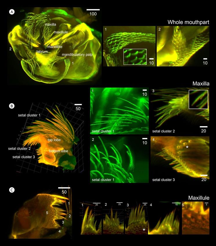
170                W. Sukparangsi et al. / Songklanakarin J. Sci. Technol. 43 (1), 169-180, 2021
Khachonpisitsak, 2017). The species richness of acorn              2.2 Taxonomic identification
barnacles found in the stations along Andaman Sea ranged
from 2-8 species while only 2-4 different species occur in the                Samples were identified based on their shell mor-
stations on the Gulf of Thailand (Pochai et al., 2017).            phology using a stereomicroscope. Taxonomic identification
           Current practices for acorn barnacles’ identification   of all acorn barnacles was performed, using shell morphology
are based on morphological studies of: i) hard shell parts, ii)    and arthropodal characters, together with keys of Pilsbry
operculum (geometry of tergum and scutum), iii) arthropodal        (1916), Newman & Ross (1976), Chan, Prabowo, Lee, & Lee
characters including 6 pairs of the biramous modified legs or      (2009), and Pochai et al., (2017). All samples from each
cirri I-VI, iv) mouthparts (two each of maxillae, maxillules,      station were deposited in the Laboratory of Zoology,
mandibles, mandibular palps and a labrum), v) a penis, and vi)     Department of Biology, Faculty of Science, Burapha
caudal appendages. The presence of caudal appendages is a          University.
unique feature found specifically in the genus Caudoeuraphia
Poltarukha, 1997 and the only species in this genus is             2.3 Sample preparation
Caudoeuraphia caudata (Pilsbry, 1916). Identification of
these fine arthropodal structures is generally conducted with                Shells with intact body tissue were stored directly in
light microscopy and high-resolution imaging with SEM              95% (v/v) ethanol at room temperature. Whole barnacle body
(Chan et al., 2008; Chan & Cheang, 2016; Shahdadi, Chan, &         tissue and arthropodal appendages were dissected from its
Sari, 2011). Information on these arthropodal characters is        shell under stereomicroscope and stored in 95% (v/v) ethanol
important for identifying acorn barnacles at the species-level     until processing. To prepare samples for lightsheet imaging,
while shell morphology is not always sufficient to clearly         body tissues were first rehydrated with sterile distilled water
distinguish among acorn barnacles due to variations based on       for 10 minutes and then immersed in warm 1% low melting
habitat, algae-furnished shell parts, and different diametric      point agarose (dissolved in Phosphate-Buffered Saline (PBS)
growth and/or ages (Chan, Chen, & Dando, 2016; Chan,               pH 7.4) for 5 minutes. The sample immersion in agarose was
Tsang, & Chu, 2007a, 2007b).                                       done in 1.5 ml microcentrifuge tube and kept warm until
           In recent years, fluorescence-based confocal light      embedding on heat block (50 oC). For embedding, the samples
sheet microscopy (LSFM), using pure optics for sectioning          in warm agarose were pulled slowly into glass capillary tube
helps to illustrate high resolution of three dimensional           (Blue cap, internal diameter 1.9 mm, Hilgenberg GmbH), and
structures of large and live or fixed specimens through            let the agarose gel harden, which should take about ten
immunofluorescence and its own autofluorescence (de                minutes. The gas capillary with sample was then assembled
Medeiros et al., 2015). Importantly, these can be done in a        into a sample holder of lightsheet station and ready for
short period of imaging and require less complicated               imaging as manufacturer’s instruction.
procedures for sample preparation, compared to laser scanning
microscopy (LSM) and SEM. In this present study, we aimed          2.4 Microscopy and imaging
to illustrate detailed morphology and three-dimensional
imaging of arthropodal characters of a new record C. caudata                 Light sheet fluorescence microscopy was performed
by exploiting the benefit of light-sheet-based fluorescence        under ZEISS Lightsheet Z1 Fluorescence illumination at the
microscopy.                                                        lightsheet laser’s wavelengths of 405, 445, 488, 515, 561, and
                                                                   638 nm. Sample positioning could be viewed by transmission
2. Materials and Methods                                           LED. Emitted light was collected by an objective lens, W
                                                                   Plan-Apochromat 20x/1.0 (water immersion). Images were
2.1 Sampling sites and sample collection                           taken by AxioCam1, AxioObserver SPIM.
                                                                             Recording time of a single section excited with both
           Our survey covered coastlines of Eastern Thailand       lasers was about 0.5-1 second. Number of optical sections
(13°20'31.8"N 100°56'34.6"E to 11°58'33.7"N 102°46'10.4            depended on various parameter settings and sample size;
"E), including Chon Buri, Rayong, Chanthaburi, and Trat            however, generally only 500-1,400 autofluorescent sections
provinces. Caudoeuraphia caudata was found in two stations         were recorded, sufficient to create 3D images of a whole
of all survey sites, including Mun Nork Island (MN), Klaeng        barnacle body. Average time of imaging was 5-8 minutes. Z-
district, Rayong province (12°34'03.5"N 101°42'05.5"E) and         stacks/sections were fused computationally to obtain 3D
Kung Wiman Beach (KW), Na Yai Am district, Chanthaburi             images without further deconvolution via maximum intensity
province (12°36'07.0"N 101°52'39.2"E) (Figure 1). A total of       projection in ZEN 2014 SP1 (Black edition version 9.2.0.0, 64
64 individuals of C. caudata were collected from rocky shores      bit, Carl Zeiss). 3D views, fluorescence intensity and
during low tides, including 32 specimens from MN and 32            brightness of arthropodal characters were edited and captured
specimens from KW. Whole acorn barnacles were removed              in ZEN 2.3 (Blue Edition, Carl Zeiss). The scale bars were
from the substratum using scalpels and immediately preserved       obtained directly from the ZEN lite software.
in 95% alcohol for further examination. All work was done                    Whole shell, shell plates and operculum images
under certified animal research protocols of W.S. and S.K.         were captured with an Olympus SZ61 Stereo Microscope and
(Certificate from Institute of Animal for Scientific Purposes      digital camera Canon EOS 700D. Images were taken with
Development-IAD, Royal Thai Government: U1-03103-2559              EOS Utility and processed with Image Frame Work
and U1-03104-2559, respectively).                                  (Tarosoft).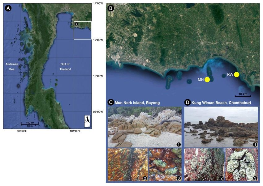
W. Sukparangsi et al. / Songklanakarin J. Sci. Technol. 43 (1), 169-180, 2021                            171

3. Results and Discussion                                             Stephenson, 1956; Endean, Stephenson, & Kenny, 1956;
                                                                      Foster, 1974; Hosie, Sampey, Davie, & Jones, 2015; Jones,
3.1 Systematic taxonomy                                               2003, 2010; Jones, Anderson, & Anderson, 1990; Pope, 1965;
                                                                      Stephenson, Endean, & Bennett, 1958), China (Liu & Ren,
           Superorder Thoracica Darwin, 1854                          2007), Japan (Chan, 2006), Singapore (Jones & Hosie, 2016),
           Order Sessilia Lamarck, 1818                               Vietnam (Poltarukha & Zvyagintsov, 2008; Zevina,
           Suborder Balanomorpha Pilsbry, 1916                        Zvyagintsev, & Negashev, 1992). In the present study, a new
           Superfamily Chthamaloidea Darwin, 1854                     location record of C. caudata from the Eastern coast of
           Family Chthamalidae Pilsbry, 1916                          Thailand, inhabiting Rayong and Chanthaburi provinces is
           Subfamily Euraphiinae Newman & Ross, 1976                  documented (Figure 1A and 1).
           Genus Caudoeuraphia Poltarukha, 1997                                  Habitat: Inside the shaded areas of rocky crevices in
           Type species. Chthamalus caudatus Pilsbry, 1916            the upper intertidal zone, periodic absence of seawater (during
           1 genus, 1 species recorded: Caudoeuraphia                 low tides), and no direct exposure to sunlight (Figure 1C and
caudata (Pilsbry, 1916).                                              D). C. caudata were found mostly in big colonies in narrow
           Caudoeuraphia caudata (Pilsbry, 1916)                      rocky crevices, in which each individual was connected by
           Chthamalus caudatus Pilsbry, 1916: 314, fig. 92 A-         shell parts (Figure 1D).
C, pl. 73 figs 1, 1a, 1b.
           Euraphia caudata – Newman & Ross, 1976: 41.                3.2 Taxonomic identification of Caudoeuraphia
           Caudoeuraphia caudata – Poltarukha, 1997: 464.                 caudata based on shell morphology
           Non-type material examined. Gulf of Thailand: 32
specimens, Rayong province, Klaeng district, Mun Nork                            Caudoeuraphia caudata lacks a peduncle, a
Island (MN), 27.I.2017, W. Sukparangsi (BUU17.CH.CC01-                distinctive feature identifying it as an acorn barnacle in the
32) and 32 specimens, Chanthaburi province, Na Yai Am                 Suborder Balanomorpha. Body length ranges from 6-10 mm.
district, Kung Wiman Beach (KW), 01.IV.2017, S.                       Shell cone is flattened or depressed (Figure 2A left). C.
Khachonpisitsak (BUU17.CH.CC33-64)                                    caudata is easily distinguished from Euraphia and Chtha-
           Diagnosis: Peduncle absent; shell conical and              malus, in that the shell cone of C. caudata is wider and flatter.
depressed, shell with 6 plates; membranous basis, non-                Densely packed colonies caused irregular shell margins and
tubiferous; caudal appendage present.                                 shapes. Shell parts adhere to rocky surfaces with a
           Distribution: Caudoeuraphia caudata is widely              membranous basis, similar to that found in other Chthamalids.
distributed in the Indo-Australian and Indo-Pacific regions. It       The edge of shell plates is solid or without a parietal tube.
has been recorded in Australia (Endean, Kenny, &                      After removal from rock surfaces, the mantle around the

                 Figure 1. Distribution map and habitat characteristics of Caudoeuraphia caudata in the Eastern Thailand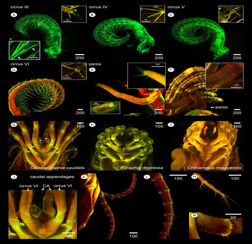
172                  W. Sukparangsi et al. / Songklanakarin J. Sci. Technol. 43 (1), 169-180, 2021

Figure 2. Shell morphology of Caudoeuraphia caudata (A) Dorsal (left panel), ventral (middle and right panel) view of external shell plate and
          intact body. (B) Variation of shell morphology. (C) External (left panel) and internal (right panel) view of tergum (upper panel) and
          scutum (lower panel). (D) External (upper panel) and internal (lower panel) view of each shell plate. Abbreviation: c, carina; cl,
          carinolateral; l, rostrolateral; r, rostrum

shell’s edge is dark grayish pink, which became black in                 of shell plates. The internal surface is yellow-light brown. The
ethanol. Color of the inner mantle around the body is white              tergum is narrow while the scutum is long, covering most of
(Figure 2A middle). On the ventral side, a long segmented                the orifice area. The tergum is deeply interlocked or
caudal appendage close to cirri VI was apparent, extending               articulated with the scutum. The scutum is triangular with a
from the posterior side of the body (Figure 2A right, Table 1).          slightly curved basal margin, and its external surface exhibits
In all sampling sites, two patterns of C. caudata shell color            shallow and horizontal striations from the occludent margin to
were found. In shaded area of rocky crevices with low                    the tergal margin. The occludent margin of the scutum is
exposure to sunlight, the shell is green-brown perhaps due to            without teeth and the tergal margin exhibits a clear sinus from
the presence of algae (Figure 2A). On the edge of rocky shore            exterior and interior view. Terga carry 4–5 lateral depressor
with higher exposure to sunlight, the shell is white-light               crests (Figure 2C). In addition, the arrangement and number
brownish (Figure 2B). The external surface of shell is usually           of shell plates around the orifice is the main characteristic for
smooth but some have an eroded surface exposing a deeper                 barnacles’ identification. As in other Chthamalids, six shell
shell surface brownish-gray in color. The orifice containing an          plates are present, including a carina, two carinolaterals, two
operculum is convex kite-shaped with rounded edges. The                  rostrolaterals and a rostrum; however, the size of each shell
opercular plates inside the orifice are symmetrical. Color and           plate is greatly varied. The junction of each shell plate has
surface of the exterior side of opercular plates resembles those         small teeth or is irregular in shape (Figure 2D).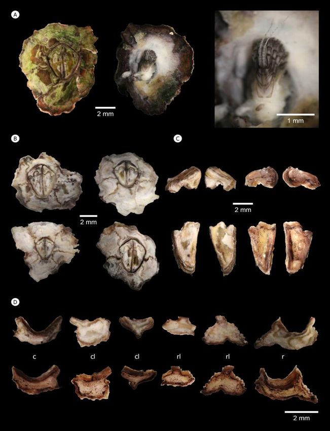
W. Sukparangsi et al. / Songklanakarin J. Sci. Technol. 43 (1), 169-180, 2021                             173

3.3 Morphological studies of acorn barnacles in high                   3.4.1 Cirri I–II
    resolution and three dimensions
                                                                                  The length and width of rami and setation of cirri I
          Without antibody staining and complication of                and cirri II are distinct from other cirri. Setal appendages
immunofluorescence, barnacle body was directly excited with            protruding from cirral limbs can be observed. Numerous setae
various wavelengths of laser light to search for the appropriate       between these maxillipeds around oral cone resemble a mesh,
wavelength capable of stimulating the barnacle’s own auto-             serving a microfiltration function in capturing small food
fluorescence. Two excitation wavelengths, 405 and 488                  particles and preventing food escapement from the mouth
appeared to be the best to visualize the structures of arthro-         (Figure 4A). Setae project in all directions from the shaft of
podal characters including soft structures. These excitation           limbs, in particular at the apex of both cirri I and II (Figure
wavelengths lead to emission/detection wavelengths at 415              4A1 and 4A2). In addition, a few short spines occur around
nm and 498 nm, respectively. Internal musculature is more              segment junctions on exopod of cirri I (Figure 4A3). If
excited at 488 nm laser, leading to a brighter red pseudocolor,        compared to conical spines of Chthamalus malayensis (Figure
while the cuticle or exoskeleton of cirri and mouthparts can be        4B), these small spines on the exopod locate more anteriorly,
excited at both 405 and 488 nm (Figure 3C). However, the               in a similar manner to those in Euraphia depressa (Figure
transparency of internal structures that can be seen through           4C).
autofluorescence varies among samples depending on planes                         In cirri I, oral setae on the anterior ramus are longer
of dissection. This slight difference in excitation enhances           than those on the posterior ramus. The posterior side of
cuticle separation from other internal soft muscles and                protopod has long setae while few shorter setae are present on
provides more detail on muscular architecture. In combination          the anterior side (Figure 4D). Only serrulate type of setae are
two channels of laser excitation of pseudocolor green and red          present around the limb of cirri I (Figure 4E and 4F).
best demonstrates whole body morphology (Figure 3C) and                Similarly, long setae occur on the oral sites of the anterior
micromorphology of specific structures of acorn barnacles and          ramus of cirri II and the protopods possess long setae on both
was used to describe fine structures.                                  anterior and posterior surfaces (Figure 4G). Unlike
                                                                       multicuspidate setae with a basal guard in C. malayensis
3.4 Comparsion of cirral and penis morphology of                       (Tsang et al., 2012), those on the oral site of all segments in
    Caudoeuraphia caudata to other Chthamalids                         the anterior ramus of cirri II in C. caudata are of the serrate
                                                                       type, composed of two rows of densely packed denticles
           Here we describe morphology and setation present            (Figure 4H). The posterior and anterior surface of protopod in
in cirri I-VI, penis, and caudal appendages of Caudoeuraphia           cirri II carry simple (Figure 4I) and serrate setae (Figure 4J),
caudata, following Chan et al. (2008) and based on lightsheet          respectively. Setae at junctions around the aboral site bear
illumination.                                                          only simple setae (Figure 4K).

Figure 3. Schematic illustrations of whole procedures for light-sheet microscopy and barnacle sample preparation (A) High-speed imaging of
          whole barnacle body with light-sheet microscopy. (B) Wavelength of laser used for visualizing the barnacle body (Caudoeuraphia
          caudata) with autofluorescence. (C) The reconstruction of the whole Chthamalid barnacle in anterior side by imaging auto-
          fluorescence. Scale bar, µm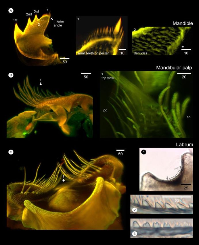
174 W. Sukparangsi et al. / Songklanakarin J. Sci. Technol. 43 (1), 169-180, 2021
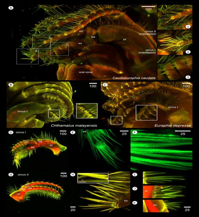
Figure 4. Light-sheet based visualization of 3D-autofluorescent cirri I and II of Caudoeuraphia caudata (A) Lateral view of C. caudata. (Inset
1) setation around anterior and (Inset 2) posterior rami of cirrus I. (Inset 3) spines protruding from anterior ramus of cirrus I. (B)
Lateral view of Chthamalus malayensis showing conical spines (inset) on the cirrus I. (C) Lateral view of Euraphia depressa showing
small spines (inset) on the anterior ramus of cirrus I. (D) overall morphology of cirrus I of C. caudata. (E) Apex of anterior ramus of
cirrus I. (F) serrulate setae on cirrus I. (G) Overall morphology of cirrus II. (H) Close-up on apex of anterior ramus of cirrus II
showing serrate setae (inset). (I) Simple setae found on the posterior side of protopod of cirrus I. (J) Serrate setae found on anterior
surfaces of protopod of cirrus I. (K) Simple type of aboral setae at the segmental junction. Abbreviation: en, endopod; ex, exopod;
basipod; pr, protopod. Scale bar, µm
Segment numbers of exopods and endopods vary in 3.4.2 Cirri III –VI
both cirri I-II and between left and right. Unequal number of
segments is common between exopods and endopods. Cirri III-VI share similar features of limb mor-
Asymmetry between right and left cirri I occurs also in some phology. Although cirri III are maxillipeds, their architecture
individuals (Table 1). Length of maxillipeds is clearly shorter resembles that of a cirral fan, except for setal type. Numerous
than that of cirri III-VI. In cirri I, the anterior ramus is longer setae between cirri III-VI are mesh-like but with larger mesh
than the posterior, whereas cirri II exhibit shorter anterior holes than those between cirri I-II, indicative of larger food
ramus than the posterior. Exopod width of both cirri I-II is particle gathering. The apex of anterior and posterior rami of
more than twice that of the endopod (Table 2). cirri III has two large spines projected toward the oral cone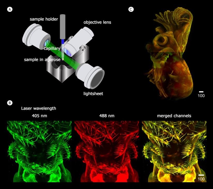
W. Sukparangsi et al. / Songklanakarin J. Sci. Technol. 43 (1), 169-180, 2021                                                    175
Table 1.     Diverse segment numbers of arthropodal characters including cirri I – VI and caudal appendages found in Caudoeuraphia caudata.

                                                                                              Number of segment
            Arthropodal characters                                                                  Right
                                                           Anterior ramus/ exopod                                        Posterior ramus/ endopod

 cirrus 1                                    511%, 622%, 756%, 811%                                         525%, 663%, 713%
 cirrus 2                                    622%, 756%, 811%                                               844%, 944%, 1111%, 1211%
 cirrus 3                                    1162.5%, 1325%, 1412.5%                                        1112.5%, 1237.5%, 1325%, 1412.5%, 1512.5%
 cirrus 4                                    1237.5%,1312.5%, 1437.5%, 1612.5%                              1325%, 1412.5%, 1550%, 1812.5%
 cirrus 5                                    1312.5%, 1425%, 1537.5%, 1612.5%, 1912.5%                      1312.5%, 1412.5%, 1537.5%, 1612.5%, 1712.5%, 1812.5%
 cirrus 6                                    1314.3%,1514.3%, 1657.1%, 2014.3%                              1528.5%,1642.9%, 1714.3%, 1914.3%
 caudal appendage                            1914.3%, 2014.3%, 2128.6%, 2314.3%, 2414.3%, 2514.3%

                                                                                                     Left
            Arthropodal characters
                                                            Anterior ramus/exopod                                        Posterior ramus/endopod
                                               44%   56%
 cirrus 1                                     6 ,7                                                          5 , 6 , 711%
                                                                                                             22%   67%

 cirrus 2                                    733%, 822%, 944%                                               822%, 967%, 1011%
 cirrus 3                                    1012.5%, 1137.5%, 1225%, 1325%                                 1112.5%, 1350%, 1437.5%
 cirrus 4                                    1337.5%, 1425%, 1525%, 1612.5%                                 1312.5%, 1450%, 1512.5%, 1612.5%, 1712.5%
 cirrus 5                                    1312.5%, 1537.5%, 1637.5%, 1912.5%                             1312.5%, 1537.5%, 1637.5%, 2012.5%
 cirrus 6                                    1416.67%,1666.66%, 1816.67%                                    1533.33%, 1616.67%, 1733.33%, 1916.67%
 caudal appendage                            2014.3%, 2142.8%, 2228.6%, 2714.3%

Table 2.     Average length and width of arthropodal characters including cirri I – VI and caudal appendages and penis found in Caudoeuraphia
             caudata.

    Arthropodal                                                              Fold
                          Endopod (endo) and exopod (exo) length                             Endopod (endo) and exopod (exo) width             Fold difference
     characters                                                           difference

       cirri 1                         exo (444 μm) >                        1.32                       exo (171 μm) >                               1.66
                                       endo (336 μm)                                                    endo (103 μm)
       cirri 2                         exo (653 μm) <                        1.37                       exo (167 μm) <                               1.80
                                       endo (893 μm)                                                     endo (93 μm)
       cirri 3                        exo (1,309 μm) <                       1.31                       exo (154 μm) =                               1.00
                                      endo (1,709 μm)                                                   endo (154 μm)
       cirri 4                        exo (2,311 μm) =                       1.00                       exo (144 μm) =                               1.00
                                      endo (2,289 μm)                                                   endo (144 μm)
       cirri 5                        exo (2,273 μm) <                       1.12                       exo (160 μm) =                               1.00
                                      endo (2,545 μm)                                                   endo (160 μm)
       cirri 6                        exo (2,080 μm) <                       1.15                exo (140 μm) = endo (140 μm)                        1.00
                                      endo (2,400 μm)
 caudal appendage                    right (2,130 μm) =                      1.00
                                       left (2,130 μm)
           penis                          2,080 μm                                -

and 2-3 thinner and shorter spines that protrude toward the                           the largest segment number in both exopod and endopod
posterior site (Figure 5A). Differently, the apex of cirri IV-VI                      (Table 1). Length of cirri III is about twice longer than that of
has three large spines: two protruding toward the oral cone                           cirri I-II. In addition, cirri III have a shorter anterior ramus
and one directed in a posterior direction (Figure 5B-5D). All                         than the posterior; whereas, both rami in cirri IV-VI are
of these cirri have long terminal oral setae and short setae at                       approximately equal in length and width (Table 2).
the junction of exopod and endopod segments. The protopods
of cirri III contain setae on both anterior and posterior                             3.4.3 Penis
surfaces; whereas, cirri IV-VI have setae only on the anterior
surface (Figure 5A-5D). All setae in Cirri III-VI are simple,                                   C. caudata has a shorter penis, relative to that of
except some serrate setae on the oral side of the anterior                            Euraphia and Chthamalus. The whole penis including its apex
ramus-Cirri III (Figure 5A, inset). Cirri III and IV exhibit                          in all dissected specimen were covered within the cirri;
unequal numbers of segments between the anterior and                                  whereas, in other Chthamalids the penis is easily seen as a
posterior rami and between the left and right limbs. Cirri V                          long appendage extending away from other cirri (Figure 5E
and VI generally have equal segment numbers on the anterior                           and 5D). C. caudata penis length is similar to those of cirri
and posterior rami; however, segment number can also vary in                          IV-VI (Table 2). The penis apex is covered with 7-10 long
C. caudata with similar body length or age. Cirri VI exhibit                          simple setae, and shorter simple setae can also be found near
176 W. Sukparangsi et al. / Songklanakarin J. Sci. Technol. 43 (1), 169-180, 2021
Figure 5. Light-sheet based visualization of 3D-autofluorescent cirri III-VI, penis and caudal appendages of Caudoeuraphia caudata (A)-(D)
Morphology of cirrus III, cirrus IV, cirrus V and cirrus VI, respectively. (A) Top inset showing setation on apex of cirrus III. Bottom
inset showing serrate type of setae found around oral site of anterior ramus of cirrus III. (B) Inset showing setation on apex of cirrus
IV. (C) Inset showing setation on apex of cirrus V. (D) Inset showing setation on apex of cirrus V. (E) morphology of penis. Top inset
illustrates pattern of setae around apex of the penis. Bottom inset shows surface of penis having pattern of irregular pattern of cuticle
rings. (F) Penis of Euraphia depressa. Inset shows regular pattern of cuticle rings around its penis. (G)-(I) Posterior view of barnacles.
(G) C. caudata carries caudal appendages (asterisks). (H)-(I) both E. depressa and Chthamalus malayensis have no caudal
appendages. (J) Posterior view of C. caudata shows the position of caudal appendages (CA) joined to the base of sixth cirri. (K)
Lateral view of caudal appendages. (L) Close-up on caudal appendage showing setae extending around segment junction. (M) Apex of
caudal appendages. (N) Surface of caudal appendage covering with denticles. Scale bar, µm
the penis close to the apex (Figure 5E inset). In addition, C. to the protopods of both sixth cirri (Figure 5J) and extends
caudata shows irregular shape of exoskeleton rings around the away from the cirral fan (Figure 5K). Simple setae (4-6) of
penis (Figure 5E inset) while that of Euraphia depressa unequal lengths occur at the junctions of segments (Figure
exhibits obvious cuticular rings of cuticle (Figure 5F inset). 5L), whereas 4 longer simple setae are present at the apex
(Figure 5M). Denticles with fine spines are present on the
3.4.4 Caudal appendages surface of caudal appendages (Figure 5N). Caudal appendages
generally have 21 segments; however, in some specimens the
The most distinctive feature of C. caudata is the number ranges from 19-27 segments (Table 1). Length is
presence of caudal appendages at the posterior side (Figure similar to that of cirri 6; however, width is much less than that
5G, asterisks), while these are absent in other chthamalids of cirri I-VI (Table 2).
(Figure 5H and 5I). The base of caudal appendages is joinedW. Sukparangsi et al. / Songklanakarin J. Sci. Technol. 43 (1), 169-180, 2021 177
3.5 Three dimensional visualization of mouth first cluster of setae faces upward away from the oral cone,
appendages of Caudoeuraphia caudata carrying long simple and long serrulate setae (Figure 6B1 and
6B2). The second cluster on the top lobe contains two types of
Mouthparts were illuminated and visualized through setae projecting toward the labrum: long-serrulate (Figure 6B3
a light-sheet microscope (Figure 6A). Fan-like shape denticles inset) and short-simple (Figure 6B3 asterisk). The last cluster
with 5-6 small spikes occur around the labrum and on the base on the bottom lobe consists of a single row of simple setae,
and body of each mouth appendage (Figure 6A1 and 6A2). close to the base of maxilla (Figure 6B4). In addition, some
Here we further describe each mouth appendage in detail small spines occur around the bottom lobe (Figure 6B4,
(Figure 6 and Figure 7). asterisk).
3.5.1 Maxillae 3.5.2 Maxillules
Maxilla has a bilobed shape (top and bottom lobes) Two cutting and notched surfaces occur on the
and setae are grouped into three clusters: two clusters on the maxillules (Figure 6C). Above the first notch, one large spine
top lobe and one cluster on the bottom lobe (Figure 6B). The is located at the top followed by two smaller spines, then four
Figure 6. Light-sheet microscopy-based visualization of whole mouthparts and each individual mouthpart structures (maxilla and maxillule) of
Caudoeuraphia caudata (A) Whole mouthpart or oral cone showing composition of mouth appendages. 1-2 is close-up of (A) and are
shown on the middle (1) and right (2) panels. Panel 1 showing fan-like denticles (inset) around the surface of labrum. Panel 2 showing
denticles around base of mandibular palp. (B) Overall morphology of maxilla. Panel 1-2 showing close-up view on setal cluster 1.
Panel 3 showing close-up view on setal cluster 2. Panel 4 showing close-up view on setal cluster 3. (C) Overall morphology of
maxillule. Number indicates the close-up pictures shown in panel 1-5. Panel 1-4 showing close-up of setae on maxillule. Panel 5
showing close-up on patterns of denticles on surface of maxillule. Scale bar, µm178 W. Sukparangsi et al. / Songklanakarin J. Sci. Technol. 43 (1), 169-180, 2021
Figure 7. Light-sheet microscopy-based visualization of mouthpart structures (mandible, mandibular palp, and labrum) of Caudoeuraphia
caudata (A) Overall morphology of mandible. 1st, 2nd and 3rd indicate teeth number. 1-2 indicate close-up view showing in the middle
and right panel. Panel 1 showing close-up view on setae on pecten. Panel 2 showing close-up view patterns of denticles on surface of
mandible. Asterisk indicates setae below the inferior angle. (B) Overall morphology of mandibular palp. Number 1 and arrow
indicates picture from top view showing in right panel 1. Panel 1 illustrates setation on the surface of mandibular palp. (C) Overall
morphology of labrum. Number 1 and arrow indicates picture a piece of labrum showing in right panel 1-3. Panel 1 illustrates whole
labrum. Boxes are close-up of teeth showing panel 2 and 3. Abbreviation: in, inferior side; ex, exterior side. Scale bar, µm
smaller spines (Figure 6C1). At the middle of maxillules, two small teeth. At an inferior angle of the mandible and located
rows of spines are visible by 3D view: a first row with 4 small next to the pecten, are one large and two smaller teeth (Figure
spines and a second row with two larger spines (Figure 6C2). 7A1). In addition, numerous fine setae occur below the
Three additional spines are very close to each other, are below inferior angle (Figure 7A2 asterisk). Several rows of fan-
the six larger spines (Figure 6C3, asterisk). Below the second shape denticles illuminated on the surface of mandible close
notch, eight small spines are located in a row. In this last lobe, to the teeth (Figure 7A2). The number of mandible teeth is a
two small notches further divide spines into a 1+4+3 pattern clear characteristic distinguishing Chthamalus from Euraphia.
(Figure 6C4). The surface on the maxillule is decorated with Previously, the presence of mandibles with three teeth in C.
denticles with small and sharp spines (Figure 6C5). This caudata led to its nomenclature as Euraphia caudata (Pilsbry,
double notched maxillule is generally absent in Chthamalus 1916).
and Euraphia.
3.5.4 Mandibular palps
3.5.3 Mandibles
Mandibular palps are rectangular in shape (Figure
Three large teeth occur on the mandibles (Figure 7B). Two types of setae are present on the superior surface.
7A). The pecten of the mandible bears two rows of 12-18 Long and serrulate setae occur at the tip and along the exteriorW. Sukparangsi et al. / Songklanakarin J. Sci. Technol. 43 (1), 169-180, 2021 179
side of the mandibular palps. Setae about one third shorter in Zootaxa, 4098(2), 201–226. doi:10.11646/zootaxa.
length are present along the inferior surface of the palps 4098.2.1.
(Figure 7B1). Chan, B. K. K., Chen, H. N., Dando, P. R., Southward, A. J,
& Southward, E. C. (2016). Biodiversity and bio-
3.5.5 Labrum geography of chthamalid barnacles from the North-
Eastern Pacific (Crustacea Cirripedia). PLoSONE,
The shape of labrum is concave (Figure 7C1). Two 11(3), Article ID e0149556. doi:10.1371/journal.
types of teeth cover the length of the labrum: sharp teeth pone.0149556.
(similar to mammalian canine teeth) and teeth with two or Chan, B. K. K., Garm, A., & Høeg, J. T. (2008). Setal mor-
three cusps (similar to mammalian premolar or molar teeth). phology and cirral setation of thoracican barnacle
The sharp teeth are located in the middle of concaved labrum cirri: Adaptations and implications for thoracican
(Figure 7C2) while the cuspidate teeth are present at both end evolution. Journal of Zoology, 275(3), 294-306.
of the labrum (Figure 7C3). This tooth pattern differs from doi:10.1111/j.1469-7998.2008.00441.x.
that in Euraphia depressa, carrying only sharp teeth at the Chan, B. K. K., Prabowo, R. E., Lee, K. S., & Lee, K. H.
middle of the concaved labrum (Lively & Raimondi, 1987). (2009). Crustacean fauna of Taiwan: Barnacles,
Volume 1-Cirripedia: Thoracica excluding the
4. Conclusions Pyrogomatidae and Acastinae. Keelung, Taiwan:
National Taiwan Ocean University.
Lightsheet Fluorescence Microscopy (LSFM) Chan, B. K. K., Tsang L. M., & Chu K. H. (2007). Cryptic
provides ultrafast 3D imaging. With this technology, we diversity of the Tetraclita squamosa complex
provide a more detailed description of Caudoeuraphia (Crustacea: Cirripedia) in Asia: description of a new
caudata found on the rocky shores locating in the Gulf of species from Singapore. Zoological Studies, 46(1),
Thailand, Eastern part of Thailand. The approach presented 46–56.
here exploits the barnacles’ own fluorescence to visualize Chan, B. K. K., Tsang, L. M., & Chu, K. H. (2007). Morpho-
anatomical details of whole barnacle body structures, and this logical and genetic differentiation of the acorn bar-
could be an alternative way to unveil nature of marine nacle Tetraclita squamosa (Crustacea, Cirripedia) in
crustaceans. East Asia and description of a new species of
Tetraclita. Zoologica Scripta, 36, 79–91. doi:10.11
Acknowledgements 11/j.1463-6409.2007.00260.x.
Chen, Y. Y., Lin, H. C., & Chan, B. K. K. (2012). Description
We would like to thank S. Suwanpairoj and of a new species of coral-inhabiting barnacle,
Rushmore Precision Co., Ltd. for Lightsheet Fluorescence Darwiniella angularis sp. n. (Cirripedia, Pyrgoma-
Microscope (LSFM) operation. We greatly appreciate Prof. B. tidae) from Taiwan. ZooKeys, 214, 43–74. doi:10.
Beamish for critical reading of the manuscript and the Faculty 3897/zookeys.214.3291.
of Science, Burapha University for technical assistance. This Chen, H. N., Tsang, L. M., Chong, V. C., & Chan, B. K. K.
work was financially supported by the Research Grant of (2014). Worldwide genetic differentiation in the
Burapha University through National Research Council of common fouling barnacle, Amphibalanus am-
Thailand (Grant no. 27/2560). phitrite,” Biofouling, 30, 1067–1078. doi:10.1080/
08927014.2014.967232.
References de Medeiros, G., Norlin, N., Gunther, S., Albert, M., Pana
vaite, L., Fiuza, U. M., . . . Hufnagel, L. (2015).
Anderson, D. T. (1994). Barnacles - structure, function, deve- Confocal multiview light-sheet microscopy. Nature
lopment and evolution. London, England: Chapman Communications, 6, 8881. doi:10.1038/ncomms98
and Hall. 81.
Brickner, I., & Høeg, J.T. (2010). Antennular specialization in Endean, R., Kenny, R., &Stephenson, W. (1956). The ecology
cyprids of coral-associated barnacles. Journal of and distribution of intertidal organisms on the rocky
Experimental Marine Biology and Ecology, 392, shores of the Queensland mainland. Australian
115–124. doi:10.1016/j.jembe.2010.04.015. Journal of Marine and Freshwater Research, 7(1),
Brickner, I., Loya, Y., & Achituv, Y. (2010). Diverse life 88-146.
strategies in two coral-inhabiting barnacles (Pyrgo- Endean, R., Stephenson, W., & Kenny, R. (1956). The
matidae) occupying the same host (Cyphastrea ecology and distribution of intertidal organisms on
chalcidicum), in the northern Gulf of Eilat. Journal certain islands off the Queensland coast. Australian
of Experimental Marine Biology and Ecology, 392, Journal of Marine and Freshwater Research, 7(3),
220–227. doi:10.1016/j.jembe.2010.04.022. 317-342.
Chan, B. K. K. (2006). Ecology and biodiversity of rocky Foster, B.A. (1974). The barnacles of Fiji with observations
intertidal barnacles along a latitudinal gradient; on the ecology of barnacles on tropical shores.
Japan, Taiwan and Hong Kong. The Nagisa World Pacific Science, 28(1), 34-56.
Congress (pp. 1-10). Frith, D. W., Tantanasiriwong, R., & Bhatia, O. (1976).
Chan, B. K. K. & Cheang, C. C. (2016). First discovery of a Zonation and abundance of macrofauna on a
new species of Newmanella Ross, 1969 (Balano- mangrove shore, Phuket Island. Phuket Marine
morpha: Tetraclitidae) in the western Pacific, with a Biological Center Research Bulletin, 10(37).
note on the new status of Neonrosella Jones 2010.180 W. Sukparangsi et al. / Songklanakarin J. Sci. Technol. 43 (1), 169-180, 2021
Hayashi, R. (2013). A checklist of turtle and whale barnacles coasts: The Andaman Sea and the Gulf of Thailand.
(Cirripedia: Thoracica: Coronuloidea). Journal of Zoosystematics and Evolution, 93(1), 13-34.
the Marine Biological Association of the United Poltarukha, O. P. (1997). Composition, phylogeny, and po-
Kingdom, 93(1), 143–182. doi:10.1017/S002531541 sition of the subfamily Euraphiinae (Crustacea,
2000847. Chthamalinae) in the system of Cirripedia. Russian
Holm, E. R. (2012). Barnacles and biofouling. Integrative and Journal of Zoology, 1, 463–470.
Comparative Biology, 52(3), 348–55. doi:10.1093/ Poltarukha, O. P. (2006). Identification Atlas of superfamily
icb/ics042. chthamaloidea (Cirripedia Thoracica) barnacles in
Hosie, A. M., Sampey, A., Davie, P. J. F, & Jones, D. S. World Ocean. Moscow, Russia: KMK Scientific
(2015). Kimberley marine biota. Historical data: Press.
Crustaceans. Records of the Western Australian Poltarukha, O. P. & Zvyagintsov, A. Y. (2008). The barnacles
Museum, 247-285. (Cirripedia, Thoracica) of Vietnam and their role in
Jones, D. S. (2003). The biogeography of Western Australian the fouling communities. Russian Academy of
shallow-water barnacles. Proceedings of the Sciences (pp. 335). Moscow, Russia: KMK Scienti-
Eleventh International Marine Biological Work- fic Press.
shop: The Marine Flora and Fauna of Dampier, Pope, E. C. (1965). A review of Australian and some
Western Australia, 2, 479-496. Indomalayan Chthamalidae (Crustacea: Cirripedia).
Jones, D. S. (2010). The littoral and shallow-water barnacles Proceedings of the Linnean Society of New South
(Crustacea: Cirripedia) of south eastern Queensland. Wales, 90, 10-77.
Proceedings of the Thirteenth International Marine Rawangkul, S., Angsupanich, S., & Panitchart, S. (1995).
Biological Workshop, the Marine Flora and Fauna Preliminary study of barnacles damaging the
of Moreton Bay, Queensland, Memoirs of the mangrove plantation Rhizophora mucronata at Tha
Queensland Museum, 54(3), 199–233. Phae canal, Nakorn Si Thammarat. The Ninth
Jones, D. S., Anderson, J. T., & Anderson, D. T. (1990). National Seminar on Mangrove Ecology, Mangrove
Checklist of the Australian Cirripedia. Technical Conservation for Thai Society in The Next Decade,
Reports of the Australian Museum, 3, 1-38. National Research Council of Thailand Bangkok III-
Jones, D. S. & Hosie, A. M. (2016). A checklist of the 06.
barnacles (Cirripedia: Thoracica) of Singapore and Shahdadi, A., Chan, B. K. K., & Sari, A. (2011). Tetraclita
neighbouring waters. The Raffles Bulletin of Zoo- ehsani sp.n. (Cirripedia, Tetraclitidae), a common
logy, 34, 241-311. intertidal barnacle from the Gulf of Oman, Iran.
Kaji, T. & Palmer, A. R. (2017). How reversible is deve- ZooKeys, 136, 1-12.
lopment? Contrast between developmentally plastic Sophia Rani, S., Pmbhu, S., & Przyadharshini, S. (2010).
gain and loss of segments in barnacle feeding legs. Infestation of barnacle (Balanus amphitrite) in the
Evolution, 71(3), 756-765. doi:10.111/evo.13152. mangrove environment. World Journal of Fish and
Liu, J. Y. & Ren X. (2007). Crustacea cirripedia thoracica. Marine Sciences, 2(4), 307–310.
Fauna Sinica: Invertebrata, Volume 42 (pp. i–xv, Stephenson, W., Endean, R., & Bennett, I. (1958). An
1–633, figure 236). Beijing, China: Science Press. ecological survey of the marine fauna of Low Isles,
Lively, C. M. & Raimondi, P. T. (1987). Desiccation, pre- Queensland. Australian Journal of Marine and
dation, and mussel-barnacle interactions in the Freshwater Research, 9(2), 261-318.
northern Gulf of California. Oecologia (Berlin), 74, Tsang, L. M., Wu, T. H., Shih, H. T., Williams, G. A., Chu,
304-309. K. H., & Chan, B. K. K. (2012). Genetic and mor-
Molnar, J. L., Gamboa, R. L., Revenga, C., & Spalding, M. D. phological differentiation of the Indo-West Pacific
(2008). Assessing the global threat of invasive intertidal barnacle Chthamalus malayensis. Integra-
species to marine biodiversity. Frontier in Ecology tive and Comparative Biology, 52, 388–409. doi:10.
and the Environment, 6(9), 485–492. doi:10.1890/ 1093/icb/ics044.
070064. Vernberg, F. J., & Vernberg, W. B. (1983). The biology of
Newman, W. A., & Ross, A. (1976). Revision of the balano- crustacea: 8. Environmental adaptations. The biolo-
morph barnacles; including a catalogue of the gy of crustacean. New York, NY: Academic Press.
species. Memoirs of the San Diego Society of Yu, M-C., Kolbasov, G. A., & Chan, B. K. K. (2016). A new
Natural History, 9, 1–108. species of sponge inhabiting barnacle Bryozobia
Pilsbry, H. A. (1916). The sessile barnacles (Cirripedia) con- (Archaeobalanidae, Bryozobiinae) in the West
tained in the collections of the United States Pacific. ZooKeys, 571, 1-20. doi:10.3897/zookeys.
National Museum; including a monograph of the 571.6894.
American species. Bulletin of the United States Zevina, G. B., Zvyagintsev, A. Y., & Negashev, S. E. (1992).
National Museum, 93, 1–366. Usonogie raki poberezh’ya V’etnama i ikh rol’v
Pochai, A., Kingtong, S., Sukparangsi, W., & Khachon obrastanii [Barnacles of the Vietnam Coast and their
pisitsak, S. (2017). The diversity of acorn barnacle role in encrustation],” Vladivostok, Dal’nauka, pp.
(Cirripedia, Balanomorpha) across Thailand’s 142, fgs. 69, tables 8. [in Russian]You can also read