Open reduction and internal fixation of quadrilateral plate fractures in the elderly: association between initial fracture pattern and outcomes
←
→
Page content transcription
If your browser does not render page correctly, please read the page content below
Wu et al. BMC Musculoskeletal Disorders    (2021) 22:122
https://doi.org/10.1186/s12891-021-04002-4

 RESEARCH ARTICLE                                                                                                                                 Open Access

Open reduction and internal fixation of
quadrilateral plate fractures in the elderly:
association between initial fracture pattern
and outcomes
Haiyang Wu1†, Qipeng Shao1†, Ranran Shang2, Chengjing Song3, Ximing Liu1 and Xianhua Cai1*

  Abstract
  Background: Acetabular fractures with medial displacement of the quadrilateral plate (QLP) are common in the
  elderly. The presence of QLP fractures greatly increase the surgical difficulty of acetabular fractures. This study aims
  to evaluate the clinical radiological outcomes of open reduction and internal fixation (ORIF) in QLP fractures in
  elderly patients and to investigate factors potentially affecting the result.
  Methods: We conducted a retrospective study. A series of 37 consecutive patients with acetabular fracture
  involving the QLP aged 60 years and older who received ORIF between January 2010 and May 2019 were included.
  QLP fractures were classified according to Walid’s classification system. Radiological outcomes were evaluated using
  Matta criteria and functional outcomes were assessed using the modified Merle d’Aubigné score. The relationships
  between Walid’s classification and radiological or functional outcomes were analyzed.
  Results: According to Walid’s classification, 18, 13, 6 were classified as QLP1, QLP2 and QLP3, respectively. The
  average follow-up was 35.5 ± 10.7 months. We obtained anatomic reduction in 48.6 % (18/37) of cases, imperfect
  reduction in 40.5 % (15/37) of cases, and poor reduction in 10.8 % (4/37) of cases. Excellent-good functional scores
  were found in 83.7 % (modified Merle d’Aubigné). There were no cases of screw entering the hip, pull-out and
  loosening or implant failure during the follow-up. Walid’s classification was positively correlated with radiological
  outcomes of reduction (r = 0.661; P < 0.001), and functional outcomes (r = 0.478; P = 0.003). Unsatisfactory reduction
  was demonstrated a correlation with the development of post-traumatic arthritis (r =-0.410; P = 0.012).
  Conclusions: ORIF may be suggested for quadrilateral plate fractures in the elderly. Walid’s classification system is
  associated with the reduction quality and functional recovery.
  Keywords: Acetabulum, Fracture fixation, Internal, Elderly, Classification

Background                                                                           Epidemiological investigations have shown that acetabu-
Acetabular fractures are relatively uncommon yet ser-                                lar fractures have a bimodal distribution with respect to
ious injuries which make up about 3–8 % of all fractures.                            age that has modes at 30–40 and 70–90 years [1]. As the
                                                                                     population ages, the incidence of osteopenic acetabular
* Correspondence: lzgkcxh@163.com
†
                                                                                     fractures resulting from low energy injuries is also in-
 Haiyang Wu and Qipeng Shao contributed equally to this work.
1                                                                                    creasing. Open reduction and internal fixation (ORIF)
 Department of Orthopaedic Surgery, General Hospital of Central Theater
Command, Wuhan Clinical Medicine College of Southern Medical University,             remain the preferred treatment for displaced acetabular
430070 Wuhan, China                                                                  fractures [2, 3]. However, controversies still exist
Full list of author information is available at the end of the article

                                      © The Author(s). 2021 Open Access This article is licensed under a Creative Commons Attribution 4.0 International License,
                                      which permits use, sharing, adaptation, distribution and reproduction in any medium or format, as long as you give
                                      appropriate credit to the original author(s) and the source, provide a link to the Creative Commons licence, and indicate if
                                      changes were made. The images or other third party material in this article are included in the article's Creative Commons
                                      licence, unless indicated otherwise in a credit line to the material. If material is not included in the article's Creative Commons
                                      licence and your intended use is not permitted by statutory regulation or exceeds the permitted use, you will need to obtain
                                      permission directly from the copyright holder. To view a copy of this licence, visit http://creativecommons.org/licenses/by/4.0/.
                                      The Creative Commons Public Domain Dedication waiver (http://creativecommons.org/publicdomain/zero/1.0/) applies to the
                                      data made available in this article, unless otherwise stated in a credit line to the data.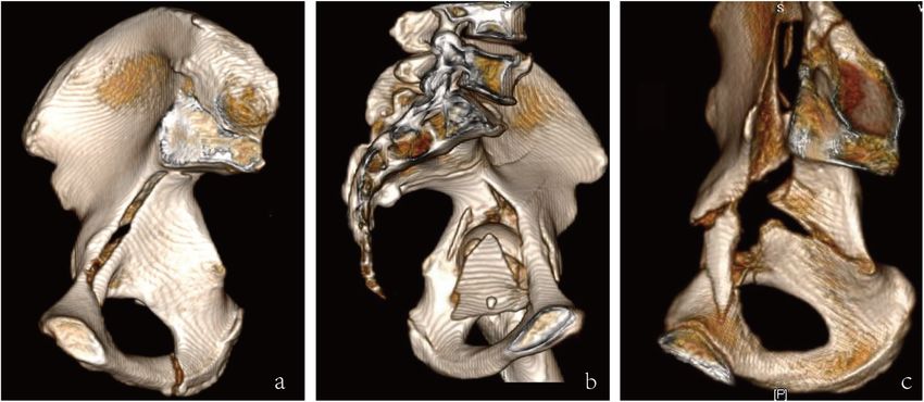
Wu et al. BMC Musculoskeletal Disorders   (2021) 22:122                                                           Page 2 of 8

regarding the optimal treatment of these fractures in           patients with acetabular fractures were identified from
older people [4, 5]. And surgical treatment represents a        the trauma database at our level I trauma center. Inclu-
great challenge for orthopedic surgeons because of the          sion criteria consisted of all types of acetabular fractures
decreased physiological compensatory capacity and the           involving the QLP, treated with Dynamic Anterior Plate-
severe osteoporosis of the elderly [6].                         Screw system for Quadrilateral plate (DAPSQ), age older
   Acetabular fractures in elderly patients frequently in-      than 60 years at the time of injury, unilateral acetabular
volve the quadrilateral plate (QLP), a deep and thin ana-       fracture, a minimum of 1-year postoperative follow up.
tomical structure constituting the medial wall of the           The exclusion criteria were open or pathologic fractures,
acetabulum. Isolated QLP fractures are rare and always          bilateral acetabular injuries, pre-existing ipsilateral hip
associated with the anterior or posterior column frac-          diseases, or femoral head fracture. The flow chart of our
tures. Injuries resulting from the force along the femoral      retrospective study was illustrated in Fig. 1. A total of 37
neck can lead to comminuted fractures of the QLP and            patients with QLP fractures were included in our study
even central dislocation of the femoral head. The QLP           eventually.
does not play a crucial role in the weight-bearing of the
hip and is not key structure of biomechanical functional-         Radiographs and medical records were collected by
ity. However, previous studies showed that a separate           two investigators who were not implicated in the initial
quadrilateral-plate component and/or central dislocation        intervention. Pre-operative and post-operative radio-
of the femoral head might adversely affect the outcomes         graphic analysis included Anterior-Posterior (AP) view
[1, 7]. Bastian et al. [8] reported that nearly two thirds of   and Judet views (iliac and obturator oblique views),
the patients in complex fracture morphologies with              along with three-dimensional (3D) CT reconstruction.
medial displacement of QLP required an additional ap-           Acetabular fractures were classified according to Judet
proach in addition to the modified Stoppa approach,             and Letournel classification system [9] and QLP frac-
which indicated that QLP fractures increased the com-           tures were classified according to Walid’s classification
plexity and difficulty of acetabular fractures surgery. The     system [10]. Walid’s classification system divides the
technical difficulty of ORIF of this area is predominantly      QLP fractures into four categories as follows (Fig. 2):
due to the complicated anatomy, deep location and nar-          QLP1, incompletely separated simple fracture; QLP2, in-
row surgical field.                                             completely separated comminuted fracture; QLP3, com-
   This study aims to evaluate the clinical radiological        pletely separated comminuted fracture; QLP4,
outcomes of ORIF in QLP fractures in older people and           completely separated simple fracture. Pre-operative im-
to investigate factors potentially affecting the result.        aging evaluation should place extra attention on the
                                                                “Gull sign”. When acetabular fractures involve the roof
Materials and Methods                                           of the acetabulum, pelvic radiographs often show two
Ethical approval was obtained from the ethical commit-          typical double arc shadows on the acetabular roof, which
tee of the hospital. Between January 2010 and May 2019,         are similar to the wings of a seagull in flight and

 Fig. 1 Study flow chart
Wu et al. BMC Musculoskeletal Disorders (2021) 22:122 Page 3 of 8
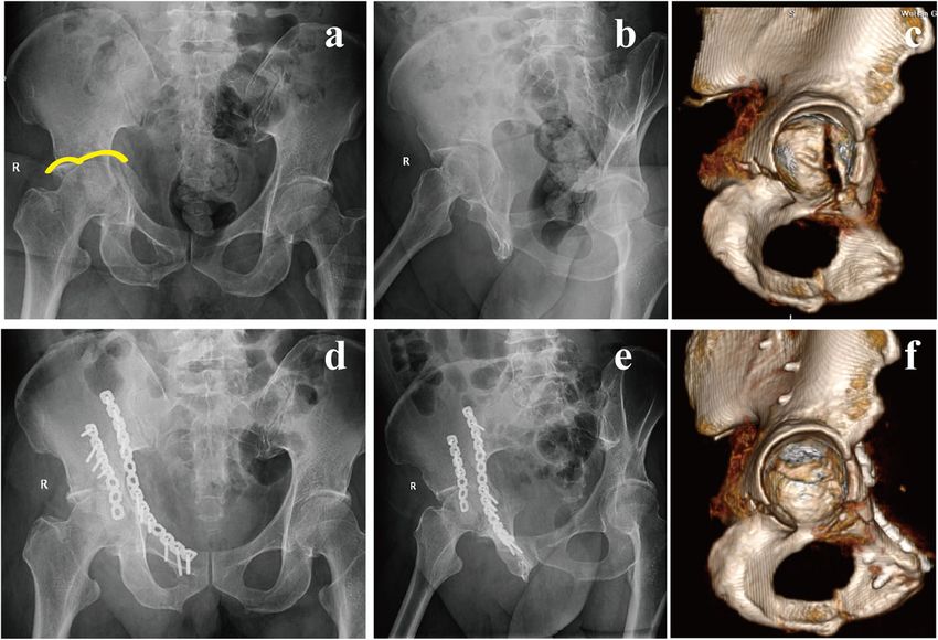
Fig. 2 Walid’s classification of quadrilateral plate fracture. (a) QLP1, incompletely separated simple fracture; (b) QLP2, incompletely separated
comminuted fracture; (c) completely separated comminuted fracture
therefore described as the “Gull sign” [11]. Medical re- estimated in advance by measuring the total anatomical
cords of all patients enrolled in this study were reviewed length of the placement trajectory of DAPSQ (Fig. 3). For
retrospectively, including gender, age, mechanism of in- comminuted and complex acetabular fractures, after
jury, concomitant injuries, fracture type, and pre-existing obtaining informed consent from the patients, the 3D
comorbidity. digital virtual model of the pelvis was imported into a 3D
printer (Makerbot Replicator 2X, Makerbot, USA), and a
rapid prototyping model of the pelvis was then printed.
Preoperative planning
Commercially available surgical simulation software
BOHOLO (Boholo Medical Technology Co., Ltd., China) Surgical technique
was used for the pre-operative planning. All patients All surgical procedures were performed by two experi-
underwent a thin-slice pelvic CT scan (Siemens Sensation enced orthopaedic surgeons. Among them, 28 patients
64, Germany). The original CT images were imported as a underwent ORIF via a classic ilioinguinal approach [12],
DICOM file format into BOHOLO software. Each pelvic and 9 patients was combined with the Kocher-
bones and fracture fragments were segmented virtually re- Langenbeck (K-L) approach. The QLP fracture was re-
moving the surrounding tissue using semiautomatic duced and temporarily fixed with various techniques, in-
thresholding tools and obtaining individual 3D digital cluding the use of ball spike, pelvic clamp, K-wires or
models. All the bone fragments were considered inde- screws. When the “Gull sign” or the compression frac-
pendent and removable. And it was beneficial to accur- ture of acetabular dome appeared on the preoperative
ately measure the rotation angle and displacement X-ray, the compression zone could be exposed by dir-
distance of the bone fragments, help surgeons to better ectly prying open the fracture fragments of the QLP or
understand the fracture type, as well as to simulate surgi- indirectly fenestration and osteotomy above the acetabu-
cal procedures to accurately perform surgeries. Moreover, lar dome, and restored by the implantation of autolo-
the required length of the reconstruction plate could be gous iliac bone or artificial bone.
Fig. 3 Computer-assisted preoperative planning. a A 3D model of acetabular fracture; b Select the bone fragments and perform a virtual fracture
operation; c Fracture model after reduction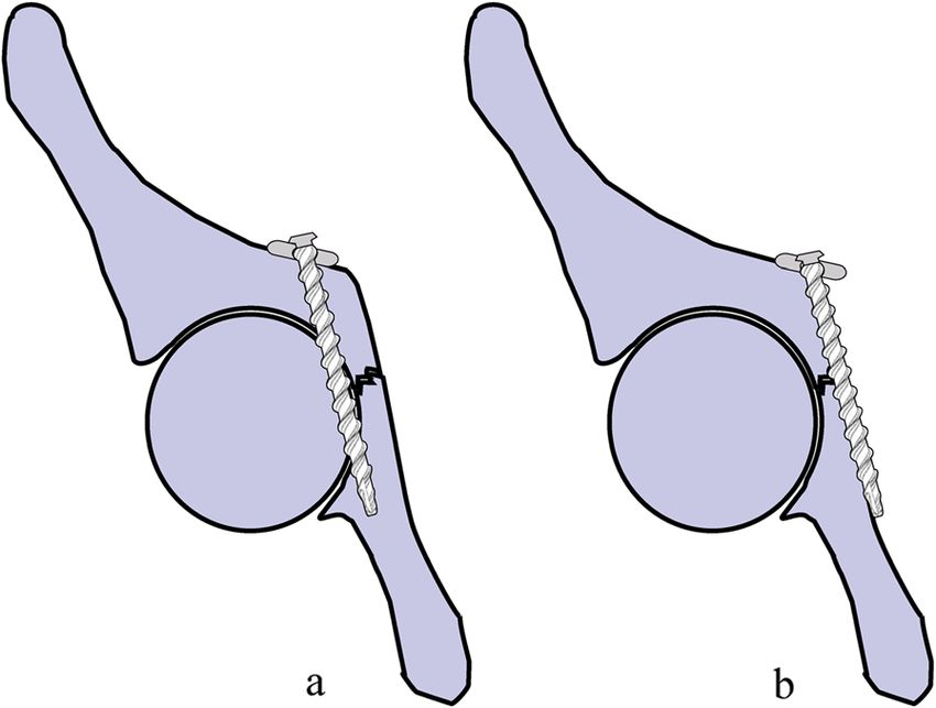
Wu et al. BMC Musculoskeletal Disorders      (2021) 22:122                                                                   Page 4 of 8

   After fracture reduction and temporary fixation, a re-                distribution. Categorical variables were presented as abso-
construction plate of the appropriate length was selected                lute (n) and relative frequencies (%). The Spearman rank
and shaped according to the preoperative planning and in-                correlation coefficient was used to measure the association
traoperative measurements. DAPSQ plate was placed on                     between Walid’s classification and radiological or func-
the superior arcuate line, and the ends extended along the               tional outcomes. The association between potential risk
iliac wing and the superior pubic ramus direction, respect-              factors and post-traumatic arthritis was also tested. Inter-
ively. The sequence of screws placement followed certain                 observer agreement was calculated using the kappa coeffi-
principles described previously (Fig. 4) [13].                           cient. P < 0.05 was considered statistically significant.

                                                                         Results
Follow‐up and evaluation criteria                                        Patient demographics are listed in Table 1. The
Radiographic and functional assessment were per-                         mean age of this group was 64.9 (SD = 3.8) years and
formed at follow-up visits through the outpatient                        there were 11 females (29.7 %) and 26 males. The
clinic. Quality of reduction was estimated by the im-                    most common fracture type was both-column frac-
mediate postoperative X-ray according to the Matta                       tures (40.5 %), followed by anterior column posterior
radiological criteria [14], and the scores were graded                   hemitransverse (21.6 %). In addition, posterior wall
as anatomic(0-1mm), imperfect (2-3mm), or poor (>
3mm) based on the millimeters of residual displace-                      Table 1 Patient demographics (n = 37)
ment on all views. Functional outcomes were evalu-                       Gender (n, %)
ated using the modified Merle d’Aubigné score [15]                       Male                                                  26(70.3)
at the last follow-up, and the scores were classified as                 Female                                                11(29.7)
excellent (18 points), good (15–17 points), fair (13 or
                                                                         Age,years (mean ± SD)                                 64.9 ± 3.8
14 points), or poor (< 13 points). All the evaluations
were performed independently by two experienced                          Mechanism of injury (n, %)
orthopedic surgeons.                                                     Fall from height (greater than standing)              11(29.7)
                                                                         Fall (from standing height)                           7(18.9)
Statistical analysis                                                     Traffic accident                                      19(51.4)
Data was collected, coded and analyzed with SPSS soft-                   Fracture side,left (n, %)                             22(56.5)
ware (version 19.0, IBM Corp). Continuous variables
                                                                         Concomitant injuries (n, %)
were presented as the means ± standard deviations (SD).
The Kolmogorov-Smirnov test (K-S) was used to test                       Head trauma                                           7(18.9)
whether all continuous variables followed normal                         Spine and sacral fracture                             7(18.9)
                                                                         Limb fracture                                         9(24.3)
                                                                         Rib or clavicle fracture                              6(16.2)
                                                                         Dislocation of hip                                    13(35.1)
                                                                         Others                                                4(10.8)
                                                                         Gull sign (n, %)                                      8(21.6)
                                                                         Pre-existing comorbidity (n, %)
                                                                         Hypertension                                          10(27)
                                                                         Diabetes mellitus                                     6(16.2)
                                                                         Lung disease                                          4(10.8)
                                                                         Fracture type (n, %)
                                                                         Both columns                                          15(40.5)
                                                                         Both columns + Posterior wall                         3(8.1)
                                                                         Anterior column                                       2(5.4)

 Fig. 4 a Traditional fixation method: a plate placed along the medial   Anterior column + Anterior wall                       1(2.7)
 edge of the pelvic brim with screws extending distally into the         ACPH                                                  8(21.6)
 posterior column. It has a high risk of screws penetrating into the
                                                                         T type                                                3(8.1)
 hip. b DAPSQ: Screws were inserted parallel to the surface of
 quadrilateral plate and only 1/3 to 1/2 transverse diameter of          Transverse and posterior wall                         2(5.4)
 quadrilateral screws entered into the bone. This visualization          Transverse                                            3(8.1)
 method could entirely avoid the risk of hip penetration
                                                                         ACPH anterior column and posterior hemitransverse
Wu et al. BMC Musculoskeletal Disorders (2021) 22:122 Page 5 of 8
fracture was involved in 5 cases and the rate of frac- complications were presented in Table 2. And one typ-
tures with a Gull sign was 21.6 %. ical case was shown in Fig. 5.
Treatment was standardized with a mean interval from According to Walid’s classification, 18 patients were
injury to surgery of 10.5 days with a range of 6–17 days. classified as QLP1, 13 patients were classified as QLP2
In regard to surgical approach, 28 (75.7 %) patients were and 6 patients were classified as QLP3. Walid’s classifi-
treated with single ilioinguinal approach, and 9 (24.3 %) cation was strongly positively correlated with radio-
patients (5 cases of posterior wall fracture, 3 cases of logical outcomes of reduction (r = 0.661; P < 0.001), and
both-column fracture and 1 case of T type fracture) were moderately positively correlated with functional out-
combined with a K-L approach. Surgical time averaged comes (r = 0.478; P = 0.003) (Table 3).
256.9 minutes (SD = 58.9). Intraoperative blood loss av-
eraged 1029.7 mL (SD = 442.8). Eventually 86 % of the In addition, the quality of reduction was strongly posi-
cases received a blood transfusion with an average of tively correlated with the functional outcomes (r = 0.701;
662.2 mL (SD = 486.7). The mean duration of hospital P < 0.001). Unsatisfactory reduction was also demon-
stay was 26.1 days (SD = 6.4). No intra-operative compli- strated a correlation with the development of post-
cations were observed. Surgery details of all included pa- traumatic arthritis (r =-0.410; P = 0.012). The associa-
tients were listed in Table 2. tions between Gull sign or Walid’s classification and
post-traumatic arthritis revealed no statistically signifi-
Kappa analysis showed a high consistency between the cant differences (P = 0.644 and P = 0.133, respectively).
two senior surgeons for the subjective score with Kappa The relationships between Letournel classification and
coefficient of 0.87. According to the definition of the radiological or functional outcomes were also analyzed.
quality of the reduction, 18 patients (48.6 %) showed The results showed that no significant correlation
anatomical reduction, 15 (40.5 %) had an imperfect re- existed between both columns, ACPHT or involved pos-
duction, and 4 (10.8 %) had a poor reduction. terior wall and radiological or functional outcomes (P all
Patients were followed-up for more than 12 months > 0.05) (Table 4).
with a mean follow-up period of 35.5 months (SD =
10.7). There were no cases of screw pull-out, screw loos-
ening or implant failure during the follow-up. At the last Discussion
follow-up, the mean modified Merle d’Aubigne score The management of acetabular fractures in elderly is
was 16 (range 10–18), categorized as excellent in 16 controversial. Both conservative and surgical treatment
cases (43.2 %), good in 15 cases (40.5 %), fair in 4 cases have been reported to be risky and potentially unsuc-
(10.8 %), and poor in 2 cases (5.4 %). Postoperative cessful [4, 5]. The current, commonly used surgical op-
tions for the elderly with acetabular fractures include
minimally invasive percutaneous internal fixation [16],
Table 2 Surgery details, followed-up time and complications ORIF [17], or hip replacement [18]. Anatomic reduction
(n = 37)
and stable fixation are primary goals of ORIF. The atten-
Time to surgery, days (mean ± SD) 10.5 ± 3.1
tion of many orthopaedic surgeons today is not only
Surgical approach (n, %) aimed at technological innovations from fixation devices
Ilioinguinal 28(75.7) and surgical approach but also at improving the effect-
Ilioinguinal + Kocher-Langenbeck 9(24.3) iveness of those already used in the clinics. In most
Surgical time, min (mean ± SD) 256.9 ± 58.9 cases, as shown in Fig. 4, placing a reconstruction plate
Blood loss, mL (mean ± SD) 1029.7 ± 442.8
along the pelvic brim of the anterior column with screws
extending distally into the posterior column can provide
Blood transfusion, mL (mean ± SD) 662.2 ± 486.7
adequate stability when used in non-osteoporotic bone,
Hospital stay time, days (mean ± SD) 26.1 ± 6.4 but at the risk of screws penetrating into the hip.
Followed-up time, years (mean ± SD) 35.5 ± 10.7 DAPSQ was designed based on the traditional anterior
Complications (n, %) reconstruction plate [13, 19]. Several biomechanical ex-
Deep venous thrombosis 1(2.7) periments have confirmed its reasonable mechanical sta-
Lateral femoral cutaneous nerve injury 3(8.1)
bility [20, 21]. We consider that this technique is more
suitable for acetabular fractures involving the anterior
Superficial wound infection 1(2.7)
column, including both-column fractures and transverse
Posttraumatic arthritis 7(18.9) fractures mainly with anterior column displacement, cer-
THA 2(5.4) tain types of T-shaped and ACPHT fractures [19]. Al-
THA total hip arthroplasty though acetabular fractures involving the anteriorWu et al. BMC Musculoskeletal Disorders (2021) 22:122 Page 6 of 8
Fig. 5 A 61-year-old man presented with acetabular fracture of the right acetabulum. Preoperative AP view (a) showed a typical feature of “Gull
sign”. Judet view (b) and 3D CT reconstruction (c) showed that the quadrilateral plate was separated from the anterior column and partially
attached to the posterior column, which was classified as QLP1. ORIF was performed via a single ilioinguinal approach, and the compression
fracture of acetabular dome was managed with artificial bone grafting. Post-operative X-rays (d, e) and 3D view (f) showing an
anatomical reduction
column are the most common fracture types in the eld- osteopenic QLP fractures. They found that 52.4 % (11/
erly, DAPSQ is not suitable for all types. For delayed ace- 21) of patients obtained anatomic reduction and 92.9 %
tabular fractures, acetabular posterior wall fractures, and with excellent and good functional outcomes by using
acetabular fractures mainly involving posterior column, fix- an infra-pectineal buttress plate. These results differ
ation of the posterior column with reconstruction or locking from our data and may be attributed to several factors
plates through a posterior approach is often needed. such as different age range (55–82 years) and choice of
The results of the present study show that 75.7 % of surgical approach (modified Stoppa).
patients were treated through a single ilioinguinal ap- Recently, Herman et al. [23] proposed a new classifica-
proach, and an additional Kocher-Langenbeck approach tion scheme for acetabular fractures, identifying three
was required in 9 patients. Reduction after operation different types based on the displacement vector and the
was considered anatomical in 48.6 % of the patients, and fractured anatomic structures. According to this classifi-
83.7 % of them had excellent or good functional out- cation, the medial displacement of the QLP was
comes. Peter et al. [3] reported 84.6 % excellent and regarded as a hallmark of the superomedial displacement
good functional outcome with the L-shaped buttress vector. Walid et al. [10] further divided the QLP frac-
plate to treat QLP fractures in 13 elderly patients, which tures into four categories. As far as we know, this is the
was similar to results seen in our study. Another study first study to explore the relationship between Walid’s
by Laflamme et al. [22]. included 21 patients with classification and radiological or functional outcomes.
Table 3 The relationships between Walid’s classification and radiological or functional outcomes
Walid’s Radiological outcomes Functional outcomes
classification
Anatomical Imperfect Poor Excellent Good Fair Poor
QLP1(18) 15 3 0 12 5 0 1
QLP2(13) 2 9 2 3 8 1 1
QLP3(6) 1 3 2 1 2 3 0
Total (37) 18(48.6) 15(40.5) 4(10.8) 16(43.2) 15(40.5) 4(10.8) 2(5.4)
QLP quadrilateral plateWu et al. BMC Musculoskeletal Disorders (2021) 22:122 Page 7 of 8
Table 4 The relationships between Letournel classification and radiological or functional outcomes
Letournel Radiological outcomes Functional outcomes
classification
r P r P
Both columns 0.237 0.159 0.318 0.055
ACPHT 0.116 0.496 0.290 0.082
Involved posterior wall 0.229 0.172 0.177 0.296
ACPH anterior column and posterior hemitransverse
The results demonstrated that Walid’s classification application of virtual and 3D printing technology in the
was strongly and positively correlated with radiological treatment of pelvic and acetabular fracture [26, 27].
outcomes of reduction and moderately and positively However, it should be noted that the basis of computer
correlated with functional outcomes. Walid’s type QLP1 assisted surgery is the 3D model of acetabular fracture,
fracture was associated with better radiological and func- either through virtual technology or 1:1 printed 3D frac-
tional outcomes, while type QLP3 fracture was associ- ture model. However, the technology is limited to the
ated with poorer outcomes. The findings suggest that bone and does not include soft tissue or ligaments, thus
Walid’s classification may reflect the severity of acetabu- being still quite different from the real setting.
lar fracture to some extent and mainly relate to the de- Our study has some limitations due to its retrospective
gree of comminuted fracture of the QLP. Thus, Walid’s design and limited number of patients. Another restric-
classification may guide the selection of an appropriate tion is the absence of a comparative group. Further ran-
surgical approach and fixation strategy. The relation- domized controlled trials and larger numbers of
ships between Letournel classification and radiological participants will be necessary to confirm these findings.
or functional outcomes were also analyzed. The results In addition, although all surgical procedures in this
showed that no significant correlation existed between study were performed by two surgeons, the technique
both columns, ACPHT or involved posterior wall and was well-standardized to eliminate the effects of different
radiological or functional outcomes. This result may re- surgeons on outcome variables.
veal that preoperative assessment of the fracture pattern
according to the Walid classification is better than the Conclusions
classic Letournel classification in predicting outcomes, ORIF is still the preferred treatment for displaced ace-
and possibly modifying the surgical management. How- tabular fractures in the elderly. DAPSQ is an optional
ever, it is worth noting that there are still some limita- and effective technique for the treatment of acetabular
tions to consider when interpreting these findings. For fracture involving the QLP and has the advantages of
example, Walid’s type QLP4 fracture seems to be a the- avoiding screws penetrating into the hip. Besides,
oretical possibility, and has not yet been reported [10]. Walid’s classification system is associated with the re-
Previous studies also have shown that “Gull sign” in pre- duction quality and functional recovery. QLP fractures
operative X-ray may be an important sign of poor func- classified as QLP3 may need a more precise and appro-
tional outcomes in the elderly with acetabular fractures priate operative strategy to improve the success of
[24]. Unlike their study, our study did not find a correl- surgery.
ation between “Gull sign” and post-traumatic arthritis. In
accordance to our view, Carroll et al. [25] and Zhuang Abbreviations
DAPSQ: Dynamic Anterior Plate-Screw System for Quadrilateral plate;
et al. [11] also considered that “Gull sign” had no correl- QLP: Quadrilateral plate; AP: Anterior-Posterior; CT: Computed Tomography;
ation with secondary THA. Even if the “Gull sign” ap- 3D: Three-Dimensional; ACPH: Anterior Column Posterior Hemitransverse;
peared in preoperative X-ray of the elderly patients, THA: Total Hip Arthroplasty
satisfactory outcomes can be obtained by sufficient reduc-
Acknowledgements
tion and bone grafting of the compressed dome. We would like to thank PhD. Yanjing Li for English language editing.
This study also introduced virtual technology into the
preoperative planning of acetabular fractures in the eld- Authors’ contributions
erly. This technique can help surgeons to understand WHY, SQP, LXM and CXH conceived and designed the study. SRR and SCJ
contributed to the data collection. WHY and SRR analyzed the data. SCJ
the fracture pattern, rotation angle and displacement drew the pictures. WHY wrote the manuscript. All authors read and
distance of fragments, and simulate the reduction approved the final manuscript and consented to publish this manuscript.
process of fracture on the computer before operation.
And design the optimal reduction sequence and further Funding
This work was supported by the technological innovation projects of hubei
provide a reference for the selection and measurement provine (Grant No. 2017ACA099) and General Projects of Health Commission
of the plate. Many scholars have advocated the of Hubei Provine (Grant No. WJ2018H0064).Wu et al. BMC Musculoskeletal Disorders (2021) 22:122 Page 8 of 8
Availability of data and materials 16. Ruan Z, Luo CF, Zeng BF, Zhang CQ. Percutaneous screw fixation for the
The datasets generated and/or analyzed during the current study are acetabular fracture with quadrilateral plate involved by three-dimensional
available from the corresponding author by reasonable request. fluoroscopy navigation: surgical technique. Injury. 2012;43:517–21.
17. Li YL, Tang YY. Displaced acetabular fractures in the elderly: results after
open reduction and internal fixation. Injury. 2014;45:1908–13.
Ethics approval and consent to participate 18. Jauregui JJ, Clayton A, Kapadia BH, Cherian JJ, Issa K, Mont MA. Total hip
This study has been approved by the appropriate ethics committee (the arthroplasty for acute acetabular fractures: a review of the literature. Expert
Scientific Board of the Department of Orthopaedic Surgery, General Hospital Rev Med Devices. 2015;12:287–95.
of Central Theater Command). Written consent was obtained from all 19. Wu H, Shang R, Liu X, Song C, Chen Y, Cai X. A novel anatomically pre-
patients who participated in this study. contoured side-specific titanium plate versus the reconstruction plate for
quadrilateral plate fractures of the acetabulum: a propensity matched
Consent for publication cohort study. J Orthop Surg Res. 2020;15:172.
Not applicable. 20. Lei J, Dong P, Li Z, Zhu F, Wang Z, Cai X. Biomechanical analysis of the
fixation systems for anterior column and posterior hemi-transverse
acetabular fractures. Acta Orthop Traumatol Turc. 2017;51:248–53.
Competing interests 21. Fan Y, Lei J, Zhu F, Li Z, Chen W, Liu X. Biomechanical Analysis of the
On behalf of all authors, the corresponding author states that there is no Fixation System for T-Shaped Acetabular Fracture. Comput Math Methods
conflict of interest. Med. 2015; 2015: 370631.
22. Laflamme GY, Hebert-Davies J, Rouleau D, Benoit B, Leduc S. Internal
Author details fixation of osteopenic acetabular fractures involving the quadrilateral plate.
1
Department of Orthopaedic Surgery, General Hospital of Central Theater Injury. 2011;42:1130–4.
Command, Wuhan Clinical Medicine College of Southern Medical University, 23. Herman A, Tenenbaum S, Ougortsin V, Shazar N. There Is No Column: A
430070 Wuhan, China. 2Department of Orthopaedic Surgery, First Hospital of New Classification for Acetabular Fractures. J Bone Joint Surg Am. 2018;100:
Wuhan, Hubei University of Chinese Medicine, 430022 Wuhan, China. e8.
3
Chengjing Song Huaiyin hospital of huai an city, 223300 Huaian, China. 24. Anglen JO, Burd TA, Hendricks KJ, Harrison P. The “Gull Sign”: a harbinger of
failure for internal fixation of geriatric acetabular fractures. J Orthop Trauma.
Received: 14 September 2020 Accepted: 20 January 2021 2003;17:625–34.
25. Carroll EA, Huber FG, Goldman AT, Virkus WW, Pagenkopf E, Lorich DG, et al.
Treatment of acetabular fractures in an older population. J Orthop Trauma.
2010;24:637–44.
References 26. Boudissa M, Oliveri H, Chabanas M, Tonetti J. Computer-assisted surgery in
1. Ferguson TA, Patel R, Bhandari M, Matta JM. Fractures of the acetabulum in acetabular fractures: Virtual reduction of acetabular fracture using the first
patients aged 60 years and older: an epidemiological and radiological patient-specific biomechanical model simulator. Orthopaedics
study. J Bone Joint Surg Br. 2010;92:250–7. Traumatology: Surgery Research. 2018;104:359–62.
2. Farid YR. Cerclage wire-plate composite for fixation of quadrilateral plate 27. Wang C, Chen Y, Wang L, Wang D, Gu C, Lin X, et al. Three-dimensional
fractures of the acetabulum: a checkrein and pulley technique. J Orthop printing of patient-specific plates for the treatment of acetabular fractures
Trauma. 2010;24:323–8. involving quadrilateral plate disruption. BMC Musculoskelet Disord. 2020;21:
3. Peter RE. Open reduction and internal fixation of osteoporotic acetabular 451.
fractures through the ilio-inguinal approach: use of buttress plates to
control medial displacement of the quadrilateral surface. Injury. 2015;46:2–7.
4. Laflamme GY, Hebert-Davies J. Direct reduction technique for superomedial Publisher’s Note
dome impaction in geriatric acetabular fractures. J Orthop Trauma. 2014;28: Springer Nature remains neutral with regard to jurisdictional claims in
e39–43. published maps and institutional affiliations.
5. Wollmerstädt J, Pieroh P, Schneider I, Zeidler S, Höch A, Josten C, et al.
Mortality, complications and long-term functional outcome in elderly
patients with fragility fractures of the acetabulum. BMC Geriatr. 2020;20:66.
6. Guerado E, Cano JR, Cruz E. Fractures of the acetabulum in elderly patients:
an update. Injury. 2012;43(Suppl 2):33–41.
7. Park MS, Yoon SJ, Park JH, Choi SM. The management of the displaced
medial wall in complex acetabular fractures using plates and additional
cerclage. Hip Int. 2013;23:323–9.
8. Bastian JD, Tannast M, Siebenrock KA, Keel MJ. Mid-term results in relation
to age and analysis of predictive factors after fixation of acetabular fractures
using the modified Stoppa approach. Injury. 2013;44:1793–8.
9. Letournel E. Acetabulum fractures: classification and management. Clin
Orthop Relat Res. 1980;151:81–106.
10. ElNahal WA, Abdel Karim M, Khaled SA, Abdelazeem AH, Abdelazeem H.
Quadrilateral plate fractures of the acetabulum: Proposition for a novel
classification system. Injury. 2018;49:296–301.
11. Zhuang Y, Lei JL, Wei X, Lu DG, Zhang K. Surgical treatment of acetabulum
top compression fracture with sea Gull sign. Orthop Surg. 2015;7:146–54.
12. Letournel E. The treatment of acetabular fractures through the ilioinguinal
approach. Clin Orthop Relat Res. 1993;292:62–76.
13. Wu H, Shang R, Cai X, Liu X, Song C, Chen Y. Single ilioinguinal approach to
treat complex acetabular fractures with quadrilateral plate involvement:
outcomes using a novel dynamic anterior plate-screw system. Orthop Surg.
2020;12:488–97.
14. Matta JM. Fractures of the acetabulum: accuracy of reduction and clinical
results in patients managed operatively within three weeks after the injury.
J Bone Joint Surg Am. 1996;78:1632–45.
15. Merle DR. Numerical classification of the function of the hip. 1970. Rev Chir
Orthop Reparatrice Appar Mot. 1990; 76: 371-4.You can also read