Validity of Wearable Sensors at the Shoulder Joint: Combining Wireless Electromyography Sensors and Inertial Measurement Units to Perform Physical ...
←
→
Page content transcription
If your browser does not render page correctly, please read the page content below
sensors
Article
Validity of Wearable Sensors at the Shoulder Joint:
Combining Wireless Electromyography Sensors and
Inertial Measurement Units to Perform Physical
Workplace Assessments
Isabelle Poitras 1,2 , Mathieu Bielmann 1,2 , Alexandre Campeau-Lecours 1,3 ,
Catherine Mercier 1,2 , Laurent J. Bouyer 1,2 and Jean-Sébastien Roy 1,2, *
 1    Centre for Interdisciplinary Research in Rehabilitation and Social Integration and Laval University, Quebec
      City, QC G1M2S8, Canada; isabelle.poitras.2@ulaval.ca (I.P.); mathieu.bielmann.1@ulaval.ca (M.B.);
      Alexandre.Campeau-Lecours@gmc.ulaval.ca (A.C.-L.); Catherine.Mercier@rea.ulaval.ca (C.M.);
      Laurent.Bouyer@rea.ulaval.ca (L.J.B.)
 2    Department of Rehabilitation, Laval University, Quebec City, QC G1V0A6, Canada
 3    Department of Mechanical Engineering, Laval University, Quebec City, QC G1V0A6, Canada
 *    Correspondence: Jean-Sebastien.Roy@fmed.ulaval.ca; Tel.: +1-418-529-9141 (ext. 6005)

 Received: 21 March 2019; Accepted: 17 April 2019; Published: 20 April 2019

 Abstract: Background: Workplace adaptation is the preferred method of intervention to diminish risk
 factors associated with the development of work-related shoulder disorders. However, the majority of
 the workplace assessments performed are subjective (e.g., questionnaires). Quantitative assessments
 are required to support workplace adaptations. The aims of this study are to assess the concurrent
 validity of inertial measurement units (IMUs; MVN, Xsens) in comparison to a motion capture system
 (Vicon) during lifting tasks, and establish the discriminative validity of a wireless electromyography
 (EMG) system for the evaluation of muscle activity. Methods: Sixteen participants performed 12
 simple tasks (shoulder flexion, abduction, scaption) and 16 complex lifting tasks (lifting crates of
 different weights at different heights). A Delsys Trigno EMG system was used to record anterior
 and middle deltoids’ EMG activity, while the Xsens and Vicon simultaneously recorded shoulder
 kinematics. Results: For IMUs, correlation coefficients were high (simple task: >0.968; complex task:
 >0.84) and RMSEs were low (simple task: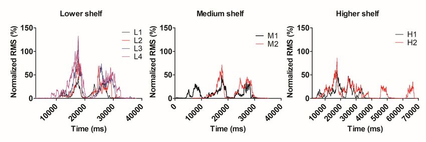
Sensors 2019, 19, 1885                                                                               2 of 14

      To prevent work-related shoulder disorders, most interventions use workplace adaptations or
educational programs to decrease the physical demands on the shoulder joint. However, the lack of
clinical objective assessments makes it difficult to determine the interventions’ efficiency to decrease
repeated or sustained arm elevations or at lowering strength requirements. The majority of workplace
assessments are based on self-administered questionnaires, interviews or clinical observations [3].
A few studies have used camera-based motion capture systems to assess simulated working tasks
in the laboratory to complement the usual workplace assessments with more objective data [8,9].
However, camera-based systems lack portability and can hardly be used outside the laboratory [10].
      Accelerometers have been used for several years to assess physical activity in rehabilitation
research; however they have been shown to lack precision for kinematics estimation (e.g., lower
accuracy for increasing segment acceleration) [11]. To overcome this limitation, gyroscopes and
magnetometers are now commonly added to the wearable units (called inertial measurement units
or IMUs). By combining the signals of each sensor through optimized data fusion algorithms, a
reasonably accurate estimate of IMU orientation can be obtained. Still, a recent systematic review on
the psychometric evidence of IMUs for the assessment of joint movement has reported highly variable
results when using IMUs to evaluate the shoulder joint. The main reason behind these findings was
the difficulty in analyzing movements in more than one plane [12].
      Recent improvements in IMUs hardware/software/data processing could result in improved
validity for the evaluation of movements at the shoulder joint. The improved systems could therefore
be an alternative to quantifying human movements outside the laboratory, i.e., in more ecological
settings, such as actual work environments. At the shoulder, IMUs have been validated in several
contexts but mostly during simple arm movements (movements performed in only one plane of
movement: sagittal, frontal or transverse) [10]. Unfortunately, such movements are not representative
of real work demands. The shoulder is the most mobile joint of the human body, and everyday tasks
require that it performs complex 3D movements [3,13]. Few studies have validated IMUs for the
shoulder joint during complex tasks (movements performed in more than one plane of movement)
and have shown that validity is highly variable [14–17], as previously mentioned.
      Other physical factors such as increases in muscle activity and muscle fatigue are also associated
with a higher risk of work-related shoulder disorders [6]. During laboratory assessments, wireless
electromyography (EMG) systems are often used to quantify muscle activity and fatigue, and these
systems are sometimes also used in the clinic. During arm elevation, shoulder muscles are highly
requested to maintain joint stability, and having an adequate muscle activation pattern plays a major
role in the prevention of injuries [18]. To the best of our knowledge, only two studies have identified the
effects of arm elevation and weight lifting on muscle activation during simulated working tasks [19,20].
However, the effect of shoulder range of motion was not accounted for, which can be an important
contributing factor to shoulder pain of manual handling workers. Also, wireless EMG systems lack
validation in work contexts, limiting their current application in clinical and work environments.
      Therefore, the aims of this study are (1) to evaluate the criterion validity of a commercial IMU
system (MVN Awinda system, Xsens) by comparing it to a camera-based laboratory motion capture
system (Vicon), during isolated shoulder movements and complex upper extremity lifting tasks; and
(2) to evaluate the discriminative validity of a wireless EMG system (Delsys Trigno; EMG activity
of the anterior and middle deltoid muscles) by looking at its discriminative capacities according to
shoulder range of motion (ROM) and lifting weights. The hypotheses are that: (1) criterion validity
will be characterized as good to excellent (0.8 ≥ r ≤ 1.0, error of measurement ≤ 15◦ ) for both simple
movements and complex tasks, but errors of measurement for arm elevation will be larger in complex
tasks; and (2) that EMG activity will increase with arm elevation and heavy weight lifting (p < 0.05),
and the increase will be larger for the anterior deltoid, as it is the main shoulder muscle agonist for
lifting in the sagittal plane. Aggregating the IMU and EMG data will provide a good estimate of
shoulder physical demands in simple and complex tasks simulating the real work environment.
Sensors 2019, 19, 1885                                                                             3 of 14

2. Materials and Methods

2.1. Participants
     Sixteen healthy participants (eight males, 12 right-handed (1 ambidextrous), 26.4 ± 4.1 years, 1.73
± 0.09 m of height) were recruited (sample size required for an effect size of 0.8, with α = 0.05 and
1-β= 0.95). Inclusion criteria were: (1) to be between 18 and 65 of age, and (2) absence of self-reported
neurological or musculoskeletal conditions (pain, mobility limitations) that could interfere with task
execution. All participants gave written informed consent prior to experiment onset; this study was
approved by the local ethics committee (CIUSSS-CN; project #217-539).

2.2. Instrumentation and Data Collection
      Shoulder movements were recorded simultaneously with nine IMUs positioned at standardized
locations on the upper body (MVN, Xsens Technologies, Enschede, The Netherlands) and nine Vicon
MX cameras (seven MT40-S and two MT10- S cameras, Vicon Motion Systems Limited, Oxford, UK),
respectively at 60 and 100 Hz. The IMUs were placed on: the head, shoulder (2), sternum, upper arm
(2), forearm (2) and pelvis. IMUs were fixed with hook and loop straps around arms and on Lycra suit
for the trunk in accordance with the sensors configuration recommended by Xsens [21]. Rigid triads
of retroreflective markers were placed on the C7 spinous process, as well as on the right and left upper
arms. Single markers were temporarily placed bilaterally on specific anatomical landmarks (sternal
notch, lateral epicondyle, medial epicondyle and glenohumeral junction) for calibration. To record
muscle activity, wireless surface EMG sensors (Trigno Wireless EMG system, Delsys, Boston, MA,
USA) were positioned bilaterally on the anterior and middle deltoid muscles according to Seniam
recommendations [22].

2.3. Study Design and Experimental Procedure
      All participants took part in one testing session. Before the experiment began, the experimenter
explained the protocol, answered questions and obtained written consent from each participant. Then,
all participants filled a sociodemographic questionnaire and the Edinburgh Handedness Inventory.
Wireless EMG sensors were placed after skin preparation (skin was cleaned by rubbing it with alcohol
for 5 seconds). A signal preview from each sensor was used to assess signal quality. Three maximal
voluntary contractions (MVC) were performed for each muscle (anterior and middle deltoids) with
one-minute breaks in between. Thereafter, Xsens sensors and Vicon markers were placed on the upper
body. Anthropometric measures (height, shoulder width, arm span, hip height, hip width, knee height,
ankle height, foot size and sole height) were gathered and filled in the MVN Studio software (MVN
studio software, v. 4.4.0, Xsens Technologies). An anatomical pose (participant standing straight
looking forward, arms along body side, palms facing forward) was performed to calibrate the Vicon
system and an N-pose (participant standing straight looking forward, 90◦ shoulder abduction, palms
facing the ground) was held to calibrate the Xsens system. Then, the protocol was divided in two parts:
1) isolated shoulder and trunk movements, and 2) complex lifting tasks.
      During part one, IMU validity was first assessed during simple shoulder movements.
Three isolated movements (flexion, abduction and scaption) were performed 5 times each at 3
different joint angles (60, 90 and 120◦ measured with an inclinometer). Then, three combined trunk
movements (anterior flexion, lateral bending and rotation) were repeated 5 times each while the
shoulder was maintained in a 90◦ flexion (12 different movements * 5 repetitions = 60 trials per
participant).
Sensors 2019, 19, 1885                                                                                 4 of 14

     A standardized handling assessment system, Valpar 19 (Valpar International Corporation, Tucson,
AZ, United States), was used to assess the validity of IMU and surface EMG during complex tasks.
This system comprises a shelf with three levels (high [H]: 1.74 m; medium [M]: 1.25 m; low [L]: 0.46 m
from the ground) where crates of different weight representing specific physical work demand levels
(sedentary [2.3kg], light [6.8kg], moderate [13.6kg] and moderate to heavy [22.7kg]) can be placed [23].
Participants were asked to lift a crate from a step located 1.8 m in front of the shelf, move it to a specific
location on the shelf, take a 3-second break with arms along the body and return the crate back to the
starting position. They were asked to take the lifting strategy of their choice and a stepladder was
made available to them to bring the crates to the high shelf, when/if necessary. Each lifting (i.e., each
given height and weight combination) was repeated twice. To ensure participant safety, only sedentary
and light weights were used for the medium and higher shelves (2 weights × 2 trials = 4 trials/shelf);
all weights were used for the lower shelf (4 weights × 2 trials = 8 trials). Height and weight were
randomly assigned for a total of sixteen trials.

2.4. Data Processing
      Kinematic data of the right arm were processed for both motion capture systems. The data
collected from Vicon was processed in the Nexus software (Vicon Motion Systems Ltd., Oxford, UK).
Each trial was manually inspected, markers were labelled and gaps of 15 or less samples were linearly
interpolated. The data were then digitally low-pass filtered at 8 Hz (Butterworth double-pass filter).
Before calculating joint angles, sensor positions were re-initialized in N-pose. Then, joint angles at the
shoulder joint were calculated relative to the position of the trunk and arm markers and by applying
the ZYZ Euler rotation sequence.
      Each Xsens sensor orientation was extracted from MVN Studio BIOMECH (Xsens Technologies,
Enschede, Netherlands) and exported in ASCII in the form of a quaternion. The data were then
imported into MATLAB R2017a (The Math Works Inc., Natick, MA, USA) and the quaternions were
converted into a rotation matrix. Joint angles were then calculated relative to the orientation of the
trunk and upper arm sensors. The rotation sequence ZYZ was also used to calculate Euler angles.
      EMG signals were processed with a custom software written in MATLAB. They were digitally
filtered with a fourth-order zero-lag Butterworth filter (band-pass 20–450 Hz), and a root-mean-square
rectangular window of 20 ms was used for rectifying and smoothing the signal. Mean RMS value of
MVC for each muscle was used to normalize EMGs. The output of accelerometers from Trigno and
Xsens sensors was used to synchronize EMG and kinematic data by performing a cross-correlation.
Then, a custom-written algorithm was used to identify the “lifting” and ”dropping” phases of the
manual handling task. Analyses were only performed for the ”lifting” phase as both muscles perform
concentric work during this phase of movement [24]. Results were calculated for the arm which was
farther away from the body when lifting (identified manually for each trial, see Figure 1). The peak
activation and the area under the burst were calculated for each weight and height.
applying the ZYZ Euler rotation sequence.
Each Xsens sensor orientation was extracted from MVN Studio BIOMECH (Xsens Technologies,
Enschede, Netherlands) and exported in ASCII in the form of a quaternion. The data were then
imported into MATLAB R2017a (The Math Works Inc., Natick, MA, USA) and the quaternions were
converted into a rotation matrix. Joint angles were then calculated relative to the orientation of the
Sensors 2019, 19, 1885 5 of 14
trunk and upper arm sensors. The rotation sequence ZYZ was also used to calculate Euler angles.
A C
B
Figure
Figure 1. (A) 1. (A) Positioning
Positioning example forexample for sensors—front
sensors—front view, (B) Positioning
view, (B) Positioning example forexample for sensors—back
sensors—back
view, (C)
view, (C) Lifting trialLifting trialfor
example; example; for this
this specific specific
trial, trial,
the left armthe
wasleft arm wasasanalyzed
analyzed it was theasarm
it was the arm which
which
was farther from the body
was farther from the body when lifting. when lifting.
2.5. Statistical Analysis
EMG signals were processed with a custom software written in MATLAB. They were digitally
filtered with aDescriptive
fourth-orderanalyses
zero-lagwere performedfilter
Butterworth on sociodemographic
(band-pass 20–450data Hz),(mean
and and standard deviation
a root-mean-
[SD]). Cross-correlation
square rectangular window of 20analyses
ms was (r) were
used forperformed
rectifying on
andVicon and Xsens
smoothing the data to Mean
signal. establish
RMSthe criterion
validity
value of MVC forofeach
the Xsens
musclefor armused
was elevation. Correlation
to normalize EMGs.wasTheconsidered
output ofasaccelerometers
poor (less thanfrom
0.5), moderate
Trigno and(between 0.5 andwas
Xsens sensors 0.75), good
used to (between 0.75EMG
synchronize and 0.9)
andand excellent
kinematic (greater
data than 0.90)a[25,26].
by performing cross- Shoulder
angles
correlation. Then,(each of the Euler angle
a custom-written separately)
algorithm calculated
was used from Vicon
to identify and Xsens
the “lifting” andfor arm elevation were
”dropping”
phases of compared
the manualusing
handling task. Analyses
root-mean-square were
error only performed
(RMSE) for the the
which represents ”lifting”
mean phase as both the angle
error between
calculated by the reference system and the IMU. For complex tasks, every trial was processed and
analyzed separately; all results were then combined for statistical analysis. A one-way analysis of
variance (ANOVA) was performed on kinematic data to test whether the RMSE differed according to
the shelf height. A one-way ANOVA was also used to calculate the effect of lifting weight from the
lower shelf on muscular activity (for peak value and root-mean-squared [RMS; area under the curve]).
A two-way ANOVA was used to calculate the effect of shelf height (3 shelves), crate weight (2.3 and
6.8 kg) and crate weight x shelf height interaction effects on EMG activity (for peak value and RMS).
3. Results
3.1. Kinematic Data
The correlation coefficients were excellent for all simple movements (range 0.917–0.999).
Correlation coefficients for complex tasks were lower but still considered good (0.846 ± 0.103).
Simple shoulder movements without combined trunk motion showed the lowest RMSE and average
error of estimate (RMSE = 2.8–6.7◦ ; error of estimate= 2.4–5.5◦ ), while more variability was present
for combined trunk movement, particularly for trunk rotation and lateral bending (Rotation [mean ±
standard deviation (SD)]: RMSE = 12.8 ±7.6◦ ; average error of estimate = 10.2 ± 6.1◦ ; Lateral bending
[mean ± SD]: RMSE = 11.6 ± 5.6◦ ; average error of estimate = 9.7 ± 4.9◦ ) and for the complex lifting
tasks ([mean ± SD]: RMSE = 11.5 ± 2.4◦ ; average error of estimate = 9.2 ± 2.0◦ ). See Table 1 for detailed
results and Figure 2 for the kinematic patterns of a typical subject.Sensors 2019, 19, x FOR PEER REVIEW 6 of 15
Sensors 2019, 19, 1885 6 of 14
Figure 2. Kinematic pattern (range of motion) of a typical subject performing simple movements (A: 90◦ shoulder flexion, B: 90◦ shoulder abduction, C: 90◦ shoulder
scaption,
Figure D: 90◦ shoulder
2. Kinematic patternflexion
(rangecombined
of motion)tooftrunk flexion,
a typical E: 90
subject ◦ shoulder flexion combined to lateral trunk bending, F: 90◦ shoulder flexion combined to trunk
performing simple movements (A: 90° shoulder flexion, B: 90° shoulder abduction, C: 90° shoulder
rotation) D:
scaption, and90°
a complex
shouldertask (G: lifting
flexion and dropping
combined of a crate
to trunk flexion, E: on
90°the medium
shoulder shelf).combined
flexion Vicon kinematic pattern
to lateral trunk is traced inF:red
bending, 90°and Xsens flexion
shoulder kinematic pattern to
combined is traced
trunk
in black.
rotation) and a complex task (G: lifting and dropping of a crate on the medium shelf). Vicon kinematic pattern is traced in red and Xsens kinematic pattern is traced
in black.Sensors 2019, 19, 1885 7 of 14
Table 1. Correlation coefficient, root-mean-square error, average error of estimate and absolute error
for simple and complex movements.
Range of Motion RMSE Arm Average Error of
Movement/Task (◦ )/Movement r (Mean [SD]) Elevation (Mean Estimate (Mean
Combined [SD]) (◦ ) [SD]) (◦ )
Flexion 60 0.968 [0.066] 5.17 [2.81] 4.38 [2.26]
90 0.998 [0.002] 4.67 [2.95] 3.86 [2.33]
120 0.997 [0.003] 6.21 [3.90] 4.97 [3.01]
Abduction 60 0.998 [0.001] 2.77 [1.28] 2.37 [1.13]
90 0.999 [0.0003] 3.75 [2.86] 2.97 [2.18]
120 0.999 [0.0004] 4.92 [3.02] 3.95 [2.35]
Scaption 60 0.997 [0.002] 3.95 [2.89] 3.17 [2.16]
90 0.998 [0.001] 5.16 [4.12] 4.18 [3.45]
120 0.999 [0.001] 6.72 [4.20] 5.46 [3.53]
Trunk movements Anterior flexion 0.974 [0.057] 7.05 [3.81] 6.08 [3.53]
Lateral bending 0.970 [0.041] 11.63 [5.56] 9.72 [4.85]
Rotation 0.917 [0.099] 12.82 [7.61] 10.15 [6.12]
Complex tasks Mean 0.846 [0.103] 11.48 [2.42] 9.18 [2.02]
Lower shelf 0.851 [0.111] 9.62 [3.79] 7.77 [2.99]
Medium shelf 0.840 [0.087] 11.33 [4.04] 9.03 [3.01]
Higher shelf 0.870 [0.057] 12.68 [2.96] 10.24 [2.35]
p-value 0.621 0.067 0.054
Legend: r: correlation coefficient; SD: standard deviation; RMSE: root-mean-square error.
3.2. EMG Activity
3.2.1. Anterior Deltoid
When comparing the four weights for the lower shelf, anterior deltoid EMG activity was found
to significantly increase with crate weight (RMS: pSensors 2019, 19, x FOR PEER REVIEW 8 of 15
Sensors 2019, 19, 1885 8 of 14
A B C
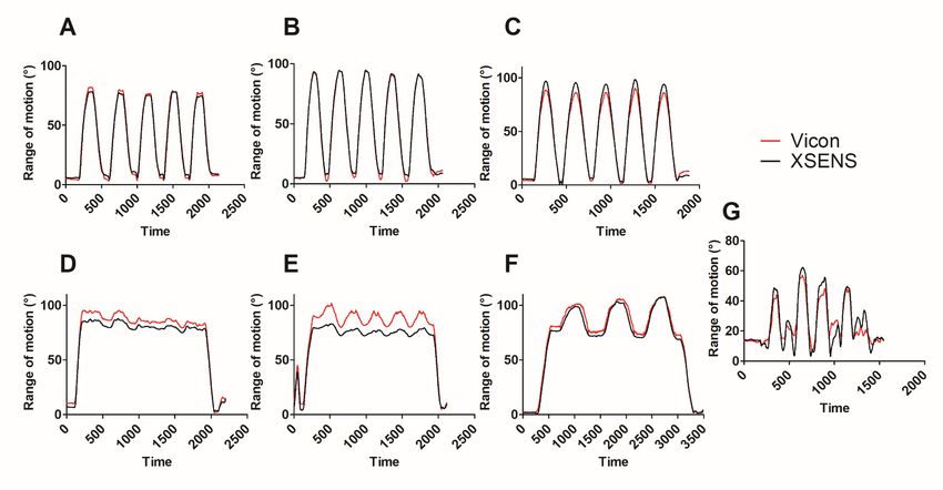
Figure 3. Anterior deltoid EMG activity (RMS) for a typical subject for different shelves (A: Lower shelf, B: Medium shelf, C: Higher shelf) and weights (Lower shelf:
Figure 3. Anterior
L1 = 2.3 deltoid
kg, L2 = 6.8 EMG
kg, L3 activity
= 13.6 kg, L4(RMS) for aMedium
= 22.7kg; typical subject for=different
shelf: M1 shelves
2.3 kg, M2 (A: Higher
= 6.8 kg; Lower shelf:
shelf, H1
B: Medium
= 2.3 kg,shelf,
H2 = C:
6.8Higher
kg). shelf) and weights (Lower
shelf: L1 = 2.3 kg, L2 = 6.8 kg, L3 = 13.6 kg, L4 = 22.7kg; Medium shelf: M1 = 2.3 kg, M2 = 6.8 kg; Higher shelf: H1 = 2.3 kg, H2 = 6.8 kg).Sensors 2019, 19, 1885 9 of 14
Table 3. Results for anterior and middle deltoids’ EMG activity (p-value and effect size for area under the curve and peak EMG activity).
One-Way Two-Way Post-Hoc Analysis
ANOVA One-Way ANOVA Two-Way L1 L1 L2 L2 M1 M1 M2 H1
ANOVA η2 ANOVA η2 L1 L1 L1 L2 L2 L3
p-value p-value vs vs vs vs vs vs vs vs
vs L2 vs L3 vs L4 vs L3 vs L4 vs L4
M1 H1 M2 H2 M2 H1 H2 H2
Weight effectSensors 2019, 19, 1885 10 of 14
3.2.2. Middle Deltoid
When comparing the four weights for the lower shelf, no significant difference was found for
middle deltoid EMG activity (RMS: p = 0.159, η2 = 0.92, peak: p = 0.24, η2 = 0.09). When comparing
results for the two lower weights on the three shelf heights, there was a significant effect found in the
weight increase (RMS: p = 0.002, η2 = 0.523) and height increase (RMS: p = 0.028, η2 = 0.241), but only
for the higher shelf (H1 vs H2: p < 0.001). Also, for the RMS, a weight, a height and a weight x height
interaction effect were observed (weight: p = 0.002, η2 = 0.523; height: p = 0.028, η2 = 0.241; weight x
height: p = 0.003, η2 = 0.352; height). For peak EMG activity, only a weight x height interaction was
observed (p = 0.001, η2 = 0.388). Post-hoc analysis identified significant differences between L2-M2 (p:
RMS = 0.002; peak = 0.013), M2-H2 (p: RMS = 0.004; peak = 0.015) and H1-H2 (p: RMS = 0.00045; peak
= 0.001) for RMS and peak EMG. Also, a significant difference for L1-L4 peak EMG was observed (p =
0.027; for details, see Table 1; Table 2).
4. Discussion
As we hypothesized, the results from this study confirm that the two commercial wireless systems
(Xsens MVN and Trigno EMG) are valid tools to assess shoulder movements and muscle activity
during simple arm elevations and complex lifting tasks. The comparison between Xsens and Vicon
shows that IMUs are valid to assess shoulder elevation ROM during simple arm movements (r ≥ 0.917;
RMSE ≤ 12.82◦ ; average error of estimate ≤ 10.15◦ ) and complex lifting tasks, regardless of the height
at which the crates are placed on the shelves of Valpar 19 (r ≥ 0.839; RMSE ≤ 12.68◦ , average error of
estimate ≤ 10.24◦ ). As for EMG activity, the anterior deltoid should be considered as an interesting
muscle to discriminate between the different levels of physical work demands (sedentary, light,
moderate and high) when performing tasks that necessitate forceful contractions in the sagittal plane.
The middle deltoid EMG activity varied depending on the weight and height at which the crates
were placed, but was less discriminant than the anterior deltoid (significant results only for L2-H2,
M2-H2 and H1-H2) for tasks performed in the sagittal plane. RMS and peak EMG activity were both
discriminative. However, the discriminative potential of RMS is larger and it should therefore be
preferred over peak activity to discriminate between the different physical work demands.
Assessing arm elevation is challenging since the shoulder is a highly mobile joint and necessitates
3D analysis. As mentioned above, IMUs could potentially be useful for workplace evaluations to
quantify physical work demands over extended periods of time. However, no clear conclusion has
previously been reached on IMU validity for arm elevation due to the heterogeneity of results and
the small number of studies addressing their validity during complex tasks [10]. Our results confirm
that IMUs are valid to assess arm elevation, although the correlations with the Vicon were higher
for simple movements than for complex tasks. These results are compatible with previous studies
in which the validity of maximal range in shoulder elevation (simple task) was evaluated [27–30].
However, two studies reported lower RMSE (ranging from 1.13 to 2.38◦ ) than those reported herein
with regards to arm elevation during simple movements [31,32]. These differences could be explained
by several factors including differences in the biomechanical models used, sensor positioning and
experimental protocols. Indeed, one study [31] reported results with the elbow constrained in the
neutral position, thereby reducing errors in sensor misalignment due to soft tissue artifact. The second
article [32] presented results during isolated wrist movements with small movements at shoulder joint.
In comparison, our results are more representative of movement patterns seen in real life since the
motion was not restricted.
For complex tasks, two previous studies evaluated lifting tasks similar to those presented herein;
however, their results were quite different [14,15]. One study [15] showed RMSE results lower than
ours in regards to MVN Xsens. A post-processing step was however added to reduce the error due to
the choice of the biomechanical model. The RMSE that they reported without removing biomechanical
model variability was much larger than the RMSE reported in this study (19.7◦ vs. 2.9◦ , respectively).
The other study [15] reported higher errors of measurement and more variability (ranging from 9.6 toSensors 2019, 19, 1885 11 of 14
33.1◦ ), but their results originate from using an older version of the XSens IMUs which could explain
the discrepancy.
It is well-known that performing physically demanding tasks at a shoulder level represents an
important risk factor for developing work-related shoulder disorders [3]. Our results demonstrate that
anterior deltoid muscle activity can be a good indicator of the physical demands on the shoulder since
RMS and peak EMG activity can discriminate between different work demand levels (sedentary, light,
moderate and heavy) [23]. These results correspond to those of a previous study from Silvetti et al,
who observed an effect of weight and height on anterior deltoid activity [33]. Unlike this work, their
study did not show a weight x height interaction, which could be explained by two main factors: 1)
they placed the crates at varying distances from the shelf edge depending on the height at which it had
to be placed (whereas we always used the same distance), and 2) the difference in weight between the
two objects carried by the participants of Silvetti’s study was smaller (Silvetti’s study: 6/8 kg compared
to 2.3/6.8 kg in this study). Furthermore, two other studies have shown results (height and weight
effects) similar to ours regarding the anterior deltoid muscle, but they also found larger differences
across weight lifted for the middle deltoid [19,20]. This difference could be explained by the fact that
we only analyzed the lifting phase; they demonstrated higher co-activation during dropping than
lifting. However, similar to this study, they have shown discriminative anterior deltoid potential.
Therefore the results of the present study show that is possible to identify physical work demands of a
specific task by analyzing muscle activity.
4.1. Technical Issues to Be Considered Prior to Clinical Implantation
Other wearable sensors are also available to collect different types of bio-signals on workers (i.e.,
photosensors (light level), heart rate and skin conductance sensors (for stress and other physiological
/psychological states), global positioning systems (GPS; mobility, environment). However, we chose to
focus on the validation of IMU and EMG sensors as we consider them to have the highest potential for
directly quantifying physical work demands at the upper extremities (e.g., number of arm elevations,
time spent with the arm elevated, forceful work and muscle fatigue) [11]. Wearable technologies have
greatly improved over the last few years. Progress has been made to improve software accuracy
(e.g., development of better algorithms) and hardware stability (e.g., lesser sensitivity to magnetic
disturbances [34]). Furthermore, real-time processing now allows to obtain more accurate 3D data
with negligible processing delay. Systems have also significantly improved their portability given the
development of smaller, lighter, wireless units. For all of these reasons, the two systems are potentially
useful tools for clinicians. Still, certain technical concerns should be addressed before implementing
them for workplace assessment. Indeed, the software used for data collection and the post-processing
needed to obtain interpretable data are complex, time-consuming and not user-friendly (requiring
technical competency in signal processing). The current technology therefore needs to be improved
before clinical uptake. Also, and more specifically for IMUs, more flexibility in the number of sensors
needed to collect meaningful and accurate data should be addressed as Xsens software requires at
least seven IMUs. Nevertheless, the results for simple and complex movements are very promising
for clinical use as the reported errors (RMSE ≤ 12.68◦ ) are lower than the errors of inclinometers and
goniometers currently used in clinic (95% limits of agreement ranged from 2◦ to 20◦ ) [35]. Moreover,
inclinometers and goniometers do not allow continuous monitoring during the task.
4.2. Study Limitations
The evaluation of shoulder kinematics is complex, and certain limitations need to be considered in
our protocol. First, the calibration of both systems had to be performed at different body positions due
to software requirements (N-pose for Xsens and anatomical calibration for Vicon) which can increase
the error of measurement as validity is dependent on the calibration method [36]. We performed a
post-processing re-initialization in N-pose to diminish the errors. Secondly, only arm elevation wasSensors 2019, 19, 1885 12 of 14
analyzed. This choice was motivated by the clinical relevance of this movement, since the range of
motion in shoulder elevation is usually targeted in workplace prevention/interventions.
However previous studies suggest that the errors are usually higher for the other two rotations [10],
and therefore the current results cannot be extended to other types of movement. For EMG activity
recording, there is one main consideration regarding the protocol used. Considering the high prevalence
of rotator cuff injury, it would have been interesting to evaluate rotator cuff muscles [37]. However,
surface EMG recordings for rotator cuff muscles is not reliable since they are deep muscles [38].
Anterior and middle deltoids were the most appropriate superficial muscles to evaluate physical
work demands as they showed greater amount of fatigue on EMG recordings in comparison to other
superficial muscles in a variety of tasks [39–41]. Finally, this study is the first step in the validation
process of these wearable sensors. Indeed, in the present research, the suitability of combining IMU
and EMG sensors has been demonstrated during simulated working tasks. The next step will be to
validate their use in the workplace, in the frame of actual work situations.
5. Conclusions
In conclusion, wireless EMG and IMU systems can be used to assess two important risk factors
during simple and complex working tasks: 1) working with arms above the head and 2) using forceful
contractions. IMUs reported lower errors of measurement compared to most tools currently used in
the clinic (goniometer and inclinometer). Still, certain improvements need to be implemented for the
systems to become more accessible and easier to use by clinicians. In addition, anterior deltoid muscle
activity was shown to be a good indicator of physical demand. It is therefore a potentially useful
clinical indicator to identify physically demanding tasks/jobs and to quantify muscle activity in the
workplace. However, although these results are promising for socio-professional rehabilitation, studies
performed in the workplace are now needed to support their suitability in actual work situations.
Author Contributions: Data curation, I.P., M.B.; Formal analysis: I.P., M.B.; Methodology, I.P., J.-S.R., L.J.B., C.M.;
Roles/Writing-original draft, I.P.; Software: M.B., L.J.B., A.C.-L.; Writing- review & editing, M.B., C.M., A.C.-L.,
L.J.B., J.-S.R.; Funding acquisition: L.J.B., J.-S.R.; Project administration: J.-S.R.
Funding: I.P. is supported by a Studentship from the Canadian Institutes of Health Research (CIHR). J.-S.R. is
supported by a salary award from CIHR and C.M. by an Emeritus Research Scholar from the Fonds de recherche
du Québec—Santé (FRQS). The study was supported in part by the Sentinel North program of Université Laval
(Canada First Research Excellence Fund) and by the Natural Sciences and Engineering Research Council of Canada
(NSERC).
Conflicts of Interest: The authors declare no conflict of interest.
References
1. Bongers, P.M.; Ijmker, S.; van den Heuvel, S.; Blatter, B.M. Epidemiology of work related neck and upper limb
problems: Psychosocial and personal risk factors (Part I) and effective interventions from a bio behavioural
perspective (Part II). J. Occup. Rehabil. 2006, 16, 272–295. [CrossRef]
2. HSE. Upper Limb Disorders in the Workplace; Health and Safety Executive (HSE): Surrey, UK, 2002; p. 89.
3. Linaker, C.H.; Walker-Bone, K. Shoulder disorders and occupation. Best Pract. Res. Clin. Rheumatol. 2015, 29,
405–423. [CrossRef] [PubMed]
4. Kennedy, C.A.; Amick Iii, B.C.; Dennerlein, J.T.; Brewer, S.; Catli, S.; Williams, R.; Serra, C.; Gerr, F.; Irvin, E.;
Mahood, Q.; et al. Systematic Review of the Role of Occupational Health and Safety Interventions in the
Prevention of Upper Extremity Musculoskeletal Symptoms, Signs, Disorders, Injuries, Claims and Lost Time.
J. Occup. Rehabil. 2010, 20, 127–162. [CrossRef] [PubMed]
5. Van Rijn, R.M.; Huisstede, B.M.; Koes, B.W.; Burdorf, A. Associations between work-related factors and
specific disorders of the shoulder—A systematic review of the literature. Scand. J. Work Environ. Health 2010,
36, 189–201. [CrossRef] [PubMed]
6. Van der Molen, H.F.; Foresti, C.; Daams, J.G.; Frings-Dresen, M.H.W.; Kuijer, P. Work-related risk factors for
specific shoulder disorders: A systematic review and meta-analysis. Occup. Environ. Med. 2017, 74, 745–755.
[CrossRef] [PubMed]Sensors 2019, 19, 1885 13 of 14
7. Hanvold, T.N.; Wærsted, M.; Mengshoel, A.M.; Bjertness, E.; Veiersted, K.B. Work with prolonged arm
elevation as a risk factor for shoulder pain: A longitudinal study among young adults. Appl. Ergon. 2015, 47,
43–51. [CrossRef]
8. Anglin, C.; Wyss, U.P. Review of arm motion analyses. Proc. Inst. Mech. Eng. Part H J. Eng. Med. 2000, 214,
541–555. [CrossRef]
9. Valevicius, A.M.; Jun, P.Y.; Hebert, J.S.; Vette, A.H. Use of optical motion capture for the analysis of normative
upper body kinematics during functional upper limb tasks: A systematic review. J. Electromyogr. Kinesiol.
2018, 40, 1–15. [CrossRef] [PubMed]
10. Cuesta-Vargas, A.I.; Galán-Mercant, A.; Williams, J.M. The use of inertial sensors system for human motion
analysis. Phys. Ther. Rev. 2010, 15, 462–473. [CrossRef]
11. Dejnabadi, H.; Jolles, B.M.; Casanova, E.; Fua, P.; Aminian, K. Estimation and visualization of sagittal
kinematics of lower limbs orientation using body-fixed sensors. IEEE Trans. Biomed. Eng. 2006, 53, 1385–1393.
[CrossRef]
12. Poitras, I.; Dupuis, F.; Bielmann, M.; Campeau-Lecours, A.; Mercier, C.; Bouyer, L.J.; Roy, J.S. Validity and
Reliability of Wearable Sensors for Joint Angle Estimation: A Systematic Review. Sensors 2019, 19, 1555.
[CrossRef] [PubMed]
13. Halder, M.A.; Kuhl, G.S.; Zobitz, E.M.; Larson, N.D.; An, N.K. Effects of the Glenoid Labrum and
Glenohumeral Abduction on Stability of the Shoulder Joint Through Concavity-Compression: An in
Vitro Study. J. Bone Jt. Surg. 2001, 83, 1062–1069. [CrossRef]
14. Robert-Lachaine, X.; Mecheri, H.; Larue, C.; Plamondon, A. Validation of inertial measurement units with an
optoelectronic system for whole-body motion analysis. Med. Biol. Eng. Comput. 2016. [CrossRef]
15. Godwin, A.; Agnew, M.; Stevenson, J. Accuracy of inertial motion sensors in static, quasistatic, and complex
dynamic motion. J. Biomech. Eng. 2009, 131, 114501. [CrossRef]
16. Fantozzi, S.; Giovanardi, A.; Magalhaes, F.A.; Di Michele, R.; Cortesi, M.; Gatta, G. Assessment of
three-dimensional joint kinematics of the upper limb during simulated swimming using wearable
inertial-magnetic measurement units. J. Sports Sci. 2016, 34, 1073–1080. [CrossRef] [PubMed]
17. Ertzgaard, P.; Öhberg, F.; Gerdle, B.; Grip, H. A new way of assessing arm function in activity using kinematic
Exposure Variation Analysis and portable inertial sensors—A validity study. Man. Ther. 2016, 21, 241–249.
[CrossRef]
18. Wattanaprakornkul, D.; Cathers, I.; Halaki, M.; Ginn, K.A. The rotator cuff muscles have a direction specific
recruitment pattern during shoulder flexion and extension exercises. J. Sci. Med. Sport 2011, 14, 376–382.
[CrossRef]
19. Blache, Y.; Dal Maso, F.; Desmoulins, L.; Plamondon, A.; Begon, M. Superficial shoulder muscle co-activations
during lifting tasks: Influence of lifting height, weight and phase. J. Electromyogr. Kinesiol. 2015, 25, 355–362.
[CrossRef]
20. Blache, Y.; Desmoulins, L.; Allard, P.; Plamondon, A.; Begon, M. Effects of height and load weight on shoulder
muscle work during overhead lifting task. Ergonomics 2014, 58, 748–761. [CrossRef] [PubMed]
21. MVN User Manual-User Guide MVN, MVN BIOMECH MVN Link, MVN Awinda; Xsens Technologies B.V.:
Enschede, The Netherlands, 2015.
22. Hermens, H.J.; Freriks, B.; Merletti, R.; Stegeman, D.; Blok, J.; Rau, G.; Disselhorst-Klug, C.; Hägg, G.
European Recommendations for Surface Electromyography: Results of the SENIAM Project; Roessingh Research
and Development: Enschede, The Netherlands, 1999.
23. Matheson, L.N. The functional capacity evaluation. In Disability Evaluation; Smith, G.A.S.D.G., Ed.; Mosby
Yearbook: Chicago, IL, USA, 2003.
24. Grabiner, M.D.; Owings, T.M. EMG differences between concentric and eccentric maximum voluntary
contractions are evident prior to movement onset. Exp. Brain Res. 2002, 145, 505–511. [CrossRef]
25. Koo, T.K.; Li, M.Y. A Guideline of Selecting and Reporting Intraclass Correlation Coefficients for Reliability
Research. J. Chiropr. Med. 2016, 15, 155–163. [CrossRef] [PubMed]
26. Mukaka, M.M. A guide to appropriate use of Correlation coefficient in medical research. Malawi Med J. 2012,
24, 69–71. [PubMed]
27. Bouvier, B.; Duprey, S.; Claudon, L.; Dumas, R.; Savescu, A. Upper Limb Kinematics Using Inertial and
Magnetic Sensors: Comparison of Sensor-to-Segment Calibrations. Sensors 2015, 15, 18813–18833. [CrossRef]
[PubMed]Sensors 2019, 19, 1885 14 of 14
28. Cutti, A.G.; Giovanardi, A.; Rocchi, L.; Davalli, A.; Sacchetti, R. Ambulatory measurement of shoulder
and elbow kinematics through inertial and magnetic sensors. Med. Biol. Eng. Comput. 2008, 46, 169–178.
[CrossRef] [PubMed]
29. Kumar, Y.; Yen, S.C.; Tay, A.; Lee, W.; Gao, F.; Zhao, Z.; Li, J.; Hon, B.; Xu, T.T.M.; Cheong, A.; et al. Wireless
wearable range-of-motion sensor system for upper and lower extremity joints: A validation study. Healthc.
Technol. Lett. 2015, 2, 12–17. [CrossRef]
30. Pérez, R.; Costa, U.; Torrent, M.; Solana, J.; Opisso, E.; Cáceres, C.; Tormos, J.M.; Medina, J.; Gómez, E.J.
Upper limb portable motion analysis system based on inertial technology for neurorehabilitation purposes.
Sensors 2010, 10, 10733–10751. [CrossRef]
31. Barraza Madrigal, J.A.; Cardiel, E.; Rogeli, P.; Leija Salas, L.; Munoz Guerrero, R. Evaluation of suitability
of a micro-processing unit of motion analysis for upper limb tracking. Med. Eng. Phys. 2016, 38, 793–800.
[CrossRef]
32. Zhou, H.; Hu, H. Reducing drifts in the inertial measurements of wrist and elbow positions. IEEE Trans.
Instrum. Meas. 2010, 59, 575–585. [CrossRef]
33. Silvetti, A.; Mar, S.; Ranavolo, A.; Forzano, F.; Iavicoli, S.; Conte, C.; Draicchio, F. Kinematic and
electromyographic assessment of manual handling on a supermarket green grocery shelf. Work 2015,
51, 261–271. [CrossRef]
34. Robert-Lachaine, X.; Mecheri, H.; Larue, C.; Plamondon, A. Effect of local magnetic field disturbances on
inertial measurement units accuracy. Appl. Erg. 2017, 63, 123–132. [CrossRef]
35. Kolber, M.J.; Hanney, W.J. The reliability and concurrent validity of shoulder mobility measurements using a
digital inclinometer and goniometer: A technical report. Int. J. Sports Phys. 2012, 7, 306–313.
36. Ligorio, G.; Zanotto, D.; Sabatini, A.M.; Agrawal, S.K. A novel functional calibration method for real-time
elbow joint angles estimation with magnetic-inertial sensors. J. Biomech. 2017, 54, 106–110. [CrossRef]
[PubMed]
37. Van der Windt, D.A.; Koes, B.W.; de Jong, B.A.; Bouter, L.M. Shoulder disorders in general practice: Incidence,
patient characteristics, and management. Ann. Rheum. Dis. 1995, 54, 959–964. [CrossRef] [PubMed]
38. Johnson, V.L.; Halaki, M.; Ginn, K.A. The use of surface electrodes to record infraspinatus activity is not
valid at low infraspinatus activation levels. J. Electromyogr. Kinesiol. 2011, 21, 112–118. [CrossRef]
39. Nieminen, H.; Takala, E.P.; Niemi, J.; Viikari-Juntura, E. Muscular synergy in the shoulder during a fatiguing
static contraction. Clin. Biomech. 1995, 10, 309–317. [CrossRef]
40. Minning, S.; Eliot, C.A.; Uhl, T.L.; Malone, T.R. EMG analysis of shoulder muscle fatigue during resisted
isometric shoulder elevation. J. Electromyogr. Kinesiol. 2007, 17, 153–159. [CrossRef]
41. Hawkes, D.H.; Alizadehkhaiyat, O.; Kemp, G.J.; Fisher, A.C.; Roebuck, M.M.; Frostick, S.P. Electromyographic
assessment of muscle fatigue in massive rotator cuff tear. J. Electromyogr. Kinesiol. 2015, 25, 93–99. [CrossRef]
[PubMed]
© 2019 by the authors. Licensee MDPI, Basel, Switzerland. This article is an open access
article distributed under the terms and conditions of the Creative Commons Attribution
(CC BY) license (http://creativecommons.org/licenses/by/4.0/).You can also read