Partial Achilles Tendon Rupture-A Neglected Entity: A Narrative Literature Review on Diagnostics and Treatment Options - MDPI
←
→
Page content transcription
If your browser does not render page correctly, please read the page content below
Journal of
           Clinical Medicine

Review
Partial Achilles Tendon Rupture—A Neglected Entity:
A Narrative Literature Review on Diagnostics and
Treatment Options
Matthias Gatz 1, *, Christoph Spang 2,3 and Håkan Alfredson 4,5
 1    Department of Orthopedics, University Hospital RWTH Aachen, 52074 Aachen, Germany
 2    Department of Integrative Medical Biology, Anatomy Section, Umeå University, 90187 Umeå, Sweden;
      christoph.spang@umu.se
 3    Alfen Spine Center, 97080 Würzburg, Germany
 4    Sports Medicine Unit, Department of Community Research and Rehabilitation, Umeå University,
      90187 Umeå, Sweden; hakan.alfredson@umu.se
 5    Institute of Sports Exercise and Health, University College London Hospitals, London W1T 7HA, UK
 *    Correspondence: mgatz@ukaachen.de; Tel.: +49-241-7501-438

 Received: 23 September 2020; Accepted: 13 October 2020; Published: 21 October 2020

 Abstract: Partial ruptures in the Achilles tendon are rather uncommon and are often misinterpreted
 as aggravated Achilles tendinopathy, and not always considered as a differential diagnosis. The aim
 of this literature review was to characterize typical symptoms, to provide an overview of available
 diagnosis and treatment options, and to give reference points for future research. There were
 few studies and sparse knowledge of scientific value, making it difficult to give evidence-based
 recommendations. Based on the few studies and the authors’ clinical experience, a diagnosis should
 be based on a patient’s history with a typical sharp onset of pain and inability to fully load the tendon.
 Previous intratendinous cortisone injections might be present. Clinical findings are a localized tender
 region in the tendon and often weakness during heel raises. Ultrasound and Doppler examinations
 show a region with an irregular and bulging superficial tendon line, often together with localized high
 blood flow. Magnetic resonance Imaging (MRI) shows a hyperintense signal in the tendon on T1 and
 T2-weighted sequences. First-line therapy should be a conservative approach using a 2 cm heel lift for
 the first 6 weeks and avoiding tendon stretching (for 12 weeks). This is followed by a reduced heel lift
 of 1 cm and progressive tendon loading at weeks 7–12. After 12 weeks, the heel lift can be removed
 if pain-free, and the patient can gradually start eccentric exercises lowering the heel below floor
 level and gradually returning to previous sport level. If conservative management has a poor effect,
 surgical exploration and the excision of the partial rupture and suturing is required. Augmentation
 procedures or anchor applications might be useful for partial ruptures in the Achilles insertion,
 but this depends on the size and exact location. After surgery, the 12 to 14-week rehabilitation
 program used in conservative management can be recommended before the patient’s return to full
 tendon loading activities.

 Keywords: partial rupture; partial tear; achilles; tendinopathy

1. Introduction
      Achilles tendinopathy is the most common reason for Achilles tendon pain and can be categorized
into insertional, midportion, or plantaris tendon-related Achilles tendinopathy [1]. A further pathology
is the acute Achilles tendon rupture with a sudden onset of pain and functional disability. For both
Achilles tendinopathy and Achilles tendon rupture, there are several evidence-based nonsurgical and

J. Clin. Med. 2020, 9, 3380; doi:10.3390/jcm9103380                               www.mdpi.com/journal/jcm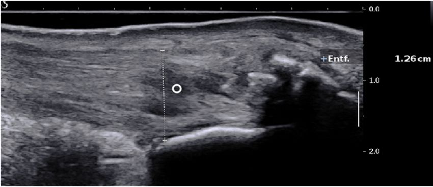
J. Clin. Med. 2020, 9, 3380                                                                           2 of 10

surgical treatment options and diagnostic pathways based on meta-analyses, randomized controlled
trials, and consensus statements [2–4].
      In comparison, a partial Achilles tendon tear is rather uncommon, is not always considered
as a differential diagnosis, and might be misinterpreted as aggravated Achilles tendinopathy [5].
While Achilles tendinopathy and Achilles tendon rupture can be easily differentiated from each other,
partial Achilles tendon rupture meets the symptom criteria of both [6,7]. This might lead to difficulties
in diagnosing and choosing the most appropriate treatment option (e.g., load vs. immobilization).
The physician might end up being caught between two stools. To the best of our knowledge, there is
no consensus regarding diagnosis and treatment for partial Achilles ruptures.
      The aim of this literature review is to characterize typical symptoms, to provide an overview of
available diagnosis and treatment options, and to give reference points for future research.

2. Methods
      The literature search included acute partial Achilles tendon rupture as well as partial tendon
tears together with a history of tendinopathy. A review of the literature was performed including all
articles published in PubMed, Scopus, and Google Scholar until August 2020. The following search
terms were used alone and in combination: “Achilles”, “partial”, “partial rupture”, and “partial tear”;
and the articles were evaluated with regard to the following research aspects: definition, epidemiology,
etiology, pathogenesis, diagnosis, and treatment.
      Articles of all clinical trial levels in English, German, Swedish, and Portuguese were included.
After assessing all abstracts (n = 388), the full text and the bibliographies of the relevant articles were
analyzed with regard to the questions posed (n = 56). Articles including animal or biomechanical studies
or outcomes without the area of interest were excluded (n = 22). Due to the advancements promoted
by the scientific progress of imaging modalities, surgical techniques, and improved understanding
of tendon load for rehabilitation, mainly articles from the years 1990–2020 were included. However,
after scanning bibliographies of the relevant articles (n = 4), articles published before 1990 were added.
The final literature research revealed 38 relevant articles.

3. Results

3.1. Definitions
      A partial tear is defined as a partial discontinuation of the Achilles tendon and usually has an acute
onset. There is often pain during loading and a feeling of weakness. Patients typically maintain the
ability to train but do not reach maximal loading [8]. Physically active patients suffer from permanent
symptoms (39%) or initial warm-up pain after a period of resting (61%), which might decrease or
increase during physical activity [6]. Tendon thickening, localized pain at a specific region, occasionally
a palpable tendon discontinuity, loss of function, and limping are clinical findings [6,9,10].

3.2. Etiology
     Most partial tears are caused by an overload of the tendon tissue and occur in areas afflicted with
tendinopathic tissue changes [11]. The precise relationship between tendinopathy and partial tears
and the pathogenesis are still not fully understood. According to recent studies, the pathogenesis
of tendinopathy is more likely to be a result of altered tissue homeostasis by repeated mechanical
overload, rather than a repair response to a partial rupture in the tendon [12]. Thus, it seems more
likely that partial ruptures appear in tissue areas that had previously developed tendinopathic features.
In fact, in recent studies non-homogenous stress at subclinical or clinically symptomatic tendinopathic
tissue is seen as the underlying mechanism for the development of an acute partial rupture, mainly in
the midportion of the Achilles tendon [13]. At the tendon insertion, partial ruptures might additionally
be caused by a tendon impingement of bony prominences in the calcaneus [14].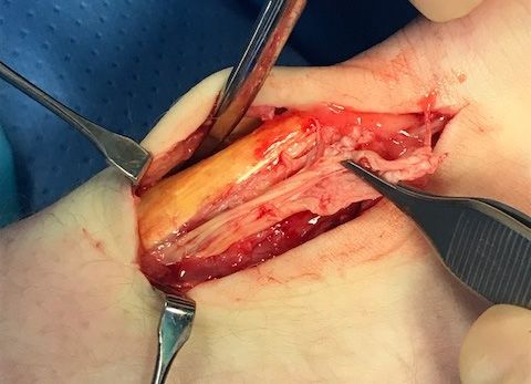
J. Clin. Med. 2020, 9, 3380 3 of 10
Injections into the tendon, especially cortisone, are known to have a significant influence on
the development of partial and total tendon ruptures by starting local degenerative processes [15].
In some studies, a previous intratendinous cortisone injection was observed in nearly 50% of partial
ruptures [9,10]. Similar figures were seen for partial tears and the use of fluorquinolone medication [16].
In general, for surgical reconstruction it has to be considered that the Achilles tendon is formed
by the confluence of the tendons of the soleus and both gastrocnemius muscles. These subtendons
intertwine and twist towards their distal insertion and have a specific arrangement in the midportion
and the insertion of the calcaneus [5,17]. Based on the anatomic region within the tendon, partial
ruptures might be assigned to the specific subtendon, which might lead to an isolated partial hypotrophy
of the gastrocsoleus muscle complex with reduced voluntary electromyography activity [17]. Partial
tears mostly occur in the posterior mid-tendon 3–4 cm above the superior calcaneus, whereas a further
study demonstrated that intratendinous tears are mostly anterior and medial, which mostly present
fibers of the soleus and lateral gastrocnemius components [8,18,19].
3.3. Classification
Smigielski classified partial injuries of the Achilles tendon based on histopathological aspects with
further subgroups based on the amount of damaged bundles and the exact origin of the gastrocsoleus
complex [5]:
(a) acute injury with fresh collagen disruption representing an acute microinjury, with detectable
extravascular erythrocytes and an early reparative process.
(b) chronic injury with fatty metaplasia and infiltrated vessels showing a reduced potential
for self-healing.
Recently, it has been suggested to distinguish between intratendinous tears and partial tears
by defining the latter as extending to the periphery of the tendon, in contrast to an intratendinous
tear, which does not reach the peripheral edge of the tendon and extends in the majority of cases
longitudinally [8].
In histopathological analysis, degenerative changes, such as collagen disorientation and fiber
separation, with increased mucoid ground substance, fibrin deposits, hypercellularity, and neovascularization
are present in partial ruptures [11]. Since these are also current findings in tendinosis, it has been
advocated that partial ruptures are rather microlesions representing an advanced stage of tendinosis [11].
However, granulation tissue, fibroblastic and myofibroblastic proliferation, and hemorrhage are more
frequently associated with partial ruptures than with tendinopathy [11].
3.4. Epidemiology
Affected patients are mostly younger males with a higher sports level than patients with midportion
tendinopathy or Achilles tendon rupture [8,9,11,20–22]. However, in top athletes, partial ruptures
might not be that common. A Champions League injury study with an 11-year follow-up stated a
total of seven total ruptures and two partial ruptures. Furthermore, at the London 2012 Olympics,
four partial ruptures and one total rupture were seen in an imaging study [23].
In some imaging studies, partial rupture might be present in up to 25% of cases with Achilles
tendinopathy, but a possible selection bias has to be considered since a further imaging study reported
an incidence rate of only 1.6% [24]. Additionally, studies on surgically treated patients with Achilles
tendinopathy have reported a high partial rupture rate of 19% and 25%, but also a lower rate of 4% in a
large cohort with 771 surgically treated tendons [1,11,22].
Therefore, the exact prevalence is unknown and might differ between several cohorts. Furthermore,
the discrepancy between the studies might be due to different definitions of a partial rupture and the
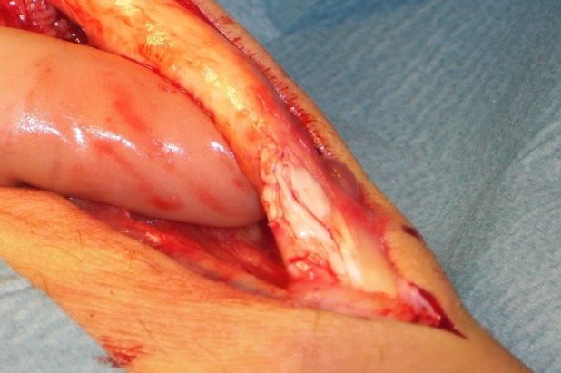
method of detection. In surgical studies, the partial rupture is often diagnosed by the surgeon based
on how the tissue looks macroscopically (Figure 1). Some studies use ultrasound or MRI for diagnosis.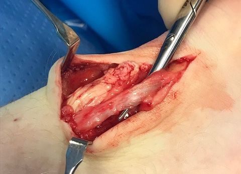
J. Clin. Med. 2020, 9, 3380 4 of 10
J. Clin. Med. 2020, 9, x 4 of 10
(a) (b)
Figure
Figure 1.
1. (a)
(a)Surgical
Surgicalobservations
observationsinin patients
patients with
with partial
partial Achilles
Achilles tendon
tendon ruptures;
ruptures; (b)
(b) higher
higher
magnification of longitudinal partial Achilles tendon rupture.
magnification of longitudinal partial Achilles tendon rupture.
3.5.Imaging
3.5. Imaging
Differentiation between
Differentiation between full-thickness
full-thicknesstears tears andand partial
partial tears
tears can
can be
be adequately
adequately assessed
assessed withwith
ultrasound (n = 26, accuracy 92%, sensitivity 100%, and specificity
ultrasound (n = 26, accuracy 92%, sensitivity 100%, and specificity 83%), showing a significant83%), showing a significant difference
in tendon in
difference thickness,
tendon an increased
thickness, anposterior
increasedacoustic
posterior shadowing at the side of
acoustic shadowing at tendon
the sideabnormality,
of tendon
and tendon retraction in case of a full-thickness tear [7]. Comparing
abnormality, and tendon retraction in case of a full-thickness tear [7]. Comparing findings of findings of preoperative ultrasound
to findings at ultrasound
preoperative surgery, Kälebo et al. reported
to findings an excellent
at surgery, Kälebodiagnostic accuracy
et al. reported anofexcellent
95% for ultrasound
diagnostic
(sensitivity
accuracy of0.94;
95%specificity 1.00) [25].
for ultrasound Further studies
(sensitivity revealed corresponding
0.94; specificity preoperative
1.00) [25]. Further studiesfindings
revealed in
8/11 patients (sensitivity 72%) or 10/11 (sensitivity 90%) [26,27]. However,
corresponding preoperative findings in 8/11 patients (sensitivity 72%) or 10/11 (sensitivity 90%) these good results might be
based on
[26,27]. a selection
However, biasgood
these in a results
surgically treated,
might be basedsmalloncollective,
a selection which might
bias in have larger
a surgically and small
treated, hence
easier-to-detect
collective, whichpartial
might ruptures.
have larger and hence easier-to-detect partial ruptures.
Themajority
The majorityof ofstudies
studiesreport
reportthat thatwith
withultrasound
ultrasoundititisis difficult
difficult to
to distinguish
distinguish partial
partialruptures
ruptures
from focal
from focal degenerative
degenerativechanges, changes,since sincepartial
partialruptures
ruptures appear
appear with a wavy,
with a wavy,irregular echoecho
irregular pattern with
pattern
accompanying focal hypoechogenic areas, detectable neovascularization,
with accompanying focal hypoechogenic areas, detectable neovascularization, and tendon and tendon thickening that
are also findings in Achilles tendinopathy [19,23,26,28,29]. A more specific
thickening that are also findings in Achilles tendinopathy [19,23,26,28,29]. A more specific finding finding might be a disrupted
dorsalbe
might Achilles tendondorsal
a disrupted borderAchilles
[19,28] (Figure
tendon 2a).borderThe[19,28]
detection of proximal
(Figure 2a). Thepartial ruptures
detection close to
of proximal
the myotendinous junction and the differentiation between older partial
partial ruptures close to the myotendinous junction and the differentiation between older partial ruptures and intratendinous
tendinopathy
ruptures are challengingtendinopathy
and intratendinous with ultrasound are[28,30] (Figurewith
challenging 2b). ultrasound
In MRI, a partial
[28,30]tear is defined
(Figure 2b). Inas
tendon
MRI, thickening
a partial tear is with a hyperintense
defined as tendon signalthickeningon T1withand aa hyperintense
strong hyperintensesignal on signal
T1 andon magnetic
a strong
resonance (MR)
hyperintense images
signal on with fluid-sensitive
magnetic resonance(T2-weighted
(MR) images and
withinversion recovery)(T2-weighted
fluid-sensitive sequences [29–31] and
(Figure 3).recovery)
inversion Typically, sequences
signal intensity [29–31] is analogous
(Figure 3). to Typically,
free fluid. A hyperintense
signal intensityarea directly located
is analogous to freeat
the tendon border should be interpreted as a partial rupture [30].
fluid. A hyperintense area directly located at the tendon border should be interpreted as a partialDue to muscle inactivity, isolated
fatty degeneration
rupture [30]. Due toand edema
muscle in the calf
inactivity, muscles
isolated might
fatty represent aand
degeneration subsequent
edema in state
the of a partial
calf muscles or
total rupture
might represent [31].a The sensitivity
subsequent of MRI
state of a in detecting
partial partial
or total tears is[31].
rupture highThe (positive predictive
sensitivity of MRI value
in
n =
detecting partial tears is high (positive predictive value 0.94, n = 18), but studies directly comparinga
0.94, 18), but studies directly comparing the accuracy of MRI and ultrasound are rare [32]. In
prospective
the accuracystudy of MRI of Kayser et al., only one-fifth
and ultrasound are rareof[32].partial
In lesions were noticed
a prospective studywith B-Modeetultrasound
of Kayser al., only
whereas of
one-fifth MRI detected
partial lesionsfive-fifths
were noticedof partial ruptures
with B-Mode[24]. ultrasound whereas MRI detected five-fifths of
partial Another
ruptures clinical
[24]. observation from our patients is that the plantaris tendon may mimic pain
fromAnother
a partialclinical
Achilles tendon rupture.
observation from ourTypically,
patients there is sudden
is that sharp pain
the plantaris on the
tendon may medial
mimicside painof
the Achilles, not seldom in the proximal or midportion Achilles tendon
from a partial Achilles tendon rupture. Typically, there is sudden sharp pain on the medial side of region (Figure 4). However,
for Achilles,
the plantaris-tendon-related
not seldom in the pain the symptoms
proximal most often
or midportion subside
Achilles within
tendon a couple
region of days
(Figure and then
4). However,
return
for during explosive plantar
plantaris-tendon-related pain the andsymptoms
dorsiflexion most activities.
often subsidePlantaris
within tendon involvement
a couple of days andcan be
then
diagnosed using ultrasound [33–35].
return during explosive plantar and dorsiflexion activities. Plantaris tendon involvement can be
diagnosed using ultrasound [33–35].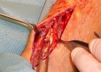
J. Clin. Med. 2020, 9, 3380 5 of 10
J.J.Clin.
Clin.Med.
Med.2020,
2020,9,9,xx 55ofof10
10
(a)
(a) (b)
(b)
Figure
Figure2. 2.2.US
USimages
imagesof
ofpartial
partialAchilles
Achillestendon
tendonruptures:
ruptures:(a) partial rupture in the dorsal part of the
Figure US images of partial Achilles ruptures: (a)
(a)partial
partialrupture
ruptureininthe
thedorsal
dorsalpart
partofofthe
the
Achilles
Achilles tendon
tendon midportion
midportion (arrows);
(arrows);(b)
(b)partial
partialrupture
ruptureinininsertional
insertionalarea
areaofofthe
theAchilles
Achillestendon
tendon
Achilles tendon midportion (arrows); (b) partial rupture in insertional area of the Achilles tendon
(circle).
(circle).US;US;ultrasound.
ultrasound.
(circle). US; ultrasound.
(a)
(a) (b)
(b)
Figure
Figure3.
Figure 3.3.MR
MRimages
MR imagesof
images ofpartial
of partialAchilles
partial Achillestendon
tendonruptures:
ruptures:(a)
ruptures: (a)partial
(a) partialrupture
partial rupturein
rupture ininthe dorsal
the
the part
dorsal
dorsal of
part
part the
ofof the
the
midportion;
midportion; (b)
midportion;(b) partial
(b)partial rupture
partial rupture in
rupture in the
in the insertion.
the insertion. MR; magnetic resonance.
magnetic resonance.
insertion. MR; magnetic resonance.
(a)
(a) (b)
(b)
Figure
Figure4. Patient
Patientwith
withaaapartial
partialrupture
rupturein close relation to aathick and compressing plantaris tendon:
Figure 4.4. Patient with partial rupture in
in close
close relation
relation to
to athick
thickand
andcompressing
compressingplantaris
plantaristendon:
tendon:
(a) thickened plantaris
thickenedplantaris
(a)thickened tendon
plantaristendon compressing
tendoncompressing onto
compressingonto the
ontothe medial
themedial side
medialside of the
sideofofthe Achilles;
theAchilles; (b)
Achilles;(b) surgical release
surgicalrelease
(b)surgical releaseof
(a)
of
ofthe
thecompressing
compressing plantaris
plantaris tendon.
tendon.
the compressing plantaris tendon.
3.6.
3.6.Treatment
3.6. TreatmentOptions
Treatment Options
Options
Based
Based
Basedonon the
onthe findings
findings
the of
of the
of the
findings the conducted
conducted systematic
systematic
conducted literature
literature
systematic research,
research,
literature there isthere
research, isis no
no clear
there no clear
consensus
clear
consensus
about about
a general
consensus a general
aboutconservative conservative or surgical
or surgical treatment
a general conservative or surgical treatment
regime. regime.
Indications
treatment Indications
regime. for for surgical
surgical treatment
Indications for surgical are
treatment
functional are
are functional
treatment limitations, such limitations,
functional as such
such as
poor single-heel
limitations, poor
raises
as andsingle-heel
poor raises
persistent pain
single-heel in and
raises and persistent
combination with
persistent pain in
positive
pain in
combination
findings with
in MRI
combination positive
withorpositive findings
ultrasound in
[6,36].
findings MRI or
In some
in MRI ultrasound [6,36].
studies, a[6,36].
or ultrasound partialIn some
Intendon studies, a partial
rupturea of
some studies, >50%
partial tendon
of the
tendon
rupture
ruptureof
of>50%
>50%
cross-sectional of
ofthe
area thecross-sectional
was cross-sectional area
area
seen as a criteria was
was
for seen
seenas
surgical asaacriteria
criteria
therapy, for
forsurgical
whereas surgical
minor therapy,
rupturewhereas
therapy, whereas minor
minor
sizes, especiallyJ. Clin. Med. 2020, 9, 3380 6 of 10
at the proximal myotendinous junction, with reduced biomechanical function might require surgical
therapy as well [30,37].
3.6.1. Nonsurgical Treatments
Studies on conservative treatments on patients with partial Achilles tendon tears are rare. The only
case series found on exercise interventions was from Masci and colleagues [38]. All patients (n = 26)
were clinically and through ultrasound examination diagnosed to have a partial Achilles tendon
rupture. They underwent a 3-month structured rehabilitation program, starting with 6 weeks (phase 1)
of heel lifts (2 cm) and avoiding calf stretching activities (for 12 weeks). Walking and moderate cycling
with pain-adapted full weight-bearing were allowed. In phase II (weeks 7–12), heel lifts were reduced
and concentric calf raises were introduced, starting with seated and progressing towards standing
bilateral and single calf raises. After 3 months, heel lifts were removed and eccentric heel drops were
introduced (Table 1). The clinical results were good for the majority of patients. In 25 of 26 patients,
there was a significant pain reduction (Visual-Analogue Scale) and an improvement in the ultrasound
and color Doppler findings after 3 months [37]. Recently, Medeiros presented in his case report of a
futsal player with a partial rupture of up to 50% of the cross-sectional area a detailed rehabilitation
protocol suggesting an initial immobilization with crutches and 2 cm heel lifts for 6 weeks combined
with a progressive loading protocol [39].
Table 1. Suggested rehabilitation program by Masci and Alfredson [38].
0–6 Weeks 7–12 Weeks >12 Weeks (When Pain-Free)
• 2 cm heel lifts • 1 cm heel lifts • Heel lifts removed
• Seated calf raises (3 × 15 reps daily)
Progression to standing bilateral calf
• Eccentric heel drops (from
• Avoiding calf muscle raises (from floor level to tiptoe, 3 × 15
tiptoe to below floor level, 3 × 15
stretching reps daily)
reps, 3 ×/week)
Progression to single calf raises (floor
level to tip toe, 3 × 15 reps daily)
• Exercises performed 3 ×/week
• Gradual return to previous
• Walking/cycling allowed together with cross training, walking,
(pre-injury) tendon loading
swimming, and cycling
Beyond physiotherapeutic approaches, extracorporeal shock wave therapy (ESWT) and
platelet-rich plasma (PRP) injections are additional options. However, clinical results are based
on single case reports and need to be evaluated in further studies [40,41]. Hence, for conservative
treatment of partial ruptures there is no evidence-based approach available. The exercise protocol
suggested by Masci and Alfredson is promising but needs to be evaluated in large-scale trials using
specific patient-related outcome parameters [38].
3.6.2. Surgical Treatments
In relation to surgical treatment, there are a few low-evidence studies in the current literature.
In general, surgical treatment differs depending on the exact location of the partial rupture (insertion
vs. midportion) within the tendon. In general, surgery can be divided into lesion excision with or
without suture adaption and tendon augmentation or tendon transfer surgery depending on the size
of the partial rupture. Using drill holes or anchors for reattachment for insertional ruptures is another
option. There is no consensus for the optimal time for surgery, but in the majority of the studies
operative treatment was initiated after a conservative approach. It is presumable that this might not
affect patient outcomes, as seen in a recent study about a late minimally invasive Achilles tendon
repair after 14 days [42]. Irrespective of the anatomic locations, there might be no relevant symptomJ. Clin. Med. 2020, 9, 3380 7 of 10
improvement 3 months postoperatively, but there may be after 6–12 months with no deterioration after
years [43].
With regard to partial ruptures in the midportion, previous studies combined heterogeneous
patient cohorts suffering from total and partial ruptures with conservative and surgical therapy in their
analyses, such that data interpretation is biased [20,44]. In a study of Denstad et al., 44 of 58 partial
ruptures were treated with excision of pathologic tendon tissue and a suture side-to-side adaptation
with a few (n = unknown) requiring a Lindholm repair with a turn down flap [20]. Overall, the results
were good with 46 pleased, 8 satisfied, and 3 unsatisfied patients (1 lost to follow-up). There was
a return-to-sports rate of (100%, n = 44) with 85% reaching an equal or better level [20]. Another
study using the equal surgical procedure on 64 patients with a mean follow-up of 6 years reported a
satisfaction rate of only 66% with a reoperation rate of 14% and more favorable results in proximal
ruptures than distal or combined ruptures [45].
Regarding surgical treatment of insertional partial ruptures, prospective studies are rare.
Jerosch et al. performed an endoscopic calcaneoplasty for insertional Achilles tendinopathy in
164 patients; 61 patients had a distal partial rupture based on MRI [46]. Based on the Ogilvie–Harris
scores, 155 patients showed good or excellent results after 46 months. Even though the outcome of
partial ruptures was not evaluated separately in this study, it indirectly indicates that suture application
of partial ruptures might not be strictly necessary for achieving good long-term results [46].
Lohrer performed operations on 36 patients with persistent insertional Achilles pain with
bursectomy and resection of Haglund exostosis; 5 out of 36 patients required a suture of a ventral
longitudinal partial rupture based on the intraoperative surgeon’s decision [14]. However, there is
no comparative data on whether or not sutures of this kind of tear are mandatory [14]. In a further
study, anterior partial tears in 20 patients were resected and a tendon reconstruction was performed
with sutures [47]. Postoperatively, patients were mobilized with a walking boot and decreasing heel
support (2.5–1 cm) between the first and sixth week. The VISA-A score increased from 46 points to 72
after 6 months and to 84 points after 12 months [47].
An additional treatment strategy might be a bone–quadriceps tendon graft for insertional ruptures,
which has been evaluated in a retrospective study on 24 patients with a high rate of satisfaction on the
AOFAS-Score. There were few side effects—one wound healing problem and one instance of deep
venous thrombosis [37]. Further treatment strategies might include augmentation with the plantaris
tendon, flaps, or a flexor hallucis longus transfer [5,9].
4. Discussion and Clinical Recommendations
For partial Achilles tendon ruptures, there are few studies and unfortunately sparse knowledge
of scientific value, making it difficult to give strict recommendations for diagnostics and treatment.
High-quality research studies on this diagnosis are warranted.
Based on the presented studies and our clinical experience in the field of Achilles tendinopathy
and partial Achilles tendon ruptures, diagnosis should be based on a patient’s history with a typical
sharp onset of pain and inability to fully load the tendon. Clinical findings are a localized tender
region in the tendon and often some weakness during heel raises. Greyscale ultrasound with a
high-resolution probe and Power Doppler show a region with an irregular and bulging superficial
tendon line. MRI shows a hyperintense signal on T1 and T2-weighted sequences. First-line therapy
should be a conservative approach, such as using a 2 cm heel lift with pain-adapted full weight-bearing
and avoiding tendon stretching for the first 6 weeks. This phase is followed by reduced heel lifts
to 1 cm and progressive tendon loading in weeks 7–12. After 12 weeks, heel lifts can be removed if
pain-free, and the patient can start eccentric exercises, lowering the heel under floor level, and gradually
returning to the patient’s previous sport level. If conservative management has a poor effect, surgical
exploration and excision of the partial rupture and suturing is required. However, this might only
be needed in patients previously treated with intratendinous injections (most commonly cortisone).
Augmentation procedures or anchor applications might be useful in some cases, but this depends onJ. Clin. Med. 2020, 9, 3380 8 of 10
the size and location. After surgery, we recommend the 12 to 14-week rehabilitation program used in
conservative management before a return to full tendon-loading activities.
Future research needs to provide basic epidemiological data about size, location, and affected
patients. Additionally, imaging studies should focus on the diagnostic accuracy and monitoring
capacities of MRI versus high-resolution ultrasound and further assessments of promising ultrasound
technologies, such as shear wave elastography or ultrasound tissue characterization [48,49].
Evidence-based decision-making for conservative approaches requires prospective trials evaluating
the effect of immobilization, heel raises, or physiotherapy [50]. Moreover, indications for surgical
treatment, augmentation procedures, or suture application need to be studied in relation to the location
and the size of the rupture.
Author Contributions: Conceptualization, M.G., C.S., and H.A.; methodology, M.G.; formal analysis, M.G. and C.S.;
original draft preparation, M.G.; review and editing, M.G., C.S., and H.A.; visualization, C.S. All authors have
read and agreed to the published version of the manuscript.
Funding: There was no specific funding that was used for this study.
Conflicts of Interest: The authors declare no conflict of interest.
References
1. Alfredson, H.; Spang, C. Clinical presentation and surgical management of chronic Achilles tendon
disorders—A retrospective observation on a set of consecutive patients being operated by the same
orthopedic surgeon. Foot Ankle Surg. 2018, 24, 490–494. [CrossRef] [PubMed]
2. Van der Vlist, A.C.; Winter, M.; Weir, A.; Ardern, C.L.; Welton, N.J.; Caldwell, D.M.; Verhaar, J.A.N.; de Vos, R.J.
Which treatment is most effective for patients with Achilles tendinopathy? A living systematic review with
network meta-analysis of 29 randomised controlled trials. Br. J. Sports Med. 2020. online ahead of print.
[CrossRef] [PubMed]
3. Gatz, M.; Driessen, A.; Eschweiler, J.; Tingart, M.; Migliorini, F. Open versus minimally-invasive surgery for
Achilles tendon rupture: A meta-analysis study. Arch. Orthop. Trauma Surg. 2020. online ahead of print.
[CrossRef] [PubMed]
4. Ochen, Y.; Beks, R.B.; van Heijl, M.; Hietbrink, F.; Leenen, L.P.H.; van der Velde, D.; Heng, M.;
van er Meijden, O.; Groenwold, R.H.H.; Houwert, R.M. Operative treatment versus nonoperative treatment of
Achilles tendon ruptures: Systematic review and meta-analysis. BMJ 2019, 364, k5120. [CrossRef] [PubMed]
5. Smigielski, R. Management of partial tears of the gastro-soleus complex. Clin. Sports Med. 2008, 27, 219–229.
[CrossRef] [PubMed]
6. Segesser, B.; Goesele, A.; Renggli, P. The Achilles tendon in sports. Orthopade 1995, 24, 252–267.
7. Hartgerink, P.; Fessel, D.P.; Jacobson, J.A.; van Holsbeeck, M.T. Full- versus partial-thickness Achilles tendon
tears: Sonographic accuracy and characterization in 26 cases with surgical correlation. Radiology 2001, 220,
406–412. [CrossRef]
8. Chan, O.; Morton, S.; Pritchard, M.; Parkes, T.; Malliaras, P.; Crisp, T.; Padhiar, N.; Maffulli, N.; King, J.;
Morrissey, D. Intratendinous tears of the Achilles tendon—A new pathology? Analysis of a large 4-year
cohort. Muscles Ligaments Tendons J. 2017, 7, 53–61. [CrossRef]
9. Ljungqvist, R. Subcutaneous partial rupture of the Achilles tendon. Acta Orthop. Scand. 1968, 39 (Suppl. 113),
1–86. [CrossRef]
10. Skeoch, D.U. Spontaneous partial subcutaneous ruptures of the tendo achillis. Review of the literature and
evaluation of 16 involved tendons. Am. J. Sports Med. 1981, 9, 20–22. [CrossRef]
11. Astrom, M.; Rausing, A. Chronic Achilles tendinopathy. A survey of surgical and histopathologic findings.
Clin. Orthop. Relat. Res. 1995, 316, 151–164. [CrossRef]
12. Tran, P.H.T.; Malmgaard-Clausen, N.M.; Puggaard, R.S.; Svensson, R.B.; Nybing, J.D.; Hansen, P.; Schjerling, P.;
Zinglersen, A.H.; Couppé, C.; Boesen, M.; et al. Early development of tendinopathy in humans: Sequence
of pathological changes in structure and tissue turnover signaling. FASEB J. 2020, 34, 776–788. [CrossRef]
[PubMed]
13. Arndt, A.; Brüggemann, G.P.; Koebke, J.; Segesser, B. Asymmetrical loading of the human triceps surae:
I. Mediolateral force differences in the Achilles tendon. Foot Ankle Int. 1999, 20, 444–449. [CrossRef] [PubMed]J. Clin. Med. 2020, 9, 3380 9 of 10
14. Lohrer, H. Minimally invasive repair of an impingement induced partial tear of the anterior Achilles tendon
in a top level athlete. Z. Orthop. Unfall. 2010, 148, 80–82. [CrossRef]
15. Mahler, F.; Fritschy, D. Partial and complete ruptures of the Achilles tendon and local corticosteroid injections.
Br. J. Sports Med. 1992, 26, 7–14. [CrossRef] [PubMed]
16. Childs, S.G. Pathogenesis of tendon rupture secondary to fluoroquinolone therapy. Orthop. Nurs. 2007, 26,
175–182. [CrossRef]
17. Mahan, J.; Damodar, D.; Trapana, E.; Barnhill, S.; Nuno, A.U.; Smyth, N.A.; Aiyer, A.; Jose, J. Achilles tendon
complex: The anatomy of its insertional footprint on the calcaneus and clinical implications. J. Orthop. 2020,
17, 221–227. [CrossRef]
18. Szaro, P.; Witkowski, G.; Smigielski, R.; Krajewski, P.; Ciszek, B. Fascicles of the adult human Achilles
tendon—An anatomical study. Ann. Anat. 2009, 191, 586–593. [CrossRef]
19. Alfredson, H.; Masci, L.; Ohberg, L. Partial mid-portion Achilles tendon ruptures: New sonographic findings
helpful for diagnosis. Br. J. Sports Med. 2011, 45, 429–432. [CrossRef]
20. Denstad, T.F.; Roaas, A. Surgical treatment of partial Achilles tendon rupture. Am. J. Sports Med. 1979, 7,
15–17. [CrossRef]
21. Astrom, M. Partial rupture in chronic achilles tendinopathy. A retrospective analysis of 342 cases.
Acta Orthop. Scand. 1998, 69, 404–407. [PubMed]
22. Johansson, K.; Lempainen, L.; Sarimo, J.; Laitala-Leinonen, T.; Orava, S. Different distributions of operative
diagnoses for Achilles tendon overuse injuries in Italian and Finnish athletes. Muscles Ligaments Tendons J.
2016, 6, 111–115. [CrossRef]
23. Elias, D.A.; Carne, A.; Bethapudi, S.; Engebretsen, L.; Budgett, R.; O’Connor, P. Imaging of plantar fascia and
Achilles injuries undertaken at the London 2012 Olympics. Skelet. Radiol. 2013, 42, 1645–1655. [CrossRef]
[PubMed]
24. Kayser, R.; Mahlfeld, K.; Heyde, C.E. Partial rupture of the proximal Achilles tendon: A differential diagnostic
problem in ultrasound imaging. Br. J. Sports Med. 2005, 39, 838–842. [CrossRef]
25. Kälebo, P.; Allenmark, C.; Peterson, L.; Swärd, L. Diagnostic value of ultrasonography in partial ruptures of
the Achilles tendon. Am. J. Sports Med. 1992, 20, 378–381. [CrossRef] [PubMed]
26. Paavola, M.; Paakkala, T.; Kannus, P.; Järvinen, M. Ultrasonography in the differential diagnosis of Achilles
tendon injuries and related disorders. A comparison between pre-operative ultrasonography and surgical
findings. Acta Radiol. 1998, 39, 612–619. [CrossRef]
27. Lehtinen, A.; Peltokallio, P.; Taavitsainen, M. Sonography of Achilles tendon correlated to operative findings.
Ann. Chir. Gynaecol. 1994, 83, 322–327.
28. Syha, R.; Springer, F.; Ketelsen, D.; Ipach, I.; Kramer, U.; Horger, M.; Schick, F.; Grosse, U. Achillodynia—
Radiological imaging of acute and chronic overuse injuries of the achilles tendon. Rofo 2013, 185, 1041–1055.
[CrossRef] [PubMed]
29. Aström, M.; Gentz, C.F.; Nilsson, P.; Rausing, A.; Sjöberg, S.; Westlin, N. Imaging in chronic achilles
tendinopathy: A comparison of ultrasonography, magnetic resonance imaging and surgical findings in
27 histologically verified cases. Skelet. Radiol. 1996, 25, 615–620. [CrossRef]
30. Heyde, C.E.; Kayser, R.; Jungmichel, D.; Melzer, C. Limitations of sonography in the diagnosis of partial
ruptures of the achilles tendon in the musculo-tendinous junction: A case report. Sportverletz. Sportschaden
2003, 17, 39–43. [CrossRef]
31. Hoffmann, A.; Mamisch, N.; Buck, F.M.; Espinosa, N.; Pfirrmann, C.W.A.; Zanetti, M. Oedema and fatty
degeneration of the soleus and gastrocnemius muscles on MR images in patients with Achilles tendon
abnormalities. Eur. Radiol. 2011, 21, 1996–2003. [CrossRef] [PubMed]
32. Husson, J.L.; De Korvin, B.; Polard, J.L.; Attali, J.Y.; Duvauferrier, R. Study of the correlation between magnetic
resonance imaging and surgery in the diagnosis of chronic Achilles tendinopathies. Acta Orthop. Belg. 1994,
60, 408–412. [PubMed]
33. Alfredson, H. Midportion Achilles tendinosis and the plantaris tendon. Br. J. Sports Med. 2011, 45, 1023–1025.
[CrossRef]
34. Alfredson, H.; Masci, L.; Spang, C. Surgical plantaris tendon removal for patients with plantaris tendon-related
pain only and a normal Achilles tendon: A case series. BMJ Open Sport Exerc. Med. 2018, 4, e000462.
[CrossRef] [PubMed]J. Clin. Med. 2020, 9, 3380 10 of 10
35. Masci, L.; Spang, C.; van Schie, H.T.M.; Alfredson, H. How to diagnose plantaris tendon involvement in
midportion Achilles tendinopathy—Clinical and imaging findings. BMC Musculoskelet. Disord. 2016, 17, 97.
[CrossRef] [PubMed]
36. Allenmark, C. Partial Achilles tendon tears. Clin. Sports Med. 1992, 11, 759–769. [CrossRef]
37. Philippot, R.; Wegrzyn, J.; Grosclaude, S.; Besse, J.L. Repair of insertional achilles tendinosis with a
bone-quadriceps tendon graft. Foot Ankle Int. 2010, 31, 802–806. [CrossRef]
38. Masci, L.; Alfredson, H. Promising results using a simple rehabilitation program to treat partial ruptures in
the Achilles midportion. J. Biomed. Graph. Comput. 2013, 3, 47. [CrossRef]
39. Medeiros, D.M. Conservative treatment of Achilles tendon partial tear in a futsal player: A case report.
Physiother. Theory Pract. 2019, 1–8. [CrossRef]
40. Hsu, Y.C.; Wu, W.T.; Chang, K.V.; Han, D.S.; Chou, L.W. Healing of Achilles tendon partial tear following
focused shockwave: A case report and literature review. J. Pain Res. 2017, 10, 1201–1206. [CrossRef]
41. Filardo, G.; Presti, M.L.; Kon, E.; Marcacci, M. Nonoperative biological treatment approach for partial
Achilles tendon lesion. Orthopedics 2010, 33, 120–123. [CrossRef] [PubMed]
42. Carmont, M.R.; Zellers, J.A.; Brorsson, A.; Silbernagel, K.G.; Karlsson, J.; Nilsson-Helander, K.
No difference in strength and clinical outcome between early and late repair after Achilles tendon rupture.
Knee Surg. Sports Traumatol. Arthrosc. 2020, 28, 1587–1594. [CrossRef] [PubMed]
43. Lohrer, H. Minimum 3.5-year outcomes of operative treatment for Achilles tendon partial tears in the
midportion and retrocalcaneal area. J. Orthop. Surg. Res. 2020, 15, 395. [CrossRef] [PubMed]
44. Persson, A.; Ljungqvist, R. Electrophysiological observations in cases of partial and total rupture of the
achilles tendon. Electroencephalogr. Clin. Neurophysiol. 1971, 31, 239–246. [CrossRef]
45. Morberg, P.; Jerre, R.; Swärd, L.; Karlsson, J. Long-term results after surgical management of partial Achilles
tendon ruptures. Scand. J. Med. Sci. Sports 1997, 7, 299–303. [CrossRef]
46. Jerosch, J.; Sokkar, S.; Dücker, M.; Donner, A. Endoscopic calcaneoplasty (ECP) in Haglund’s syndrome.
Indication, surgical technique, surgical findings and results. Z. Orthop. Unfall. 2012, 150, 250–256. [CrossRef]
47. Lohrer, H.; Nauck, T. Results of operative treatment for recalcitrant retrocalcaneal bursitis and midportion
Achilles tendinopathy in athletes. Arch. Orthop. Trauma Surg. 2014, 134, 1073–1081. [CrossRef]
48. Gatz, M.; Betsch, M.; Bode, D.; Schweda, S.; Dirrichs, T.; Migliorini, F.; Tingart, M.; Quack, V.
Intra individual comparison of unilateral Achilles tendinopathy using B-mode, power doppler, ultrasound
tissue characterization and shear wave elastography. J. Sports Med. Phys. Fitness 2020. online ahead of print.
49. Frankewycz, B.; Henssler, L.; Weber, J.; Silva, N.; Koch, M.; Jung, E.M.; Docheva, D.; Alt, V.; Pfeifer, C.G.
Changes of Material Elastic Properties during Healing of Ruptured Achilles Tendons Measured with Shear
Wave Elastography: A Pilot Study. Int. J. Mol. Sci. 2020, 21, 3427. [CrossRef]
50. Gatz, M.; Betsch, M.; Dirrichs, T.; Schrading, S.; Tingart, M.; Michalik, R.; Quack, V. Eccentric and Isometric
Exercises in Achilles Tendinopathy Evaluated by the VISA-A Score and Shear Wave Elastography. Sports Health
2020, 12, 373–381. [CrossRef]
Publisher’s Note: MDPI stays neutral with regard to jurisdictional claims in published maps and institutional
affiliations.
© 2020 by the authors. Licensee MDPI, Basel, Switzerland. This article is an open access
article distributed under the terms and conditions of the Creative Commons Attribution
(CC BY) license (http://creativecommons.org/licenses/by/4.0/).You can also read