The Effect of Cranial Cruciate Ligament Rupture on Range of Motion in Dogs
←
→
Page content transcription
If your browser does not render page correctly, please read the page content below
veterinary
              sciences

Article
The Effect of Cranial Cruciate Ligament Rupture on Range of
Motion in Dogs
Stefania Pinna * , Francesco Lanzi and Chiara Tassani

                                          Department of Veterinary Medical Sciences, University of Bologna, Via Tolara di Sopra 50,
                                          40064 Ozzano dell’Emilia (BO), Italy; fralanz@gmail.com (F.L.); chiara.tassani2@unibo.it (C.T.)
                                          * Correspondence: stefania.pinna@unibo.it; Tel.: +39-051-2097535

                                          Abstract: Range of motion (ROM) is a measure often reported as an indicator of joint functionality.
                                          Both the angle of extension and that of flexion were measured in 234 stifle joints of dogs with cranial
                                          cruciate ligament (CCL) rupture. The aims of this study were to investigate the correlation between
                                          CCL rupture and alterations in the range of stifle joint motion and to determine whether there
                                          was a prevalence modification of one of the two angles. All the extension and flexion angles were
                                          obtained from clinical records and were analysed in various combinations. A significant relationship
                                          was found between normal angles and abnormal angles; concerning the reduction in the ROM, a
                                          significant prevalence in the alteration extension angle was found. Of the 234 stifles, 33 (13.7%) were
                                          normal in both angles. These results could offer important insights regarding the influence of CCL
                                          rupture on compromising the ROM. This awareness could be a baseline for understanding the ability
                                          of surgical treatment to restore one angle rather than another angle, to address the choice of treatment
                                          and to help physiotherapists in their rehabilitation program.


                                          Keywords: range of motion; cranial cruciate ligament; extension angle; flexion angle; dog
Citation: Pinna, S.; Lanzi, F.;
Tassani, C. The Effect of Cranial
Cruciate Ligament Rupture on Range
of Motion in Dogs. Vet. Sci. 2021, 8,
                                          1. Introduction
119. https://doi.org/10.3390/
vetsci8070119
                                                Range of motion (ROM) is the ability of a joint to move between positions to its
                                          maximum potential in relation to the three anatomic planes, the sagittal, frontal and
Academic Editor: Aristidis                transverse planes. Concerning the stifle joint, it is the movement in the sagittal plane,
S. Veskoukis                              defined as either flexion or extension, which is usually studied [1]. The measurement of
                                          the joint angle is achieved using clinical goniometry, which gives an indication of the joint
Received: 19 May 2021                     function and the eventual severity of the pathology [1,2].
Accepted: 22 June 2021                          Goniometry has been validated in humans [3–6], and several methods of measure-
Published: 24 June 2021                   ments have been documented and compared [7–9]. In veterinary medicine, goniometry
                                          has been studied in different species, such as cats [10], horses [11–13] and sheep [14]. The
Publisher’s Note: MDPI stays neutral      reliability of goniometry in dogs was documented by Jaegger in 2002, comparing the
with regard to jurisdictional claims in   measurements made with radiography with the measurements made by a transparent
published maps and institutional affil-   plastic goniometer in 16 healthy Labrador Retrievers [2].
iations.                                        Physiological and/or pathological causes can modify the ROM. Variations can be
                                          based on activity and the breed of dog; the body’s condition and joint health can affect
                                          the ROM as a result of changes in the shapes of the joints, capsules, periarticular tendons,
                                          muscles and ligaments [1,15–17]. Therefore, a lack of joint motion can occur following
Copyright: © 2021 by the authors.         injury or surgical treatment or in patients with chronic diseases, such as osteoarthritis
Licensee MDPI, Basel, Switzerland.        (OA) or muscle contracture. During the OA process, osteophytes, capsular adhesions and
This article is an open access article    shortened ligaments are generated and lead to a mechanical reduction of the ROM. Another
distributed under the terms and           cause of ROM abnormality could be pain, as maximum joint motions lead to a stretching
conditions of the Creative Commons        of the joint capsule and, consequently, increases the nociceptor activity [18,19]. Changes in
Attribution (CC BY) license (https://     the ROM can be due to reducing the extension angle or increasing the flexion angle or both
creativecommons.org/licenses/by/          alterations. The result is obviously a reduction in the ROM.
4.0/).

Vet. Sci. 2021, 8, 119. https://doi.org/10.3390/vetsci8070119                                                    https://www.mdpi.com/journal/vetsci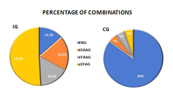
Vet. Sci. 2021, 8, 119                                                                                              2 of 11

                              Reducing the ROM could lead to lameness, which is greater when the loss of ROM is
                         greater. The incomplete extension or flexion of a joint is associated with some functional
                         disabilities, such as jumping into a car, rising from a recumbent position or climbing
                         stairs [19].
                              Goniometry has been used in canine orthopaedics [20] to assess the treatment efficacy of
                         diseases involving shoulders [21], elbows [19,22,23], stifles [19,24–29] and hip joints [30,31].
                         The most common injury involving the canine stifle joint is cranial cruciate ligament (CCL)
                         rupture [32,33]. The incorrect use of the affected limb, i.e., due to pain, chronic disease, joint
                         involved or OA, leads to muscle atrophy and a reduced ROM [32]. Several studies have
                         used the measurements of the ROM in dogs with a CCL rupture to evaluate the functional
                         outcome after surgical repair [32–41] or postoperative rehabilitation. It is currently known
                         that physiotherapy is useful for improving the functioning of a restricted joint [24,42,43].
                              The aim of the present study was to investigate the relationship between a CCL
                         rupture at the time of diagnosis and the changes in stifle joint motion using the values of
                         the extension and flexion angles. The hypotheses were that there was a correlation between
                         the two angles and the loss of extension was more frequent than the alteration of flexion in
                         dogs with a CCL rupture.

                         2. Materials and Methods
                              The Bologna Healing Stifle Injury Index—Clinical Record (BHSII—CR) of dogs with a
                         spontaneous CCL rupture was obtained from the archives of the University Hospital of
                         the Department of Veterinary Medical Sciences, University of Bologna, Ozzano dell’Emilia
                         (Bologna), Italy from 2011 to 2018, and they were retrospectively reviewed. Of the various
                         items in the index, the assessment of both extension and flexion movements was retrieved.
                         The diagnosis of a CCL rupture was made at the time of the compilation of the BHSII using
                         a complete orthopaedic examination.
                              All items of the BHSII concern a damaged stifle joint; the assessment method selected
                         was the Likert scale, which has five levels of answers (scores from 0 to 4) [44]. Based on
                         the median values published by Jaegger [2], such as extension angle = 162◦ and flexion
                         angle = 41◦ , which referred to the stifle joints of healthy dogs, the authors of the BHSII [44]
                         created the following scale of values. The extension angle (EA) was set as scores 0 = 162–158◦ ,
                         1 = 157–153◦ , 2 = 152–148◦ , 3 = 147–143◦ and 4 = 142–138◦ and the flexion angle (FA) as
                         scores 0 = 41–45◦ , 1 = 46–50◦ , 2 = 51–55◦ , 3 = 56–60◦ and 4 = 61–65◦ (see https://www.
                         frontiersin.org/articles/10.3389/fvets.2019.00065/full#supplementary-material Accessed on
                         1 April 2021) [44]. These scores were indicative of no alteration (score 0) or a slight, mild,
                         moderate or severe reduction in the degree of extension and/or increase in flexion (scores 1, 2,
                         3 and 4, respectively). Each angle of extension and flexion was assessed individually.
                              At the time of the BHSII-CR compilation, all the dogs were sedated and placed in
                         lateral recumbency. Sedation was required to obtain adequate relaxation of the patient
                         in order to perform specific measurements and manualities, i.e., angle of motion and
                         drawer movement, without interference due to abnormalities, namely pain or muscle
                         contracture. The same protocol was used for each patient. All the dogs were sedated
                         with dexmedetomidine (Dextroquillan; ATI, Bologna, Italy) 3–5 mcg/kg or medetomidine
                         (Sedastart; Ecupharma s.r.l, Milano, Italy) 5–10 mcg/kg intramuscularly (IM) in combi-
                         nation with butorphanol (Nargesic; Acme S.r.l., Reggio Emilia, Italy) 0.1–0.2 mg/kg. The
                         dexmedetomidine or medetomidine were administered at the anaesthetist’s discretion.
                         The range of joint motion was measured as the degree of extension and flexion using a
                         plastic goniometer. As described by Jaegger, the arms of the goniometer were aligned
                         with the tibia shaft and with the longitudinal axis of the femur, which were the line that
                         joins the lateral femur epicondyle and the greater trochanter, respectively [2]. Measure-
                         ments of the extension and flexion angles were reported in the BHSII-CR of each dog. The
                         measurements were taken three times by the veterinarians of the orthopaedics staff of the
                         University Hospital and were averaged [33,36,45], and the corresponding score was then
                         selected on the BHSII-CR.
Vet. Sci. 2021, 8, 119 3 of 11
The inclusion criteria were a BHSI-CR completely filled out without limit by age, sex,
breed, acute or chronic disease, duration of lameness, partial or complete CCL tears or
signs of OA. If the BHSH-CR was missing information regarding the ROM, and other
items of the clinical record, the dogs were excluded. The dogs with a BHSII that met the
inclusion criteria were included in the injury group (IG), meaning those with a spontaneous
CCL rupture.
2.1. Collection Data
All demographic data and the scores of stifle motion of each dog were collected in a
spreadsheet (Office Excel 2019, Microsoft Corporation, Redmond, WA 98052-6399 United
States). The groups of combination scores were classified as follows:
• NG = the normal group that included dogs having a score of 0 in both extension and
flexion;
• EFAG = the extension and flexion abnormal group that included dogs having scores
1–4 in both extension and flexion;
• EAAG = the extension angle (reduced) abnormal group that included dogs with stifles
having an extension angle with scores 1 to 4 and a flexion angle having a score of 0;
• FAAG = the flexion angle (increased) abnormal group that included dogs with stifles
having an extension angle with a score of 0 and a flexion angle with scores 1–4.
The control group (CG) included twenty healthy dogs without a history or clinical
signs of orthopaedic diseases in order to verify the reliability of the scores.
Based on body weight, the dogs in the IG were divided into three groups: Group A <
20 kg, Group B from 21 to 30 kg and Group C > 31 kg.
The ROM in the IG was assessed on the basis of the duration of the lameness and/or
the time passed from the trauma, if known. An acute group (AG) and a chronic group
(ChG) were created. The ROM in the IG was also assessed on the basis of muscle mass, and
the normal muscle group (NMG) and the decreased muscle group (DMG) were obtained.
2.2. Statistical Analysis
The continuous data were evaluated using the Kolmogorov–Smirnov test for normal
distribution; if they were rejected, nonparametric tests were carried out. The data were
reported as the median, range (minimum and maximum values) and 95% confidence
interval (CI). The categorical or discrete variables were evaluated as frequencies and/or
percentages and processed using the chi-square test for trends. Spearman’s rank correlation
coefficient was calculated to measure the relationship between the angles of extension
and flexion. All the data were analysed using a statistical software program (MedCalcR
Software 16.8.4, Ostend, Belgium). Significance for all the analyses was set at p < 0.05.
3. Results
3.1. Descriptive Analysis
A total of 234 dogs with a CCL rupture met the inclusion criteria (IG)). The age and
body weight were reported as the median, range and 95% CI. All the demographic data
are reported in Table 1.
The most common breeds included 67 crossbreed dogs, 28 Labrador Retrievers, 18
Boxers, 11 German Shepherds, 11 Rottweilers, 9 Maremma Sheepdogs, 8 American Stafford-
shire Terriers, 8 Cane Corso, 6 Beagles, 6 Yorkshire Terries, 4 Epagneul Bretons, 4 Jack
Russell Terriers, 4 Maltesi, 3 Australian Shepherds, 3 Barboncini, 3 Bernese Mountain
dogs, 3 Border Collies, 3 Doberman Pinchers and 3 Volpino Italiano. There were also
two dogs of each of the following breeds: Bolognese, Bullmastiff, Chow Chow, Golden
Retriever, Leonberger, Pyrenean Mastiff and Tosa Inu. There was also one dog each for
the following breeds: Akita Inu, American Pit Bull Terrier, Bracco Italiano, Caucasian
Shepherd, Cavalier King Charles Spaniel, Dogue de Bordeaux, English Setter, Fox Terrier,
Great Dane, Lagotto Romagnolo, Miniature Pinscher, Pyrenean Shepherd, Riesenschnauzer,
Saint Bernard, Samoyed, Siberian Husky, Toy Poodle and West Highland Terrier.Vet. Sci. 2021, 8, 119 4 of 11
Table 1. Descriptive characteristics of the dogs. Median, range (minimum and maximum values) and 95% CI for the
medians are reported for age (y) and weight (kg).
Age Weight
Dog/Stifle n. %
Median Range 95% CI Median Range 95% CI
IG 234 100 6 1–13 6–7 30 4–75 28–32
Male 99 42.3 6 1–13 5–7 35 * 4–75 30–38
Female 135 57.7 7 1–13 6–7 28 4–56 26–30
Group A 64 27.4 8 2–13 7–9.3 10 4–18 8–11
Group B 91 38.9 6 1–12 5–6.3 29 21–35 28–30
Group C 79 33.8 5 1–13 5–6 42 36–75 40–44.9
CG 20 100 6 1–12 3–7.8 28 14–55 22–29.8
IG, Injury Group; Group A, 31kg; CG, Control Group; * significant p-value.
In the IG, the Mann–Whitney test was applied to evaluate the differences in age
and/or weight between the male and female groups. Only the weight distribution was
statistically significant (p = 0.0009).
The CG included six Golden Retrievers; three crossbreeds; three Border Collies; two
Dachshunds and one dog each of the following breeds: Basenji, Belgian Malinois, Labrador
Retriever, Lagotto Romagnolo, Newfoundland and Weimaraner.
3.2. Statistical Analysis
• In the IG, the Spearman’s rank correlation coefficient revealed a significant level of
correlation (p = 0.0031; rho = 0.192) between the extension and flexion angles.
• In the IG, each injured stifle angle of each dog was investigated. The scores of motion
in both extension and flexion were then assessed as a normal angle (NA = score 0) or
an abnormal angle (AA = scores 1, 2, 3 or 4). The data are reported in Table 2.
• In the IG, the chi-square test revealed a statistically significant relationship of the
NAs and AAs between the two angle motion groups (EA and FA) (p = 0.01). There
was a reduced ROM in 86.3% of the cases, and there was a significant prevalence in
the alteration of the extension angle. The combinations of the scores were analysed:
13.7% of the dogs had a normal ROM, 50.9% of stifles had a reduced ROM due to an
alteration of both movements, 19.2% had abnormal extension with normal flexion and
16.2% had abnormal flexion and normal extension (Figure 1 and Table 3). There was a
prevalence of abnormal extension, having a contingency coefficient of 0.16.
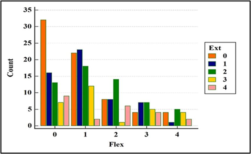
• All five scores (0 to 4) were then evaluated to obtain more details. In the IG, the
combinations of the scores of the EAs and FAs were statistically significant, with
p = 0.0248 (Figure 2). Only 13.7% (n.33) of the stifles were normal. The stifles with
normal EAs (score 0) were associated with abnormal FAs distributed in the slight
prevalence score. The stifles with normal FAs (score 0) were associated with abnormal
EAs mainly distributed in the slight and the mild scores. Overall, the alterations of the
EAs were found in all the scores of severity; instead, the prevalence of the frequency
of the alterations of the FA was found in score 1 (slight). The contingency coefficient
was 0.33.
• The dogs were grouped based on weight: Group A < 20 kg, Group B from 21 to 30 kg
and Group C > 31 kg. The demographic data are listed in Table 1. The chi-square test
for the trends revealed a statistically significant difference in the EA among the dogs
in Groups B and C, having greater alterations (p = 0.0098 and p = 0.0124, groups B and
C, respectively). The alterations in the FA were not significant.
• No significant differences were found between males and females in either the exten-
sion or the flexion angles.
• All ruptures were present for more than 2 weeks. Dogs with known lameness and/or
trauma occurrence within the past 30 days were classified as the acute group (AG).
There were 16 dogs (6.84%) (out of 234) with acute injuries. The chi-square test for the
relationship of NAs and AAs was not significant (p = 0.36). There were 218 (93.16%)Vet. Sci. 2021, 8, 119 5 of 11
dogs with chronic disease (Chronic Group: ChG), and the chi-square test revealed a
significant difference in the normal and abnormal angles of motion (NAs and AAs),
p = 0.027. In the ChG, the alteration of extension was prevalent at flexion, having a
percentage of 19.3%, which was the same as the value found in the IG (Table 3).
• The IG was divided into two groups of normal and abnormal muscle mass. The
investigation revealed that NMG included 22 dogs, and there was not a significant
difference between normal and abnormal angles (p = 0.55). The DMG consisted of
212 dogs, and there was a significative difference between NAs and AAs (p = 0.019).
The percentage of the combination of normal and abnormal extension and flexion had
the same trend of IG (Table 3).
• In the CG, 20 healthy dogs showed 85% of the dogs having a normal ROM. The
remaining stifles (15%) had scores slight or mild; the latter dogs weighed > 20 kg and
Vet. Sci. 2021, 8, x FOR PEER REVIEW were >7 years of age (Figure 1 and Tables 2 and 3). 5
Table 2. Distribution in frequency (n.) and percentage (%) of the extension and flexion angle scores in both the CCL injury
group and the control group.
• In the IG, the chi-square test revealed a statistically significant relationship o
Angle Motion NAs and AAs between Scores the two angle motion groups (EA and Total
FA)AA(p = 0.01). T
0 (normal)
was a reduced 1 (slight)
ROM in 286.3% (mild) of the3 (moderate)
cases, and there4 (severe)
was a significant prevalen
EA 70 (30%)
the alteration of the extension angle. The combinations of the164
54 (23%) 58 (25%) 29 (12%) 23(10%) (70%) were anal
scores
IG
FA 77 (33%) 76 (32%) 38 (16%) 27 (12%) 16 (7%) 157 (67%)
13.7% of the dogs had a normal ROM, 50.9% of stifles had a reduced ROM due
EA 17 (85%) 2 (10%) 1 (5%) -
CG alteration of both movements, 19.2% had abnormal- extension3 (15%) with normal fle
FA 17 (85%) 3 (15%) - - - 3 (15%)
and 16.2% had abnormal flexion and normal extension (Figure 1 and Table 3). T
IG: Injury Group; CG: Control Group; EA: extension angle; FA: flexion angle; AA: abnormal angle (scores 1, 2, 3 or 4).
was a prevalence of abnormal extension, having a contingency coefficient of 0.1
Figure
Figure 1. 1.
PiePie chart
chart showing
showing the distribution
the distribution of the combinations
of the combinations of and/or
of the normal the normal and/or abn
abnormal
angles
angles of extension
of extension and flexion
and flexion in the IGin
andthe
theIG
CG.and the CG.
IG: injury IG:CG:
group; injury group;
control group; CG: control group
NG: normal
normal
group; group;
EAAG: EAAG:
extension extension
angle abnormalangle abnormal
(reduced) (reduced)
group; FAAG: group;
flexion FAAG: flexion
angle abnormal angle abn
(increased)
(increased)
group; group; EFAG:
EFAG: extension extension
and flexion and flexion
angle abnormal angle abnormal group.
group.
Table 3. Percentage of score combinations of the normal and abnormal angles in the various gr
IG CG AG ChG NMG DM
NG (0 + 0) 32 (13.7%) 17 (85%) 4 (25%) 28 (12.8) 4 (18.2%) 28 (13.
EAAG (1 + 0) 45 (19.2) 1 (5%) 3 (18.8%) 42 (19.3%) 5 (22.7%) 40 (18.
FAAG (0 + 1) 38 (16.2%) 1 (5%) 2 (12.5%) 36 (16.5%) 3 (13.6%) 35 (16.
EFAG (1 + 1) 119 (50.9%) 1 (5%) 7 (43.8%) 112 (51.4%) 10 (45.5%) 109 (51
Total dog/group 234 20 16 218 22 212EER REVIEW
Vet. Sci. 2021, 8, 119 6 of 11
6 of 11
Figure
Figure 2.2.The
The chart
chart shows
shows the(0–4)
the scores scores (0–4)
of the ofangle
flexion the flexion anglewith
in combination in combination
extension angle.with
Count:extension angle.
number of stifles.
Count: number of stifles.
Table 3. Percentage of score combinations of the normal and abnormal angles in the various groups.
• The dogs were IG grouped based CG on weight: AG Group A < ChG20 kg, Group NMGB from 21 toDMG 30 kg
NG (0 +and0) Group C > 31 kg. The
32 (13.7%) demographic
17 (85%) data are listed
4 (25%) in Table 1.
28 (12.8) The chi-square
4 (18.2%) test
28 (13.2%)
EAAG (1for + 0)the trends revealed a statistically significant difference in the EA among the dogs
45 (19.2) 1 (5%) 3 (18.8%) 42 (19.3%) 5 (22.7%) 40 (18.9%)
FAAG (0 + 1) 38 (16.2%) 1 (5%) 2 (12.5%) 36 (16.5%) 3 (13.6%) 35 (16.5%)
EFAG (1in Groups
+ 1) 119B(50.9%)
and C, having greater alterations
1 (5%) 7 (43.8%) (p =1120.0098
(51.4%)and p =100.0124,
(45.5%) groups 109B and
(51.4%)
TotalC, respectively). The alterations in the FA were not significant.
234 20 16 218 22 212
dog/group
• No significant differences were found between males and females in either the exten-
IG: injury group; CG: control group; AG: acute group; ChG: chronic group; NMG: normal muscle group; DMG: decrease muscle group; NG:
normalsion
group;or theextension
EAAG: flexion angles.
angle abnormal (reduced) group; FAAG: flexion angle abnormal (increased) group; EFAG: extension and
•flexion angle abnormal group.
All ruptures were present for more than 2 weeks. Dogs with known lameness and/or
trauma occurrence within the past 30 days were classified as the acute group (AG).
4. Discussion
There were 16 dogsThe (6.84%) (out
aim of the of 234)
present studywith
was toacute injuries.investigate
retrospectively The chi-square
a correlationtest for a
between
the relationshipCCL of rupture
NAs and and the
AAsalterations
was not in thesignificant
ROM in stifle(p joints. The analysis
= 0.36). Thereof were
234 stifle joints
218
revealed that the CCL rupture led to a reduced ROM, likely due to a modification of the
(93.16%) dogs with chronic disease
joint biomechanics and/or(Chronic
modification Group: ChG), andstructures
of the periarticular the chi-square test
[32]. The authors’
revealed a significant hypothesesdifference
concerningin athe normalbetween
correlation and abnormal angles
the alterations of theoftwo
motion
angles (NAs
of motion
and AAs), p = 0.027. In the ChG, the alteration of extension was prevalent at flexion,and
and the fact that the alterations in the extension angle were statistically significant
greater than those in the flexion angle were confirmed. A previous study [46] carried out on
having a percentage of 19.3%, which was the same as the value found in the IG (Table
healthy dogs reported that the movement across the joint is flexor from ground contact until
3). mid-stance and then extensor until the end of the stance phase. In a dog with a CCL rupture
• The IG was divided in whichintothetwo groups
extension of normal
is limited, and also
its gait could abnormal muscleand
be compromised, mass. The in-that,
it is assumed
as in human knees, abnormal extension is less
vestigation revealed that NMG included 22 dogs, and there was not a significant tolerable than abnormal flexion [36,47].
dif-The
above-mentioned considerations provided the purpose of the investigation in the present
ference betweenstudy, normal and abnormal angles (p = 0.55). The DMG consisted of 212
as, to the best of the authors’ knowledge, no previous reports have examined the
dogs, and there incidence was a significative
and prevalencedifference
in movementbetweenchanges inNAs and
dogs as AAs (p = 0.019).
a consequence of a CCL Therupture.
percentage of the combination of normal and abnormal extension and flexion had the
same trend of IG (Table 3).
• In the CG, 20 healthy dogs showed 85% of the dogs having a normal ROM. The re-
maining stifles (15%) had scores slight or mild; the latter dogs weighed > 20 kg andVet. Sci. 2021, 8, 119 7 of 11
Not investigating the relationship of abnormal ROM with other parameters, such
as the severity or duration of lameness and scores of OA, was a limitation and potential
flaw or shortcoming of this study. It is known that the duration of lameness could be a
cause of OA progression [48] and that OA could lead to stiffness, a mechanical block from
osteophytes [49,50] and a loss of ROM [18,19]. Furthermore, in the literature, the lack of a
relationship between the functional capacity of dogs with OA and radiographic evidence
of disease has been described [51]. The aforementioned considerations were addressed
in the present study in order to verify whether a CCL rupture could be responsible for
the modification of one of the two angles of motion, or both, by means of evaluating a
large number of cases without limits of recruitment. Another limitation could be the breed
itself, keeping in mind that goniometric measurements of the stifle joints could be different
based on different breeds of healthy dogs [1,15–17]; however, in the present study, the most
common breed was the crossbreed.
The present study assessed the angles using a Likert scale from 0 to 4 in place of the
exact degree of each angle, obtained from the BHSII. The EA and FA measurements were
taken three times and averaged [33,36,45], and the value obtained was put into a range
of one of the five scores. Each score had a range of five degrees, so the authors assumed
that intra-observer and interobserver variability could be avoided. As the range of each
scale is known, anyone can deduce the degree of the angle in order to compare it with
other studies.
As expected, the authors found a correlation between the angles of extension and
flexion, as they are movements of the same joint. Despite this, there was a percentage
prevalence of the extension angle at the time of the diagnosis of CCL ruptures in dogs
weighing > 21 kg. This prevalence was in contrast with Sabanci’s study in which the body
weight of healthy dogs was the most influential factor regarding the flexion angle. It is
likely that this difference could have been due to the joint disease [16].
An investigation regarding the length of time of the disease found a difference in
sample sizes between the acute and the chronic groups, (16 vs. 218, respectively), which
did not allow the application of correct statistical tests. However, the results obtained from
the chi-square test assumed that a longer duration of the lesion could affect the alteration
of the ROM; the prevalence in the frequency of extension alteration was similar in the
entire IG.
Muscle mass has an influence on the ROM; in particular, an atrophied muscle can
modify the flexion angle. In other words, it can produce a reduction in the flexion degree.
This consideration was in contrast with the results of the present study, in which the
alteration of the extension was prevalent in the flexion angle. Therefore, the authors
assumed that the CCL rupture led to an alteration of extension angle due to other regions.
Several studies have investigated variations in ROM based on pain and muscle
mass [32–41]. Knowledge of the causes of a reduced flexion angle is associated with
muscle disuse and atrophy, while causes of an increased flexion angle are associated with
changes in the joint structures. Concerning extension, joint alterations can be acute (i.e.,
capsular effusion) [50,52,53], which, if treated quickly, could favour the recovery of normal
extension. When the alterations are chronic, they have already developed OA [50,52,53]
and are responsible for biomechanical influences that could result in a greater difficulty of
being resolved and in the consequent persistence of the reduction of the extension angle.
Pain is always a contributing factor to alterations in both angles [18,19]. All these variables
can be concomitant and consequent to the CCL rupture; therefore, the authors assumed
only the certain biomechanics and degenerative alterations that occur in the stifle joints
with a CCL rupture as the main parameter of ROM modification, reporting a prevalence
of reduced extension. Despite this, it was found that 13.7% (n. 33) of the stifles had no
change in ROM in contrast to what was expected. Additional studies carried out on a
greater number of stifles are necessary to investigate the possible maintenance of normal
angles of motion in dogs with a CCL rupture. Concerning the evaluation of the correlation
between the ROM and OA, the authors consider this report to be a preliminary study. OnceVet. Sci. 2021, 8, 119 8 of 11
the hypothesis is confirmed, it will be possible to carry out future studies to investigate the
influence of OA on the variations in the angles of extension and/or flexion.
As expected, the control group was helpful in showing that in the absence of cruciate
rupture ROM may be normal, except for older or overweight animals, which can be
assumed to have other influencing factors, e.g., OA.
In the present study, all the measurements were taken with the dogs sedated in order
to eliminate the interference of pain or tendon, ligament, capsule and muscle contractures.
In diseased joints, the ROM may be restricted in response to a painful sensation in awake
patients [2]. In effect, in Jaegger’s study, the measurements of the ROM in dogs awake and
sedated were identical [2]. However, the authors pointed out that their study regarded
a healthy population, and these results would be unlikely in dogs with degenerative
joints [2]. For the same reason, in 2010, Mostafa [38] also considered the ROM for all joints
in dogs under sedation in order to minimise the possible restriction of a painful response.
The present study confirmed the results of Mostafa’s study [38], in which Labradors
with and without CCL deficiency were compared and the goniometric measurements of
extension and flexion were abnormal and statistically significant for the extension, as in the
present study.
The results of this study were discordant with the Jandy report [36], which assessed
412 dogs with a CCL rupture that had both the angles of extension and flexion as normal [2].
These normal values were reported prior to surgical treatment in several dogs and, also,
after tibial plateau levelling osteotomy (TPLO) treatment, and there was no significant
correlation between flexion and extension in the dogs examined. Moreover, a loss of
flexion or extension greater than 10◦ was associated with greater lameness [36]. Other
studies have reported alterations in the ROM after surgical treatment and have often only
stated whether or not there was an improvement, without reporting the degree of the angle
measurements [32,33,35,37–39,41]. In 2010, Au reported the pre- and postoperative ROM as
not being significant [36]; however, he did not report the degree of the angles. The Moeller
and Mölsä studies reported the angles measured after TPLO treatment and compared
them to the contralateral limbs [32,33]; however, they did not indicate the degrees of the
angles prior to surgery. In 2013, McDonald reported that the ROM in an affected limb
was significantly less than that of a healthy contralateral limb [41]; however, he did not
compare the flexion and extension, as was done in the present study. The papers in the
literature did not use the same criteria for evaluating the ROM; therefore, the results of the
above-mentioned reports could not be compared with those of the present study, which
reported degree scores for both angles.
Given that, for the most part, gait is performed with the limb in weight-bearing
and in the stance phase of extension, this information and that mentioned above could
provide input for a screening program and preventive rehabilitation. Additional studies
are important for understanding each of the most frequently used surgical techniques that
can compromise and/or favour one of the angles of motion or both in order to address the
choice of treatment.
5. Conclusions
In conclusion, the authors believe that the results of this study could offer important
information regarding the influence of a CCL rupture in compromising the ROM. This
awareness could be a baseline for additional studies regarding the ability of surgical
treatments to restore one angle rather than another. Based on these findings, physiotherapy
could also be helpful with a novel approach in a nonsurgical and/or postsurgical treatment
program or in preparing dogs prior to surgery.
Author Contributions: Conceptualisation, S.P.; data curation, S.P. and C.T.; formal analysis, S.P.;
funding acquisition, S.P., F.L. and C.T.; methodology, S.P.; resources, F.L. and C.T.; supervision, S.P.
and F.L.; writing—original draft, S.P. and C.T. and writing—review and editing, S.P., F.L. and C.T. All
authors have read and agreed to the published version of the manuscript.Vet. Sci. 2021, 8, 119 9 of 11
Funding: This research received no external funding.
Institutional Review Board Statement: Not applicable.
Informed Consent Statement: Not applicable.
Conflicts of Interest: The authors declare no conflict of interest.
References
1. Hady, L.L.; Fosgate, G.T.; Weh, J.M. Comparison of range of motion in Labrador Retrievers and Border Collies. J. Vet. Med. Anim.
Health 2015, 7, 122–127.
2. Jaegger, G.; Marcellin-Little, D.J.; Levine, D. Reliability of goniometry in Labrador Retrievers. Am. J. Vet. Res. 2002, 63, 979–986.
[CrossRef] [PubMed]
3. Mitchell, W.S.; Millar, J.; Sturrock, R.D. An evaluation of goniometry as an objective parameter for measuring joint motion. Scott
Med. J. 1975, 20, 57–59. [CrossRef] [PubMed]
4. Boone, D.C.; Azen, S.P.; Lin, C.M.; Spence, C.; Baron, C.; Lee, L. Reliability of goniometric measurements. Phys. Ther. 1978, 58,
1355–1360. [CrossRef] [PubMed]
5. Goodwin, J.; Clark, C.; Deakes, J.; Burdon, D.; Lawrence, C. Clinical methods of goniometry: A comparative study. J. Phys. Ther
Sci. 1992, 14, 10–15. [CrossRef]
6. Svensson, M.; Lind, V.; Löfgren Harringe, M. Measurement of knee joint range of motion with a digital goniometer: A reliability
study. Physiother. Res. Int. 2019, 24, e1765. [CrossRef]
7. Grohmann, J.E.L. Comparison of two methods of goniometry. Phys. Ther. 1983, 63, 922–925. [CrossRef]
8. Clapper, M.P.; Wolf, S.L. Comparison of the reliability of the Orthoranger and the standard goniometer for assessing active lower
extremity range of motion. Phys. Ther. 1988, 68, 214–218. [CrossRef]
9. Fraeulin, L.; Holzgreve, F.; Brinkbäumer, M.; Dziuba, A.; Friebe, D.; Klemz, S.; Schmitt, M.; Theis, A.A.L.; Tenberg, S.;
Van Mark, A.; et al. Intra-and inter-rater reliability of joint range of motion tests using tape measure, digital inclinometer and
inertial motion capturing. PLoS ONE 2020, 15, e0243646. [CrossRef]
10. Jaeger, G.H.; Marcellin-Little, D.J.; DePuy, V.; Lascelles, B.D.X. Validity of goniometric joint measurements in cats. Am. J. Vet. Res.
2007, 68, 822–826. [CrossRef]
11. Liljebrink, Y.; Bergh, A. Goniometry: Is it a reliable tool to monitor passive joint range of motion in horses? Equine Vet. J. 2010, 42,
676–682. [CrossRef]
12. Adair, H.S., III; Marcellin-Little, D.J.; Levine, D. Validity and repeatability of goniometry in normal horses. Vet. Comp. Orthop.
Traumatol. 2016, 29, 314–319. [CrossRef]
13. Bergh, A.; Lauridsen, N.G.; Hesbach, A.L. Concurrent Validity of Equine Joint Range of Motion Measurement: A Novel Digital
Goniometer versus Universal Goniometer. Animals 2020, 10, 2436. [CrossRef]
14. Govoni, V.M.; Rahal, S.C.; Agostinho, F.S.; Conceição, R.T.; Tsunemi, M.H.; El-Warrak, A.O. Goniometric measurements of the
forelimb and hindlimb joints in sheep. Vet. Comp. Orthop. Traumatol. 2012, 25, 297–300. [CrossRef]
15. Thomovsky, S.A.; Chen, A.V.; Kiszonas, A.M.; Lutskas, L.A. Goniometry and limb girth in miniature Dachshunds. J. Vet. Med.
2016, 2016, 5846052. [CrossRef] [PubMed]
16. Sabanci, S.S.; Ocal, M.K. Comparison of goniometric measurements of the stifle joint in seven breeds of normal dogs. Vet. Comp.
Orthop. Traumatol. 2016, 29, 214–219. [PubMed]
17. Formenton, M.R.; de Lima, L.G.; Vassalo, F.G.; Joaquim, J.G.F.; Rosseto, L.P.; Fantoni, D.T. Goniometric Assessment in French
Bulldogs. Front. Vet. Sci. 2019, 6, 424. [CrossRef] [PubMed]
18. Johnston, S.A. Osteoarthritis: Joint anatomy, physiology, and pathobiology. Vet. Clin. N. Am. Small Anim. Pract. 1997, 27, 699–723.
[CrossRef]
19. Crook, T.; McGowan, C.; Pead, M. Effect of passive stretching on the range of motion of osteoarthritic joints in 10 labrador
retrievers. Vet. Rec. 2007, 160, 545–547. [CrossRef]
20. Marcellin-Little, D.J.; Levine, D. Principles and application of range of motion and stretching in companion animals. Vet. Clin. N.
Am. Small Anim. Pract. 2015, 45, 57–72. [CrossRef]
21. Montasell, X.; Dupuis, J.; Huneault, L.; Ragetly, G.R. Short-and long-term outcomes after shoulder excision arthroplasty in 7
small breed dogs. Can. Vet. J. 2018, 59, 277–283.
22. Clarke, E.; Aulakh, K.S.; Hudson, C.; Barnes, K.; Gines, J.A.; Liu, C.C.; Aulakh, H.K. Effect of sedation or general anesthesia on
elbow goniometry and thoracic limb circumference measurements in dogs with naturally occurring elbow osteoarthritis. Vet.
Surg. 2020, 49, 1428–1436. [CrossRef] [PubMed]
23. Huňáková, K.; Hluchý, M.; Špaková, T.; Matejová, J.; Mudroňová, D.; Kuricová, M.; Rosocha, J.; Ledecký, V. Study of bilateral
elbow joint osteoarthritis treatment using conditioned medium from allogeneic adipose tissue-derived MSCs in Labrador
retrievers. Res. Vet. Sci. 2020, 132, 513–520. [CrossRef]
24. Bertocci, G.; Smalley, C.; Brown, N.; Bialczak, K.; Carroll, D. Aquatic treadmill water level influence on pelvic limb kinematics in
cranial cruciate ligament-deficient dogs with surgically stabilised stifles. J. Small Anim. Pract. 2018, 59, 121–127. [CrossRef]
25. Barnes, K.; Faludi, A.; Takawira, C.; Aulakh, K.; Rademacher, N.; Liu, C.C.; Lopez, M.J. Extracorporeal shock wave therapy
improves short-term limb use after canine tibial plateau leveling osteotomy. Vet. Surg. 2019, 48, 1382–1390. [CrossRef] [PubMed]Vet. Sci. 2021, 8, 119 10 of 11
26. Roh, Y.H.; Jung, J.H.; Lee, J.H.; Jeong, J.M.; Jeong, S.M.; Lee, H. Clinical Results of Distal Femoral Osteotomy for Treatment of
Grade 4 Medial Patella Luxation with Concurrent Distal Femoral Varus in Small Breeds Dogs: 13 Cases. J. Vet. Clin. 2020, 37,
135–140. [CrossRef]
27. Pinna, S.; Lanzi, F.; Tassani, C.; Mian, G. Intra-articular replacement of a ruptured cranial cruciate ligament using the Mini-
TightRope in the dog: A preliminary study. J. Vet. Sci. 2020, 21, e53. [CrossRef]
28. Pinna, S.; Lanzi, F.; Grassato, L. Bologna Healing Stifle Injury Index: A Comparison of Three Surgical Techniques for the Treatment
of Cranial Cruciate Ligament Rupture in Dogs. Front. Vet. Sci. 2020, 7, 567473. [CrossRef]
29. Piras, L.A.; Mancusi, D.; Olimpo, M.; Gastaldi, L.; Rosso, V.; Panero, E.; Staffieri, B.; Peirone, B. Post-operative analgesia following
TPLO surgery: A comparison between cimicoxib and tramadol. Res. Vet. Sci. 2021, 136, 351–359. [CrossRef]
30. Chomsiriwat, P.; Ma, A. Comparison of the Effects of Electro-acupuncture and Laser Acupuncture on Pain Relief and Joint Range
of Motion in Dogs with Coxofemoral Degenerative Joint Disease. Am. J. Tradit. Chin. Vet. Med. 2019, 14, 11–20.
31. Alves, J.C.; Santos, A.; Jorge, P.; Lavrador, C.; Carreira, L.M. Clinical and diagnostic imaging findings in police working dogs
referred for hip osteoarthritis. BMC Vet. Res. 2020, 16, 425. [CrossRef]
32. Moeller, E.M.; Allen, D.A.; Wilson, E.R.; Lineberger, J.A.; Lehenbauer, T. Long-term outcomes of thigh circumference, stifle
range-of-motion, and lameness after unilateral tibial plateau levelling osteotomy. Vet. Comp. Orthop. Traumatol. 2010, 23, 37–42.
[PubMed]
33. Mölsä, S.H.; Hyytiäinen, H.K.; Hielm-Björkman, A.K.; Laitinen-Vapaavuori, O.M. Long-term functional outcome after surgical
repair of cranial cruciate ligament disease in dogs. BMC Vet. Res. 2014, 10, 266. [CrossRef] [PubMed]
34. Marsolais, G.S.; Dvorak, G.; Conzemius, M.G. Effects of postoperative rehabilitation on limb function after cranial cruciate
ligament repair in dogs. J. Am. Vet. Med. Assoc. 2002, 220, 1325–1330. [CrossRef] [PubMed]
35. Hoelzler, M.G.; Millis, D.L.; Francis, D.A.; Weigel, J.P. Results of arthroscopic versus open arthrotomy for surgical management of
cranial cruciate ligament deficiency in dogs. Vet. Surg. 2004, 33, 146–153. [CrossRef]
36. Jandi, A.S.; Schulman, A.J. Incidence of motion loss of the stifle joint in dogs with naturally occurring cranial cruciate ligament
rupture surgically treated with tibial plateau leveling osteotomy: Longitudinal clinical study of 412 cases. Vet. Surg. 2007, 36,
114–121. [CrossRef] [PubMed]
37. Au, K.K.; Gordon-Evan, W.J.; Dunning, D.; O’Dell-Anderson, K.J.; Knap, K.E.; Griffon, D.; Johnson, A.L. Comparison of short-and
long-term function and radiographic osteoarthrosis in dogs after postoperative physical rehabilitation and tibial plateau leveling
osteotomy or lateral fabellar suture stabilization. Vet. Surg. 2010, 39, 173–180. [CrossRef]
38. Mostafa, A.A.; Griffon, D.J.; Thomas, M.W.; Constable, P.D. Morphometric characteristics of the pelvic limb musculature of
Labrador Retrievers with and without cranial cruciate ligament deficiency. Vet. Surg. 2010, 39, 380–389. [CrossRef]
39. Drygas, K.A.; McClure, S.R.; Goring, R.L.; Pozzi, A.; Robertson, S.A.; Wang, C. Effect of cold compression therapy on postoperative
pain, swelling, range of motion, and lameness after tibial plateau leveling osteotomy in dogs. J. Am. Vet. Med. Assoc. 2011, 238,
1284–1291. [CrossRef]
40. Gordon-Evans, W.J.; Dunning, D.; Johnson, A.L.; Knap, K.E. Effect of the use of carprofen in dogs undergoing intense rehabilitation
after lateral fabellar suture stabilization. J. Am. Vet. Med. Assoc. 2011, 239, 75–80. [CrossRef]
41. MacDonald, T.L.; Allen, D.A.; Monteith, G.J. Clinical assessment following tibial tuberosity advancement in 28 stifles at 6 months
and 1 year after surgery. Can. Vet. J. 2013, 54, 249–254.
42. Monk, M.L.; Preston, C.A.; McGowan, C.M. Effects of early intensive postoperative physiotherapy on limb function after tibial
plateau leveling osteotomy in dogs with deficiency of the cranial cruciate ligament. Am. J. Vet. Res. 2006, 67, 529–536. [CrossRef]
43. Marsolais, G.S.; McLean, S.; Derrick, T.; Conzemius, M.G. Kinematic analysis of the hind limb during swimming and walking in
healthy dogs and dogs with surgically corrected cranial cruciate ligament rupture. J. Am. Vet. Med. Assoc. 2003, 222, 739–743.
[CrossRef] [PubMed]
44. Pinna, S.; Lanzi, F.; Grassato, L. Evidence-Based Veterinary Medicine: A Tool for Evaluating the Healing Process After Surgical
Treatment for Cranial Cruciate Ligament Rupture in Dogs. Front. Vet. Sci. 2020, 6, 65. [CrossRef] [PubMed]
45. Hyytiäinen, H.K.; Mölsä, S.H.; Junnila, J.T.; Laitinen-Vapaavuori, O.M.; Hielm-Björkman, A.K. Ranking of physiotherapeutic
evaluation methods as outcome measures of stifle functionality in dogs. Acta Vet. Scand. 2013, 55, 29. [CrossRef]
46. Colborne, G.R.; Innes, J.F.; Comerford, E.J.; Owen, M.R.; Fuller, C.J. Distribution of power across the hind limb joints in Labrador
Retrievers and Greyhounds. Am. J. Vet. Res. 2005, 66, 1563–1571. [CrossRef]
47. Irrgang, J.J.; Harner, C.D. Loss of motion following knee ligament reconstruction. Sports Med. 1995, 19, 150–159. [CrossRef]
48. Pinna, S.; Lanzi, F.; Cordella, A.; Diana, A. Relationship between the stage of osteoarthritis before and six months after tibial
tuberosity advancement procedure in dogs. PLoS ONE 2019, 14, e0219849. [CrossRef]
49. Pinna, S.; Landucci, F.; Tribuiani, A.M.; Carli, F.; Venturini, A. The effects of pulsed electromagnetic field in the treatment of
osteoarthritis in dogs: Clinical study. Pak. Vet. J. 2013, 33, 96–100.
50. Cook, J.L.; Luther, J.K.; Beetem, J.; Karnes, J.; Cook, C.R. Clinical comparison of a novel extracapsular stabilization procedure
and tibial plateau leveling osteotomy for treatment of cranial cruciate ligament deficiency in dogs. Vet. Surg. 2010, 39, 315–323.
[CrossRef]
51. Gordon, W.J.; Conzemius, M.G.; Riedesel, E.; Besancon, M.F.; Evans, R.; Wilke, V.; Ritter, M.J. The relationship between limb
function and radiographic osteoarthrosis in dogs with stifle osteoarthrosis. Vet. Surg. 2003, 32, 451–454. [CrossRef] [PubMed]Vet. Sci. 2021, 8, 119 11 of 11
52. Johnson, J.M.; Johnson, A.L. Cranial cruciate ligament rupture: Pathogenesis, diagnosis, and postoperative rehabilitation. Vet.
Clin. N. Am. Small Anim. Pract. 1993, 23, 717–733. [CrossRef]
53. Moore, E.V.; Weeren, R.; Paek, M. Extended long-term radiographic and functional comparison of tibial plateau leveling
osteotomy vs tibial tuberosity advancement for cranial cruciate ligament rupture in the dog. Vet. Surg. 2020, 49, 146–154.
[CrossRef] [PubMed]You can also read