Peptide Binding Sites of Connexin Proteins - MDPI
←
→
Page content transcription
If your browser does not render page correctly, please read the page content below
Communication
Peptide Binding Sites of Connexin Proteins
Ágnes Simon 1 , Csaba Magyar 2 , László Héja 1, * and Julianna Kardos 1, *
 1   Functional Pharmacology Research Group, Institute of Organic Chemistry, Research Centre for Natural
     Sciences, Hungarian Academy of Sciences, 1519 Budapest, Hungary; simon.agnes@ttk.mta.hu
 2   Institute of Enzymology, Research Centre for Natural Sciences, 1519 Budapest, Hungary;
     magyar.csaba@ttk.mta.hu
 *   Correspondence: heja.laszlo@ttk.mta.hu (L.H.); kardos.julianna@ttk.mta.hu (J.K.)

 Received: 1 June 2020; Accepted: 3 July 2020; Published: 14 July 2020

 Abstract: Intercellular gap junction (GJ) contacts formed by the coupling of connexin (Cx)
 hemichannels (HCs) embedded into the plasma membranes of neighboring cells play significant
 role in the development, signaling and malfunctions of mammalian tissues. Understanding and
 targeting GJ functions, however, calls for finding valid Cx subtype-specific inhibitors. We conjecture
 the lack of information about binding interactions between the GJ interface forming extracellular EL1
 and EL2 loops and peptide mimetics designed to specifically inhibit Cx43HC coupling to Cx43GJ.
 Here, we explore active spots at the GJ interface using known peptide inhibitors that mimic various
 segments of EL1 and EL2. Binding interactions of these peptide inhibitors and the non-peptide
 inhibitor quinine has been modelled in combination with the use of blind docking molecular mechanics
 (MM). The neuron-specific Cx36HC and astrocyte-specific Cx43HC subtypes were modelled with a
 template derived from the high-resolution structure of Cx26GJ. GJ-coupled and free Cx36HC and
 Cx43HC models were obtained by dissection of GJs (GJ-coupled) followed by 50 ns molecular
 dynamics (free). Molecular mechanics (MM) calculations were performed by the docking of inhibitors,
 explicitly the designed Cx43 EL1 or EL2 loop sequence mimetics (GAP26, P5 or P180–195, GAP27,
 Peptide5, respectively) and the Cx36 subtype-specific quinine into the model structures. In order
 to explore specific binding interactions between inhibitors and CxHC subtypes, MM/Generalized
 Born Surface Area (MM/GBSA) ∆Gbind values for representative conformers of peptide mimetics and
 quinine were evaluated by mapping the binding surface of Cx36HC and Cx43HC for all inhibitors.
 Quinine specifically contacts Cx36 EL1 residues V54-C55-N56-T57-L58, P60 and N63. Blocking the
 vestibule by the side of Cx36HC entry, quinine explicitly interacts with the non-conserved V54, L58,
 N63 residues of Cx36 EL1. In addition, our work challenges the predicted specificity of peptide
 mimetics, showing that the docking site of peptides is unrelated to the location of the sequence they
 mimic. Binding features, such as unaffected EL2 residues and the lack of Cx43 subtype-specificity of
 peptide mimetics, suggest critical roles for peptide stringency and dimension, possibly pertaining to
 the Cx subtype-specificity of peptide inhibitors.

 Keywords: connexin; gap junction; hemichannel; subtype-specific inhibitors; peptide mimetics; quinine

1. Introduction
     Gap junctions (GJs) formed by the coupling of various connexin (Cx) subtype hemichannels
(CxHCs, connexons) maintain adhesion and conduction between adjacent cells [1–3]. They are recognized
as critical players in the development and disease of mammalian tissues [4–6] (and reference cited).
All Cx subtypes contain two extracellular loops (EL1 and EL2) which are supposed to participate in GJ
formation [7]. Previously, Warner et al. [8] conjectured HC coupling to GJ and conserved amino acid
(AA) motifs, with QPG of EL1 being among the residues that possibly intervene HC coupling to GJ.

Chemistry 2020, 2, 662–673; doi:10.3390/chemistry2030042                     www.mdpi.com/journal/chemistry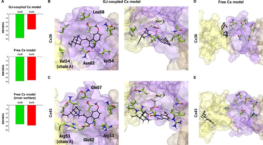
Chemistry 2020, 2                                                                                    663

Significantly, the consensus sequence of the conserved AAs of EL1 DEQSxFxCNTxQPGCxNVCYDxx
highlights sequences of fully (bold) or in at least 50% identical residues in all of some twenty human Cx
proteins [9]. One third of EL1 AAs (x) are less conserved, providing opportunity for subtype-specific
inhibitor design. An exposed tetrapeptide sequence within the EL2 sequence has also been emphasized
concerning the specificity of inter-connexon interaction [10,11].
     As a result, several EL1 or EL2 mimicking peptides targeted to inhibit the coupling of HC to GJ
have been developed [12–15]. Unexpectedly, GJ inhibitor peptide mimetics do block CxGJ-facilitated
intercellular communications, but in unanticipated ways [16]. We and others [2] have sought to develop
a more detailed understanding of HC coupling to GJ, first made apparent at the molecular level by the
discovery of the X-ray structure of Cx26 GJ at 3.5 Å resolution [17].
     As major subclasses of GJs shaping brain signaling, the astrocytic and neuronal GJs are formed
by astrocytic Cx43 and neuronal connexin36 (Cx36) [18]. Indeed, GJs consisting of Cx43 protomer
chains participate in long-range synchronized neural activity, underlying epilepsy [19,20] or slow-wave
sleep [21]. Our undertaking expects insights into the structural quality associated with Cx43 versus
Cx36 selectivity of peptide mimetic inhibitors. It is conceivable that, being identical with various
sequences of EL1 or EL2 of Cx43 (P5, GAP26 or Peptide5, GAP27, P180-195, respectively; Figure 1),
the peptide mimetic inhibitors designed to date shall meet the criteria of Cx43 subtype selectivity.
Importantly, however, the literature relating the functional relevance of peptide mimetics does not
validate the specificity [14,15,22–26]. Furthermore, the subtype specificity of these peptides is also
challenged by the fact that the protein segments they mimic are widely shared by other Cx subtypes,
as outlined above. Furthermore, subtype selectivity of another widely accepted selective GJ inhibitor,
the Cx36-specific (R)-(6-methoxyquinolin-4-yl)((2S,4S,8R)-8-vinylquinuclidin-2-yl)methanol (quinine)
has not been strengthened either [6,27–30]. We hypothesize that Cx subtype-specific coupling of HC to
GJ shall become true by undertaking the validation of the binding of mimetic peptides and quinine at
the extracellular interface of Cx43HC and/or Cx36HC.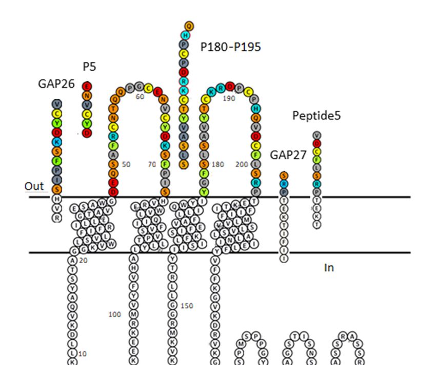
Chemistry 2020,
  Chemistry     2 2, x
            2020,                                                                                                 3664

      Figure 1. Plot of Cx43 protomer explaining sequence identity of peptide inhibitors GAP26, P5, GAP27,
         Figure 1. Plot of Cx43 protomer explaining sequence identity of peptide inhibitors GAP26, P5, GAP27,
      P180–195 and Peptide5. The membrane-embedded residues, including four transmembrane (TM)
         P180–195 and Peptide5. The membrane-embedded residues, including four transmembrane (TM)
      helices are recognized using the “positioning proteins in membrane” (PPM) server by the “orientation
         helices are recognized using the “positioning proteins in membrane” (PPM) server by the “orientation
      of of
         proteins  in membrane” (OPM) database [31]. AA colour code: aromatic—green; hydrophobic—gray;
            proteins in membrane” (OPM) database [31]. AA colour code: aromatic—green; hydrophobic—
      basic—blue;    acidic—red;
         gray; basic—blue;         polar neutral—orange;
                             acidic—red;                  Cys—lemon.
                                          polar neutral—orange;          Figure
                                                                 Cys—lemon.     was generated
                                                                              Figure           by Protter
                                                                                     was generated        [32].
                                                                                                    by Protter
         [32].
      We began this study by identifying the contact area between the peptide inhibitors and GJ-coupled
and freeWe Cx36/Cx43
              began thisconnexon
                           study bymodels     usingthe
                                       identifying   an contact
                                                         iterativearea
                                                                    blind  docking
                                                                        between   theapproach   that did not
                                                                                      peptide inhibitors   andrequire
                                                                                                                GJ-
existing
   coupledexperimental    bindingconnexon
              and free Cx36/Cx43     data [33]. models
                                                By blind   docking
                                                        using        peptideblind
                                                               an iterative  sequence    mimetics,
                                                                                   docking          wethat
                                                                                             approach   alsodid
                                                                                                             explored
                                                                                                                not
therequire
     valid existing
             bindingexperimental
                       area. Calculations      of the
                                      binding data     Molecular
                                                    [33].            Mechanics/Generalized
                                                          By blind docking                       Born Surface
                                                                             peptide sequence mimetics,          Area
                                                                                                            we also
(MM/GBSA)       ∆G
   explored the valid    values   were  performed   [34]  on the 30  best scoring  poses  of the blind
                    bind binding area. Calculations of the Molecular Mechanics/Generalized Born Surface docking  trials
to Area
   fit CxHC    peptide ΔG
         (MM/GBSA)       inhibitors   and
                            bind values    quinine.
                                        were         This[34]
                                              performed     method
                                                               on thehas  proved
                                                                       30 best     to beposes
                                                                               scoring   a powerful    tooldocking
                                                                                              of the blind  to predict
   trials to
binding      fit CxHC
           affinities   peptide
                      and         inhibitors
                           identify           and binding
                                      the correct  quinine.poses
                                                             This method    has proved tocomplexes
                                                                    for protein–peptide      be a powerful
                                                                                                        [35].tool to
   predict binding affinities and identify the correct binding poses for protein–peptide complexes [35].
2. Results and Discussion
   2. Results and Discussion
2.1. Homology Modelling of Homomeric GJs Formed by Cx36 or Cx43
   2.1. Homology Modelling of Homomeric GJs Formed by Cx36 or Cx43
      Homomeric GJ models built up exclusively by Cx36 or Cx43 protomers were constructed using the
X-ray structure (2zw3) of homologous Cx26 GJ [17] as a template and the Swiss-Model server facility [36].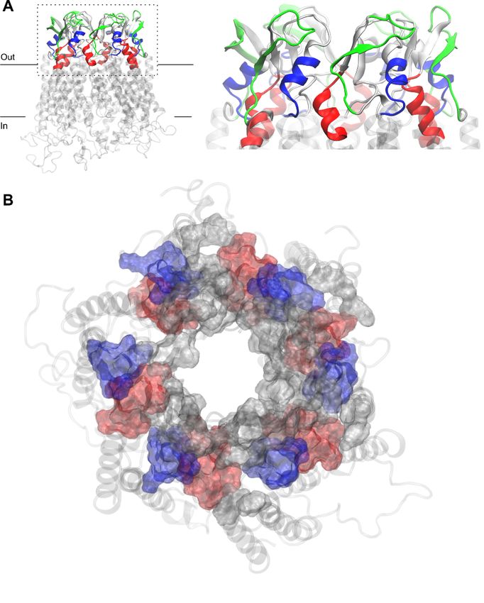
Chemistry 2020, 2, x                                                                                                4
Chemistry 2020, 2                                                                                                  665

       Homomeric GJ models built up exclusively by Cx36 or Cx43 protomers were constructed using
This initialstructure
the   X-ray            (2zw3)
             PDB structure        of homologous
                               includes   a whole GJ,Cx26   GJ [17]chains
                                                        explicitly   as a template     and the Swiss-Model
                                                                           A-B-C-D-E-F-G-H-I-J-K-L,            server
                                                                                                          shaping  the
facility [36]. This initial PDB    structure  includes   a whole   GJ, explicitly   chains A-B-C-D-E-F-G-H-I-J-K-
extracellular (interface) and transmembrane (TM) regions, except the large intracellular region, due to
L, potentially
its  shaping the     extracellular
                 disordered    nature.(interface)
                                         Sequencesand      transmembrane
                                                     of cytosolic               (TM) regions,
                                                                   residues, without               except determined
                                                                                         experimentally    the large
intracellular  region,  due  to  its potentially  disordered   nature.  Sequences     of cytosolic residues,
coordinates in the crystal structure, were utilized to connect individual TM helices intracellularly         without
experimentally    determined       coordinates   in the  crystal structure,  were    utilized to
using the built-in protocol of Swiss-Model server. Our model GJ structures enfold two hexameric  connect  individual
TM helices
HCs          intracellularly
       that are                 using theprotomer
                formed by apposed           built-in protocol
                                                       chains of  Swiss-Model
                                                               A-B-C-D-E-F         server. Our
                                                                                 coupled   withmodel
                                                                                                 chainsGJG-H-I-J-K-L.
                                                                                                           structures
enfold   two  hexameric   HCs     that  are formed   by  apposed   protomer     chains   A-B-C-D-E-F
Since there are no significant insertions or deletions among Cx subtypes in the extracellular and      coupled   with
                                                                                                                   TM
chains   G-H-I-J-K-L.   Since   there   are no  significant  insertions  or deletions    among   Cx
regions, the built-in automated alignment of Swiss-Model was used for creating individual homology   subtypes  in the
extracellular
models.    Theand   TM regions,
                modelling     server theputs
                                         built-in automatedatoms
                                             on hydrogen       alignment    of Swiss-Model
                                                                      and arranges     amino was
                                                                                               acidused
                                                                                                     (AA)for creating
                                                                                                           sidechains
individual homology models. The modelling server puts on hydrogen atoms and arranges amino
so that no clashes appear in the structure. Otherwise, the alpha carbon backbone of homomeric GJs
acid (AA) sidechains so that no clashes appear in the structure. Otherwise, the alpha carbon backbone
formed by Cx36 or Cx43 resembles the Cx26 structure, reflecting the 6-fold symmetry. Due to these
of homomeric GJs formed by Cx36 or Cx43 resembles the Cx26 structure, reflecting the 6-fold
preparatory steps, homology models were ready for the separation of the A-F chains from the whole
symmetry. Due to these preparatory steps, homology models were ready for the separation of the A-
of GJ (A-L). Hence, homomeric Cx36GJ and Cx43GJ models were used to cut off solo connexons,
F chains from the whole of GJ (A-L). Hence, homomeric Cx36GJ and Cx43GJ models were used to cut
i.e., Cx36HC or Cx43HC. To this end, G-L protomer chains of both Cx36 and Cx43 GJs were removed by
off solo connexons, i.e., Cx36HC or Cx43HC. To this end, G-L protomer chains of both Cx36 and Cx43
means of Schrödinger’s Maestro module [37]. The remaining A-F chains, representing the GJ-coupled
GJs were removed by means of Schrödinger’s Maestro module [37]. The remaining A-F chains,
model of Cx36HC and Cx43HC, are characterized by their extracellular front (Figure 2A) and top
representing the GJ-coupled model of Cx36HC and Cx43HC, are characterized by their extracellular
(Figure 2B) views.
front (Figure 2A) and top (Figure 2B) views.

      Figure 2. (A,B) Gap junction (GJ)-coupled homology model of Cx43HC with extracellular loops shown
      Figure 2. (A,B) Gap junction (GJ)-coupled homology model of Cx43HC with extracellular loops
      in cartoon representation in front view (A) and surf representation in top view (B). For the latter,
      shown in cartoon representation in front view (A) and surf representation in top view (B). For the
      the z-axis of the coordinate system was set up to point towards the channel. EL1 and EL2 loops of each
      latter, the z-axis of the coordinate system was set up to point towards the channel. EL1 and EL2 loops
      chain, depicted using the “positioning proteins in membrane” (PPM) server by the “orientation of
      of each chain, depicted using the “positioning proteins in membrane” (PPM) server by the
      proteins in membrane” (OPM) database [31] as detailed in Section 2.2, are colored by lighter and darker
      “orientation of proteins in membrane” (OPM) database [31] as detailed in Section 2.2, are colored by
      versions of the same color, respectively. Color codes for the different chains are as follows: yellow
      lighter and darker versions of the same color, respectively. Color codes for the different chains are as
      (A), violet (B), brown (C), blue (D), magenta (E), green (F). TM helices and intracellular segments are
      follows: yellow (A), violet (B), brown (C), blue (D), magenta (E), green (F). TM helices and intracellular
      shown in transparent light gray cartoon representation. (C,D) Free connexon models were obtained
      segments are shown in transparent light gray cartoon representation. (C,D) Free connexon models
      from the homology model of GJ-coupled Cx43HC (A,B) by applying 50 ns molecular dynamics to the
      were obtained from the homology model of GJ-coupled Cx43HC (A,B) by applying 50 ns molecular
      hemichannel. Views and color codes are the same as in A–B.
      dynamics to the hemichannel. Views and color codes are the same as in A–B.
     Since this structure represents the conformation of connexins in the full GJ form and peptides are
     Sincetothis
expected         structure
             interact with represents the conformation
                            the hemichannel            of connexins
                                            form, we applied         in the fulldynamics
                                                             50 ns molecular     GJ form and
                                                                                         (MD)peptides
                                                                                               to allow
are expected   to interact with the hemichannel form, we  applied 50  ns molecular   dynamics (MD)
the protein adopting the free hemichannel-like form not to resemble the prior presence of the apposedto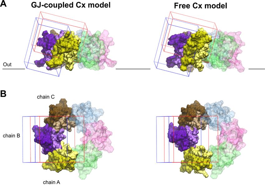
Chemistry 2020, 2                                                                                   666

connexon (G-L chains). Molecular dynamics (MD) calculations were performed using the Desmond
software package obtained from DE Shaw Research [37]. Cx36HC and Cx43HC transmembrane regions
were defined according to Section 2.2. These HCs were then prepared via the Protein Preparation
Wizard, and membranes were added using the System Builder menu by placing the membrane on the
pre-aligned structure obtained from PPM. The model was relaxed before simulation and molecular
dynamics was run at 300 K for a total simulation time of 50 ns at the supercomputer facility of the
Governmental Agency for IT Development (KIFU), Hungary. The resulting free Cx36HC and Cx43HC
model structures (Figure 2C,D) were used to dock mimetic peptides.

2.2. Determination of the Position of TM Regions
     Successful prediction of inhibitor binding at extracellular CxHC areas necessitates the realistic
arrangement of membrane bilayers. Explicitly, the claim concerns the genuine arrangement of
extracellular loops EL1 and EL2 of CxHC subtypes, critically depending on the valid position of
the membrane bilayer. Initially, the determination of the position of TM regions was focused on
the primary sequence of the Cx43 protein. According to Delvaeye et al. [15], we also sampled the
sequence-based prediction of TM regions based on Uniprot’s TM assignment as being 14–36, 77–99,
155–177, 209–231. These data were based on the prediction of the Transmembrane Protein Topology
with a Hidden Markov Model (TMHMM) server [38], although the prediction of the first TM region
of Cx43 was mistakenly identified as 14–36, and is currently under correction (Uniprot—personal
communication). When TM regions were assigned according to 3D structures of our CxHC models,
a significantly different TM1 region 21–46 was obtained, using the “positioning proteins in membrane”
(PPM) server by the “orientation of proteins in membrane” (OPM) database [31]. To clarify the issue of
real membrane boundaries determining extracellular EL1/EL2 domains, a series of sequence-based
predictions of the TM regions from the CCTOP server [39] were weighed against the 3D structure-based
predictions of the TM regions from the PPM server by the OPM database [31] in addition to an earlier
TMDET server [40]. The procedure of the 3D structure-based assessments of the TM regions via the
PPM and OPM approach seems to provide more detailed information about the membrane-embedded
amino acids (AAs). This way, the membrane boundaries were determined, providing the consensus
Cx36-membrane embedded amino acids (AAs) 3–9, 12, 23–49, 67, 71–95/94, 198, 200–225/198–225,
246–274/273. These AAs comprise secondary structures such as TM helices 23–49, 75–92/95, 200–220,
247–273. Similarly, the embedded AAs of Cx43 display comparable arrangement of AAs 3–9, 12/12–13,
21–46, 48, 71–94, 154–180, 202, 204–230, 232 shaping TM helices 21–46, 74–93, 156–176, 204–229/230.
     TM domains, characterized by two shorter and two longer TM helices in succession together with
the embedded residues near the N-terminal, are recognized. It is worth mentioning that structural motifs
of CxHCs [2,17,41,42] (also this work) may reveal similar mechanistic clues such as the ball-and-chain
inactivation in the K+ channel [43]. In fact, the ball-and-chain mechanism was further confirmed by
the recently determined 3D structure of Cx26 [44].

2.3. Topological Arrangements of Cx43 EL1 and EL2 Sequences Identical with EL1-Mimetic GAP26 and
EL2-Mimetic GAP27 or P180-195
     The question arises as to whether the peptide mimetics designed to specifically inhibit Cx43HC
coupling to Cx43GJ shall act along the GJ-interface surface. Figure 3 shows that AA residues of
GAP26 (blue), GAP27 (red) and P180–195 (green) shape extracellular Cx43HC interfaces. Explicitly,
the AAs corresponding to mimetic peptides GAP26, GAP27 and P180–195 are principally located at
interfaces between inner EL1 and outer EL2 or at the peripheral boundary interface of EL2 (Figure 3).
The arrangement illustrates that permutations of the vertical inter-loop interface joined with the
horizontal loop-periphery interface are beyond the most exposed EL1 loop sequences, lying on the
front of the CxHC coupling reaction. These findings conclusively suggest that the location of interfaces
identified by matching AA sequences of designed peptide mimetics argue against the notion that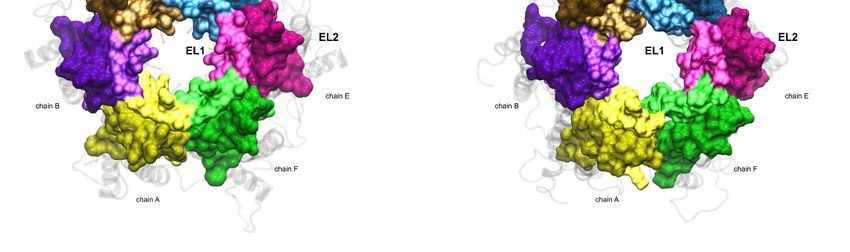
Chemistry 2020, 2                                                                                                        667

Chemistry 2020, 2, x                                                                                                       6

mimetics could directly inhibit connexon coupling to GJ. Notably, EL1 sequences contiguous to channel
notion that mimetics could directly inhibit connexon coupling to GJ. Notably, EL1 sequences
forming TM helices take the shape of a vestibule by the side of HC entry.
contiguous to channel forming TM helices take the shape of a vestibule by the side of HC entry.

      Figure 3. Residues of free Cx43HC model corresponding to three commonly used peptide mimetic
      inhibitors  derived from
      Figure 3. Residues          different
                             of free  Cx43HC EL1  and EL2
                                               model       sequences, to
                                                       corresponding   GAP26
                                                                          three(blue),
                                                                                 commonlyGAP27   (red)
                                                                                              used     and P180–195
                                                                                                     peptide  mimetic
      (green) in front
      inhibitors       (A) from
                  derived  and top    (B) views
                                  different       andz-axis
                                             EL1with        pointing towards
                                                       EL2 sequences,  GAP26the     channel.
                                                                                (blue),  GAP27P180–195   labeling
                                                                                                 (red) and        was
                                                                                                             P180–195
      removed    from  (B) for  clarity. Peptide  residues are shown   in surf representation   on   the
      (green) in front (A) and top (B) views with z-axis pointing towards the channel. P180–195 labeling extracellular
      region  of Cx43. from
      was removed       Other(B)residues  of EL1 and
                                     for clarity.      EL2,residues
                                                   Peptide  not corresponding
                                                                     are shown   to in
                                                                                    peptide
                                                                                        surf mimetic   inhibitors,
                                                                                             representation    on are
                                                                                                                   the
      shown   in gray  surf. The   membrane-embedded       residues, including   four  TM  helices  recognized
      extracellular region of Cx43. Other residues of EL1 and EL2, not corresponding to peptide mimetic         using
      the PPM server
      inhibitors,       by OPM
                   are shown    indatabase   [31],
                                    gray surf.  Thearemembrane-embedded
                                                       shown in the transparent   light gray
                                                                             residues,        cartoon.
                                                                                         including   four TM helices
      recognized using the PPM server by OPM database [31], are shown in the transparent light gray
2.4. Validation of Blind Docking Procedure via Optimization of MM/GBSA ∆Gbind Values
      cartoon.
     Docking calculations were performed using the Schrödinger Small-Molecules Drug Discovery Suite
2020-1 softwareofpackage
2.4. Validation          [45]. The
                  Blind Docking    in silicovia
                                Procedure    calculations were
                                                Optimization    performed based
                                                             of MM/GBSA   ΔGbind on the recommendations
                                                                                 Values
of Tubert-Brohman et al. [46] for peptide docking with increased accuracy. The Glide SP-Peptide mode
      Docking calculations were performed using the Schrödinger Small-Molecules Drug Discovery
was used for docking with subsequent MM/GBSA ∆Gbind ranking calculations [34]. The structures of
Suite 2020-1 software package [45]. The in silico calculations were performed based on the
the connexins Cx36 and Cx43 were prepared using the Protein Preparation Wizard program, while the
recommendations of Tubert-Brohman et al. [46] for peptide docking with increased accuracy. The
quinine structure was prepared using the Ligprep module. Two binding area definitions were used
Glide SP-Peptide mode was used for docking with subsequent MM/GBSA ΔGbind ranking calculations
around the EL1–EL2 loop regions, and docking grids suitable for peptide docking were generated
[34]. The structures of the connexins Cx36 and Cx43 were prepared using the Protein Preparation
(Figure 4). The ligand diameter midpoint box sizes were increased from the default to values between
Wizard program, while the quinine structure was prepared using the Ligprep module. Two binding
30 and 40 Angströms to cover the whole binding site region. During the Glide SP-Peptide mode docking
area definitions were used around the EL1–EL2 loop regions, and docking grids suitable for peptide
calculations, sampling was enhanced by a factor of 2 and the expanded sampling option was used as
docking were generated (Figure 4). The ligand diameter midpoint box sizes were increased from the
well. Because of the increased volume of the binding site region, instead of the default number 1000,
default to values between 30 and 40 Angströms to cover the whole binding site region. During the
the 3000 best poses were used for energy minimization to assure an exhaustive search of the possible
Glide SP-Peptide mode docking calculations, sampling was enhanced by a factor of 2 and the
expanded sampling option was used as well. Because of the increased volume of the binding site
region, instead of the default number 1000, the 3000 best poses were used for energy minimization to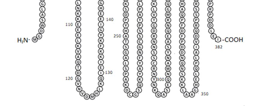
Chemistry 2020,
Chemistry 2 x
2020, 2, 668
7
assure an exhaustive search of the possible binding poses. The Schrödinger peptide docking protocol
binding poses. The Schrödinger peptide docking protocol uses an initial Macromodel conformational
uses an initial Macromodel conformational search instead of the Confgen conformational search
search instead of the Confgen conformational search performed by the Ligand docking protocol.
performed by the Ligand docking protocol. To model the peptide docking procedure, a Macromodel
To model the peptide docking procedure, a Macromodel conformational search was performed on
conformational search was performed on all ligand structures. The resulting conformers were
all ligand structures. The resulting conformers were clustered into five structurally different clusters.
clustered into five structurally different clusters. Based on visual inspection, only representative
Based on visual inspection, only representative structures for the first three clusters were taken into
structures for the first three clusters were taken into account during docking calculations with the
account during docking calculations with the “Canonicalize input conformation” Glide option turned
“Canonicalize input conformation” Glide option turned off. The MM/GBSA ΔGbind calculations were
off. The MM/GBSA ∆Gbind calculations were performed on the best 30 poses. The resulting poses from
performed on the best 30 poses. The resulting poses from the two docking grids were combined and
the two docking grids were combined and analyzed together.
analyzed together.
Figure 4.
Figure Validation of
4. Validation of blind
blinddocking
dockingsearch
searchforforthe binding
the binding crevices of Cx
crevices inhibitors.
of Cx Front
inhibitors. (A) (A)
Front andand
top
(B) views of boundaries limiting docking grids in the GJ-coupled (left) and the free (right)
top (B) views of boundaries limiting docking grids in the GJ-coupled (left) and the free (right) connexon connexon
models. The
models. The grid
grid positioned
positioned atat the
the water-facing,
water-facing, outer
outer side
side of
of EL2
EL2 is
is shown
shown in in blue.
blue. The
The other
other grid,
grid,
covering the EL1–EL2 interface as well as the top region of the pore-facing EL1 and
covering the EL1–EL2 interface as well as the top region of the pore-facing EL1 and water-facing EL2 water-facing EL2
loops, is shown in red. The significant overlap between the two grids allows cross-validation
loops, is shown in red. The significant overlap between the two grids allows cross-validation of of docking
results. Only
docking EL1Only
results. and EL1
EL2 loops
and EL2of Cx43
loopsare
of displayed. EL1 and EL2
Cx43 are displayed. EL1loops of each
and EL2 chain
loops are colored
of each by
chain are
lighter and darker versions of the same color, respectively. Color codes for the different
colored by lighter and darker versions of the same color, respectively. Color codes for the different chains are as
follows: yellow (A), violet (B), brown (C), blue (D), magenta (E), green (F). Chains not included in the
chains are as follows: yellow (A), violet (B), brown (C), blue (D), magenta (E), green (F). Chains not
docking grids (D–F) are displayed in transparent colors. For the position of the outer membrane, see
included in the docking grids (D–F) are displayed in transparent colors. For the position of the outer
legends to Figures 1–3 and Section 2.2.
membrane, see legends to Figures 1–3 and Section 2.2.
2.5. Mapping Binding Interactions of Inhibitory Peptide Mimetics and Quinine in Model CxHC Structures
2.5. Mapping Binding Interactions of Inhibitory Peptide Mimetics and Quinine in Model CxHC Structures
MM/GBSA ∆Gbind values obtained for the best 30 blind docking runs (see previous paragraph)
for quinine and ΔG
MM/GBSA eachbind values obtained for the best 30 blind docking runs (see previous paragraph) for
peptide mimetic inhibitors have been evaluated. The best MM/GBSA ∆Gbind
quinine and each from
values obtained peptide mimetic
docking inhibitors
quinine haveor
to Cx36 been
Cx43evaluated.
GJ-coupledThe models
best MM/GBSA
indicate ΔG bind values
that quinine
obtained from docking quinine to Cx36 or Cx43 GJ-coupled models indicate
prefers binding to Cx36 over Cx43 (Figure 5A–C). However, docking quinine into the free Cx36HC that quinine prefers
binding
and Cx43HCto Cx36 over Cx43
structures (Figure
did not show5A–C). However, docking
subtype-specificity (Figurequinine
5A,D,E).into the structure,
In this free Cx36HC and
quinine
Cx43HC structures did not show subtype-specificity (Figure 5A,D,E). In this structure,
was not able to dock onto the same surface identified in docking to the GJ-coupled Cx36 structure, quinine was
not
andable to dock
instead onto the
preferred sametosurface
docking identified
the outer surfaceinofdocking to the GJ-coupled
the extracellular region (FigureCx365D,E).
structure, and
Filtering
instead preferred
the docking docking
results for posesto the outer
on the surface
inner of the
surface stillextracellular
did not revealregion (Figure 5D,E). Filtering
any subtype-specific the
interaction
docking results for poses on the inner surface still did not reveal any subtype-specific
(Figure 5A, bottom). These data suggest that quinine shall exert its subtype-specificity by entering the interaction
(Figure
GJ from5A,thebottom).
cytosol.These data suggest
Altogether, that quinine
the claimed shall exert its of
subtype-specificity subtype-specificity
neuronal-type Cx36GJ by entering the
inhibitor
GJ from the cytosol. Altogether, the claimed subtype-specificity of neuronal-type Cx36GJ inhibitor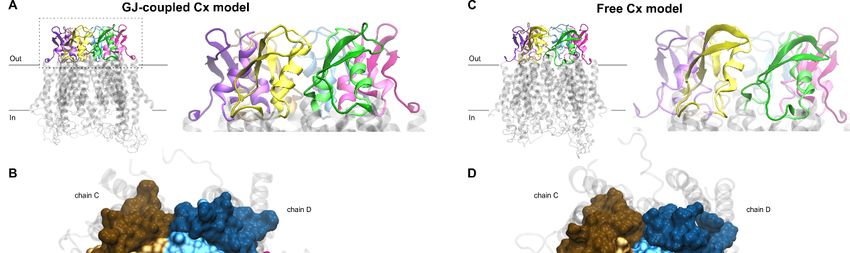
Chemistry 2020, 2                                                                                                                               669

Chemistry 2020, 2, x                                                                                                                            8

quinine [6,27–30] has been substantiated by molecular modelling of binding at the GJ-coupled, but not
 quinine [6,27–30] has been substantiated by molecular modelling of binding at the GJ-coupled, but
in the free Cx36HC versus Cx43HC model structures (Figure 5).
 not in the free Cx36HC versus Cx43HC model structures (Figure 5).

       Figure5.5.Docking
      Figure        Dockingresults
                                resultsconfirm
                                          confirmCx36-specificity
                                                    Cx36-specificity     ofof  quinine
                                                                            quinine    in in
                                                                                          thethe   GJ-coupled
                                                                                                GJ-coupled         connexin
                                                                                                               connexin        model.
                                                                                                                          model.    (A)(A)
                                                                                                                                         Best
       Best MM/GBSA
      MM/GBSA         ∆Gbind     bind scores
                             ΔGscores         of quinine
                                         of quinine    afterafter
                                                              dockingdocking
                                                                         to Cx36to Cx36
                                                                                    or Cx43or Cx43
                                                                                                in theinGJ-coupled
                                                                                                         the GJ-coupledmodel model
                                                                                                                                (top),(top),
                                                                                                                                       in the
       in the
      free     free connexin
           connexin      model model
                                   (middle)(middle)    and filtering
                                             and filtering               the results
                                                                the results              of the
                                                                                of the free       free connexin
                                                                                               connexin    model model       to thesurface
                                                                                                                     to the inner     inner
       surface   (bottom).    (B,C)   Best  docking     conformation        of  quinine    in   Cx36   (B)
      (bottom). (B,C) Best docking conformation of quinine in Cx36 (B) and Cx43 (C) GJ-coupled models in    and   Cx43   (C)  GJ-coupled
       models
      side  viewinfrom
                     side inside
                          view from      inside
                                    the pore     theand
                                              (left)   poretop(left)
                                                                 viewand(right)
                                                                          top view
                                                                                 based (right)  based on MM/GBSA
                                                                                          on MM/GBSA                       ΔGbind
                                                                                                              ∆Gbind values.       values.is
                                                                                                                                 Quinine
       Quinine     is shown     in  sticks  (black).   Residue     sidechains     in the   vicinity   (
Chemistry 2020, 2 670
The best MM/GBSA ∆Gbind scores of peptide mimetic inhibitors after docking to the free Cx36HC
Chemistry 2020, 2, x 9
or Cx43HC models indicate that most peptides bind to both Cx36 and Cx43 subtypes (Figure 6A),
although the Cx43 EL2 sequence mimetic P180–195 seems to be rather Cx36 subtype-specific.
although the Cx43 EL2 sequence mimetic P180–195 seems to be rather Cx36 subtype-specific.
Importantly, however, binding surfaces of peptide mimetic inhibitors do not correspond to the
Importantly, however, binding surfaces of peptide mimetic inhibitors do not correspond to the
sequence mimicked by the particular peptides (Figure 6B,C). In fact, the possible appearance of novel
sequence mimicked by the particular peptides (Figure 6B,C). In fact, the possible appearance of novel
binding hotspots for Cx43 peptide mimetic inhibitors has already been predicted just by the reduced
binding hotspots for Cx43 peptide mimetic inhibitors has already been predicted just by the reduced
accessibility of the pertinent EL1–EL2 regions (see Section 2.3, Figure 3). Peptides mimicking the EL1
accessibility of the pertinent EL1–EL2 regions (see Section 2.3, Figure 3). Peptides mimicking the EL1
loop (GAP26, P5) or the EL2 loop (GAP27, P180–195, peptide 5) all bind at the inner EL1 surface and the
loop (GAP26, P5) or the EL2 loop (GAP27, P180–195, peptide 5) all bind at the inner EL1 surface and
EL1–EL2 interface (Figure 6B,C), irrespective of the derivation of their sequences. Indeed, the subtype
the EL1–EL2 interface (Figure 6B,C), irrespective of the derivation of their sequences. Indeed, the
specificity of these peptides is also challenged by the fact that the protein segments they mimic are
subtype specificity of these peptides is also challenged by the fact that the protein segments they
widely shared by other Cx subtypes, as previously introduced. Consequently, these structural and
mimic are widely shared by other Cx subtypes, as previously introduced. Consequently, these
docking data
structural andsuggest
dockingthat
datathe rationale
suggest that behind the design
the rationale behindofthe
peptidomimetics may not be may
design of peptidomimetics validnot
for
connexin gap junction proteins.
be valid for connexin gap junction proteins.
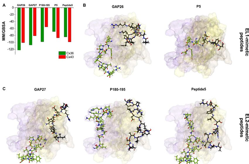
Figure 6. Docking positions of peptide mimetic inhibitors do not correspond to the mimicked
Figure 6. Docking positions of peptide mimetic inhibitors do not correspond to the mimicked sequences.
sequences. (A) Best MM/GBSA ΔGbind values of peptide mimetic inhibitors after docking to Cx36 or
(A) Best MM/GBSA ∆Gbind values of peptide mimetic inhibitors after docking to Cx36 or Cx43 free
Cx43 free connexon models. (B,C) Best docking conformations of peptide inhibitors (black) and
connexon models. (B,C) Best docking conformations of peptide inhibitors (black) and mimicked Cx43
mimicked Cx43 sequence (green) are shown as sticks from front view in the free Cx43HC structure.
sequence (green) are shown as sticks from front view in the free Cx43HC structure. Only EL1 and EL2
Only EL1 and EL2 loops of Cx43 A–C chains are displayed. Color codes for the different chains are as
loops of Cx43 A–C chains are displayed. Color codes for the different chains are as follows: yellow (A),
follows: yellow (A), violet (B), brown (C). For the definition of the position of the EL1 and EL2 see
violet (B), brown (C). For the definition of the position of the EL1 and EL2 see Section 2.2.
Section 2.2.
Our present understanding is that the particular reduction in the extracellular conformational
Our
freedom of present understanding
connexons during GJis formation
that the particular
increasesreduction in thesubtype-specificity
the connexon extracellular conformational
of quinine.
freedom of connexons during GJ formation increases the connexon subtype-specificity of quinine.
The principle predicts the “quinine paradox” (see above) but does not explain the lack of subtype-specificity
The principle
in the predicts
case of the thesequence
EL1/EL2 “quininemimetic
paradox” (see above)
peptides. but does
Checking notposition
up the explain of theEL1/EL2
lack of sequences
subtype-
specificity
mimicked by in the case of
peptide the EL1/EL2
mimetics sequence
reveals, however,mimetic peptides.
that the Checking
GAP26/GAP27 up the position
matching segmentsofare
EL1/EL2
buried
sequences mimicked by peptide mimetics reveals, however, that the GAP26/GAP27
in the connexon structure both in the GJ-coupled and the free Cx43HC (Figure 3). Of note, the P180–195 matching
segments are buried
matching EL2 segmentincomprising
the connexon
threestructure
basic (K,both
R, H)inand
theone
GJ-coupled
acidic (D)and
AAsthe(cf.free Cx43HC
Figure 1) may(Figure
lead to
3). Of note, the P180–195 matching EL2 segment comprising three basic (K, R, H) and one
a partially protonated peripheral surface at physiological pH, explaining the relatively weak P180–195 acidic (D)
AAs (cf. Figure 1) may lead to a partially protonated peripheral surface at physiological pH,
explaining the relatively weak P180–195 interaction by the Cx43HC setting (Figure 6A). Instead,
peptide mimetics seem docking to more variable EL1 surface (Figure 6), thus gaining flexible contactChemistry 2020, 2 671
interaction by the Cx43HC setting (Figure 6A). Instead, peptide mimetics seem docking to more variable
EL1 surface (Figure 6), thus gaining flexible contact areas for binding. Peptide binding to these surface
hotspots may be size-dependent but not particularly subtype structure-specific.
3. Conclusions
The validation of connexon binding interactions suggests that the design principle of peptide
mimetics based on selected primary EL1/EL2 sequences may fail in predicting the mechanism of
inhibitory action. Instead, we put forward the new rationale of modelling 3D binding hotspots. Of note,
quinone moieties of antimalarial therapeutics quinine and hydroxychloroquine may substantiate a
search for medications to treat COVID-19 via furthering the awareness of a possible relationship
between connexons and COVID-19. To this end, future studies should be set up for both in silico
molecular docking of additional antimalarial drugs to Cx36HC connexon subtype and testing hits
against COVID-19.
Author Contributions: Conceptualization, Á.S., L.H., and J.K.; modelling, Á.S.; blind docking, C.M.; mapping of
binding surface, L.H.; writing—original draft preparation, J.K. and L.H.; supervision, J.K. All authors have read
and agreed to the published version of the manuscript.
Funding: This research was supported by grants of the Hungarian National Research, Development and Innovation
Office VEKOP-2.1.1-15-2016-00156, OTKA K124558 and OTKA K115698.
Conflicts of Interest: The authors declare no conflicts of interest. The funders had no role in the design of the
study; in the collection, analyses, or interpretation of data; in the writing of the manuscript, or in the decision to
publish the results.
References
1. Nielsen, M.S.; Axelsen, L.N.; Sorgen, P.L.; Verma, V.; Delmar, M.; Holstein-Rathlou, N.H. Gap Junctions.
Compr. Physiol. 2012, 2, 1981–2035.
2. Bai, D.; Yue, B.; Aoyama, H. Crucial Motifs and Residues in the Extracellular Loops Influence the Formation
and Specificity of Connexin Docking. Biochim. Biophys. Acta Biomembr. 2018, 1860, 9–21. [CrossRef]
3. Smith, S.; Grima, R. Single-Cell Variability in Multicellular Organisms. Nat. Commun. 2018, 9, 1–8. [CrossRef]
[PubMed]
4. Laird, D.W.; Lampe, P.D. Therapeutic Strategies Targeting Connexins. Nat. Rev. Drug Discov. 2018, 17,
905–921. [CrossRef]
5. Martin, P.E.; Kwak, B.R. An Overview of the Focus of the International Gap Junction Conference 2017 and
Future Perspectives. Int. J. Mol. Sci. 2018, 19, 2823. [CrossRef] [PubMed]
6. Medina-Ceja, L.; Salazar-Sánchez, J.C.; Ortega-Ibarra, J.; Morales-Villagrán, A. Connexins-Based Hemichannels/
Channels and Their Relationship with Inflammation, Seizures and Epilepsy. Int. J. Mol. Sci. 2019, 20, 5976.
[CrossRef]
7. Dahl, G.; Werner, R.; Levine, E.; Rabadan-Diehl, C. Mutational Analysis of Gap Junction Formation. Biophys. J.
1992, 62, 172–182. [CrossRef]
8. Warner, A.; Clements, D.K.; Parikh, S.; Evans, W.H.; DeHaan, R.L. Specific Motifs in the External Loops of
Connexin Proteins Can Determine Gap Junction Formation between Chick Heart Myocytes. J. Physiol. 1995,
488, 721–728. [CrossRef]
9. Beyer, E.C.; Berthoud, V.M. The Family of Connexin Genes. In Connexins: A Guide; Humana Press Inc.:
Totowa, NJ, USA, 2009; pp. 3–26.
10. Bruzzone, R.; White, T.W.; Paul, D.L. Expression of Chimeric Connexins Reveals New Properties of the
Formation and Gating Behavior of Gap Junction Channels. J. Cell Sci. 1994, 107, 955–967. [PubMed]
11. Harris, A.L. Emerging Issues of Connexin Channels: Biophysics Fills the Gap. Q. Rev. Biophys. 2001, 34,
325–472. [CrossRef]
12. Howard Evans, W.; Leybaert, L. Mimetic Peptides as Blockers of Connexin Channel-Facilitated Intercellular
Communication. Cell Commun. Adhes. 2007, 14, 265–273. [CrossRef]
13. Rico, F.; Oshima, A.; Hinterdorfer, P.; Fujiyoshi, Y.; Scheuring, S. Two-Dimensional Kinetics of Inter-Connexin
Interactions from Single-Molecule Force Spectroscopy. J. Mol. Biol. 2011, 412, 72–79. [CrossRef] [PubMed]Chemistry 2020, 2 672
14. Leybaert, L.; Lampe, P.D.; Dhein, S.; Kwak, B.R.; Ferdinandy, P.; Beyer, E.C.; Laird, D.W.; Naus, C.C.;
Green, C.R.; Schulz, R. Connexins in Cardiovascular and Neurovascular Health and Disease: Pharmacological
Implications. Pharmacol. Rev. 2017, 69, 396–478. [CrossRef] [PubMed]
15. Delvaeye, T.; Vandenabeele, P.; Bultynck, G.; Leybaert, L.; Krysko, D.V. Therapeutic Targeting of Connexin
Channels: New Views and Challenges. Trends Mol. Med. 2018, 24, 1036–1053. [CrossRef] [PubMed]
16. Dahl, G. Gap Junction-Mimetic Peptides Do Work, but in Unexpected Ways. Cell Commun. Adhes. 2007, 14,
259–264. [CrossRef] [PubMed]
17. Maeda, S.; Nakagawa, S.; Suga, M.; Yamashita, E.; Oshima, A.; Fujiyoshi, Y.; Tsukihara, T. Structure of the
Connexin 26 Gap Junction Channel at 3.5 A Resolution. Nature 2009, 458, 597–602. [CrossRef]
18. Dermietzel, R.; Meier, C. Gap Junction Expression in Brain Tissues with Focus on Development. In Gap
Junctions in Development and Disease; Springer: Berlin/Heidelberg, Germany, 2005; pp. 83–110.
19. Kékesi, O.; Ioja, E.; Szabó, Z.; Kardos, J.; Héja, L. Recurrent Seizure-like Events Are Associated with Coupled
Astroglial Synchronization. Front. Cell. Neurosci. 2015, 9, 215.
20. Vincze, R.; Péter, M.; Szabó, Z.; Kardos, J.; Héja, L.; Kovács, Z. Connexin 43 Differentially Regulates
Epileptiform Activity in Models of Convulsive and Non-Convulsive Epilepsies. Front. Cell. Neurosci. 2019,
13, 173. [CrossRef]
21. Szabó, Z.; Héja, L.; Szalay, G.; Kékesi, O.; Füredi, A.; Szebényi, K.; Dobolyi, Á.; Orbán, T.I.; Kolacsek, O.;
Tompa, T.; et al. Extensive Astrocyte Synchronization Advances Neuronal Coupling in Slow Wave Activity
in Vivo. Sci. Rep. 2017, 7, 6018.
22. Kwak, B.R.; Jongsma, H.J. Selective Inhibition of Gap Junction Channel Activity by Synthetic Peptides.
J. Physiol. 1999, 516, 679–685. [CrossRef]
23. Martin, P.E.M.; Wall, C.; Griffith, T.M. Effects of Connexin-Mimetic Peptides on Gap Junction Functionality
and Connexin Expression in Cultured Vascular Cells. Br. J. Pharmacol. 2005, 144, 617–627. [CrossRef]
[PubMed]
24. Wang, J.; Ma, M.; Locovei, S.; Keane, R.W.; Dahl, G. Modulation of Membrane Channel Currents by Gap
Junction Protein Mimetic Peptides: Size Matters. Am. J. Physiol. Cell Physiol. 2007, 293, C1112–C1119.
[CrossRef] [PubMed]
25. O’Carroll, S.J.; Alkadhi, M.; Nicholson, L.F.B.; Green, C.R. Connexin43 Mimetic Peptides Reduce Swelling,
Astrogliosis, and Neuronal Cell Death after Spinal Cord Injury. Cell Commun. Adhes. 2008, 15, 27–42.
[CrossRef] [PubMed]
26. Kim, Y.; Griffin, J.M.; Harris, P.W.R.; Chan, S.H.C.; Nicholson, L.F.B.; Brimble, M.A.; O’Carroll, S.J.; Green, C.R.
Characterizing the Mode of Action of Extracellular Connexin43 Channel Blocking Mimetic Peptides in an in
Vitro Ischemia Injury Model. Biochim. Biophys. Acta Gen. Subj. 2017, 1861, 68–78. [CrossRef] [PubMed]
27. Srinivas, M.; Hopperstad, M.G.; Spray, D.C. Quinine Blocks Specific Gap Junction Channel Subtypes.
Proc. Natl. Acad. Sci. USA 2001, 98, 10942–10947. [CrossRef] [PubMed]
28. Gajda, Z.; Szupera, Z.; Blazsó, G.; Szente, M. Quinine, a Blocker of Neuronal Cx36 Channels, Suppresses
Seizure Activity in Rat Neocortex in Vivo. Epilepsia 2005, 46, 1581–1591. [CrossRef] [PubMed]
29. Medina-Ceja, L.; Ventura-Mejía, C. Differential Effects of Trimethylamine and Quinine on Seizures Induced
by 4-Aminopyridine Administration in the Entorhinal Cortex of Vigilant Rats. Seizure 2010, 19, 507–513.
[CrossRef]
30. Franco-Pérez, J.; Manjarrez-Marmolejo, J.; Rodríguez-Balderas, C.; Castro, N.; Ballesteros-Zebadua, P. Quinine
and Carbenoxolone Enhance the Anticonvulsant Activity of Some Classical Antiepileptic Drugs. Neurol. Res.
2018, 40, 26–33. [CrossRef]
31. Lomize, M.A.; Pogozheva, I.D.; Joo, H.; Mosberg, H.I.; Lomize, A.L. OPM Database and PPM Web Server:
Resources for Positioning of Proteins in Membranes. Nucleic Acids Res. 2012, 40, D370–D376. [CrossRef]
32. Omasits, U.; Ahrens, C.H.; Müller, S.; Wollscheid, B. Protter: Interactive Protein Feature Visualization and
Integration with Experimental Proteomic Data. Bioinformatics 2014, 30, 884–886. [CrossRef]
33. Ciemny, M.; Kurcinski, M.; Kamel, K.; Kolinski, A.; Alam, N.; Schueler-Furman, O.; Kmiecik, S.
Protein–Peptide Docking: Opportunities and Challenges. Drug Discov. Today 2018, 23, 1530–1537. [CrossRef]
[PubMed]
34. Genheden, S.; Ryde, U. The MM/PBSA and MM/GBSA Methods to Estimate Ligand-Binding Affinities.
Expert Opin. Drug Discov. 2015, 10, 449–461. [CrossRef] [PubMed]Chemistry 2020, 2 673
35. Weng, G.; Wang, E.; Chen, F.; Sun, H.; Wang, Z.; Hou, T. Assessing the Performance of MM/PBSA and
MM/GBSA Methods. 9. Prediction Reliability of Binding Affinities and Binding Poses for Protein-Peptide
Complexes. Phys. Chem. Chem. Phys. 2019, 21, 10135–10145. [CrossRef] [PubMed]
36. Guex, N.; Peitsch, M.C.; Schwede, T. Automated Comparative Protein Structure Modeling with
SWISS-MODEL and Swiss-PdbViewer: A Historical Perspective. Electrophoresis 2009, 30, S162–S173.
[CrossRef]
37. Bowers, K.J.; Chow, E.; Xu, H.; Dror, R.O.; Eastwood, M.P.; Gregersen, B.A.; Klepeis, J.L.; Kolossvary, I.;
Moraes, M.A.; Sacerdoti, F.D.; et al. Scalable Algorithms for Molecular Dynamics Simulations on Commodity
Clusters. In Proceedings of the 2006 ACM/IEEE Conference on Supercomputing, Tampa, FL, USA, 11–17
November 2006.
38. Krogh, A.; Larsson, B.; Von Heijne, G.; Sonnhammer, E.L.L. Predicting Transmembrane Protein Topology with
a Hidden Markov Model: Application to Complete Genomes. J. Mol. Biol. 2001, 305, 567–580. [CrossRef]
39. Dobson, L.; Reményi, I.; Tusnády, G.E. CCTOP: A Consensus Constrained TOPology Prediction Web Server.
Nucleic Acids Res. 2015, 43, W408–W412. [CrossRef]
40. Tusnády, G.E.; Dosztányi, Z.; Simon, I. TMDET: Web Server for Detecting Transmembrane Regions of Proteins
by Using Their 3D Coordinates. Bioinformatics 2005, 21, 1276–1277. [CrossRef]
41. Lopez, W.; Ramachandran, J.; Alsamarah, A.; Luo, Y.; Harris, A.L.; Contreras, J.E. Mechanism of Gating by
Calcium in Connexin Hemichannels. Proc. Natl. Acad. Sci. USA 2016, 113, E7986–E7995. [CrossRef]
42. Unwin, P.N.T.; Ennis, P.D. Calcium-Mediated Changes in Gap Junction Structure: Evidence from the Low
Angle x-Ray Pattern. J. Cell Biol. 1983, 97, 1459–1466. [CrossRef]
43. Fan, C.; Sukomon, N.; Flood, E.; Rheinberger, J.; Allen, T.W.; Nimigean, C.M. Ball-and-Chain Inactivation in
a Calcium-Gated Potassium Channel. Nature 2020, 580, 288–293. [CrossRef]
44. Khan, A.K.; Jagielnicki, M.; McIntire, W.E.; Purdy, M.D.; Dharmarajan, V.; Griffin, P.R.; Yeager, M. A Steric
“Ball-and-Chain” Mechanism for PH-Mediated Regulation of Gap Junction Channels. Cell Rep. 2020,
31, 107482. [CrossRef] [PubMed]
45. Schrödinger. Schrödinger Small-Molecules Drug Discovery Suite 2020-1; Schrödinger, LLC: New York, NY,
USA, 2020.
46. Tubert-Brohman, I.; Sherman, W.; Repasky, M.; Beuming, T. Improved Docking of Polypeptides with Glide.
J. Chem. Inf. Model. 2013, 53, 1689–1699. [CrossRef] [PubMed]
© 2020 by the authors. Licensee MDPI, Basel, Switzerland. This article is an open access
article distributed under the terms and conditions of the Creative Commons Attribution
(CC BY) license (http://creativecommons.org/licenses/by/4.0/).You can also read