Platinum-Palladium Core-Shell Nanoflower Catalyst with Improved Activity and Excellent Durability for the Oxygen Reduction Reaction
←
→
Page content transcription
If your browser does not render page correctly, please read the page content below
COMMUNICATION
Electrocatalyst
                                                                                                                        www.advmatinterfaces.de

Platinum-Palladium Core–Shell Nanoflower Catalyst
with Improved Activity and Excellent Durability
for the Oxygen Reduction Reaction
Altamash M. Jauhar, Fathy M. Hassan, Zachary P. Cano, Md Ariful Hoque,
and Zhongwei Chen*

                                                                                                   are strongly adsorbed, this reduces the
In this work, highly active and stable platinum–palladium core–shell nano-                         availability of active sites and negatively
flowers supported on sulfur-doped graphene (PtPd-NF/SG) with a polyol                              affects ORR activity. The addition of sec-
reduction method are synthesized. Platinum is decorated on palladium seeds                         ondary metals such as palladium (Pd) to
                                                                                                   Pt can change the d-band center, surface
to form core–shell structured floral petals to improve surface activity and give
                                                                                                   atomic arrangement, and decrease chem-
high electrochemically active surface area and stability. The catalyst is depos-                   isorption of oxygen-containing species
ited on sulfur-doped graphene to induce highly favorable catalyst-support                          (mostly hydroxyl ions), thus positively
interactions to ensure long-term electrochemical stability. The specific activity                  impacting the ORR kinetics.[2] However,
and mass activity of the synthesized core–shell nanocatalysts are 3.2 and                          the binding energy of oxygen on Pd is
4.7 times higher than commercial Pt/C toward oxygen reduction reaction,                            0.4 eV higher than Pt, which results in
                                                                                                   lower ORR activity of Pd and highlights
respectively. After 10 000 testing cycles, the mass and specific activity of                       the importance of limiting surface expo-
the catalyst is ≈25 and ≈18 times higher than the Pt/C benchmark catalyst,                         sure of pure Pd in Pt–Pd catalysts. It has
respectively. The enhanced electrochemical activity and excellent stability                        also been recognized that strain develop-
of PtPd-NF/SG can be attributed to the 2D core–shell nanoflower structure,                         ment in Pt-based materials can decrease
weak binding of hydroxyl groups to the platinum metal deposited on palla-                          the binding affinity of oxygen groups.
                                                                                                   Therefore, core–shell nanostructure cata-
dium, and robust sulfur-doped graphene support.
                                                                                                   lysts such as Pt–Pd structures can be
                                                                                                   engineered to create internal strain to
                                                                                                   improve ORR activity.[3] Pt–Pd catalysts
Platinum (Pt) is the most critical component of the cata-                   can also exhibit better durability than pure Pt, as addition of Pd
lysts used to facilitate the oxygen reduction reaction (ORR) in             could prevent the corrosion of Pt by sacrificing itself to provide
polymer electrolyte membrane fuel cells (PEMFCs). While it                  cathodic protection.[4]
offers a higher catalytic activity for ORR than any other metal,               Careful design of Pt nanostructures is another effective way
the sluggish ORR kinetics, low durability, and persistently high            to modulate the activity and protect against loss of electrochem-
price of Pt limits large scale commercialization of PEMFCs.                 ically active surface area (ECSA).[5–7] Pt-based catalysts currently
At present, desired fuel cell performance and stability under               used in PEMFCs consist of 2–5 nm nanoparticles having ill-
simulated drive cycles requires significantly higher Pt loading             defined shapes and facet structures.[8] 0D Pt/C nanoparticles,
compared to the Department of Energy (DOE) guidelines.[1]                   while offering high surface area, have diminished electrocata-
Therefore, reducing Pt loading and improving durability is a                lytic activity due to their significant number of defect sites, lat-
central focus of industrial and academic research. At cathode in            tice boundaries, and low atomic coordination on the surface.
PEMFCs, in the first step oxygen gas is adsorbed at the active              During PEMFC operation, which is typically in the voltage
sites, where it reacts with an electron and proton to form OOH              range of 0.8–0.9 V or above, Pt nanoparticles are also suscep-
species. This oxygen group further reacts with protons and                  tible to agglomeration, dissolution, and Ostwald ripening due to
electrons and leads to the formation of H2O. If oxygen species              their high surface energy and carbon support corrosion, which
                                                                            ultimately results in detachment of Pt nanoparticles from the
                                                                            support and an ensuing decline in performance.[9–11] Therefore,
A. M. Jauhar, Dr. F. M. Hassan, Z. P. Cano,
                                                                            Pt particle properties and the interaction between Pt and the
Dr. M. A. Hoque, Dr. Z. Chen
Department of Chemical Engineering                                          support material play an important role in the catalyst’s activity
University of Waterloo                                                      and stability. 2D Pt nanostructures such as plate or flower-like
200 University Ave. W, Waterloo, Ontario N2L 3G1, Canada                    structures can be engineered to improve durability by growing
E-mail: zhwchen@uwaterloo.ca                                                onto stable nanostructured support materials such as graphene
     The ORCID identification number(s) for the author(s) of this article   or carbon nanotubes.[11–13] When compared with commercial
     can be found under https://doi.org/10.1002/admi.201701508.
                                                                            carbon black, graphene possesses an incredible improvement
DOI: 10.1002/admi.201701508                                                 in chemical, mechanical, and electrical properties due to its

Adv. Mater. Interfaces 2018, 1701508                             1701508 (1 of 8)              © 2018 WILEY-VCH Verlag GmbH & Co. KGaA, Weinheim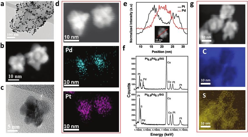
www.advancedsciencenews.com
www.advmatinterfaces.de
unique 2D sp2 hybridized carbon structure.[11–13] Traditional a Pt–Pd core–shell nanoflower supported on sulfur-doped gra-
carbon can be heated to high temperatures (≈1000 °C), leading phene (SG) with improved activity and excellent durability.
to lattice rearrangement and an increase in graphitic nature of The 2D nanoarchitecture of the core–shell nanoflowers pro-
the material. Graphitization produces a highly resistant mate- vides enhanced surface activity and improved electron kinetics,
rial to oxidation and carbon corrosion. To further improve while sulfur atoms doped on graphene support provide a strong
performance, several studies have incorporated heteroatoms anchor for the nanoflowers through tethering behavior, leading
doping of graphene as an efficient way to tune intrinsic proper- to excellent durability under very aggressive test conditions.[18]
ties, which is helpful in improving electrocatalyst activity and The electrochemical activity and stability of the prepared elec-
stability.[12,14] trocatalyst was investigated. Enhancement in electrochemical
Recent research efforts have been focused on the devel- activity can be attributed to the nanoflower core–shell Pt–Pd
opment of less expensive and improved electrochemically nanostructure and weak OH binding to Pt in comparison
active catalysts such as advanced Pt alloys, nonprecious metal to Pd. The morphology of the catalyst was responsible for
catalysts, and core–shell catalysts. Among these, core–shell high ECSA, while its core–shell structure and the interaction
structure catalysts consisting of a thin layer of Pt deposited between the catalyst and highly robust SG facilitate long-term
on low cost seed material have attracted considerable atten- stability to high potential cycling.
tion.[4,6,15,16] Deposition of an ultrathin Pt shell on Pd offers a The catalyst synthesis procedure is demonstrated in Figure 1,
great opportunity to enhance catalytic activity and durability and the detailed synthesis route and reaction mechanism is pro-
while reducing its loading.[15,17] However, nonuniform deposi- vided in Figure S1 (Supporting Information). Pd nanoparticle
tion of the Pt shell and long-term durability at high potential seeds were selectively synthesized through manipulation of the
range is still a problem with core–shell structures. This moti- reduction kinetics of the polyol process,[19,20] by reducing Pd
vated us to carefully design a Pt–Pd electrocatalyst and inte- salt (PdCl42−) with diethylene glycol and polyvinyl pyrrolidone
grated uniquely carbon support to improve durability and (PVP) (Figure 1a,b). Generally, metallic particles are thermo-
achieve the DOE targets. To improve Pt-based ORR activity dynamically favored to form into bulk shapes to lower surface-
and more importantly long-term durability, herein, we report free energies. The addition of PVP reduced the reduction rate
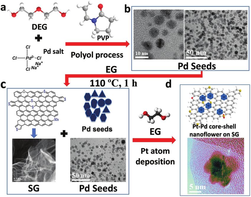
Figure 1. Schematic illustrations of synthesis of PtxPdy-NF/SG core–shell nanoflower (here x, y subscript represents atomic percentage) synthesis.
Synthesis followed as Pd seed synthesis, Pd deposition on SG, with in situ Pt deposition on Pd seed. Here light blue (Pt), dark blue (Pd), and yellow
(S) in Pt–Pd core–shell nanoflower.
Adv. Mater. Interfaces 2018, 1701508 1701508 (2 of 8) © 2018 WILEY-VCH Verlag GmbH & Co. KGaA, Weinheim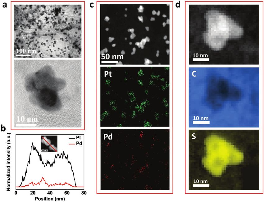
www.advancedsciencenews.com
                                                                                                                            www.advmatinterfaces.de

and allowed the nucleation and growth to be kinetically con-                 distribution of elements (particularly Pt and Pd) in the material,
trolled,[21] which prevented the nanoparticles from growing                  while electron energy loss spectroscopy (EELS) was employed
too large and agglomerating. Separately, graphene oxide and                  to detect light elements (i.e., carbon and sulfur) with high
phenyl disulfide (PDS) were mixed and subjected to a thermal                 spatial resolution. EDX elemental mapping of two particles of
shock (1000 °C) and quenching process to synthesize SG,                      Pt0.9Pd0.1/SG (Figure 2d) confirm the Pd core–Pt shell struc-
which exhibits a multilayer sheet-like structure (Figure 1c). The            tural distribution of these two elements. To further confirm the
Pd seeds, which served as a template for Pt deposition, were                 core–shell structure, an EDX line scan profile of Pd (red) and
well dispersed and attached onto SG (Figure 1c) in a solution                Pt (black) across a Pt–Pd nanoflower is shown in Figure 2e,
of ethylene glycol, PVP, potassium bromide (KBr), and ascorbic               which displays normalized intensities of Pd and Pt. The two
acid and heated at 110 °C for an hour. The final product, Pt–Pd              high-Pt intensity peaks at the sides with the high-Pd intensity
core–shell nanoflowers on SG (Pt–Pd NF/SG) (Figure 1d), was                  peak in between indicate the presence of a Pd core and Pt shell.
synthesized by an in situ continuous feeding method. A syringe               The size of the core and entire core–shell structure is 5–10 and
pump was used to introduce a solution of Na2PtCl6 · 6H2O and                 15–20 nm, respectively. The EDX spectra of sulfur, Pt and Pd
ethylene glycol (EG) into the growth solution used for attaching             in Pt0.9Pd0.1/SG and Pt0.5Pd0.5/SG are compared in Figure 2f,
Pd seeds on SG. Addition of the Pt precursor into the growth                 with the larger Pt peak in Pt0.9Pd0.1/SG and larger Pd peak in
solution caused immediate reduction to metallic Pt by ascorbic               Pt0.5Pd0.5/SG indicating the successful control of the relative
acid and EG. A low injection rate was used to maintain low con-              elements in each structure. In Figure S3 (Supporting Infor-
centrations of Pt, thus preventing self-nucleation and agglom-               mation), a low magnification HAADF-STEM image and corre-
eration while promoting highly dispersed Pt deposition.                      sponding EDX maps show the same Pd core-Pt shell structure
   The transmission electron microscopy (TEM) images in                      across a large number of Pt0.9Pd0.1/SG particles, while sulfur
Figure 2a–c show well attached and dispersed nanoflowers on                  is homogenously distributed across the SG sheet. Figure S4
SG sheets. PtPd-NF/SG was synthesized with different Pt:Pd                   (Supporting Information) shows a lower magnification
atomic ratios, with 90 at% Pt (Pt0.9Pd0.1/SG, Figure 2a–c) and               HAADF-STEM overview of Pt0.9Pd0.1 nanoflower distribution
50 at% Pt (Pt0.5Pd0.5/SG), (Figure S2, Supporting Informa-                   on SG, with EDX elemental mapping indicating homogenous
tion). Pt0.9Pd0.1/SG appears well dispersed on SG (Figure 2a),               distribution of sulfur atoms and PtPd nanoflowers across the
and their nanoflower morphology is confirmed in the high-                    graphene sheet. Figure 2g displays a HAADF-STEM image of
angle annular dark-field scanning TEM (HAADF-STEM) and                       Pt0.9Pd0.1/SG and associated EELS maps of sulfur and carbon.
high-resolution TEM (HR-TEM) images in Figure 2b,c. Energy                   Sulfur is concentrated particularly underneath the Pt petals of
dispersive X-ray spectroscopy (EDX) was used to detect the                   the nanoflower, indicating the desirable anchoring interaction

Figure 2. a) TEM image of Pt0.9Pd0.1/SG (Pt–Pd core–shell nanoflower). b) HAADF-STEM image of Pt0.9Pd0.1/SG Pt–Pd core–shell nanoflower.
c) HR-TEM image of Pt–Pd core–shell nanoflower. d) HAADF-STEM image of two Pt–Pd core–shell nanoflower particle and their corresponding EDX
maps of Pd and Pt. e) EDX line scan of profiles of Pt (black) and Pd (red) of a single Pt–Pd core shell nanoflower particle. f) EDX spectra comparison
of Pt0.5Pd0.5/SG and Pt0.9Pd0.1/SG with Pt, Pd, and S peaks (Cu peaks are attributed to the TEM grid). HAADF-STEM image of Pt–Pd core–shell nano-
flowers and corresponding EELS spectra of sulfur (S) and carbon (C).

Adv. Mater. Interfaces 2018, 1701508                             1701508 (3 of 8)                 © 2018 WILEY-VCH Verlag GmbH & Co. KGaA, Weinheim
www.advancedsciencenews.com
www.advmatinterfaces.de
between these two elements as demonstrated in our previous To investigate activity and durability, ORR kinetics and
works.[18,22] accelerated durability testing (ADT) was performed using a
Figure S5 (Supporting Information) shows that the X-ray rotating disc electrode to imitate the harsh, corrosive, and
diffraction (XRD) patterns of Pt0.9Pd0.1/SG and Pt0.5Pd0.5/SG potentiodynamic states encountered at the cathode of PEMFCs.
reveal typical Pt-fcc peaks (JCPDS No. 04-0802), with diffraction Figure 3a–c shows cyclic voltammograms of Pt0.9Pd0.1/SG and
pattern of Pd/SG and commercial Pt/C shown for comparison. Pt0.5Pd0.5/SG catalysts together with commercial Pt/C (TKK,
The (111) crystal face, which delivers increased Pt mass-based 28.8 wt% Pt). The cyclic voltammograms were performed in
ORR activity in comparison to commercial Pt/C,[23–25] is promi- N2-saturated 0.1 m HClO4 solution at a sweep rate of 50 mV s−1 in
nent in Pt0.9Pd0.1/SG and Pt0.5Pd0.5/SG relative to the other the potential range of 0.05–1.20 V versus a reversible hydrogen
characteristic peaks. There was no elemental sulfur, indicated electrode (RHE) at room temperature. This range was selected
by the lack of characteristic sulfur peaks. Pt and Pd have the considering Pt dissolution (and subsequent agglomeration) can
same crystal structure and similar lattice constants, which occur above 0.8 V versus RHE and carbon corrosion occurs
makes them difficult to distinguish in regular XRD measure- above 0.9 V versus RHE.[37–39] The ECSA of the catalysts was
ments. X-ray photoelectron spectroscopy (XPS) can be used to calculated from the charge associated with the hydrogen
investigate the electronic state of a catalyst material surface, desorption peak and normalized with the Pt mass. Before
which is a significant factor in the catalytic activity and dura- ADT, the specific ECSA of Pt0.9Pd0.1/SG and Pt0.5Pd0.5/SG
bility of Pt.[26] Figure S6 (Supporting Information) displays the are 76 and 72 m2 g−1Pt, respectively, which are higher in com-
full XPS spectra of SG, Pd/SG, Pt0.9Pd0.1/SG, and Pt0.5Pd0.5/ parison to the commercial Pt/C (65 m2 g−1Pt) (Figure 3g).
SG. The C1s peaks for SG are deconvoluted into separate peaks Therefore, the fact that Pt–PdNF/SG possesses higher specific
corresponding to sp2 CC (284.41 eV), sp3 CC (285.82 eV), ECSA despite 3–4 times larger particle sizes in comparison to
CO (287.90 eV), and π–π* (290.65 eV) (Figure S7a, Sup- commercial Pt/C nanoparticles (2–5 nm) supports the claim
porting Information),[27–30] with the results showing that that the dispersion of active Pt surfaces can be improved with
SG largely comprises sp2 CC bonds. After deposition of Pt on larger particles. In this case, the high ECSA of Pt-PdNF/SG
SG to create Pt0.9Pd0.1/SG, the CC peak is shifted to 284.57 eV can be attributed to its 2D core–shell nanoflower morphology,
and the full width at half maximum increases from 1.0 eV (SG) which includes higher dispersion of the catalyst, planar, and
to 1.29 eV (Pt0.9Pd0.1/SG); hence, the broadened peak can be porous catalyst morphology and seamless contact between the
convoluted into another peak at 285.68 eV corresponding to catalyst and the SG support material. The very small changes in
the sp3 CC bond (Figure S7b, Supporting Information). The hydrogen adsorption and desorption peaks during ADT and the
π–π* peak is also negatively shifted from 290.65 to 289.25 eV. lack of large oxidative currents at potentials above 1.0 V versus
These differences between SG and Pt0.9Pd0.1/SG can be attrib- RHE indicate that sulfur oxides released from SG do not have
uted to the interaction between the Pt d-orbital and π–π carbon a negative effect on the synthesized materials.[40] For reference,
in graphene. CV behavior and ORR activity of Pd/SG was also investigated
The sulfur content of SG is 3.16 at% (Table S1, Supporting (Figure S8, Supporting Information) to show that Pd alone is
Information), mostly consisting of the thiophenic groups not sufficient to achieve high electrochemical activity toward
(Figure S7c, Supporting Information). The high-resolution sulfur ORR. Comparing the double layer capacitance regions of the
signal (S2p) was deconvoluted into two major peaks located at CV curves before ADT, both Pt–Pd/SG catalysts display higher
163.74 and 164.92 eV and two minor peaks at 165.09 and 166.27 eV. double layer thickness in comparison to Pt/C. It is well known
The two major peaks resulting from the S2p spin–orbit doublet that electrochemical double layer capacitance is affected by the
of the CSC bond (S2p1/2 at 163.74 eV and S2p3/2 at 164.92 eV) specific surface area, pore structures, and surface activity of the
were separated by 1.18 eV, which is consistent in value with the support materials. Graphene-based materials possess large sur-
theoretical spin doublet separation of 1.13 eV.[31] The two minor face area and higher capacitance capabilities, and these phenom-
peaks can be attributed to carbon bonded with SOx species enon can explain the increase in double layer capacitance.[4,8,9]
(CSOx–).[32,33] Thiophene species incorporate with graphene After ADT, a relatively higher increase of the double-layer
in a pentagonal configuration, therefore residing at the edge and capacity under the CV curves was observed for Pt0.5Pd0.5/SG
defected sites of SG. Therefore, sulfur in SG can serve as a plat- (Figure 3b) in comparison to Pt0.9Pd0.1/SG (Figure 3a).
form for anchoring nucleation and growth of Pt ions.[10] In addi- Pt0.5Pd0.5/SG undergoes more leaching away of Pd compared to
tion to sulfur peak analysis, the fitted Pt 4f7/2 and Pt 4f5/2 peaks the Pt0.9Pd0.1/SG due to the higher initial presence of Pd. This
for Pt0.9Pd0.1/SG are observed at 71.23 and 74.56 eV, respectively results in higher exposed surface area of highly porous SG,
(Figure S7d, Supporting Information), while those for Pt0.5Pd0.5/ leading to enhanced double layer capacitance.
SG (50–50% Pt and Pd) are observed at 71.34 and 74.67 eV, The ORR polarization curves of the catalysts are shown in
respectively (Figure S7e, Supporting Information). The shift in Figure 3d–f. The ORR curves were recorded at room tempera-
peaks of 0.11 eV can be attributed to the interaction of Pt and the ture in an O2-saturated 0.1 m HClO4 solution. The kinetic cur-
support material. This is further supported by the positive shift rent for each ORR polarization curve was calculated using the
of 0.09 eV of S2p peak from SG to Pt0.9Pd0.1/SG (Figure S5f, Koutecký–Levich equation and normalized against Pt mass
Supporting Information). It has been previously demonstrated and ECSA for mass and specific activities (jk,mass and jk,specific),
that the interaction of deposited metal on support material plays respectively. Before ADT, the mass and specific activities of
a vital role in the stability of supported catalysts.[34] The shifting Pt0.9Pd0.1/SG and Pt0.5Pd0.5/SG are significantly improved
in peaks can be attributed to the sulfur bonding directly with the in the 0.86–0.94 V versus RHE region relative to Pt/C. At
carbon atoms in the heterocyclic configuration.[35,36] 0.9 V versus RHE, the mass activities of Pt0.9Pd0.1/SG and
Adv. Mater. Interfaces 2018, 1701508 1701508 (4 of 8) © 2018 WILEY-VCH Verlag GmbH & Co. KGaA, Weinheimwww.advancedsciencenews.com
www.advmatinterfaces.de
Figure 3. Cyclic voltammetry curves for a) Pt0.9Pd0.1/SG, b) Pt0.5Pd0.5/SG, and c) Pt/C before and 3000 cycles. ORR polarization curve of
d) Pt0.9Pd0.1/SG, e) Pt0.5Pd0.5/SG, and f) Pt/C before and after 3000 cycles. g) ECSA, h) mass activity, and i) specific activity bar chart of Pt0.9Pd0.1/SG,
Pt0.5Pd0.5/SG, and Pt/C before and after 3000 cycles. j) CV curves. k) ORR polarization curves. l) Bar chart comparing ECSA, mass activity, and specific
activity of Pt0.9Pd0.5/SG with commercial Pt/C before and after 10 000 cycles.
Pt0.5Pd0.5/SG are 371 and 115 mA mg−1Pt, respectively, with the structure also lowers its d-band center energy,[44] which pro-
former representing a 3.12-fold improvement over commercial vides a weaker Pt-OHads interaction[45,46] and leads to a decrease
Pt/C (119 mA mg−1Pt) (Figure 3h). The specific activities at 0.9 V in OH coverage at a given potential and a resulting increase
versus RHE for Pt0.9Pd0.1/SG and Pt0.5Pd0.5/SG are 0.491 and in the number of sites available for O2 adsorption, dissociation,
0.197 mA cm−2Pt, respectively, with the former representing and reduction. Previous work has demonstrated that the vacant
a 2.71-fold increase over commercial Pt/C (0.183 mA cm−2Pt) d-orbital of individual atoms play a vital role in the catalyst
(Figure 3i). The high activities and ECSA of Pt0.9Pd0.1/SG could activity and are the basis for excellent catalytic activity.[47] The
be attributed to the higher exposure of the (111) facets of Pt, addition of another metal to the Pt downshifts its d-band center
which has comparatively better activity than the (100) facet and and therefore reduces oxygen binding energy and increases
core–shell structure.[41–43] The addition of Pd to the core–shell catalytic activity compared to pure Pt.[48–50] Generally, bimetallic
Adv. Mater. Interfaces 2018, 1701508 1701508 (5 of 8) © 2018 WILEY-VCH Verlag GmbH & Co. KGaA, Weinheimwww.advancedsciencenews.com
www.advmatinterfaces.de
structures form weaker bonds with oxygen in comparison with under the same condition (0.05 –1.2 V vs RHE) (Figure 3j–l),
pure Pt, as the variation in oxygen metal bond depends to a the specific ECSA of Pt0.9Pd0.1/SG (54 m2 g−1Pt) remains
large extent on the coupling between the oxygen 2p states and higher than Pt/C after 3000 cycles (43 m2 g−1Pt) and
metal 3d states. This leads to a lower position of the d-band 10 000 cycles (39 m2 g−1Pt). Moreover, the mass and specific
center relative to the Fermi level, which affects the metal– activities of Pt0.9Pd0.1/SG after 10 000 cycles (282 mA mg−1Pt
oxygen bond interaction and thus benefits ORR activity.[49] In and 0.525 mA cm−2Pt) remain higher than the initial Pt/C
the case of Pt0.9Pd0.1/SG, although the thickness of the Pt shell activity (119 mA mg−1Pt and 0.183 mA cm−2Pt), and are 25 and
is relatively high, defects in the core–shell structure can allow 18 times higher than the mass and specific activities of Pt/C
for sites where Pd atoms modify the electronic structure of after 10 000 cycles (11 mA mg−1Pt and 0.0285 mA cm−2Pt),
the Pt surface. Moreover, the high mass and specific activity of respectively (Figure 3l). To further emphasize, Pt0.9Pd0.1/SG
Pt0.9Pd0.1/SG could be associated to its nanoflower morphology, displays a mass activity loss of only 24% compared to the 91%
with its well-attached 2D structure providing improved electron mass activity loss of commercial Pt/C, which is critically impor-
transfer kinetics. tant to the success of prospective ORR catalysts in PEMFCs.
The electrocatalytic durability of the catalysts was evaluated The durability of Pt0.9Pd0.1/SG, given the aggressive ADT
through accelerated tests applying linear potential sweeps in potential range (0.05–1.2 V vs RHE) used in this work in com-
the range of 0.05–1.2 V versus RHE at a rate of 50 mV s−1 in parison to other recent Pt-based catalyst reports, is particularly
an N2 saturation 0.1 m HClO4 solution at room temperature. impressive.[18,22,51,52]
After 3000 ADT cycles, Pt0.9Pd0.1/SG retains most of its ECSA The exceptional stability of Pt0.9Pd0.1/SG is partly attributed
and ORR activity. The ECSA of Pt0.9Pd0.1/SG, Pt0.5Pd0.5/SG, and to its high graphitic content, the core–shell nanoflower archi-
Pt/C after 3000 cycles was 72, 45, and 43 m2 g−1Pt (Figure 3g), tecture of Pt–Pd, and the presence of functional anchoring
representing decreases of 5, 38, and 34%, respectively. In terms groups and strong interactions between SG and Pt.[22] Addition-
of catalytic durability after 3000 cycles, the Pt0.9Pd0.1/SG only ally, the Pd core delivers long term stability to the Pt shell layer,
exhibits a 20% loss in mass activity (297 mA mg−1Pt) and 14% since Pd has a lower oxidation potential (0.92 V vs RHE) than
loss of specific activity (0.421 mA cm−2), while Pt0.5Pd0.5/SG Pt (1.19 V vs RHE), therefore preventing the cathode potential
loses 63% (42 mA mg−1Pt) and 52% (0.095 mA cm−2Pt) reaching a value where Pt oxidation can take place.[4] Since it
and commercial Pt/C loses 81% (23 mA mg−1Pt) and 71% is difficult to synthesize a pore-free Pt shell, the Pd core will
(0.053 mA cm−2Pt) of its mass and specific activity, respectively establish contact with the electrolyte and gradually oxidize
(Figure 3h,i). Indeed, after 3000 ADT cycles, the mass and spe- when the potential rises above its oxidation potential. Oxi-
cific activities of Pt0.9Pd0.1/SG are 13 and eight times higher than dized Pd+2 will subsequently diffuse through any pores in the
commercial Pt/C, respectively. Even after 10 000 ADT cycles Pt shell, leaving empty space in the core. After prolonged
Figure 4. a) TEM and HR-TEM images of Pt0.9Pd0.1/SG after 10 000 cycles. b) EDX line scan profiles of Pt (black) and Pd (red) of two Pt–Pd core–shell
nanoflower particles after 10 000 cycles. c) Low magnification HAADF-STEM image and corresponding EDX maps of Pt0.9Pd0.1/SG after 10 000 cycles.
d) HAADF-STEM image of Pt0.9Pd0.1/SG after 10 000 cycles and corresponding EELS maps of carbon and sulfur.
Adv. Mater. Interfaces 2018, 1701508 1701508 (6 of 8) © 2018 WILEY-VCH Verlag GmbH & Co. KGaA, Weinheimwww.advancedsciencenews.com
www.advmatinterfaces.de
potential cycling, the empty space left causes the Pt shell to Conflict of Interest
undergo small contractions and become even less reactive.[44]
The authors declare no conflict of interest.
Therefore, the Pd core increases the stability of the Pt shell by
inducing lattice contractions and by acting as a sacrificial anode
to provide cathodic protection to the Pt shell. Evidence of this
protection mechanism is provided by postcycling TEM analysis. Keywords
After 10 000 ADT cycles, Pt0.9Pd0.1/SG was removed from the electrocatalyst, oxygen reduction, PEMFCs, platinum
glassy carbon electrode by rinsing in ethanol and analyzed with
TEM to search for the presence of agglomeration or dissolution Received: November 17, 2017
of Pt particles, and any other catalyst morphological changes. Revised: December 21, 2017
The Pt–Pd particles remained well-dispersed on SG, while no Published online:
significant agglomeration or overall changes in its morphology
were observed (Figure 4a). However, in comparison to the cata-
lyst before cycling (Figure 2f), the EDX line spectra in Figure 4b
[1] United States Department of Energy: Technical Plan – Fuel Cells,
shows a clear decrease of Pd intensity relative to the Pt inten- 2012, http://www1.eere.energy.gov/hydrogenandfuelcells/mypp/
sity, thus demonstrating that Pd indeed dissolves during pdfs/fuel_cells.pdf (accessed: November 2013).
cycling due to defects in the core–shell structure. However, [2] J. Rossmeisl, G. S. Karlberg, T. Jaramillo, J. K. Nørskov, Faraday
it should be noted that part of Pd remains after ADT testing. Discuss. 2008, 140, 337.
This can be attributed to the parting limit, where higher [3] W. Tang, G. Henkelman, J. Chem. Phys. 2009, 130, 194504.
atomic ratio of the more noble component (Pt in this case) [4] M. A. Hoque, F. M. Hassan, D. Higgins, J.-Y. Choi, M. Pritzker,
diminishes dissolution of the other or both metals in a binary S. Knights, S. Ye, Z. Chen, Adv. Mater. 2015, 27, 1229.
systems.[53] EDX elemental mapping of individual nano- [5] K. Sasaki, H. Naohara, Y. Cai, Y. M. Choi, P. Liu, M. B. Vukmirovic,
J. X. Wang, R. R. Adzic, Angew. Chem., Int. Ed. Engl. 2010, 49,
flowers in Pt0.9Pd0.1/SG shows the retention of the Pd core-Pt
8602.
shell structure after ADT (Figure 4c), while mapping at lower
[6] Z. Chen, M. Waje, W. Li, Y. Yan, Angew. Chem., Int. Ed. Engl. 2007,
magnification shows the homogenous distribution of carbon, 46, 4060.
sulfur, Pt, and Pd is also maintained (Figure S9, Supporting [7] C. Koenigsmann, A. C. Santulli, K. Gong, M. B. Vukmirovic,
Information). EELS elemental mapping (Figure 4d) shows W. P. Zhou, E. Sutter, S. S. Wong, R. R. Adzic, J. Am. Chem. Soc.
that sulfur stays concentrated at the edge of core–shell nano- 2011, 133, 9783.
flower catalyst, indicating the interaction between sulfur and [8] B. Y. Xia, W. T. Ng, H. Bin Wu, X. Wang, X. W. D. Lou, Angew. Chem.,
Pt remains after ADT. Int. Ed. Engl. 2012, 51, 7213.
In summary, we have designed and synthesized a novel [9] N. S. Porter, H. Wu, Z. Quan, J. Fang, Acc. Chem. Res. 2013, 46,
ORR catalyst composed of Pt–Pd core shell nanoflower-like 1867.
[10] S. J. Guo, S. Zhang, D. Su, S. H. Sun, J. Am, Chem. Soc. 2013, 135,
structures deposited on sulfur-doped graphene. The core–shell
13879.
nanoflower structures exhibit a substantial enhancement in
[11] R. Wang, D. C. Higgins, M. A. Hoque, D. Lee, F. Hassan, Z. Chen,
electrochemical activity and remarkable long-term stability Sci. Rep. 2013, 3, 1.
compared with commercial Pt/C. After 3000 ADT cycles, the [12] S. Sun, G. Zhang, D. Geng, Y. Chen, R. Li, M. Cai, X. Sun, Angew.
Pt0.9Pd0.1/SG catalyst retains 80 and 86% of its mass and spe- Chem., Int. Ed. 2011, 50, 422.
cific activity, respectively, while Pt/C only retains 19 and 28%. [13] Y. Zhou, K. Neyerlin, T. S. Olson, S. Pylypenko, J. Bult, H. N. Dinh,
Even after longer-term ADT (10 000 cycles), the mass activity T. Gennett, Z. Shao, R. O'Hayre, Energy Environ. Sci. 2010, 3, 1437.
of Pt0.9Pd0.1/SG was 2.4 times higher than the initial activity [14] S. Sun, G. Zhang, D. Geng, Y. Chen, M. N. Banis, R. Li, M. Cai,
of Pt/C, demonstrating its exceptional stability. Through the X. Sun, Chemistry 2010, 16, 829.
combination of a unique SG support and nanostructured [15] Y. Shao, J. Sui, G. Yin, Y. Gao, Appl. Catal., B 2008, 79, 89.
[16] L. Zhang, S. Zhu, Q. Chang, D. Su, J. Yue, Z. Du, M. Shao,
2D core–shell structure, this work provides an attractive strategy
ACS Catal. 2016, 6, 3428.
for obtaining excellent catalyst performance and durability to
[17] D. Wang, Y. Yu, J. Zhu, S. Liu, D. A. Muller, H. D. Abruña, Nano
overcome the performance barriers of traditional PEMFC cath- Lett. 2015, 15, 1343.
odes electrocatalyst. [18] X. Wang, S.-I. Choi, L. T. Roling, M. Luo, C. Ma, L. Zhang, M. Chi,
J. Liu, Z. Xie, J. A. Herron, M. Mavrikakis, Y. Xia, Nat. Commun.
2015, 6, 7594.
Supporting Information [19] D. Higgins, M. A. Hoque, M. H. Seo, R. Wang, F. Hassan, J.-Y. Choi,
Supporting Information is available from the Wiley Online Library or M. Pritzker, A. Yu, J. Zhang, Z. Chen, Adv. Funct. Mater. Butterworth-
from the author. Heinemann 2014, 24, 4325.
[20] J. W. Mullin, Crystallization, Elsevier,2001, pp. 216–288.
[21] F. Fievet, J. P. Lagier, M. Figlarz, MRS Bull. 1989, 14, 29.
[22] Y. Xiong, Y. Xia, Adv. Mater. 2007, 19, 3385.
Acknowledgements [23] I. Herrmann, U. I. Kramm, J. Radnik, S. Fiechter, P. Bogdanoff,
A.M.J. and F.M.H. contributed equally to this work. This research was J. Electrochem. Soc. 2009, 156, B1283.
supported by the Natural Sciences and Engineering Research Council of [24] J. Liang, X. Du, C. Gibson, X. W. Du, S. Z. Qiao, Adv. Mater. 2013,
Canada (NSERC). The TEM and EELS characterizations were conducted 25, 6226.
at the Canadian Center for Electron Microscopy (CCEM) at McMaster [25] S. Yang, L. Zhi, K. Tang, X. Feng, J. Maier, K. Müllen, Adv. Funct.
University. Mater. 2012, 22, 3634.
Adv. Mater. Interfaces 2018, 1701508 1701508 (7 of 8) © 2018 WILEY-VCH Verlag GmbH & Co. KGaA, Weinheimwww.advancedsciencenews.com
www.advmatinterfaces.de
[26] S. Shrestha, Y. Liu, W. E. Mustain, Catal. Rev.: Sci. Eng. 2011, 53, [41] D. F. van der Vliet, C. Wang, D. Tripkovic, D. Strmcnik, X. F. Zhang,
256. M. K. Debe, R. T. Atanasoski, N. M. Markovic, V. R. Stamenkovic,
[27] X. H. Yang, J. W. Guo, S. Yang, Y. Hou, B. Zhang, H. G. Yang, Nat. Mater. 2012, 11, 1051.
J. Mater. Chem. A 2014, 2, 614. [42] N. Markovic, J. Electrochem. Soc. 1997, 144, 1591.
[28] S. A. Best, P. Brant, R. D. Feltham, T. B. Rauchfuss, D. M. Roundhill, [43] J. Zhang, M. B. Vukmirovic, Y. Xu, M. Mavrikakis, R. R. Adzic,
R. A. Walton, Inorg. Chem. 1977, 16, 1976. Angew. Chem., Int. Ed. 2005, 44, 2132.
[29] N. A. Travlou, G. Z. Kyzas, N. K. Lazaridis, E. A. Deliyanni, Langmuir [44] M. B. Vukmirovic, J. Zhang, K. Sasaki, A. U. Nilekar, F. Uribe,
2013, 29, 1657. M. Mavrikakis, R. R. Adzic, Electrochim. Acta 2007, 52, 2257.
[30] T. I. T. Okpalugo, P. Papakonstantinou, H. Murphy, J. McLaughlin, [45] J. X. Wang, J. Zhang, R. R. Adzic, J. Phys. Chem. A 2007, 111, 12702.
N. M. D. Brown, Carbon 2005, 43, 153. [46] A. U. Nilekar, M. Mavrikakis, Surf. Sci. 2008, 602, L89.
[31] A. Barrie, I. W. Drummond, Q. C. Herd, J. Electron Spectrosc. Relat. [47] P. Hu, Z. Huang, Z. Amghouz, M. Makkee, F. Xu, F. Kapteijn,
Phenom. 1974, 5, 217. A. Dikhtiarenko, Y. Chen, X. Gu, X. Tang, Angew. Chem., Int. Ed.
[32] C. H. Choi, S. H. Park, S. I. Woo, Green Chem. 2011, 13, 406. 2014, 53, 3418.
[33] Z. Liu, H. Nie, Z. Yang, J. Zhang, Z. Jin, Y. Lu, Z. Xiao, S. Huang, [48] V. Stamenkovic, B. S. Mun, K. J. J. Mayrhofer, P. N. Ross,
Nanoscale 2013, 5, 3283. N. M. Markovic, J. Rossmeisl, J. Greeley, J. K. Nørskov, Angew.
[34] B. Qiao, J.-X. Liang, A. Wang, C.-Q. Xu, J. Li, T. Zhang, J. J. Liu, Chem., Int. Ed. 2006, 45, 2897.
Nano Res. 2015, 8, 2913. [49] V. R. Stamenkovic, B. S. Mun, M. Arenz, K. J. J. Mayrhofer, C. A. Lucas,
[35] M. E. Labib, J. H. Thomas, D. D. Embert, Carbon 1984, 22, 445. G. Wang, P. N. Ross, N. M. Markovic, Nat. Mater. 2007, 6, 241.
[36] Y. Chang, F. Hong, C. He, Q. Zhang, J. Liu, Adv. Mater. 2013, 25, [50] J. Greeley, I. E. L. Stephens, A. S. Bondarenko, T. P. Johansson,
4794. H. A. Hansen, T. F. Jaramillo, J. Rossmeisl, I. Chorkendorff,
[37] Y. Shao-Horn, W. C. Sheng, S. Chen, P. J. Ferreira, E. F. Holby, J. K. Nørskov, Nat. Chem. 2009, 1, 552.
D. Morgan, Top. Catal. 2007, 46, 285. [51] M. A. Hoque, F. M. Hassan, M.-H. Seo, J.-Y. Choi, M. Pritzker,
[38] H. A. Gasteiger, S. S. Kocha, B. Sompalli, F. T. Wagner, Appl. Catal., S. Knights, S. Ye, Z. Chen, Nano Energy 2016, 19, 27.
B 2005, 56, 9. [52] J. Hu, L. Wu, K. A. Kuttiyiel, K. R. Goodman, C. Zhang, Y. Zhu,
[39] P. J. Ferreira, G. J. la O’, Y. Shao-Horn, D. Morgan, R. Makharia, M. B. Vukmirovic, M. G. White, K. Sasaki, R. R. Adzic, J. Am. Chem.
S. Kocha, H. A. Gasteiger, J. Electrochem. Soc. 2005, 152, Soc. 2016, 138, 9294.
A2256. [53] R. Newman, S. Corcoran, J. Erlebacher, M. J. Aziz, K. Sieradzki,
[40] J. Fu, M. Hou, C. Du, Z. Shao, B. Yi, J. Power Sources 2009, 187, 32. MRS Bull. 1999, 24, 24.
Adv. Mater. Interfaces 2018, 1701508 1701508 (8 of 8) © 2018 WILEY-VCH Verlag GmbH & Co. KGaA, WeinheimYou can also read


