Dopamine Receptors Gene Expression in Male Rat Hippocampus after Administration of MDMA (Ecstasy)
←
→
Page content transcription
If your browser does not render page correctly, please read the page content below
Int. J. Morphol.,
33(1):301-308, 2015.

          Dopamine Receptors Gene Expression in Male Rat
         Hippocampus after Administration of MDMA (Ecstasy)

                       La Expresión Génica de Receptores de Dopamina en el Hipocampo
                       de Ratas Macho Después de la Administración de MDMA (Éxtasis)

                       Mahakizadeh Simin*; Jahanshahi Mehrdad**; Haidari Kamran*** & Shahbazi Majid***

SIMIN, M.; MEHRDAD, J.; KAMRAN, H. & MAJID, S. Dopamine receptors gene expression in male rat hippocampus after
administration of MDMA (Ecstasy). Int. J. Morphol., 33(1):301-308, 2015.

         SUMMARY: Ecstasy is one of the most popular amusing drugs among young people. Documents indicate some effects of
Ecstasy on hippocampus and close relations between dopaminergic functions with reward learning. Therefore, the aim of this study was
evaluation of the chronic effects of Ecstasy on memory in male Wistar rats and determination of dopamine receptors' gene expression in
hippocampus. Forty adult male Wistar rats randomly distributed in five groups: Control, sham (received 1 ml/kg 0.9% saline) and three
experimental groups were: Exp. 1 (2.5 mg/kg), Exp. 2 (5 mg/kg), and Exp. 3 (10 mg/kg) received MDMA intraperitoneally once every
7 days (3 times a day, 3 hours apart) for 4 weeks. Before the first injection animals trained in Shuttle Box memory and tested after the last
injection. 24 hours after the final testing, brains of rats were dissected and hippocampus was removed and homogenized. After total
RNA extraction and cDNA synthesis, expression of dopamine receptor genes in the hippocampus determined with Real-Time PCR. Our
results showed that 2.5 and 5 mg/kg MDMA-treated groups had memory impairment. Also we found that MDMA increased the mRNA
expression of dopamine receptors in hippocampus and the highest increase found in dopamine D1 receptors in the 5 mg/kg experimental
group. We concluded that low doses of Ecstasy could increase Dopamine takers gene expression in hippocampus and disorder avoidance
memory. But in high doses the increase in Dopamine takers gene expression was not as much as that in low doses and avoidance memory
disorder was not observed.

        KEY WORDS: MDMA; Dopamine Receptors; Gene expression; Hippocampus; Rat.

INTRODUCTION

       Today        MDMA          (3,4-methylenedioxy-                           Some documents indicate a close relations in
metamphetamine) is one of the most popular drugs among                    hippocampus between dopaminergic functions of reward
the young, especially at dance parties because of its ability             related-associative learning (Di Chiara, 1999), reward
to induce attraction and increasing energy (Cohen 1995;                   prediction and incentive salience (Berridge & Robinson,
Kindlundh-Högberg et al., 2006). Ecstasy (MDMA) acts as                   1998; Schultz 1998; Everitt et al., 1999), and serotonergic
a stimulant and mescaline-like hallucinogen, and is known                 functions of disinhibition and impulsivity (Linnoila et al.,
to suppress the appetite (Cohen; Green et al., 2003).                     1983; Af Klinteberg et al., 1990; Winstanley et al., 2004)
                                                                          because of numerous specific interactions between the two
        Frequent and prolonged use of MDMA causes                         systems.
depression, anxiety and aggression, but it has also been
accompanied with cognitive changes, loss of shyness,                             The issue whether compulsive MDMA-taking
impulsivity, memory loss, and eating disorders in human                   behavior mostly is regulated by dopamine or serotonin
and rats (Cohen; Morgan, 1998; McCann et al., 1999; Morley                related functions is very important to understand the
et al., 2001; Curran et al., 2004; McCardle et al., 2004;                 implication and role of MDMA in mechanisms of addictive
Gouzoulis-Mayfrank et al., 2005; Kindlundh-Högberg et al.).               behaviors (Kindlundh-Högberg et al.).

*
    Department of Anatomy, Neuroscience Research Center, Golestan University of Medical Sciences, Gorgan, Iran.
**
    Associate Professor of Anatomy, Department of Anatomy, Neuroscience Research Center, Golestan University of Medical Sciences, Gorgan, Iran.
***
    Cellular and Molecular Research Center, Golestan University of Medical Sciences, Gorgan, Iran.

                                                                                                                                                  301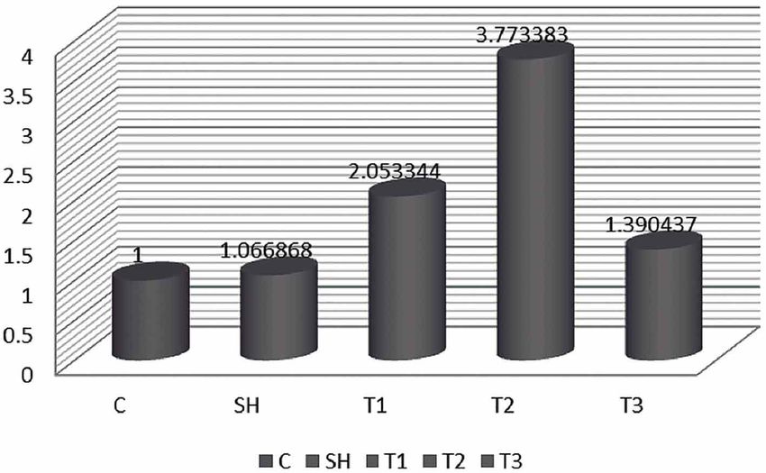
SIMIN, M.; MEHRDAD, J.; KAMRAN, H. & MAJID, S. Dopamine receptors gene expression in male rat hippocampus after administration of MDMA (Ecstasy). Int. J. Morphol., 33(1):301-308, 2015.

        There are several limitations in accompanying                                         MDMA stems from human conditions. MDMA is taken at
researches on MDMA-taking; for example, ecstasy is                                            doses of 50-60 mg in humans. One single dose of 5 mg/kg
administered in dose regimes that poorly imitate the                                          per rat corresponds to 65 mg in a 65 kg human according to
behaviors observed among young person's taking this drug                                      the interspecies scaling technique: D human = D animal (W
on a weekly or at least intermittent basis at dance clubs.                                    human/W animal) 0.7 (Mordenti et al., 1991). The Golestan
Instead MDMA, in most previous studies, was either                                            University of Medical sciences Guidelines for the Care and
administered as a single dose or multiple doses for 17 days                                   Use of Animals in Research were followed.
(Gold & Koob, 1989; Taylor & Jentsch, 2001; Fone et al.,
2002; McGregor et al., 2003). Secondly, studies of MDMA                                       Inhibitory Avoidance Apparatus. The step-through
caused long-term effects have focused on mechanisms that                                      inhibitory avoidance apparatus consisted of two
occur some days after drug discontinuation reflecting                                         compartments of the same size (20 x 20 x 30 cm3). In the
peripheral or withdrawal effects rather than effects of                                       middle of a dividing wall, a guillotine door (7.9 cm2) could
repeated long-term intake (Green et al.). Third, several                                      be lifted manually. The walls and floor of one compartment
studies have reported effects upon behavior (locomotion,                                      consisted of white opaque resin and the walls of the other
conditioned place preference, self-administration), protein                                   compartment were dark. Stainless steel bars (3 mm in
density (autoradiography), neurotransmitters and their                                        diameter and 1 cm intervals) constituted the floor of the dark
metabolites (micro dialysis) (Green et al.; Piper & Meyer,                                    compartment. Intermittent electric shocks (50 Hz, 3 s, 1.5
2004), but it still remains unclear whether observed                                          mA intensity) were delivered to the grid floor of the dark
alterations also involve changes in mRNA contents of                                          compartment by an isolated stimulator.
included markers. Finally, many studies have focused on
single or a few markers in single brain structures, and there                                 Behavioral Procedures. Our previous study (Azami et al.,
is no comprehensive testing of dopaminergic and                                               2010) described passive avoidance as follows: All animals
serotonergic markers simultaneously in multiple brain                                         were allowed to habituate in the experimental room (with
regions (Kindlundh-Högberg et al.).                                                           light and sound attenuated) for at least 30 min prior to the
                                                                                              experiments. Then, each animal was gently placed in the
       We hypothesize that alterations of gene transcript                                     brightly lit compartment of the apparatus; after 5 s the
levels of markers implicated in mechanisms regulating                                         guillotine door was opened and the animal was allowed to
rewarding properties underlie the psychiatric changes that                                    enter the dark module.
have been reported to be associated with MDMA intake.
The specific aim of the present study was to investigate how                                        The latency with which the animal entered the dark
the repeated intermittent administration of MDMA affects                                      chamber was recorded. Animals that waited more than 120
the gene-transcript content of dopamine receptors in regions                                  s to enter the dark chamber were excluded from the
of the male rat hippocampus implicated in memory loss.                                        experiments.

                                                                                                       Once the animal entered with all four-paws to the
MATERIAL AND METHOD                                                                           next chamber, the guillotine door was closed and the rat was
                                                                                              immediately withdrawn from the compartment. This trial
                                                                                              was repeated after 30 min. As in the acquisition trial, after 5
Animals. Eight week male Wistar rats (Pasteur Institute,                                      s the guillotine door was opened, and as soon as the animal
Tehran, Iran) weight 200±20 g at the initiation of the study,                                 entered the dark (shock) compartment the door was closed;
served as subjects and were housed pair-wise in air                                           and a foot shock (50 Hz, 1 mA and 3 s) was immediately
conditioned rooms (12:12-h dark/light cycle) at 22±3 °C and                                   delivered to the grid floor of the dark room. After 20 s, the
a humidity of 53%. The rats were randomly distributed into                                    rat was removed from the apparatus and placed temporarily
three MDMA-treated (-3,4-methylenedioxy-N-                                                    into its home cage. Two minutes later, the animal was retested
methamphetamine-HCl), Sigma Pharmaceutical) and Sham                                          in the same way as in the previous trials; if the rat did not
groups. All animals received three intraperitoneal injections                                 enter the dark compartment during 120 s, a successful
(3 h apart; a challenge) every 7 day for 4 weeks. The MDMA                                    acquisition of inhibitory avoidance response was recorded.
was dissolved into the vehicle on the day of testing. During                                  Otherwise, when the rat entered the dark compartment
the treatment day the MDMA low-dose (n=8) received 3                                          (before 120 s) a second time, the door was closed and the
2.5 mg/kg MDMA, the MDMA middle dose (n=8) 3 x 5                                              animal received the shock again. After retesting, if the rat
mg/kg, the MDMA high dose rats (n=8) 3 x 10 mg/kg                                             learned inhibitory avoidance response successfully, it was
whereas Sham group received the vehicle of sterile 0.9%                                       moved to the cage. On the test day each animal was gently
saline solution (1 ml/kg). The rationale for these doses of                                   placed in the light compartment and after 5 s the door was
302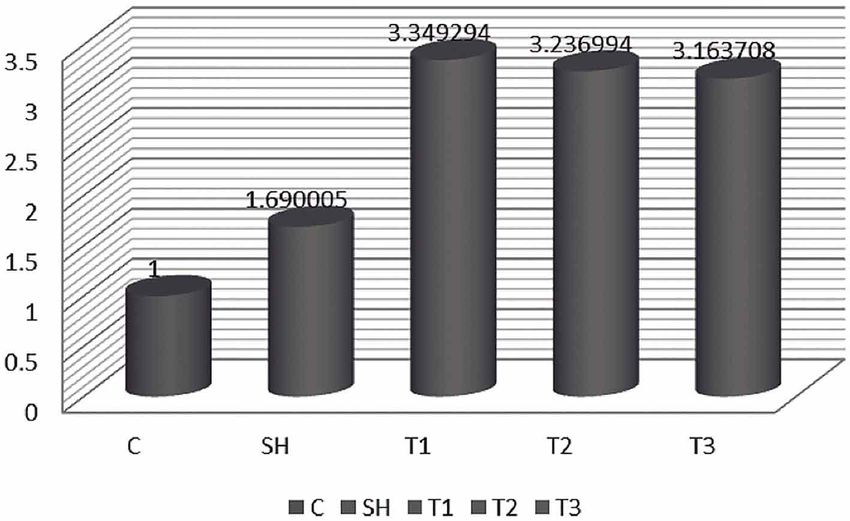
SIMIN, M.; MEHRDAD, J.; KAMRAN, H. & MAJID, S. Dopamine receptors gene expression in male rat hippocampus after administration of MDMA (Ecstasy). Int. J. Morphol., 33(1):301-308, 2015.
opened, and step through latency (sec) was recorded in the Stage 4: 95 °C for 15 seconds, 60 °C for 30 seconds and 95
absence of electric foot shocks, as indicator of inhibitory °C for 15 seconds.
avoidance behavior.
Primers used for Q RT-PCR. Primers pairs of the internal
Thirty minutes after the last injection of MDMA the housekeeping gene (s18 RNA) and the dopamine receptors
rats tested in Shuttle box and 24 h after the final test, the were designed to Primers-3 software and National Center
brains of rats were dissected.Brain region of interest for Biotechnology Information (NCBI):
hippocampus (at bregma 4.8 to 5.6), were dissected using a Primers of s18 RNA; gtgatccccgagaagtttca;
rat brain matrix (Paxinos & Watson, 2007), rapidly frozen ctgctttcctcaacaccaca
on dry ice, immersed in RNA later (Ambion) for 1 h, and Dopamine D1 receptor primer; tccttcaagagggagacgaa;
then stored at -80 °C. All procedures were performed in ccacacaaacacatcgaagg
accordance with institutional guidelines for animal care and Dopamine D4 receptor primer; gatgtgttggacgcctttct;
use. tcggcattgaagatggtgta
Dopamine D5 receptor primer; ccacatgataccgaatgcag;
Isolation of total RNA and reverse transcriptase. After cacagtcaagctcccagaca
homogenizing of the hippocampus tissue, total RNA were
isolated with RNeasy Mini Kits (Qiagen, Germany), Statistical analysis. Statistical analyses for memory
following the manufacturers protocol and recovered in 20 experiment were performed using one-way ANOVA. Gene
µL elution solution. Then, reverse transcription was done expression fold was calculated relative to the level of each
with equal amounts of RNA using Omniscript kit Qiagen, sample of the housekeeping gene, S18 RNA using Microsoft
Germany) to generate cDNA template for real-time Excel® 2-∆∆ct = PRODUCT(2^-((A4-B4)-(C4-D4))) . The
polymerase chain reaction (PCR) according to the differences with P< .05 were considered statistically
manufacturer’s protocol. The cDNA templates were significant.
evaluated by PCR and gel-electrophoresis.
Quantitative Real-Time PCR. Quantitative real-time PCR RESULTS
(Q RT-PCR) was used blindly on the samples (ABI 7300;
USA) using the Power SYBR Green PCR Master Mix
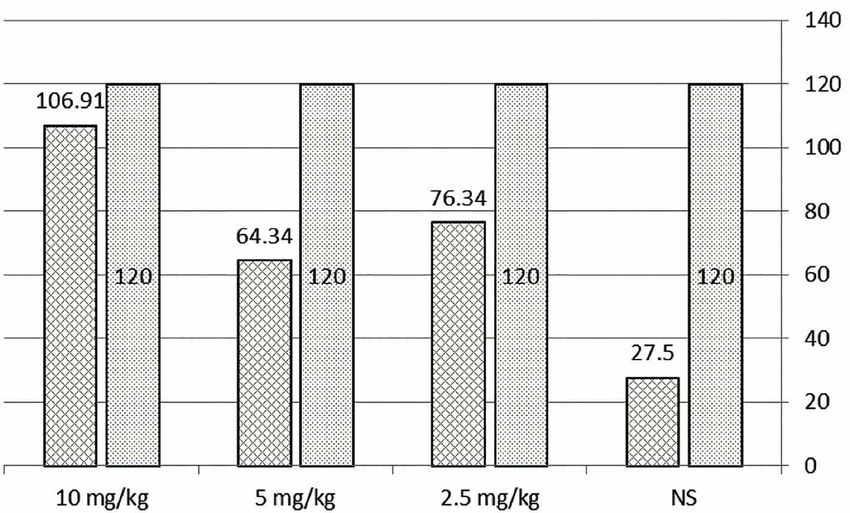
(Applied Biosystems) to analyze the transcript levels of As we show in Figure 1, the present study showed
dopamine receptors in total volume 25 µl. that the Saline group got amnesia due to the lapse of time
with the average of 27.5 s delay in entering the black house/
Amplification was done for 40 cycles at four stages: cell. But it seemed that the Ecstasy effected the recalling of
Stage 1: 50 °C for 2 minutes; Stage 2: 95 °C for 1 minute; the memory in experimental group and because of the amount
Stage 3: 95 °C for 15 seconds and 62 °C for 1.5 minutes; of dose rats got less amnesia. In other words most of rats
Fig. 1. Delay time (per second) to entrance to dark chamber after one month.
303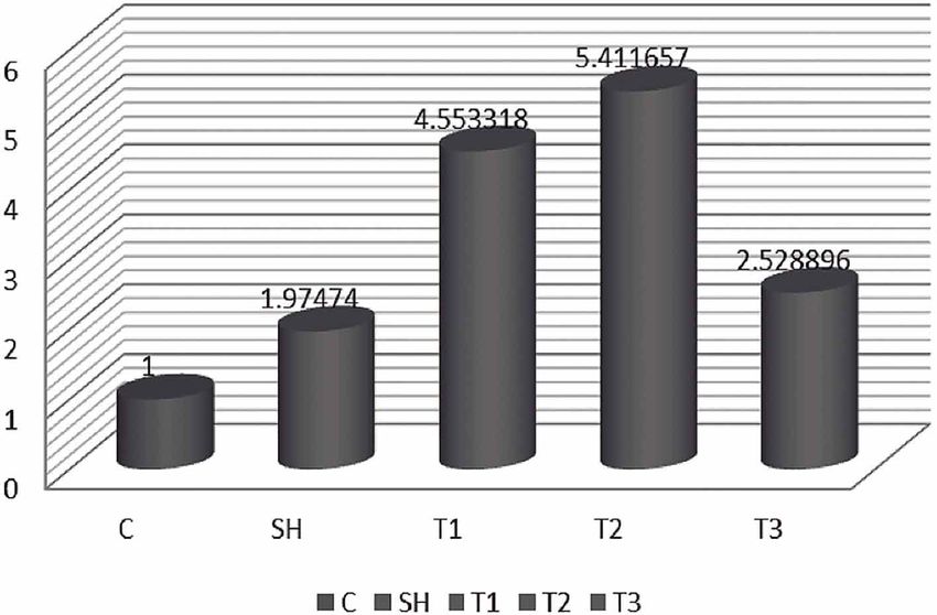
SIMIN, M.; MEHRDAD, J.; KAMRAN, H. & MAJID, S. Dopamine receptors gene expression in male rat hippocampus after administration of MDMA (Ecstasy). Int. J. Morphol., 33(1):301-308, 2015.

                                                                                                                 with complete memory/recall were observed
                                                                                                                 in 10 mg/kg dose (Fig. 1).

                                                                                                                         The expression profiles of these three
                                                                                                                 genes relative to s18 RNA showed that the
                                                                                                                 injection of MDMA increase the D1, D4, and
                                                                                                                 D5 Dopamine receptors gene expression in
                                                                                                                 comparison with control and control-Saline
                                                                                                                 groups.

                                                                                                                       Considering this point is essential that
                                                                                                                 diagram of this gene expression changes is
                                                                                                                 sinuous and its peak is in T2 (5 mg/kg
                                                                                                                 MDMA). In other words the most increase of
                                                                                                                 gene expression was observed for all three
                                                                                                                 Dopamine takers in this group.
Fig. 2. D1 Dopamine receptor genes expression in all groups. C= Control, SH=
Sham, T1= 2.5 mg/kg, T2= 5 mg/kg and T3= 10 mg/kg MDMA.                                                                 D1, D4 and D5 receptors gene
                                                                                                                 expression in 2.5 mg/kg group were increased
                                                                                                                 in comparison with Sham. The highest
                                                                                                                 expression of the receptor revealed in D1 re-
                                                                                                                 ceptor (Figs. 2–4).

                                                                                                                        D1, D4 and D5 receptors gene expression
                                                                                                                 in 5 mg/kg group had a higher level in
                                                                                                                 comparison with Sham. Also the highest
                                                                                                                 expression of the receptors revealed in D1 re-
                                                                                                                 ceptor while D4 receptor gene expression in this
                                                                                                                 group is not much difference in comparison with
                                                                                                                 low dose group (Fig. 2–4).

                                                                                                                 D1, D4 and D5 receptors gene expression in 10
                                                                                                                 mg/kg group were increased. Also the highest
                                                                                                                 expression of the receptors revealed in D4 re-
Fig. 3. D4 Dopamine receptor genes expression in all groups. C= Control, SH=
Sham, T1= 2.5 mg/kg, T2= 5 mg/kg and T3= 10 mg/kg MDMA.                                                          ceptor and also D4 receptor gene expression in
                                                                                                                 this group is not much difference in comparison
                                                                                                                 with 2.5 and 5 mg/kg dose group (Figs. 2–4).

                                                                                                                 DISCUSSION

                                                                                                                        There are several behavioral patterns to
                                                                                                                 test memory and learning in laboratory animals.
                                                                                                                 But during recent years passive avoidance
                                                                                                                 memory as a learning model has been used
                                                                                                                 extensively (Azami et al.). The present study
                                                                                                                 showed that taking 2.5 and 5 mg/kg doses of
                                                                                                                 MDMA over long periods (4 weeks) destroyed
                                                                                                                 memory on one hand, and increased the gene
                                                                                                                 expression Dopamine receptors, although taking
Fig. 4. D5 Dopamine receptor genes expression in all groups. C= Control, SH=                                     10 mg/kg of MDMA over long periods (4 weeks)
Sham, T1= 2.5 mg/kg, T2= 5 mg/kg and T3= 10 mg/kg MDMA.                                                          had no destructive effects on memory in spite
304
SIMIN, M.; MEHRDAD, J.; KAMRAN, H. & MAJID, S. Dopamine receptors gene expression in male rat hippocampus after administration of MDMA (Ecstasy). Int. J. Morphol., 33(1):301-308, 2015.
of decreasing in the gene expression Dopamine receptors, The results of this study also confirmed that
comparing to other experimental groups; although, it was Amphetamine derivatives could disorder the avoidance
more than the performance of gene expression Dopamine memory. Although no memory disorder was observed in high
receptors group which showed that beside the instruction of MDMA dose (10 mg/kg).
animals, taking MDMA was effective on memory and the
level of gene expression. Those who take sustain prolonged habits (Williamson
et al., 1997). It is essential to consider this point that although
Another considerable point was our given memory and learning disorders exist among MDMA takers
Dopamine receptors different reactions to MDMA. (Kalechstein et al., 2007), but the results of the present study
Dopamine receptors were divided into two groups of D1- did not completely match with the previous studies; this
like (D1, D5) and D2-like (D2, D3, D4) (Brennan et al., might be due to the memory and learning complicated
2009). In this study we observed that the Maximum level process and the point that each Amphetamine compounds
of gene expression Dopamine receptors of D2-like (D2, causes special disorder (Murnane et al., 2012).
D3, D4) meant D4 sustained little change by the increase
of MDMA dose. But the level of gene expression Regarding to this point it is hard to generalize the
Dopamine takers of D1-like (D1, D5) had considerable results of an animal study to one of people addicted to
changes by the alternation of MDMA dose. Therefore, Amphetamine. On the other hand, avoidance memory can
probably, the takers expression of D1-like (D1, D5) was show memory disorders but it can be used just as a preclinical
largely related to the dose of drug while the maximum of investigation (Kalechstein et al.). Therefore, comparing the
the takers expression of D2-like (D2, D3, D4) was in results of this study to the reports about addiction can be useful
specific dose and with the increase of drug not so much in finding a way to treat the disorders (Murnane et al.).
changes did not observed. Behavioral disorders were
reported among people who took Amphetamine for 3 It was shown in previous studies that MDMA takers
months (Richards et al., 1993). had neurotic disorders in Hippocampus. Its long term intake
affected Serotonin and lead to hippocampus memory
Amphetamine caused permanent behavioral disorders disorders (Gudelsky & Yamamoto, 2008). Moreover
Morris Maze Water (MMW) among animals that took Serotonergic neurons in median raphe which are targeted
(Friedman et al., 1998), Also METH and by hippocampus neurons activates 5HT1A-rec in neurogenesis
parachloroamphetamine (PCA) caused drawback in passive in dentate gyrus (Azami et al., 2009).
avoidance (PA) learning and considerable decrease in their
Dopamine, Serotonin, and their metabolisms in different Recently in post mortem studies it has been reported
regions of brain. In contrast, taking MDMA decreased that individuals with long term Met Amphetamine (METH)
Dopamine in anterior striatum and cingulated and did not background sustained low level of Dopamine, Tyrosine,
disorder avoidance memory (Murnane et al., 2012). Hydroxylase, and Dopamine vectors (Wilson et al., 1996).
In similar research in vivo the decrease in the amount of
MDMA, METH and PCA have different abilities Dopamine vectors among METH takers were reported
in avoidance memory due to their different neuro-chemical (McCann et al., 1998). But considering the possibility of
effects. In this regard METH and PCA destruct avoidance operational differences in various Amphetamines in the
memory while MDMA neither caused the secretion of present study, the researchers showed that MDMA increased
neurotransmitters nor avoidance memory disorders Amphetamine receptors which were related to dose, taking
(Murnane et al.). The researchers also showed in the present high doses of Amphetamine compounds of Dopaminergic
that higher doses of MDMA did not destruct avoidance and Serotonergic systems in lab animals' brain (Bittner et
memory, but they did not find similar cases regarding the al., 1981). Investigations have showed that prolonged
Dopamine receptors gene expression. Previous studies Amphetamine (6-8 months) decreased Dopamine and
showed that avoidance memory did not dependent on Serotonin levels in brain tissue (Sanders-Bush et al., 1975;
Serotonin (Santucci et al., 1996; Barrionuevo et al., 2000; Friedman et al.; Cass & Manning, 1999).
Myhrer, 2003). Moreover, Serotonin did not decrease by
METH and PCA. The present report confirmed the In several previous studies it has been shown that PVA
previous studies which showed that avoidance memory specifically provokes Serotonin secretion (Steranka et al.,
was mediated by Dopaminergic system (Sugimoto et al., 1977; Steranka & Sanders-Bush, 1980; Adriani et al., 1998),
2001). On the other hand, processes related to Dopamine meanwhile some other studies showed that PCA effects were
which were related to memory and learning might be mediated by Dopaminergic activities (O'Callaghan & Miller,
disordered because of Amphetamine (Murnane et al.). 1994; Itzhak et al., 2004). Moreover, PCA dose influenced
305SIMIN, M.; MEHRDAD, J.; KAMRAN, H. & MAJID, S. Dopamine receptors gene expression in male rat hippocampus after administration of MDMA (Ecstasy). Int. J. Morphol., 33(1):301-308, 2015.
the amount of Dopamine and Serotonin secretion (Stone et In vitro studies demonstrated that the addiction to
al., 1987). Studies on the effects of MDMA and METH on MDMA differentiated neuron cells from neural stem cells
small mice showed the influence of Dopaminergic (Hirata et in granular layer in dentate gyrus (Azami et al., 2009); while
al., 1995; Renoir et al., 2008) and Serotonergic (Scatton et other studies showed that MDMA could cause neurogenesis
al., 1980; Shankaran & Gudelsky, 1998) systems. (Catlow et al.).
It is not clear yet why such discrepancies are observed
but it seems the obtained results are affected by laboratory CONCLUSION
conditions such as drug dose, amount of training to animals,
race differences and etc. (Murnane et al.).
This study revealed that low doses of Ecstasy could
PCA had high capacity to secrete Serotonin while increase Dopamine takers gene expression in hippocampus
both MDMA and METH decreased Serotonin level in six and disorder avoidance memory. But in high doses the
regions of brain (hippocampus, hypothalamus, PFC, NACs, increase in Dopamine takers gene expression was not as
striatum, brain stem) (Murnane et al.). much as that in low doses and avoidance memory disorder
was not observed.
These three compounds cause secretion of Dopamine
in post striatum but METH and PCA specifically decrease
Dopamine level in ant striatum. Although these three ACKNOWLEDGMENTS
compounds decrease Dopamine metabolite; Dopamine in
post striatum, but only METH and PCA specifically cause
the secretion of DOPAC in ant striatum and increase of The authors would like to thank of the Neuroscience
hydroxyl-vanillic acid (HVA) in post striatum. In the same Research Center and the Cell and Molecular research center.
way only METH and PCA increase the textual level of We are also thankful for financial support of Research and
metabolic Serotonin, 5-HLAA in frontal cortex and cingulate Technology department of Golestan University of Medical
(Murnane et al.). Sciences.
Although Hippocampus plays a main role in memory
and learning process, but doesn't change in Serotonin SIMIN, M.; MEHRDAD, J.; KAMRAN, H. & MAJID, S. La
expresión génica de receptores de dopamina en el hipocampo de
receptors gene expression as a consequence of MDMA taking
ratas macho después de la administración de MDMA (éxtasis). Int.
in this region of the brain. But in hypothalamus the increase J. Morphol., 33(1):301-308, 2015.
in 5HT2C and 5HT3 receptors gene expression is observed
and in this region some changes in Dopamine receptors gene RESUMEN: El éxtasis es una de las drogas de diversión
expression by increasing expression in D3-rec (in high and más populares entre los jóvenes. La investigación reporta algu-
low dose of MDMA taking). Actually, Dopamine receptors nos de los efectos del éxtasis sobre el hipocampo y la relación
gene expression in limbic system and nigrostriatum are not entre las funciones dopaminérgicas con la recompensa en el apren-
influenced by MDMA (Kindlundh-Högberg et al.). dizaje. El objetivo de este estudio fue la evaluación de los efec-
tos crónicos del éxtasis en la memoria de ratas macho Wistar y la
determinación de la expresión de genes receptores de dopamina
Hippocampus is innervated by Dopamine neurons
en el hipocampo. Cuarenta ratas macho adultas fueron distribui-
(Scatton et al.) and addiction to MDMA is also the result of das al azar en cinco grupos: grupo control, simulado (a 1 ml/kg
progressive increase in Dopamine in hippocampus 0,9% de solución salina) y tres grupos experimentales: Grupo
(Shankaran & Gudelsky), but this increase does not happen exp. 1 (2,5 mg/kg), Exp. 2 (5 mg/kg), y Exp. 3 (10 mg/kg) reci-
in striatum. It was also shown that nervous noradrenergic bió MDMA vía intraperitoneal cada 7 días (3 veces al día, con 3
terminals were the sources of increase in outer cell surface horas de diferencia) durante 4 semanas. Antes de la primera in-
of Dopamine in Hippocampus on the influence MDMA yección los animales fueron entrenados en memoria Shuttle Box
which this mechanism itself was influenced by the increase y examinados después de la última inyección. Veinticuatro horas
of amine tyrosine acid in brain (Shankaran & Gudelsky). después de la prueba final, los cerebros de las ratas fueron
diseccionados, el hipocampo fue separado y homogeneizado.
Después de la extracción total de ARN y síntesis de ADNc, la
MDMA in mature mice increased proliferation in expresión de genes de los receptores de dopamina en el
dentate gyrus and, of course, decrease progenitor cells. But hipocampo fue determinado con PCR en tiempo real. Nuestros
those results did not show the same effects in animal took resultados mostraron que los grupos de 2,5 kg y 5 mg/MDMA
low dose (1.25 mg/kg) and medium dose (2.5 mg/kg) tratados tenían deterioro de la memoria. Además, encontramos
(Catlow et al., 2010). que la MDMA aumentó la expresión de ARNm de los receptores
306SIMIN, M.; MEHRDAD, J.; KAMRAN, H. & MAJID, S. Dopamine receptors gene expression in male rat hippocampus after administration of MDMA (Ecstasy). Int. J. Morphol., 33(1):301-308, 2015.
de dopamina en el hipocampo y el aumento mayor se observó Cohen, R. S. Subjective reports on the effects of the MDMA ('ecstasy')
en los receptores D1 de dopamina en el 5 mg/kg Grupo experi- experience in humans. Prog. Neuropsychopharmacol. Biol. Psychiatry,
mental. En conclusión, las dosis bajas de éxtasis podrían aumen- 19(7):1137-45, 1995.
tar tomadores de expresión génica de la dopamina en el
Curran, H. V.; Rees, H.; Hoare, T.; Hoshi, R. & Bond, A. Empathy and
hipocampo y trastornos de la memoria. Sin embargo, en dosis aggression: two faces of ecstasy? A study of interpretative cognitive
altas el aumento de la expresión génica no mostró un aumento bias and mood change in ecstasy users. Psychopharmacology (Berl.),
significativo, a diferencia de los resultados con dosis bajas, tam- 173(3-4):425-33, 2004.
poco se observaron trastornos disociativos de memoria.
Di Chiara, G. Drug addiction as dopamine-dependent associative learning
PALABRAS CLAVE: MDMA; Receptores de disorder. Eur. J. Pharmacol., 375(1-3):13-30, 1999.
dopamina; Expresión génica; Hipocampo; Rata.
Everitt, B. J.; Parkinson, J. A.; Olmstead, M. C.; Arroyo, M.; Robledo, P. &
Robbins, T. W. Associative processes in addiction and reward. The
role ofamygdala-ventral striatal subsystems. Ann. N. Y. Acad. Sci.,
877:412-38, 1999.
REFERENCES
Fone, K. C.; Beckett, S. R.; Topham, I. A.; Swettenham, J.; Ball, M. &
Maddocks, L. Long-term changes in social interaction and reward
Adriani, W.; Felici, A.; Sargolini, F.; Roullet, P.; Usiello, A.; Oliverio A. & following repeated MDMA administration to adolescent rats without
Mele A. N-methyl-D-aspartate and dopamine receptor involvement accompanying serotonergic neurotoxicity. Psychopharmacology
in the modulation of locomotor activity and memory processes. Exp. (Berl.), 159(4):437-44, 2002.
Brain Res., 123(1-2):52-9, 1998.
Friedman, S. D.; Castañeda, E. & Hodge, G. K. Long-term monoamine
af Klinteberg, B.; Oreland, L.; Hallman, J.; Wirsén, A.; Levander, S. E. & depletion, differential recovery, and subtle behavioral impairment
Schalling, D. Exploring the connections between platelet monoamine following methamphetamine-induced neurotoxicity. Pharmacol.
oxidase activity and behavior: relationships with performance in Biochem. Behav., 61(1):35-44, 1998.
neuropsychological tasks. Neuropsychobiology, 23(4):188-96, 1990.
Gold, L. H. & Koob, G. F. MDMA produces stimulant-like conditioned
Azami, A.; Pasbakhsh, P.; Akbari, M.; Barbarestani, M.; Ghahremani, M.; locomotor activity. Psychopharmacology (Berl.), 99(3):352-6, 1989.
Shokrgozar, M. & Hassanzadeh, G. Dual effects of 3, 4-
methylenedioxymethamphetamine (ecstasy) on survival and apoptosis Gouzoulis-Mayfrank, E.; Fischermann, T.; Rezk, M.; Thimm, B.; Hensen,
of primary hippocampal neurons. Neural Regen. Res., 4(12):1068-72, G. & Daumann, J. Memory performance in polyvalent MDMA
2009. (ecstasy) users who continue or discontinue MDMA use. Drug Alco-
hol Depend., 78(3):317-23, 2005.
Azami, N. S.; Piri, M.; Oryan, S.; Jahanshahi, M.; Babapour, V. & Zarrindast,
M. R. Involvement of dorsal hippocampal alpha-adrenergic receptors Green, A. R.; Mechan, A. O.; Elliott, J. M.; O'Shea, E. & Colado, M. I. The
in the effect of scopolamine on memory retrieval in inhibitory pharmacology and clinical pharmacology of 3,4-
avoidance task. Neurobiol. Learn. Mem., 93(4):455-62, 2010. methylenedioxymethamphetamine (MDMA, "ecstasy"). Pharmacol.
Rev., 55(3):463-508, 2003.
Barrionuevo, M.; Aguirre, N.; Del Rı´o, J. D. & Lasheras, B. Serotonergic
deficits and impaired passive-avoidance learning in rats by MDEA: a Gudelsky, G. A. & Yamamoto, B. K. Actions of 3,4-
comparison with MDMA. Pharmacol. Biochem. Behav., 65(2):233- methylenedioxymethamphetamine (MDMA) on cerebral
40, 2000. dopaminergic, serotonergic and cholinergic neurons. Pharmacol.
Biochem. Behav., 90(2):198-207, 2008.
Berridge, K. C. & Robinson, T. E. What is the role of dopamine in reward:
hedonic impact, reward learning, or incentive salience? Brain Res. Hirata, H.; Ladenheim, B.; Rothman, R. B.; Epstein, C. & Cadet, J. L.
Brain Res. Rev., 28(3):309-69, 1998. Methamphetamine-induced serotonin neurotoxicity is mediated by
superoxide radicals. Brain Res., 677(2):345-7, 1995.
Bittner, S. E.; Wagner, G. C.; Aigner, T. G. & Seiden, L. S. Effects of a
high-dose treatment of methamphetamine on caudate dopamine and Itzhak, Y.; Achat-Mendes, C. N.; Ali, S. F. & Anderson, K. L. Long-lasting
anorexia in rats. Pharmacol. Biochem. Behav., 14(4):481-6, 1981. behavioral sensitization to psychostimulants following p-
chloroamphetamine-induced neurotoxicity in mice.
Brennan, K. A.; Carati, C.; Lea, R. A.; Fitzmaurice, P. S. & Schenk, S. Neuropharmacology, 46(1):74-84, 2004.
Effect of D1-like and D2-like receptor antagonists on
methamphetamine and 3,4-methylenedioxymethamphetamine self- Kalechstein, A. D.; De La Garza, R. 2nd.; Mahoney, J. J. 3rd.; Fantegrossi,
administration in rats. Behav. Pharmacol., 20(8):688-94, 2009. W. E. & Newton, T. F. MDMA use and neurocognition: a meta-analytic
review. Psychopharmacology (Berl.), 189(4):531-7, 2007.
Cass, W. A. & Manning, M. W. Recovery of presynaptic dopaminergic
functioning in rats treated with neurotoxic doses of methamphetamine. Kindlundh-Högberg, A. M.; Svenningsson, P. & Schiöth, H. B. Quantitative
J. Neurosci., 19(17):7653-60, 1999. mapping shows that serotonin rather than dopamine receptor mRNA
expressions are affected after repeated intermittent administration of
Catlow, B. J.; Badanich, K. A.; Sponaugle, A. E.; Rowe, A. R.; Song, S.; MDMA in rat brain. Neuropharmacology, 51(4):838-47, 2006.
Rafalovich, I.; Sava, V.; Kirstein, C. L. & Sanchez-Ramos, J. Effects
of MDMA ("ecstasy") during adolescence on place conditioning and Linnoila, M.; Virkkunen, M.; Scheinin, M.; Nuutila, A.; Rimon, R. &
hippocampal neurogenesis. Eur. J. Pharmacol., 628(1-3):96-103, 2010. Goodwin, F. K. Low cerebrospinal fluid 5-hydroxyindoleacetic acid
307SIMIN, M.; MEHRDAD, J.; KAMRAN, H. & MAJID, S. Dopamine receptors gene expression in male rat hippocampus after administration of MDMA (Ecstasy). Int. J. Morphol., 33(1):301-308, 2015.
concentration differentiates impulsive from nonimpulsive violent Sanders-Bush, E.; Bushing, J. A. & Sulser, F. Long-term effects of p-
behavior. Life Sci., 33(26):2609-14, 1983. chloroamphetamine and related drugs on central serotonergic
mechanisms. J. Pharmacol. Exp. Ther., 192(1):33-41, 1975.
McCann, U. D.; Mertl, M.; Eligulashvili, V. & Ricaurte, G. A. Cognitive
performance in (+/-) 3,4-methylenedioxymethamphetamine (MDMA, Santucci, A. C.; Knott, P. J. & Haroutunian, V. Excessive serotonin release,
"ecstasy") users: a controlled study. Psychopharmacology (Berl.), not depletion, leads to memory impairments in rats. Eur. J. Pharmacol.,
143(4):417-25, 1999. 295(1):7-17, 1996.
McCann, U. D.; Wong, D. F.; Yokoi, F.; Villemagne, V.; Dannals, R. F. & Scatton, B.; Simon, H.; Le Moal M. & Bischoff, S. Origin of dopaminergic
Ricaurte, G. A. Reduced striatal dopamine transporter density in abstinent innervation of the rat hippocampal formation. Neurosci. Lett., 18(2):125-
methamphetamine and methcathinone users: evidence from positron 31, 1980.
emission tomography studies with [11C]WIN-35,428. J. Neurosci.,
18(20):8417-22, 1998. Schultz, W. Predictive reward signal of dopamine neurons. J. Neurophysiol.,
80(1):1-27, 1998.
McCardle, K.; Luebbers, S.; Carter, J. D.; Croft, R. J. & Stough, C. Chronic
MDMA (ecstasy) use, cognition and mood. Psychopharmacology (Berl.), Shankaran, M. & Gudelsky, G. A. Effect of 3,4-
173(3-4):434-9, 2004. methylenedioxymethamphetamine (MDMA) on hippocampal dopamine
and serotonin. Pharmacol. Biochem. Behav., 61(4):361-6, 1998.
McGregor, I. S.; Clemens, K. J.; Van der Plasse, G.; Li, K. M.; Hunt, G. E.;
Chen, F. & Lawrence, A. J. Increased anxiety 3 months after brief Steranka, L.; Bessent, R. & Sanders-Bush, E. Reversible and irreversible
exposure to MDMA ("Ecstasy") in rats: association with altered 5-HT effects of p-chloroamphetamine on brain serotonin in mice. Commun.
transporter and receptor density. Neuropsychopharmacology, 28(8):1472- Psychopharmacol., 1(5):447-54, 1977.
84, 2003.
Steranka, L. R. & Sanders-Bush, E. Long-term effects of continuous exposure
Mordenti, J.; Chen, S. A.; Moore, J. A.; Ferraiolo, B. L. & Green, J. D. to amphetamine on brain dopamine concentration and synaptosomal
Interspecies scaling of clearance and volume of distribution data for uptake in mice. Eur. J. Pharmacol., 65(4):439-43, 1980.
five therapeutic proteins. Pharm. Res., 8(11):1351-9, 1991.
Stone, D. M.; Hanson, G. R. & Gibb, J. W. Differences in the central
Morgan, M. J. Recreational use of "ecstasy" (MDMA) is associated with serotonergic effects of methylenedioxymethamphetamine (MDMA) in
elevated impulsivity. Neuropsychopharmacology, 19(4):252-64, 1998. mice and rats. Neuropharmacology, 26(11):1657-61, 1987.
Morley, K. C.; Gallate, J. E.; Hunt, G. E.; Mallet, P. E. & McGregor, I. S. Sugimoto, Y.; Ohkura, M.; Inoue, K. & Yamada, J. Involvement of serotonergic
Increased anxiety and impaired memory in rats 3 months after and dopaminergic mechanisms in hyperthermia induced by a serotonin-
administration of 3,4-methylenedioxymethamphetamine ("ecstasy"). Eur. releasing drug, p-chloroamphetamine in mice. Eur. J. Pharmacol., 430(2-
J. Pharmacol., 433(1):91-9, 2001. 3):265-8, 2001.
Murnane, K. S.; Perrine, S. A.; Finton, B. J.; Galloway, M. P.; Howell, L. L. Taylor, J. R. & Jentsch, J. D. Repeated intermittent administration of
& Fantegrossi, W. E. Effects of exposure to amphetamine derivatives on psychomotor stimulant drugs alters the acquisition of Pavlovian approach
passive avoidance performance and the central levels of monoamines behavior in rats: differential effects of cocaine, d-amphetamine and 3,4-
and their metabolites in mice: correlations between behavior and methylenedioxymethamphetamine ("Ecstasy"). Biol. Psychiatry,
neurochemistry. Psychopharmacology (Berl.), 220(3):495-508, 2012. 50(2):137-43, 2001.
Myhrer, T. Neurotransmitter systems involved in learning and memory in the Williamson, S.; Gossop, M.; Powis, B.; Griffiths, P.; Fountain, J. & Strang, J.
rat: a meta-analysis based on studies of four behavioral tasks. Brain Adverse effects of stimulant drugs in a community sample of drug users.
Res. Brain Res. Rev., 41(2-3):268-87, 2003. Drug Alcohol Depend., 44(2-3):87-94, 1997.
O'Callaghan, J. P. & Miller, D. B. Neurotoxicity profiles of substituted Wilson, J. M.; Kalasinsky, K. S.; Levey, A. I.; Bergeron, C.; Reiber, G.;
amphetamines in the C57BL/6J mouse. J. Pharmacol. Exp. Ther., Anthony, R. M.; Schmunk, G. A.; Shannak, K.; Haycock, J. W. & Kish,
270(2):741-51, 1994. S. J. Striatal dopamine nerve terminal markers in human, chronic
methamphetamine users. Nat. Med., 2(6):699-703, 1996.
Paxinos, G. & Watson, C. The Rat Brain in Stereotaxic Coordinates. 6th ed.
New York, Academic Press, 2007. Winstanley, C. A.; Dalley, J. W.; Theobald, D. E. & Robbins, T. W.
Fractionating impulsivity: contrasting effects of central 5-HT depletion
Piper, B. J. & Meyer, J. S. Memory deficit and reduced anxiety in young on different measures of impulsive behavior. Neuropsychopharmacology,
adult rats given repeated intermittent MDMA treatment during the 29(7):1331-43, 2004.
periadolescent period. Pharmacol. Biochem. Behav., 79(4):723-31, 2004.
Correspondence to:
Renoir, T.; Païzanis, E.; El Yacoubi, M.; Saurini, F.; Hanoun, N.; Melfort, M.;
Dr. Mehrdad Jahanshahi
Lesch, K. P.; Hamon, M. & Lanfumey, L. Differential long-term effects
of MDMA on the serotoninergic system and hippocampal cell
Department of Anatomy, Neuroscience Research Center
proliferation in 5-HTT knock-out vs. wild-type mice. Int. J. Faculty of Medicine
Neuropsychopharmacol., 11(8):1149-62, 2008. Golestan University of Medical Sciences
Km 4 Gorgan-Sari road (Shastkola)
Richards, J. B.; Baggott, M. J.; Sabol, K. E. & Seiden, L. S. A high-dose Gorgan - IRAN Received: 16-10-2014
methamphetamine regimen results in long-lasting deficits on performance Accepted: 20-01-2015
of a reaction-time task. Brain Res., 627(2):254-60, 1993. Email: mejahanshahi@yahoo.com
308You can also read