Minimally Invasive Embedding of Saturated MSU Induces Persistent Gouty Arthritis in Modified Rat Model
←
→
Page content transcription
If your browser does not render page correctly, please read the page content below
Hindawi
BioMed Research International
Volume 2021, Article ID 6641701, 9 pages
https://doi.org/10.1155/2021/6641701

Research Article
Minimally Invasive Embedding of Saturated MSU Induces
Persistent Gouty Arthritis in Modified Rat Model

 Han-Lin Xu, Sheng-Kun Li, Xiao-Ao Xue, Zi-Yi Chen, and Ying-Hui Hua
 Department of Sports Medicine, Huashan Hospital, Fudan University, No. 12 Urumqi Middle Rd., Shanghai 200040, China

 Correspondence should be addressed to Ying-Hui Hua; hua_cosm@aliyun.com

 Received 5 December 2020; Accepted 28 May 2021; Published 9 June 2021

 Academic Editor: Jun Zou

 Copyright © 2021 Han-Lin Xu et al. This is an open access article distributed under the Creative Commons Attribution License,
 which permits unrestricted use, distribution, and reproduction in any medium, provided the original work is properly cited.

 Introduction. Animal models are valid for in vivo research on the pathophysiological process and drug screening of gout arthritis.
 Intra-articular injection of monosodium urate (MSU) is the most common method, while stable MSU deposition enveloped by
 inflammatory cells was rarely reported. Objective. To develop a modified gouty arthritis rat model characterized by intra-
 articular MSU deposition and continuous joint pain with a minimally invasive method. Method. A total of twenty-four rats were
 randomly allocated into six groups. Three intervention groups of rats received intra-articular MSU embedment. Sham groups
 received pseudosurgeries with equal normal saline (NS). Gross parameters and pathological features of synovium harvested
 from anterior capsule were estimated. Mechanical pain threshold tests were conducted over a 96-hour period postoperatively.
 Moreover, quantitative immunofluorescence was conducted to assess tissue inflammation. Result. After MSU embedding, rats
 got more persistent arthritic symptoms as well as tissue MSU deposition. More significant synovial swelling was detected in the
 MSU group compared to sham groups (P < 0:025). Behavioral tests showed that the embedding of MSU resulted in prolonged
 mechanical hyperalgesia during 2 hours to 96 hours postoperatively (P < 0:05). MSU depositions enveloped by inflammatory
 cells that express IL-1β and TNF-α were detected in embedding groups. Quantitative immunofluorescence suggested that the
 frequencies of MSU interventions upregulated expression of proinflammatory factors including IL-1β and TNF-α (P < 0:05).
 Conclusion. A minimally invasive method was developed to establish modified rat model of intra-articular MSU deposition. This
 model was proved to be a simple reproducible method to mimic the pathological characteristics of persistent gouty arthritis.

1. Introduction MSU suspension was injected to mimic gout flares, which
 was proved feasible for researches on local inflammatory
Gout is a systematic disease of uric acid disturbance with response [11–14]. However, it was proved that uricase, which
increasing incidence and burden currently [1–3]. The central metabolizes and excretes urates rapidly, is present in most
pathological mechanism of gout refers to increased serum common mammal experimental animals including rats,
uric acid concentration (more than 6.8 mg/dl) and crystals mice, or rabbits. Hence, since self-limiting symptoms and
deposition in joint cavities [3]. Oversaturated urate clusters, pathological findings are dominated by acute inflammatory
nucleates, and grows in synovial fluid and deposits on surface cell infiltration, the injection model was used primarily to
of the cartilage, synovium, or tendons [4, 5].MSU crystal in measure the therapeutic effects of acute inflammation and
synovial fluid induces neutrophil-dominated inflammation anti-inflammatory drugs [11, 15, 16] Wu et al. introduced a
characterized by typical NET structure of MSU crystals method of homologous recombination in mouse embryonic
enveloped densely by neutrophils [6, 7]. Structures in joints stem cells to establish a mouse model lacking urate oxidase
were gradually eroded by repeated inflammatory response, [17]. Congenital MSU deposition in the kidney was observed
resulting in joint pain and impaired function [8]. in mutated mice, but the mortality rate exceeded 50 percent
 Previous animal models of gout are mainly classified into in the first four weeks after birth. Due to the low survival rate,
a hyperuricemia model, local injection model, and gene the difficulty of gene editing, and cost of experiment, it is
knockout model [9, 10]. Intra-articular or subcutaneous hard to be comprehensively applied for further studies. Other
2 BioMed Research International

researchers performed intraperitoneal or subcutaneous injec- 2.2. Gross Measurement. Rats were sacrificed by cervical dis-
tion of MSU to simulate gout synovitis and have achieved location on the five days after the last surgery. The synovium
good results, but this air-pouched method is not suitable of joint cavity was dissected and harvested for further analy-
for the study of chronic gouty arthritis focusing the changes sis. The width of the patellar ligament and the thickness of
of the synovial membrane, cartilage, and bone [18–20]. the synovium were measured by a micrometer caliper (I.
 In this report, we assume multiple interventions with suf- QUIP, China) to evaluate effects of MSU on local tissues.
ficient MSU could simulate the pathophysiological process of The anterolateral synovium (5 mm × 5 mm) on the lateral
recurrent gout flares even when uricase is not inhibited. By side of the patellar tendon is harvested, and the area of inci-
redesigning approach and frequency of MSU embedment, sions and sutures is avoided. The area below the patella is
we demonstrated that MSU embedment resulted in persis- used as the patellar tendon sampling area (10 mm × 3 mm).
tent symptoms, including tissue swelling and mechanical Each specimen will be measured three times and averaged
hyperalgesia in rat. Moreover, pathological changes mani- as the final result. All gross measurements are performed
fested as crystal deposition densely enveloped by inflamma- and recorded by a blinded researcher.
tory cells that express IL- (interleukin-) 1β and TNF-
(Tumor Necrosis Factor-) α were detected as well. This
 2.3. Histologic Examination. The tissues were fixed in 4%
report indicates a producible, economical, and accurate
 paraformaldehyde. Dehydration was processed and embed-
model for in vivo studies on the deposition of MSU and local
 ded in paraffin. The synovium rats were processed for H&E
structure erosion in the future.
 staining and immunofluorescence staining as previously
 described [21]. For immunofluorescent staining, the syno-
2. Methods vium specimens were stained for IL-1β (ProteinTech,
 16806-1-AP) and TNF-α (ProteinTech, 60291-1-lg). The
2.1. Animal Study. A total of twenty-four male SD rats
 required reagents were purchased from Sigma-Aldrich, St.
(Shanghai Lab. Animal Research Center, Shanghai, China)
 Louis, MO, USA, and Servicebio, Wuhan, China. An ortho-
aged 8 weeks and weighted from 160 to 180 grams were used
 fluorescent microscope (Nikon, Japan) and image system
for the present study grams. Experimental procedures passed
 (Nikon, Japan) were used for imaging of samples.
a review by the Animal Welfare and Ethics Group, Depart-
 For quantitative analysis, Image-Pro Plus 6.0 software
ment of Experimental Animal Science, Shanghai Medical
 (Media Cybernetics, Inc., Rockville, MD, USA) was used to
College of Fudan University, Shanghai, China (Approval
 convert green/red fluorescence monochrome photos into
Number 2019020405). The MSU crystal was prepared by
 black and white images, and then the unified standard for
pH titration of 99% uric acid (Sigma-Aldrich, USA) accord-
 judging all positive photos was determined. The integral opti-
ing to the method proposed by a previous study [12]. A
 cal density (IOD) and pixel AREA of tissues of positive
MSU crystal suspension of 100 mg/ml was prepared then.
 images were obtained by analyzing each photo. The average
All the rats were randomly separated into four groups.
 optical (AO) density value was calculated, and AO = IOD/
MSU suspension was embedded into joint cavity at 0.2 ml
 AREA. The higher the AO value indicated, the higher the
in MSU intervention groups, respectively, while 0.2 ml nor-
 positive expression level.
mal saline (NS) was injected into the joint cavity in sham
groups. Minimally invasive procedures were performed three
to five times for each rat, depending on the group, and the 2.4. Behavioral Tests. The mechanical allodynia was deter-
intervals between operations were five days. (Figure 1). mined using a series of calibrated von Frey filaments (North
 The animals were housed in plastic cages in a 12-hour Coast, USA). Fifteen minutes before the test, the rats were
light and dark cycle with free access to food and water. Anes- individually habituated in a transparent plexiglass chamber
thesia was induced by inhaling 3% isoflurane and maintained on an elevated mesh floor. The filaments were applied per-
by inhaling 1.5% isoflurane. Bilateral knee joints of each rat pendicularly to the plantar surface of the hind paw until the
were operated under anesthesia. An 8 mm incision was made filaments buckled slightly for 3 seconds. A sharp withdrawal
on the lateral side of the joints. The joint capsule was incised of the paw and licking in response to the application or
longitudinally lateral to patellar tendon to expose the femoral removal of stimulation were considered positive response
trochlear. A chondral defect was drilled by a Kirschner wire [15, 16]. The paw withdrawal threshold (PWT) was deter-
(1.5 mm in diameter, 2 mm in depth) in the femoral trochlear mined using the Up-and-Down Method as described in pre-
groove. All incisions were cleaned and sutured after surger- vious studies [22].
ies, and there were no restrictions on animal activities After measuring the baseline (6:33 ± 1:58 g) in the pre-
(Figure 2). liminary experiment, the fiber filaments of 4 grams were
 The dose and the test period are determined based on the selected as the first stimulus. In formal trials, rats that
preliminary experiments. Longer intervention interval could did not respond to filaments of 16 grams or more were
lead to poor deposition effect, while shorter intervention excluded. This is because when the filaments stimulated
intervals are detrimental to animals’ recovery and welfare. more than 10% of the rats’ body weight, the stimulation
The 5-day interval ensures the healing of the incision. The was more likely lifting the hindfoot than stinging it. All
experimental results also showed that the activity and behavior tests are performed and recorded by an indepen-
50%PWT of the sham group recovered normal 5 days after dent researcher, and the interventions on rats were blind
surgeries. to her.
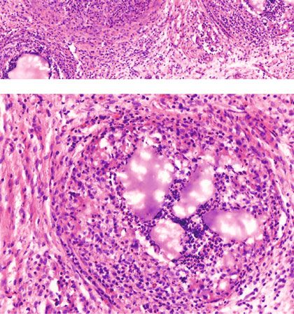
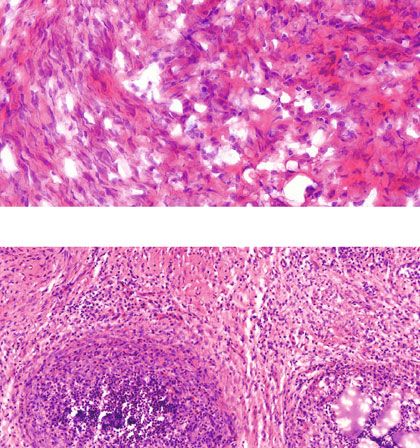
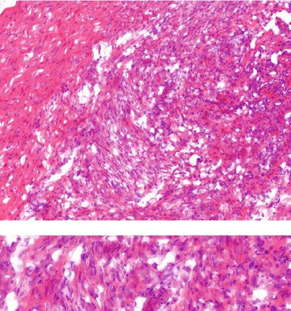
BioMed Research International 3

 Rats (n = 24) randomized for 2 groups

 Embedding material Saline (n = 12) MSU (n = 12)

 Saline MSU

 Number of operations
 3 (n =4) 4 (n = 4) 5 (n = 4) 3 (n = 4) 4 (n = 4) 5 (n = 4)

 Harvesting at 5 days after the last operation

 Further analysis Gross measurement H&E sections Immunofluorescence Behavioral experiments

 Figure 1: Flow diagram of animal study.

2.5. Statistical Analysis. Statistical analyses were performed proliferation, and collagen fiber disorder increased with the
by GraphPad Prism 8.0.1 software. All datasets were tested frequencies of interventions (Figure 4(a)). Different from
for normality for a t-test, and if the normality test failed, the previous acute gout arthritis model, recruited neutrophil
the Mann–Whitney rank-sum test was used for an packed with MSU crystal deposition was observed in the
intragroup comparison. Results are expressed as the mean MSU group. The H&E stain clearly demonstrates the eosino-
 ± SD. P value < 0.05 is considered significant. philic acellular structure (MSU crystals) and multiple sur-
 rounding layers of lobulated neutrophils (Figure 4(b)).
3. Result Immunofluorescence revealed the expression level of
 inflammatory factors in synovial tissue after MSU implanta-
All the rats received surgical interventions according to the tion. IL-1β and TNF-α are highly coexpressed in aggregated
experimental protocols. No incision-related infection, MSU inflammatory cells (Figure 5(a)). Further quantitative aver-
leakage, or other complications occurred. age optical (AO) analysis showed that the expression levels
 Gross measurements and behavioral tests were con- of two cytokines were upregulated with the increase of inter-
ducted to evaluate the local effects of MSU-mediated inflam- vention frequencies, which also demonstrated the typical
mation. Significant synovial swelling was detected in the MSU-mediated synovial inflammation in vivo (Figure 5(b)).
MSU group compared to the sham group five days after the
last intervention (P = 0:0250 after three interventions, P < 4. Discussion
0:0001 after four or five interventions) (Figure 3), while
patellar ligament was not affected by MSU inflammation This study designed a reliable and economical animal model
(P > 0:05 in all groups). for persistent gout arthritis manifested by significantly pro-
 Behavior tests revealed abnormal reductions of the PWT longed gross and behavioral abnormalities, as well as intra-
in MSU groups compared to the sham groups started with 2 articular MSU deposition and the tophi formation.
hours after the intervention (P < 0:05). Within 24 to 48 hours Animal models of gout are necessary basis for researchers
after the operation, the pain threshold of the rats in the sham to understand the disease progress and to determine poten-
groups gradually returned to the normal level. The pain tial treatments and prevention measures. A literature search
threshold of the MSU groups also recovered, but it was signif- based on the PubMed database was conducted among studies
icantly lower than that in the sham group within 96 hours of gouty arthritis over the past decade. More than 120 articles
(P < 0:05 during 2 hours to 96 hours postoperatively). are applying the injection model to study the mechanism of
 The histopathological differences between the MSU inflammatory cytokines regulation or acute inflammatory
group and the sham group were demonstrated by H&E stain- responses. Nevertheless, uric acid oxidase is lost in human
ing. In the saline groups, simple synovitis presented as but presenting in most mammals (rats, rabbits, etc.), which
inflammatory cell infiltrations. In the MSU intervention causes that serum uric acid levels of other mammals are only
group, the sections showed serious synovial hyperplasia, cell one-tenth of human [23, 24]. Hence, a limited dose of MSU
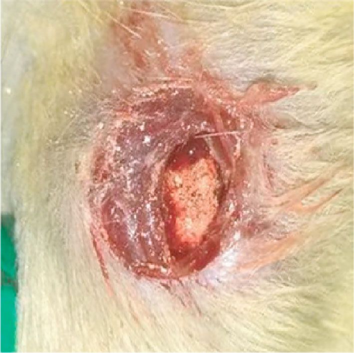
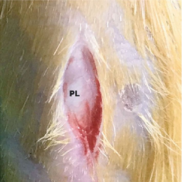
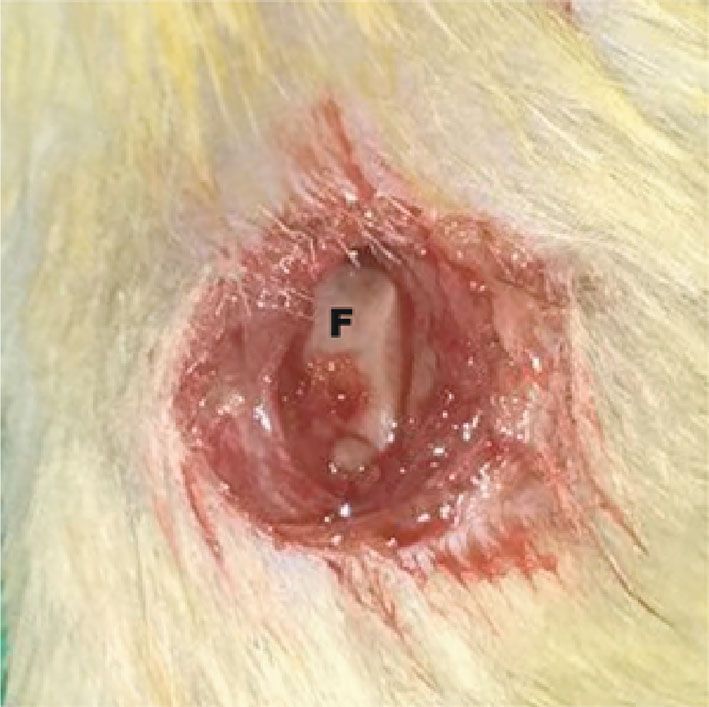
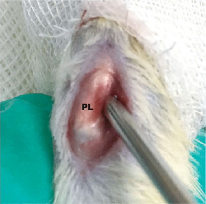
4 BioMed Research International

 (a) (b)

 (c) (d)

 (e)

Figure 2: Operating procedures of MSU embedment on rats. Protocol for establishing MSU embedment models. (a, b) The skin and capsule
of the joint were incised layer by layer along the lateral side of the patellar ligament, and the joint cavity of the knee was exposed. (c) The
intercondylar fossa cartilage of femur was damaged by a Kirschner wire. (d) The MSU crystal was embedded. (e) The incisions of the
articular capsule and skin were closed. PL: patellar ligament; F: femur.

injection often causes acute joint inflammation, while the deposition of MSU as well as interaction between crystals
threshold of tophi formation and crystal deposition is rarely and joint tissue in vivo [25].
reached. Without appropriate animal model, few studies are The present study puts accurate repeatable implantation
available regarding the formation, growth, attachment, and of larger doses of MSU into the articular cavity and maintains
BioMed Research International 5

 4 ns
 ⁎⁎⁎⁎ ⁎⁎⁎⁎ 5 ns ns
 ⁎
 Synovium thickness (mm)

 Patellar ligament width
 3 4

 3
 2

 2

 1
 1

 0 0
 3 4 5 3 4 5
 Operation times Operation times
 MSU MSU
 Sham Sham
 (a) (b)
 10

 ns
 8 ns ⁎⁎

 ⁎⁎⁎ ⁎
 ⁎
50% PWT (g)

 6 ⁎ 5
 ns
 ⁎ 4 ns ns ns
 ns
 50% PWT (g)

 4 ns
 3
 ns
 2
 2
 1

 0 0
 Pre 1 2 4 24 48 73 96 1 2 4 24 48 72 96
 Time (h) Time (h)

 Control 3 times
 MSU 4 times
 Sham 5 times
 (c) (d)

Figure 3: Gross changes in joint tissue and mechanical pain threshold in rats after MSU embedding. (a) The thickness of the synovium lateral
to the patellar ligament in the rats. The synovium thickness of the MSU groups was greater than that of the sham groups (n = 8 for each group;
two-tailed t-test, ∗ P = 0:0250, ∗∗∗∗ P < 0:0001). (b) No significant (NS) differences were detected between the sham groups and MSU groups
(n = 8 for each group; two-tailed t-test). (c) The 50%PWT of the wild-type littermates (n = 8) is shown between the dotted lines (6:33 ± 1:58
g). The pain threshold of the MSU groups was significantly lower than that in the sham group within 96 hours (n = 7 for sham group; n = 10
for MSU group; the two-tailed Mann–Whitney test was applied in the time point of 4 hours and 24 hours; a two-tailed t-test was applied for
other time points; Pre: P > 0:05; 1 hour: P > 0:05, 2 hours: P = 0:0461, 4 hours: P = 0:0164, 24 hours: P = 0:0311; 48 hours: P = 0:0005; 72
hours: P = 0:0010; 96 hours: P = 0:0355). (d) No significant differences were detected between the MSU groups with different operation
frequencies at all time points (n = 4 for each group; one-way ANOVA was applied for each time point; P > 0:05 at all time points with
different operation frequencies). The x-axis represents time points before and after operations. Data shown as the mean ± SD.

a continuous high concentration of MSU in the articular cant synovial swelling and mechanical hyperalgesia more
cavity to resist the effect of uric acid oxidase. Pain and than five days after interventions. This outcome is caused
joint swelling are typical symptoms of gout arthritis. In by MSU embedding rather than surgical incision due to
the injection model, local symptoms are proved to present the presence of a control group. In terms of pathology,
as self-limiting and relive within 72 hours [16]. Consis- MSU crystal deposition was observed surrounded by IL-
tently, the reduction of the PWT in MSU injection models 1β and TNF-α-positive inflammatory cell. This is the first
was reported to maintain within 48 to 72 hours in previ- report of exogenous MSU deposition in synovium in ani-
ous researches [16]. In contrast, this study significantly mal models into providing potential tools for clinical iden-
prolonged the persistence of local symptoms, with signifi- tification and debridement. The model also provides an
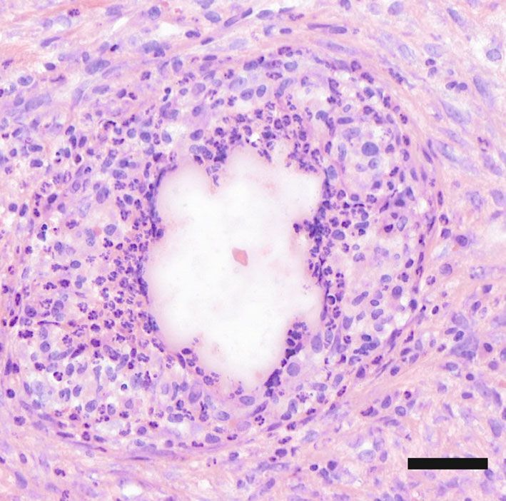
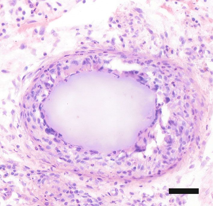
6 BioMed Research International

 Operation frequencies
 3 times 4 times 5 times

 400×
 Sham groups

 800×

 400×
 MSU groups

 800×

 (a)

 (b) (c)

Figure 4: Inflammatory cell infiltration and deposition of MSU in synovial tissue. Representative images of pathological section showed a
progressive MSU-mediated inflammation. (a) Top, in the sham groups, the synovial inflammation and disordered collagenous fibers were
observed. Bottom, MSU deposited in tissues mediating the immune response and recruiting a large number of inflammatory cells. MSU
deposition was surrounded by the inflammatory cells and collagen fibers. (b, c) Large-scale images of MSU deposition surrounded by
inflammatory cells in MSU groups received 4-5x interventions. Deposition of MSU was mainly surrounded by lobulated neutrophils. Scale
bars, 50 μm.

ideal approach for further studying the pathophysiological Compared with the injection modeling method, more
changes of MSU deposition in vivo and accurate removal persistent joint inflammation and crystal deposition are
of MSU deposition in the tissue. observed in this study for three main reasons, including
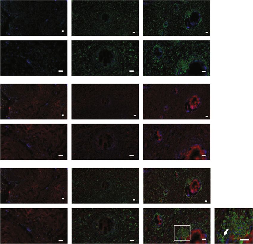
BioMed Research International 7

 MSU group (3 times) MSU group (4 times) MSU group (5 times)

 400 ×

 IL-1

 800 ×

 MSU group (3 times) MSU group (4 times) MSU group (5 times)

 400 ×

 TNF-

 800 ×

 MSU group (3 times) MSU group (4 times) MSU group (5 times)

 400 × IL-1 &TNF-

 800 ×

 (a)
 0.0025

 0.0020
 Average optical

 0.0015

 0.0010

 0.0005

 0.0000
 3 4 5
 Operation times
 IL-1
 TNF-
 (b)

Figure 5: Immunofluorescence revealed upregulation of IL-1β expression in the MSU embedding rat model. (a) Representative images of
immunofluorescence showed regulation of cytokine expression in MSU embedding groups. IL-1β (green) expression was significantly
upregulated in tissues. Similarly, TNF-α (red) is also highly expressed around MSU deposition. The arrow indicates a significant
expression of IL-1β in cells surrounding MSU. (b) Further quantitative average optical (AO) analysis showed that the expression levels of
two cytokines were upregulated with the increase of intervention frequencies (n = 4 for each group; one-way ANOVA and Tukey’s
multiple comparisons tests were applied for each cytokine). Data shown as the mean ± SD. Scale bars, 50 μm.
8 BioMed Research International

higher dose, more accurate implantation, and combination Ethical Approval
with artificial defect. (1) The implantation dose was signif-
icantly more sufficient than the traditional injection Experimental procedures passed a review by the Animal
method. Injection dose was distributed between 0.5 mg Welfare and Ethics Group, Department of Experimental
and 5.0 mg in previous research [12, 15, 16, 26], while Animal Science, Shanghai Medical College of Fudan Univer-
the total embedding dose of MSU in this experiment sity, Shanghai, China (Approval Number 2019020405).
reached 60 to 100 mg. High-dose MSU intervention,
though larger than other injection models, was shown to Disclosure
be safe, and no lesions or symptoms outside the joint were
found. The results proved that MSU crystals were depos- For the purpose of sharing scientific findings, this manuscript
ited in the synovium by inflammatory cells before being was submitted as a preprint in the link https://www
decomposed by uricase. (2) The space of rats’ knee articu- .researchsquare.com/article/rs-73068/v1.
lar cavity is limited with multiple contents including the
ligaments, cartilage, and meniscus. In addition, saturated Conflicts of Interest
MSU crystals tend to precipitate at room temperature
and then block injection needles. Swell and effusion of tis- The authors declare that there is no conflict of interest
sues induced by MSU could further aggravate the stenosis regarding the publication of this paper.
of cavity and increase the failure rate of repeated injection.
Minimally invasive approach solved this problem by
exposing the joint cavity and locating position of MSU Acknowledgments
crystals accurately. (3) Previous studies suggested that We thank the financial support from the Shanghai Municipal
physic impact would result in traumatic arthritis in rabbits Science and Technology Project (17441900900).
[27, 28]. A radiographic study also found a strong rela-
tionship between bone erosion and tophi formation [29].
Therefore, an impaired cartilage accelerates the establish- References
ment of a gouty arthritis model and then shortens the trial
 [1] E. Roddy and H. K. Choi, “Epidemiology of gout,” Rheumatic
period [30]. Diseases Clinics of North America, vol. 40, no. 2, pp. 155–175,
 There are some limitations in the present study as fol- 2014.
lows: (1) the procedures of this minimally invasive operation [2] T. R. Mikuls, J. T. Farrar, W. B. Bilker, S. Fernandes, Schuma-
required practice and repetition in order to get a satisfied cher HR Jr, and K. G. Saag, “Gout epidemiology: results from
result of MSU embedding. Basic surgical training is required the UK General Practice Research Database, 1990-1999,”
to meet the operational requirements. (2) The duration of Annals of the Rheumatic Diseases, vol. 64, no. 2, pp. 267–272,
this model was still much less than the natural course of 2005.
chronic gouty arthritis in human lasting for a few years. Fur- [3] G. Ragab, M. Elshahaly, and T. Bardin, “Gout: an old disease in
ther studies have to include a more convenient way to embed new perspective - a review,” Journal of Advanced Research,
MSU and increase the sessions of embedment to extend the vol. 8, no. 5, pp. 495–511, 2017.
affect time. It is not verified how long the model will last [4] P. O'Connor, “Crystal deposition disease and psoriatic arthri-
without further MSU embedding, which will be one of the tis,” Seminars in Musculoskeletal Radiology, vol. 17, no. 1,
research directions in the future. (3) In this study, synovial pp. 74–79, 2013.
crystal deposition was observed, but the tendon, ligament, [5] N. Schlesinger, “Management of acute and chronic gouty
cartilage, or bone was not involved. Due to the process of sec- arthritis: present state-of-the-art,” Drugs, vol. 64, no. 21,
tion decalcification, some technical difficulties of preserving pp. 2399–2416, 2004.
crystals from strong acid need to be overcome. Sections of [6] G. Schett, C. Schauer, M. Hoffmann, and M. Herrmann, “Why
osteochondral specimens should also be analyzed in further does the gout attack stop? A roadmap for the immune patho-
researches. genesis of gout,” RMD Open, vol. 1, article e000046, Suppl 1,
 2015.
 [7] C. Schauer, C. Janko, L. E. Munoz et al., “Aggregated neutro-
5. Conclusion phil extracellular traps limit inflammation by degrading cyto-
 kines and chemokines,” Nature Medicine, vol. 20, no. 5,
A minimally invasive surgical method was developed to pp. 511–517, 2014.
establish the novel rat model of chronic gouty arthritis. This [8] N. Schlesinger and R. G. Thiele, “The pathogenesis of bone
model was proved to be a simple repeatable method and to erosions in gouty arthritis,” Annals of the Rheumatic Diseases,
mimic the pathological characteristics of human persistent vol. 69, no. 11, pp. 1907–1912, 2010.
gouty arthritis. [9] Z. Liu, T. Chen, H. Niu et al., “The establishment and charac-
 teristics of rat model of atherosclerosis induced by hyperurice-
 mia,” Stem Cells International, vol. 2016, Article ID 1365257, 7
Data Availability pages, 2016.
 [10] R. Bluestone, J. Waisman, and J. R. Klinenberg, “Chronic
The authors declare that all experimental data of this study experimental hyperuricemic nephropathy,” Laboratory Inves-
are available by contacting the corresponding author. tigation, vol. 33, no. 3, pp. 273–279, 1975.
BioMed Research International 9

[11] C. Pineda, A. J. Fuentes-Gómez, C. Hernández-Díaz et al., [25] A. Chhana, G. Lee, and N. Dalbeth, “Factors influencing the
 “Animal model of acute gout reproduces the inflammatory crystallization of monosodium urate: a systematic literature
 and ultrasonographic joint changes of human gout,” Arthritis review,” BMC Musculoskeletal Disorders, vol. 16, no. 1, 2015.
 Research & Therapy, vol. 17, no. 1, p. 37, 2015. [26] R. M. dos Santos, S. M. Oliveira, C. R. Silva, C. Hoffmeister,
[12] T. J. Coderre and P. D. Wall, “Ankle joint urate arthritis J. Ferreira, and J. Assreuy, “Anti-nociceptive and anti-
 (AJUA) in rats: an alternative animal model of arthritis to that edematogenic effects of glibenclamide in a model of acute
 produced by Freund's adjuvant,” Pain, vol. 28, no. 3, pp. 379– gouty attack in rats,” Inflammation Research, vol. 62, no. 6,
 393, 1987. pp. 617–625, 2013.
[13] Y. Hu, Q. Yang, Y. Gao et al., “Better understanding of acute [27] D. Milentijevic, I. F. Rubel, A. S. Liew, D. L. Helfet, and P. A.
 gouty attack using CT perfusion in a rabbit model,” European Torzilli, “An in vivo rabbit model for cartilage trauma: a pre-
 Radiology, vol. 29, no. 6, pp. 3308–3316, 2019. liminary study of the influence of impact stress magnitude
[14] D. J. McCarty Jr., P. Phelps, and J. Pyenson, “Crystal-induced on chondrocyte death and matrix damage,” Journal of Ortho-
 inflammation in canine joints. I. An experimental model with paedic Trauma, vol. 19, no. 7, pp. 466–473, 2005.
 quantification of the host response,” Journal of Experimental [28] S. D. Fening, M. H. Jones, V. Moutzouros, B. Downs, and
 Medicine, vol. 124, no. 1, pp. 99–114, 1966. A. Miniaci, “Method for delivering a controlled impact to
[15] A. Marcotti, A. Miralles, E. Dominguez et al., “Joint nociceptor articular cartilage in the rabbit knee,” Cartilage, vol. 1, no. 3,
 nerve activity and pain in an animal model of acute gout and pp. 211–216, 2010.
 its modulation by intra-articular hyaluronan,” Pain, vol. 159, [29] N. Dalbeth, B. Clark, K. Gregory et al., “Mechanisms of bone
 no. 4, pp. 739–748, 2018. erosion in gout: a quantitative analysis using plain radiography
[16] W. Chai, Y. Tai, X. Shao et al., “Electroacupuncture alleviates and computed tomography,” Annals of the Rheumatic Dis-
 pain responses and inflammation in a rat model of acute gout eases, vol. 68, no. 8, pp. 1290–1295, 2009.
 arthritis,” Evidence-based Complementary and Alternative [30] C. F. Kuo, M. J. Grainge, W. Zhang, and M. Doherty, “Global
 Medicine, vol. 2018, Article ID 2598975, 15 pages, 2018. epidemiology of gout: prevalence, incidence and risk factors,”
[17] X. Wu, M. Wakamiya, S. Vaishnav et al., “Hyperuricemia and Nature Reviews Rheumatology, vol. 11, no. 11, pp. 649–662,
 urate nephropathy in urate oxidase-deficient mice,” Proceed- 2015.
 ings of the National Academy of Sciences of the United States
 of America, vol. 91, no. 2, pp. 742–746, 1994.
[18] C. Ryckman, S. R. McColl, K. Vandal et al., “Role of S100A8
 and S100A9 in neutrophil recruitment in response to monoso-
 dium urate monohydrate crystals in the air-pouch model of
 acute gouty arthritis,” Arthritis and Rheumatism, vol. 48,
 no. 8, pp. 2310–2320, 2003.
[19] W. J. Martin, O. Shaw, X. Liu, S. Steiger, and J. L. Harper,
 “Monosodium urate monohydrate crystal-recruited nonin-
 flammatory monocytes differentiate into M1-like proinflam-
 matory macrophages in a peritoneal murine model of gout,”
 Arthritis and Rheumatism, vol. 63, no. 5, pp. 1322–1332, 2011.
[20] M. Rull, G. Clayburne, M. Sieck, and H. R. Schumacher,
 “Intra-articular corticosteroid preparations: different charac-
 teristics and their effect during inflammation induced by
 monosodium urate crystals in the rat subcutaneous air pouch,”
 Rheumatology (Oxford), vol. 42, no. 9, pp. 1093–1100, 2003.
[21] M. A. al-Madol, M. Shaqura, T. John et al., “Comparative
 expression analyses of pro- versus anti-inflammatory media-
 tors within synovium of patients with joint trauma, osteoar-
 thritis, and rheumatoid arthritis,” Mediators of Inflammation,
 vol. 2017, 11 pages, 2017.
[22] W. J. Dixon, “The up-and-down method for small samples,”
 Publications of the American Statistical Association, vol. 60,
 no. 312, pp. 967–978, 1965.
[23] X. W. Wu, D. M. Muzny, C. Chi Lee, and C. Thomas Caskey,
 “Two independent mutational events in the loss of urate oxi-
 dase during hominoid evolution,” Journal of Molecular Evolu-
 tion, vol. 34, no. 1, pp. 78–84, 1992.
[24] T. B. Friedman, G. E. Polanco, J. C. Appold, and J. E. Mayle,
 “On the loss of uricolytic activity during primate evolution—I.
 Silencing of urate oxidase in a hominoid ancestor,” Compara-
 tive Biochemistry and Physiology Part B: Comparative Bio-
 chemistry, vol. 81, no. 3, pp. 653–659, 1985.
You can also read