Comparison of biotechnological culture of hypoxia-conditioned rat mesenchymal stem cells with conventional in vitro culture of ...
←
→
Page content transcription
If your browser does not render page correctly, please read the page content below
Veterinary World, EISSN: 2231-0916                                                                    RESEARCH ARTICLE
Available at www.veterinaryworld.org/Vol.12/June-2019/29.pdf                                               Open Access

     Comparison of biotechnological culture of hypoxia-conditioned
    rat mesenchymal stem cells with conventional in vitro culture of
 normoxia-conditioned rat mesenchymal stem cells for testicular failure
                    therapy with low libido in rats
                                               Erma Safitri1,2 and Mas’ud Hariadi1

   1. Department of Veterinary Reproduction, Faculty of Veterinary Medicine, Universitas Airlangga, Surabaya 60115,
Indonesia; 2. Stem Cells Research Division, Institute Tropical Disease, Universitas Airlangga, Surabaya 60115, Indonesia.
                     Corresponding author: Mas’ud Hariadi, e-mail: masudhariadi@yahoo.co.id
                                        Co-author: ES: erma-s@fkh.unair.ac.id
                   Received: 27-12-2018, Accepted: 15-04-2019, Published online: 28-06-2019

doi: 10.14202/vetworld.2019.916-924 How to cite this article: Safitri E, Hariadi M (2019) Comparison of biotechnological
culture of hypoxia-conditioned rat mesenchymal stem cells with conventional in vitro culture of normoxia-conditioned rat
mesenchymal stem cells for testicular failure therapy with low libido in rats, Veterinary World, 12(6): 916-924.

                                                            Abstract
Aim: Biotechnological culture of hypoxia-conditioned (CH) rat mesenchymal stem cells (rMSC-CH) for testicular failure
therapy with low libido improves the functional outcome of the testicle for producing spermatogenic cells and repairs
Leydig cells in rats (Rattus norvegicus).
Materials and Methods: In the first group (T1), rats with testicular failure and low libido were injected with normoxia-
conditioned (CN) rMSCs (21% oxygen); in the second group (T2), rats with testicular failure and low libido were injected
with rMSC-CH (1% oxygen); in the negative control group (T−), rats with normal testis were injected with 0.1 mL
phosphate-buffered saline (PBS); and in the sham group (TS), rats with testicular failure and low libido were injected with
0.1 mL of PBS.
Results: Vascular endothelial growth factor expression, as the homing signal, in the groups T2, T−, T1, and TS was
2.00±0.5%, 2.95±0.4%, 0.33±0.48%, and 0±0%, respectively. The number of cluster of differentiation (CD)34+ and
CD45+ cells in the groups T− and TS was 30% and >80%, respectively,
showing the mobilization of hematopoietic stem cells (HSCs). The number of spermatogenic cells (spermatogonia, primary
spermatocytes, secondary spermatocytes, and spermatid) decreased significantly (p0.05) decrease compared to that in T−. The improvement
in libido, based on the number of Leydig cells producing the hormone testosterone for libido expression, did not increase in
T1, whereas T2 was able to maintain the number of Leydig cells significantly compared to that between TS and T1.
Conclusion: rMSC-CH culture for testicular failure with low libido showed improvement in the functional outcome of the
testicle and in repairing Leydig cells.
Keywords: hypoxia-conditioned rat mesenchymal stem cells, low libido, normoxia-conditioned rat mesenchymal stem
cells, rat, testicular failure.
Introduction                                                       has yielded very promising results in regeneration
      Oligospermia, a failure of the testicle, represents          of spermatogenesis process in testicular tissue with
a major reproductive health problem in males, causing              infertility issues, such as oligospermia [5]. The effi-
low fertility and libido [1,2]. Oligospermia is a con-             cacy of the therapy was limited due to low viability
dition in which low numbers of spermatozoa are pro-                of stem cells after transplantation [6,7], which is due
duced by testicular seminiferous tubules, resulting in             to the conventional in vitro culture of normoxia-con-
infertility in males, meaning failure to reproduce [3].            ditioned rat MSCs (rMSC-CN) with oxygen concen-
Oligospermia for male is the main cause of infertil-               tration of >21%. The conventional culture causes cell
ity with varying etiologies, such as heredity, trauma,             senescence [8], apoptosis [9], and gene mutation,
neoplastic tumor, and degenerative disorders due to                such as G: C to T: A [10]. Some researchers showed
malnutrition [4].                                                  that >93% of stem cells perish between 1 and 7 days
      Transplantation therapy with mesenchymal                     after transplantation [11-15]. Therefore, stem cells
stem cells (MSCs) derived from the bone marrow                     are required in high quantities with several booster
                                                                   doses for the effectiveness of therapy using conven-
Copyright: Safitri and Hariadi. Open Access. This article is       tional culture like normoxia-conditioned (CN), thus
distributed under the terms of the Creative Commons Attribution
4.0 International License (http://creativecommons.org/licenses/    considerably increasing the cost [16]. To remain via-
by/4.0/), which permits unrestricted use, distribution, and        ble, in vitro culture of stem cells must be adapted for
reproduction in any medium, provided you give appropriate credit
to the original author(s) and the source, provide a link to the    a niche or microenvironment wherein the stem cells
Creative Commons license, and indicate if changes were made.       reside in the bone marrow. In normal conditions, the
The Creative Commons Public Domain Dedication waiver (http://
creativecommons.org/publicdomain/zero/1.0/) applies to the data
                                                                   niche of stem cells in the bone marrow is in a low
made available in this article, unless otherwise stated.           oxygen concentration conditioned hypoxia (CH)]
Veterinary World, EISSN: 2231-0916                                                                                     916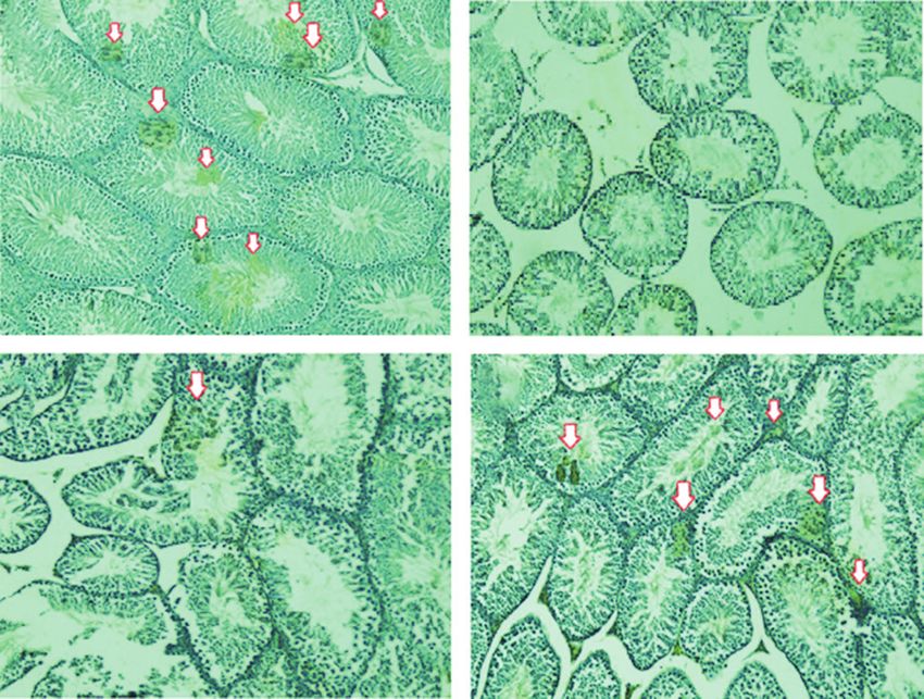
Available at www.veterinaryworld.org/Vol.12/June-2019/29.pdf

[5,8,10]. Therefore, biotechnological modification               The sample was diluted with PBS to a total
of rMSC-CH in vitro culture is required for homing         volume of 15 mL, with the tube being turned
signal and mobilization of stem cells to improve tes-      3–5 times as a means of achieving an even mix. At the
ticular function for producing sperms. The homing          next stage, centrifugation at 1600 rpm for 15 min at
signal of stem cells in the testicle tissue is based on    room temperature of 37°C was performed for 10 min
the expression of vascular endothelial growth factor       at a speed of 1600 rpm (287 rcf). Before heating, the
(VEGF), whereas mobilization is based on the expres-       supernatant was discarded, and the cells were resus-
sion of cluster of differentiation (CD) such as CD34+,     pended in 6 mL of α-MEM (M0894, Sigma Aldrich®,
CD45+, and CD105− cells [5-7].                             Burlington, Massachusetts USA). The cell suspen-
      The objective of the study was utilization of        sion was placed in 10-cm2 plate (Falcon™, Thermo
biotechnological culture of rMSC-CH for testicu-           Fisher Scientific, Pittsburgh, PA, USA) and incubated
lar failure therapy with low libido. It was revealed       at 37°C for 24 h in a humidified atmosphere contain-
that biotechnological culture of rMSC-CH improved          ing 5% CO2 until cells adhered on the surface of the
the functional outcome of the testicle for producing       plate. After 24 h, media and non-adherent cells were
spermatogenic cells and repairing Leydig cells of rat      discarded. The adhered cells were rinsed twice using
(R. norvegicus).                                           5 mL of PBS and shaken before heating the culture.
Materials and Methods                                      The supernatant was discarded, and the plate was
                                                           washed again twice with PBS. After 10 min, 10 mL of
Ethical approval
                                                           fresh α-MEM (M0894, Sigma Aldrich®, Burlington,
     Animal studies were performed using a protocol
approved by the Animal Care and Ethical Clearance          Massachusetts, USA) was added to the dish before
Committee of Faculty of Veterinary Medicine,               incubation. The cells were incubated at 37°C with
Universitas Airlangga, and were in conformance with        5% CO2, and the culture was observed daily using
the guidelines of the National Research Council (239-      an inverted microscope. (MXD-400 Phase Contrast,
KE) through ethical seminar. The research was con-         Nanjing BW Optics and Instrument Co., Ltd)
ducted at the laboratory in the Institute of Tropical            Every four days, the media were discarded, and
Diseases and Faculty of Veterinary Medicine,               cells were rinsed with 5 or 10 mL of 1× PBS before
Universitas Airlangga.                                     heating. PBS was subsequently discarded, and the dish
                                                           was filled with 10 mL of fresh α-MEM (M0894, Sigma
Isolation and culture of stem cells
                                                           Aldrich®, Burlington, Massachusetts, USA). The cells
      MSCs were collected from the bone marrow             were cultured continuously until approximately 75%–
of iliac crest of male Wistar rats (R. norvegicus) at      80% confluence was attained. The cells were then
3 months of age. The aspirate was collected in hep-        passaged into several dishes for subculture [17].
arinized tubes and stored at 4°C for transportation to           Passaging was conducted three times, and then
the laboratory at the Institute of Tropical Diseases,      cells were divided into two groups, viz., rMSC-CH
Universitas Airlangga, for in vitro culturing [17].        treatment with 1% O2 in a hypoxia chamber inside a
      The aspirate of MSCs was collected in 15-mL          5% CO2 incubator and rMSC-CN with 21% O2; both
heparin tube (Z181099, Sigma Aldrich®, Burlington,         were incubated for four days. The three gases needed
Massachusetts, USA), which were previously filled
                                                           for 1% low oxygen precondition are 5% CO2 to flow
with 3 mL of α-modified Eagle medium (α-MEM)
                                                           at 37°C in the incubator, 100% O2 only for calibra-
(M0894, Sigma Aldrich®, Burlington, Massachusetts,
                                                           tion, and N2. N2 was used to replace O2 to maintain the
USA). The aspirate was transferred to a 15-mL sterile
                                                           desired concentration.
blue cap tube and sterile 1× phosphate-buffered saline
(PBS; MFCD00131855, Sigma Aldrich®, Burlington,            Animal model of testicular failure and low libido
Massachusetts, USA) was added to a total volume of               Testicular failure in rats was obtained by fast-
10 mL. The tube with the aspirate solution was then        ing for five days, but drinking water was provided
rinsed twice with 5 mL of PBS. The diluted sam-            ad libitum. [1]. The condition of fasting for five days
ple was added with equal volume of Ficoll (F9378,          induced testicular failure in the rats because the func-
Sigma Aldrich®, Burlington, Massachusetts, USA) at         tion of adrenal cortex was suboptimal in producing
room temperature of 37°C in a separate 15-mL tube.         dehydroepiandrosterone (DHEA) due to malnutrition.
Furthermore, each aspirate was mixed with Ficoll           Low levels of DHEA in the blood can be a cause of
before centrifugation (Sorvall™ MX Series Floor            fatigue and decreased sperm concentration. DHEA is
Model Micro-Ultracentrifuge, Thermo Fisher, Grand          the most potent precursor of steroid hormones, such
Island, USA) at 1600 rpm [287 relative centrifugal         as testosterone, which is produced by the renal adrenal
force (rcf)] for 15 min at room temperature of 37°C.       cortex [18] and Leydig cells of testis [3]. Low testos-
After centrifugation, mononucleated cells were col-        terone production can lead to decreased spermatogen-
lected in the form of “buffy coat” located on the sur-     esis and libido in males.
face of Ficoll–PBS using a sterile Pasteur pipette and           The animal model used in this study was male
taken in a 15-mL tube (Sigma Aldrich®, Burlington,         Wistar rats (R. norvegicus), 250–300 g body weight,
Massachusetts, USA).                                       3 months of age, and healthy condition. Individual rats
Veterinary World, EISSN: 2231-0916                                                                            917
Available at www.veterinaryworld.org/Vol.12/June-2019/29.pdf

were placed in plastic cages at the Animal Laboratory      The cells were observed using a regular luminescence
Experimental, Faculty of Veterinary Medicine,              microscope Nikon H600L (Nikon Instuments Inc.,
Universitas Airlangga.                                     America) equipped with a digital camera (DS Fi2, 300
Biotechnological culture of rMSC-CH compared with          megapixels) and image processing software and cell
rMSC-CN culture                                            count Nikon Image System.
      Biotechnology stem cell treatment was per-           Flow cytometry
formed by injecting MSCs directly from either con-               Flow cytometry (BD FACSCalibur™, BD
ditioned rMSC-CH or rMSC-CN in the medial part             Biosciences, San Jose, USA) observation of stem cell
of both testes, following anesthesia using ketamine        mobilization was based on the calculated number of
(87 mg/kg of body weight) and xylazine (13 mg/             CD34+, CD45+, and CD105− cells. After the treatment,
kg of body weight) at 10-15 min after simultaneous         whole blood was collected in a heparin tube via car-
injection and lasting for 15-30 min, followed by a rel-    diac puncture to prevent coagulation. Flow cytometric
atively long period of immobility (mean, 3.8 h) and        observation revealed the numbers of CD34+, CD45+,
reduced responsiveness to external stimuli [19].           and CD105− cells. In brief, whole blood was centri-
      MSCs injected into male rats induced testicular      fuged at 6000 rpm (4032 rcf) at 4°C for 15 min. The
failure, such as oligospermia with low libido, compared    cellular precipitate obtained after centrifugation was
with that in the negative and sham group. In the first     mixed with cytoperm/cytofix (554714, BD Cytofix/
group (T1), rats with testicular failure and low libido    Cytoperm™, BD Biosciences, San Jose, USA) at
were injected with 200 million rMSC-CN/rat from            twice the volume of that of the obtained cell volume.
high oxygen culture (21% O2); in the second group          This mixture was then centrifuged at 1600 rpm for
(T2), rats with testicular failure and low libido were     15 min at room temperature of 37°C.
injected with 200 million rMSC-CH/rat from hypoxia               To separate the supernatant and pellet, BD wash
culture (1% O2); in the negative control group (T−),       was added to the pellet at four-times the volume of
rats with normal testicle were injected with 0.1 mL of     the obtained cell volume from the first centrifugation,
PBS; and in the sham group (TS), rats with testicular      and then lysis buffer was added at twice the volume
failure and low libido were injected with 0.1 mL of        of the first obtained cell volume. Subsequently, the
PBS.                                                       labeled antibody was added to each sample, and five
      After two cycles of spermatogenesis, male            tubes were arranged and processed simultaneously:
rats were operated to collect the testicle tissue.         (1) single staining with CD34 PE (Rabbit Anti-CD34/
Immunohistochemistry (IHC) analysis was performed          HCAM/PGP1 Polyclonal Antibody, PE-conjugated
to determine the expression of VEGF as the homing          Conjugated Primary Antibodies-bs-0521R-PE; Bioss,
signal of stem cells in the testicle tissue. Flow cytom-   USA); (2) double staining with CD34 PE and CD45
etry was performed to calculate the number of CD34+,       PerCP (PerCP-Cy5.5 anti-human cross anti-rabbit
CD45+, and CD105− cells, as an indicator of stem cell      CD45, BD Biosciences, USA); (3) double staining
mobilization, to improve testicular disorders.             with CD34 PE; (4) double staining with CD45 PerCP;
      The improvement in the testicle for producing        and (5) double staining with CD105 fluorescein iso-
sperm cells was observed by histopathological analy-       thiocyanate (FITC) (Rabbit Anti-Thy-1/CD105/
sis by hematoxylin-eosin (HE) staining (B8438, Sigma       Thy1.1 Polyclonal Antibody, 0778R-FITC, Bioss,
Aldrich®, Burlington, Massachusetts, USA), and then        USA) in true count tubes. All samples were then
the number of spermatogenic cells (spermatogonia,          stored at 4°C in a dark room and analyzed by flow
primary spermatocytes, secondary spermatocytes, and        cytometry for 1 h [21].
spermatid) and Leydig cells were calculated.               Histopathology observation
IHC analysis for VEGF expression                                The improvement in testicular function with
      IHC analysis was performed to determine VEGF         low libido for producing sperm was observed by
expression. Histological preparation was done before       histopathological preparation by HE staining and
IHC. A transverse incision was made in the paraffin        calculating the number of spermatogenic cells
block containing the testicular tissue. Further exam-      (spermatogonia, primary spermatocytes, second-
ination was performed by determining the external          ary spermatocytes, and spermatid) and Leydig cells.
expression of VEGF by IHC using VEGF monoclo-              Histopathological examination was performed using
nal antibody (A183C 13G8, Thermo Fisher Scientific,        a light microscope (Oxford BCM 400, monotaro.id,
Pittsburgh, PA, USA) with PBS (MFCD00131855,               Indonesia) under magnification of 200×. Observations
Sigma Aldrich®, Burlington, Massachusetts, USA)            were made after one cycle of spermatogenesis process
dilution. VEGF expression was observed using a             (35 days) during which food was provided normally;
regular light microscope (Oxford BCM 400, mono-            male rats were operated to collect the testicle tissue.
taro.id, Indonesia) under 200× magnification, and          The calculation of the number of spermatogenic cells
the expression of each variable was indicated by the       was based on the total number of four cell types (sper-
number of cells with the development of brownish           matogonia, primary spermatocytes, secondary sper-
discoloration of the chromogen in each incision [20].      matocytes, and spermatid) located on the five lumens
Veterinary World, EISSN: 2231-0916                                                                           918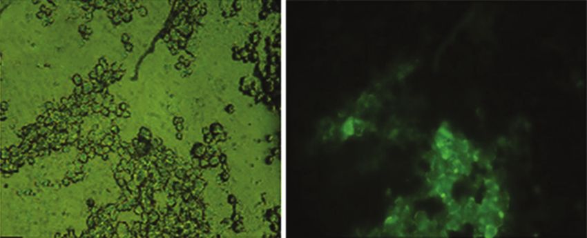
Available at www.veterinaryworld.org/Vol.12/June-2019/29.pdf

of the seminiferous tubules from five preparate slides.     was noted in TS 0a±0% (0%; Figure-3b). All scores of
Furthermore, the total number of the four cell types of     VEGF expression are summarized in Table-1.
the five tubules was divided by 5 [1,2].
      The testicular tissue of the rat was fixed in 10%
formalin, and 1 h later, mid-testis was injected with 10%
formalin. Rats testes then were dehydrated in gradually
higher concentration of alcohol, i.e. from 70%, 80%,
90%, and 96%. Then, the testes were cleaned with
xylol. Next, the testes were embedded in liquid paraf-
fin and put into molds. Before staining and sectioning,
an incision was made using a microtome and mounted          a                            b
on glass slides. Furthermore, staining was performed        Figure-1: Positive expression of cluster of differentiation
by removing paraffin with xylol, followed by treating       (CD)105 in mesenchymal stem cells (MSCs) using a
with decreased concentration of alcohol, i.e. from 96%,     fluorescent microscope at a magnification of 400×.
                                                            (a) Without filter, MSCs; (b) with green filter, fluorescent
90%, 80%, and 70%, and then subjected to HE stain-          MSCs (CD105).
ing. Finally, mounting was performed after treating the
slides with increasing concentration of alcohol, from
70%, 80%, 90%, and 96%, to remove excess stain and
then treated with xylol. The preparations were then cov-
ered with a cover glass and mounted with Canada bal-
sam (Cas No: 8007-47-4, Sigma Aldrich®, Burlington,
Massachusetts, USA)[22].
Statistical analysis
     VEGF expression, calculated numbers of CD34+
                                                            a                              b
and CD45+ cells, and number of spermatogenic and
Leydig cells were statistically analyzed using SPSS         Figure-2: Negative expression of cluster of differentiation
                                                            (CD)45 in mesenchymal stem cells (MSCs) using a fluorescent
17 for Windows XP (IBM SPSS Statistic Application,          microscope at a magnification of 400×. (a) Without filter,
America) with the confidence level of 99% (α=0.01)          MSCs; (b) with green filter, non-fluorescent MSCs (CD45).
and level of significance of 0.05 (p=0.05). The steps
of hypothesis testing were as follows: normality
data were tested by the Kolmogorov–Smirnov test,
homogeneity of variance by analysis of variance, and
post hoc test using the Tukey’s honest significant dif-
ference test with 5% as the least significant difference.
Results
                                                             a                               b
      The purity of MSCs derived bone marrow
was determined by immunofluorescence through
the identification of CD105+ and CD45− cells [17].
Immunofluorescence identification for CD105 was
positive expression and CD45 was negative expres-
sion. CD105+ and CD45− cells were determined
using specific cell surface markers of MSCs using
the monoclonal antibody FITC anti-rabbit CD105               c                               d
(Biolegend) and FITC anti-rabbit CD45 (Biolegend).          Figure-3: Immunohistochemistry analysis of homing
Immunofluorescence results showed that cultured             signal of stem cells based on vascular endothelial growth
                                                            factor (VEGF) expression (brown chromogen) with
cells were true MSCs (Figures-1 and 2).                     different treatments at 200× magnification (Nikon H600L
IHC analysis                                                Microscope; digital camera DS Fi2 300 megapixel, Nicon
                                                            Instruments Inc., America). Different superscripts indicate
     The IHC analysis was performed by scoring 0            significant difference at p50%; Figure-3a), whereas the percent-          (d) second group (T2), rats with testicular failure and
age was still higher than that in T1, with the score        low libido injected with 200 million hypoxia-conditioned
being 0.33a±0.48% (
Available at www.veterinaryworld.org/Vol.12/June-2019/29.pdf
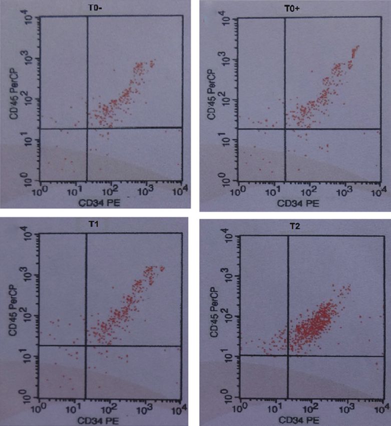
Flow cytometry observation of CD34+ and CD45+ cells calculated in T− and TS was
Flow cytometry observation of stem cell 30% (Figure-4c) and >80% in T2 (Figure-4d),
CD34+, CD45+, and CD105− cells. The average number indicating the mobilization of hematopoietic stem cells
Table-1: Score of VEGF expression by IHC analysis and the calculated number of CD34+and CD45+cell by flow cytometry
in rat testicle tissue after different treatments (mean%±SD).
Treatments Average VEGF Average number of calculated
expression score±SD CD34+and CD45+cells (%)±SD
Negative control group (T−): Rats with normal 2.95c±0.40 18.25a±0.50
testicle were injected with 0.1 mL of PBS
Sham group (TS): Rats with testicular failure and low 0a±0 19.45a±0.35
libido were injected with 0.1 mL of PBS
First group (T1): Rats with testicular failure and low 0.33a±0.48 32.15b±1.65
libido were injected with 200 million rMSC‑CN/rat
Second group (T2): Rats with testicular failure and 2.00b±0.50 83.65c±1.50
low libido were injected with 200 million rMSC‑CH/rat
Values in the same column with different superscripts indicate significant difference at pAvailable at www.veterinaryworld.org/Vol.12/June-2019/29.pdf
(HSCs). There was a significant difference (p0.05) between the T− and TS, VEGF expression, as a marker of homing sig-
although both groups were statistically different from nal, was determined through IHC analysis. VEGF
T1 (Figure-4 and Table-1). is a component of the extracellular matrix of stem
Histopathology and libido observation cells. Moreover, it plays a role in supporting a con-
The improvement in testicular function with oli- ducive microenvironment for stem cells to remain
gospermia was observed by histopathological obser- viable [26]. The low oxygen culture used for stem
vation based on the number of spermatogenic cells cells in this study provides a supportive niche and
(spermatogonia, primary spermatocytes, secondary triggers expression of VEGF-1, which is a homing
spermatocytes, and spermatid). The libido of male signal. Furthermore, VEGF-1 binds to VEGF recep-
rats was assessed based on the number of Leydig tor-1, thus, activating a series of signaling events that
cells (Table-2), the function of which is production activate stem cell factor (SCF). SCF is physiologi-
of testosterone to stimulate libido [21,22]. The results cally vital and is in the niche of protein expression
showed that although spermatogenic cell production that helps in further communication [27]. The SCF
in T2 decreased significantly (p>0.05) compared to receptor complex progresses into the cell nucleus to
that in T−, and it was significantly (pAvailable at www.veterinaryworld.org/Vol.12/June-2019/29.pdf
signals, such as stromal cell-derived factor-1, C-X-C in turgor from the reproduction organs and erection
motif chemokine ligand 12 (CXCL12), VEGF, hepato- occurs [30].
cyte growth factor, platelet-derived growth factor, and The lack of glucose in both TS and T1 led to no
integrin, that act for recruitment of stem cells [17]. or less fuel and energy sources, in this case glucose,
In this study, as mentioned in point “f”, mobilization which is universal for all cells, including spermato-
occurred due to the expression of homing signal, such genic and sperm cells. No carbon source is available
as VEGF. due to unavailability of glucose for the synthesis of
Libido in male rats was assessed by determining several compounds, such as fatty acids; cholesterol;
the number of Leydig cells. Decreased libido in TS amino acids; nucleic acids; and steroid hormones,
occurred because the adrenal cortex was ineffective such as testosterone. Glucose is also needed as a pre-
in producing DHEA due to malnutrition (rats expe- cursor to a variety of other sugars, such as lactose,
rienced fasting for five days). Low levels of DHEA nucleotides, and glycosaminoglycans [31].
in the blood can be a cause of fatigue and decreased Glucose in the cytoplasm of spermatozoa
body stamina. DHEA is the most potent precursor of undergoes glycolysis, wherein glucose is catabo-
steroid hormones, such as testosterone, produced by lized to form pyruvate to produce two molecules of
the renal adrenal cortex [18] and Leydig cells or inter- ATP by phosphorylation at the substrate level. In this
stitial cells between seminiferous tubules of testis [3]. process, nicotinamide adenine dinucleotide (NAD+)
In T2, rats with oligospermia and low libido were is converted to nicotinamide adenine dinucleotide
injected with 200 million stem cells/rat from low oxy- hydrogen (NADH), and NADH is then transferred
gen culture (1% O2). The results suggested that 200 to the mitochondrial electron chain to form pyruvic
million stem cells/rat from low oxygen culture (1% acid in the citric acid cycle for complete oxidation
O2) maintained libido same as that observed in T−. to CO2. Citric acid cycle, known as the tricarbox-
Increased DHEA as a precursor of steroid hormones, ylic cycle, is a catabolism reaction path occurring
such as the hormone testosterone, is also responsible in the matrix of mitochondria. As the process of
for fat metabolism and acts as an enzyme inhibitor the citric acid cycle and glycolysis proceeds in the
of glucose 6-phosphate dehydrogenase, which acts mitochondria inner membrane, oxidative phosphor-
as a biocatalyst for converting glucose to fat. Thus, ylation occurs in the mitochondria to use high-en-
increase in DHEA allows for an increase in the amount ergy ATP [32]. The entire process series is known as
of free adenosine triphosphate (ATP) in the body and aerobic glucose oxidation, which produces 38 mol-
reproduction organ, thus, increasing the stamina of ecules of ATP [31].
the body [29] and health of reproduction organs [3]. In this study, results of T1 suggested that rats
Further, increases in free ATP in the body and repro- injected with 200 million rMSC-CN/rat from normoxia
duction organ in addition to increased stamina also culture (21% O2) were unable to stimulate libido.
support an increase in libido. It can be explained that However, rats injected with 200 million rMSC-CH/rats
libido can be considered suitable if the body is in a from hypoxia culture (1% O2) increased the number of
state of excellent stamina, thus, allowing the process Leydig cells significantly, which was different from that
of reproduction, such as spermatogenesis. in TS and T1. However, the increase in Leydig cells in
In T1, rats with testicular failure and low libido the T2 group was different from that in T− (Table-2).
were injected with 200 million stem cells/rat from
high oxygen culture (21% O2). The results suggested Conclusion
that 200 million stem cells/rat from high oxygen cul- The efficacy of rMSC-CH biotechnological
ture (21% concentration of O2) were unable to main- culture for testicular failure therapy with low libido
tain libido same to that observed in T−. A decrease was determined based on the expression of VEGF,
in Leydig cells reduces testosterone production and increase in the number of CD34+ and CD45+ cells,
leads to decreased libido in males. improvement in the testis to produce spermatogenic
The libido process begins with the stimulation cells, and improvement in libido based on the number
of the central nervous system in the hypothalamus, of Leydig cells.
where dopamine is produced as a neurotransmitter
Authors’ Contributions
and neurohormone that affects sexual behavior and
activity in individuals. The stimuli received by the ES: Research project leader, research and
sensory nerve trigger acetaminophen that stimulates ethical clearance preparation, IHC and flow cytom-
endothelial cells to secrete nitric oxide for activat- etry analysis, stem cell isolation from rat bone mar-
ing cyclic guanosine monophosphate (cGMP). This row, rMSC-CH and rMSC-CN procedure, stem cell
process causes the muscles of corpus cavernosum transplantation, statistical procedure, and drafting the
in the penis to swell, leading to the constriction of manuscript preparation (wrote the paper). MH: Rat
penis arterioles so that the blood flow is increased. testicular failure and low libido model, observation
This, in turn, causes the erection of penis due to full of improvement in testicular function based on the
blood flow by depressing the veins and inhibiting calculated number of spermatogenic and sperm cells,
the release of blood flow so that there is an increase observation of libido (based on the number of Leydig
Veterinary World, EISSN: 2231-0916 922Available at www.veterinaryworld.org/Vol.12/June-2019/29.pdf
cells), designing the study, analyzing statistical data, 11. Cai B., Li, X., Wang N., Liu Y., Yang F., Chen H., Yin K.,
proofreading, and corresponding author. All the Tan X., Zhu J., Pan Z., Wang B. and Lu Y. (2013) Apoptosis
of bone marrow mesenchymal stem cells caused by homo-
authors have read and approved the final version of cysteine via activating JNK signal. PLoS One, 8(5): 1-9.
the manuscript. 12. Chen T. L., Zhu G. L., Wang J. A., Wang Y., He X. L. and
Jiang J. (2014) Apoptosis of bone marrow mesenchymal
Acknowledgments stem cells caused by hypoxia/reoxygenation via multiple
The study was supported by funding from The pathways. Int. J. Clin. Exp. Med., 7(12): 4686-4697.
13. Teixeira F. G., Panchalingam K. M., Anjo S. I., Manadas B.,
Directorate General of Higher Education of The Pereira R., Sousa N., Salgado A. J. and Behie, L. A. (2015)
National Education Ministry, Republic of Indonesia, Do hypoxia/normoxia culturing conditions change the neu-
Grant number: 004/ADD/SP2H/LT/DRPM/VIII/2017. roregulatory profile of Wharton jelly mesenchymal stem
cell secretome? Stem Cell Res. Ther., 6(2): 133-147.
Competing Interests 14. Qin H. H., Filippi C., Sun S., Lehec S., Dhawan A. and
The authors declare that they have no competing Hughes, R. D. (2015) Hypoxic preconditioning potentiates
the trophic effects of mesenchymal stem cells on co-cul-
interests. tured human primary hepatocytes. Stem Cell Res. Ther.,
Publisher’s Note 7(4): 1-12.
15. Zhou H., Li D., Shi C., Xin T., Yang J., Zhou Z., Hu S.,
Veterinary World remains neutral with regard Tian F., Wang J. and Chen Y. (2015) Effects of exendin-4 on
to jurisdictional claims in published institutional bone marrow mesenchymal stem cell proliferation, migra-
tion and apoptosis in vitro. Sci. Rep., 5(12898): 1-12.
affiliation. 16. Safitri E., Widiyatno T. V. and Prasetyo R. H. (2016)
References Honeybee product therapeutic as stem cells homing for
ovary failure. Vet. World, 9(11): 1324-1330.
1. Safitri E., Utama S., Widiyatno T. V., Sandhika W. and 17. Eleotério R. B., Sepúlveda R. V., Reis E. C. C., Valente F. L.
Prasetyo, R. H. (2016) Auto-regeneration of mice testicle and Borges A. P. B. (2016) Isolation, expansion and differ-
seminiferous tubules due to malnutrition based on stem entiation of mesenchymal stromal cells from rabbits’ bone
cells mobilization using honey. Asian Pac. J. Reprod., 5(1): marrow. Pesq. Vet. Bras., 36(5): 423-430.
31-35. 18. Hackbert L. and Heiman J. R. (2002) Acute dehydroepi-
2. Cakici C., Buyrukcu B., Duruksu G., Haliloglu A. H., androsterone (DHEA) effect on sexual arousal in postmeno-
Aksoy A., IsJk A., Uludag O., Ustun H., Subas J. C. and pausal women. J. Womens Health Gend. Based Med., 11(2):
Karaoz E. (2013) Recovery of fertility in azoospermia rats 155-162.
after injection of adipose-tissue-derived mesenchymal stem 19. Struct M. B., Andrutis K. A., Ramirez H. E. and Battles A.H.
cells: The sperm generation. Bio. Med. Res. Int., 13(1): 1-18. (2011) Effect of a short-term fast on ketamine-xylazine
3. Hafez E. S. E. and Hafez B. (2013) Reproduction in Farm anesthesia in rats. J. Am. Assoc. Lab. Anim. Sci., 50(3):
Animals. 7th ed. Wiley-Blackwell, Philadelphia, PA, USA. 344-348.
157-198. 20. Kumar G. L. and Rudbeck L. (2009) Immunohistochemical
4. Prasetyo R. H. and Safitri E. (2016) Effects of honey to Staining Methods. 5th ed. DAKO North America, Carpinteria
mobilize endogenous stem cells in efforts intestinal and CA, USA. 11-14.
ovarian tissue regeneration in rats with protein-energy mal- 21. Bushnel T. and Hanke I. (2014) Modern Flow Cytometry
nutrition. Asian Pac. J. Reprod., 5(3): 198-203. Ebook. Mission Ave Ste, Liberty Lake, United States. 123.
5. Safitri E., Utama S., Bumi C., Mulyani S. W. M., 22. Razi M., Najafi G., Feyzi S., Karimi A., Shahmohamad S.
Retnowati E., Prasetyo R. H., Hariadi M., Mahyudin F. and and Nejati V. (2012) Histological and histochemical effects
Rantam, F. A. (2014) Hypoxic preconditioning for viable of Glyphosate on testicular tissue and function. Iran J.
and self-renewing mesenchymal stem cells (MSCs) as the Reprod. Med., 10(3): 181-192.
regeneration of spermatogenesis process. Adv. Nat. Appl. 23. Trapani M. D., Bassi G., Midolo M., Gatti A., Kamga P. T.,
Sci., 8(8): 42-46. Cassaro A., Carusone R., Adamo A. and Krampera M. (2016)
6. Safitri E., Utama S., Bumi C., Mulyani S. W. M., Differential and transferable modulatory effects of mesen-
Susilowati H., Retnowati E., Purwati A., Prasetyo R. H., chymal stromal cell-derived extracellular vesicles on T, B
Hariadi M. and. Rantam F. A. (2013) The role of adaptive and NK cell functions. Nat. Sci. Rep., 6(24120): 1-13.
MSCs an attempt regeneration of spermatogenesis process 24. Kaundal U., Bagai U. and Rakha A. (2018)
using by hypoxia precondition in vitro. J. Anim. Prod. Adv., Immunomodulatory plasticity of mesenchymal stem cells:
3(11): 318-322. A potential key to successful solid organ transplantation.
7. Scaradavou A., Smith K. M., Hawke R., Schaible A., Rev. J. Transl. Med., 16(31): 1-16.
Abboud M., Kernan N. A., Young J.W. and Barker J. N. 25. Nejad N. A., Amidi F., Hoseini M. A., Nia K. N., Habibi M.,
(2015) Cord blood units with low CD34+ cell viability have Kajbafzadeh A. M., Mazaheri Z. and Yamini N. (2015) Male
a low probability of engraftment after double-unit transplan- germ-like cell differentiation potential of human umbilical
tation. Biol. Blood Marrow Transplant., 16(4): 500-508. cord Wharton’s jelly-derived mesenchymal stem cells in
8. Tsai C. C., Chen Y. J., Yew T. L., Chen L. L., Wang J. Y., co-culture with human placenta cells in presence of BMP4
Chiu C. H. and Hung S.C. (2011) Hypoxia inhibits senes- and retinoic acid. Iran J. Basic. Med. Sci., 18(4): 325-333.
cence and maintains mesenchymal stem cell properties 26. Dennis E. A. and Bradshaw R. A. (2011) Intercellular
through down-regulation of E2A-p21 by HIF-TWIST. Signaling in Development and Disease. 1st ed. Elsevier Inc.,
Blood J. Hematol., 117(2): 459-469. San Diego CA, USA. 121-129.
9. Mulyani S. W. M., Setiawati E. M., Safitri E. and Astuti E. R. 27. Jones D. L. and Wagers A. J. (2008) No place like home:
(2014) The role of heat shock protein 27 (HSP 27) as inhib- Anatomy and function of the stem cell niche. Nat. Rev. Mol.
itor apoptosis in hypoxic conditions of bone marrow stem Cell Biol., 9(1): 11-21.
cell culture. Dent. J. Maj. Ked. Gigi., 47(1): 41-44. 28. Arai F., Hirao A., Ohmura M., Sato H., Matsuoka S.,
10. Szablowska-Gadomska I., Zayat V. and Buzanska L. (2011) Takubo K., Ito K., Koh G. Y. and Suda T. (2007) Tie2/
Influence of low oxygen tension on expression of plurip- Angiopoietin-1 signaling regulates hematopoietic stem
otency genes in stem cells. Acta Neurobiol. Exp., 71(1): cells quiescence in the bone marrow niche. Cell, 118(2):
86-93. 149-161.
Veterinary World, EISSN: 2231-0916 923Available at www.veterinaryworld.org/Vol.12/June-2019/29.pdf
29. Maggio M., DeVita F., Lauretani F., Nouvenne A., Meschi T., ECG, Jakarta, Indonesia. 1283-1288.
Ticinesi A., Dominguez L. J., Barbagallo M., Dall’Aglio E. 31. Marks D. B., Allan D. M. and Collen M. S. (2016) Biokmia
and Ceda G. P. (2014) Review article the interplay between Kedokteran Dasar. Sebuah Pendekatan Klinis. 2nd ed. Penerbit
magnesium and testosterone in modulating physical func- Buku Kedokteran ECG, Jakarta, Indonesia. 165-177.
tion in men. Int. J. Endocrinol. 2014(ID 525249): 1-9. 32. Lehningger A. L. (2014) Dasar-Dasar Biokimia, Alih
30. Guyton A. and Hall J.E. (2013) Buku Ajar Fisiologi Bahasa Maggy Thenawidjaja. 1st ed. Penerbit Airlangga,
Kedokteran; Setiawan I. 12th ed. Penerbit Buku Kedokteran Jakarta, Indonesia. 73-93.
********
Veterinary World, EISSN: 2231-0916 924You can also read