Role of exosomal miRNAs in brain metastasis affected by radiotherapy
←
→
Page content transcription
If your browser does not render page correctly, please read the page content below
Translational Neuroscience 2021; 12: 127–137
Research Article
Zihuang Li*, Hongli Yang, Ling Ye, Rencui Quan, Meili Chen
Role of exosomal miRNAs in brain metastasis
affected by radiotherapy
https://doi.org/10.1515/tnsci-2020-0163 plasma exosomal miRNAs in five patients with brain meta-
received January 15, 2021; accepted March 15, 2021 stasis before and after radiotherapy. This study is the begin-
Abstract: In oncogenesis and development of malignant ning; more specimen and further research are needed to
tumor, microRNAs (miRNAs) regulate the complex gene explore the functional role of specific miRNAs and their
expression associated with the tumor pathogenesis. potential as therapeutic targets for brain metastasis.
Currently, only few studies have been conducted to identify Keywords: radiotherapy, brain metastasis, exosome,
miRNAs and the potential pathways involved in the patho- microRNAs, malignant tumors
genesis of brain metastasis in patients who underwent
radiotherapy, especially miRNAs in the plasma exosomes.
Therefore, this study is aimed to use small RNA analysis to
identify miRNAs and their potential target genes in plasma 1 Introduction
exosomes during the initiation and development of brain
metastasis in patients who underwent radiotherapy. Using Brain metastases are the most common intracranial malig-
high-throughput sequencing technologies, we identified nant tumors, and their primary foci typically include lung
35 differentially expressed miRNAs in patients with brain cancer (20–56%), breast cancer (5–20%), and melanoma
metastasis who had undergone radiotherapy. In annota- (7–16%), as well as colorectal cancer and renal cell carci-
tion of miRNA targets, gene ontology enrichment analysis noma [1,2]. Brain metastasis occurs because of the prolif-
revealed that the targets of the differentially expressed eration of cells. In addition, it can occur because of the
miRNAs were significantly enriched in the regulation of cel- transport of the primary tumors to cerebral vessels through
lular processes. Kyoto Encyclopedia of Genes and Genomes the blood. Complex niche tumor interactions in the micro-
revealed that most of the miRNA targets were cancer- environment, neuroinflammatory cascade, and possible
related, including genes involved in the mitogen-activated neovascularization are involved in new metastasis [3].
protein kinase signaling pathway, cancer-related pathways, Once brain metastasis occurred in patients with cancer,
phosphatidylinositol 3-kinase-protein kinase B signaling they induce various central nervous system (CNS) symp-
pathway, microtubule-associated protein kinase signaling toms that severely reduce the quality of life of patients,
pathway, Ras signaling pathway, regulation of the actin resulting in poor prognosis and high mortality. If untreated,
cytoskeleton, and axon guidance. In conclusion, this study the survival time is approximately 1–2 months, while the
provides a new perspective to understand the possible func- median survival time is only 4–12 months after active treat-
tion of these miRNAs in the pathogenesis of brain meta- ment [4]. To solve the problem of poor prognosis in patients
stasis. This was the first time that a pilot study identified with brain metastasis, we need to understand the disease
complexity at the molecular level. Some hypotheses have
been proposed to explain the unique metastasis patterns
of different primary cancers, including the importance
* Corresponding author: Zihuang Li, Department of Radiation
of “seeds” (cancer cells) and “soil” (microenvironment of
Oncology, The Second Clinical Medical College of Jinan University,
Shenzhen Municipal People’s Hospital, 1017 Dongmen North Road, receiving organs) or changes in circulation patterns
Shenzhen, Guangdong, 518020, China, between primary and metastatic tumors. Evidence shows
e-mail: li.zihuang@szhospital.com, tel: +86+134-2429-1813 that metastatic cells and tumor microenvironment are essen-
Hongli Yang, Rencui Quan, Meili Chen: Department of Radiation tial for tumor growth [3]. It is important to understand the
Oncology, The Second Clinical Medical College of Jinan University,
unique biological sensitivity of each tumor and the mole-
Shenzhen Municipal People’s Hospital, 1017 Dongmen North Road,
Shenzhen, Guangdong, 518020, China
cular differences occurring because of the corresponding
Ling Ye: Department of oncology, The First Affiliated Hospital of Ji treatment of brain metastasis. The CNS is protected by var-
Nan University, Guangzhou, Guangdong, China ious functional barriers, including the blood–brain barrier
Open Access. © 2021 Zihuang Li et al., published by De Gruyter. This work is licensed under the Creative Commons Attribution 4.0
International License.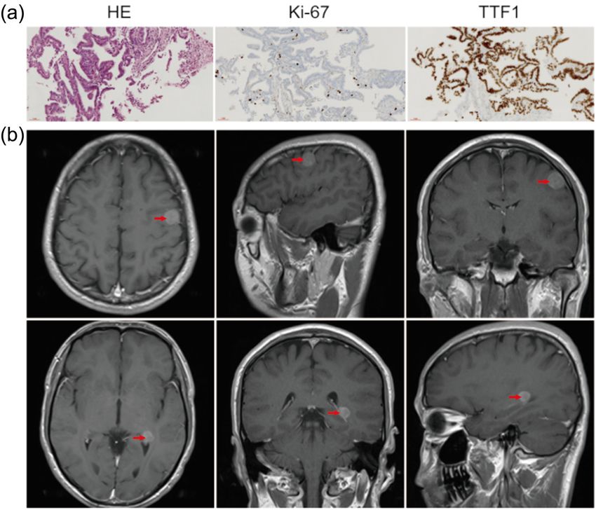
128 Zihuang Li et al.
(BBB) and blood-cerebrospinal fluid (CSF) barrier. Studies age between 18 and 75 years old; Karnofsky performance
have shown that circulating monocytes can interfere with status (KPS) score ≥60; number of brain metastasis ≤4;
brain homeostasis through the BBB [5,6], and circulating maximum diameter of metastasis ≤3 cm; primary tumor
tumor cells may use this mechanism to enter the brain [7]. was stable or controlled within 2 months after drug treat-
The blood-CSF barrier is formed by the epithelial cells of ment; no other extracranial metastasis; no hard (soft)
the choroid plexus that form tight junctions. The capillary meningeal metastasis; no history of brain radiotherapy.
window and intercellular space of the choroid plexus allow Exclusion criteria: primary tumor was not controlled; extra-
molecules to move freely in these compartments [8]. The cranial metastasis occurred simultaneously; history of cra-
expression of a complement protein (C3) in primary cancer niocerebral radiotherapy; pregnant or lactating women;
cells can destroy the blood-CSF barrier, causing mitogens to patients not suitable for large fraction irradiation; patients
enter the CSF [9]. These factors may lead to brain metastasis. with active liver, kidney, and heart diseases; drug abuse,
Exosomes are heterogeneous nanoscale vesicles found long-term alcoholism, and AIDS. Withdrawal criteria:
in blood and other body fluids. These disperse a variety of unable to carry out treatment according to the require-
bioactive molecules, such as proteins, mRNA, microRNAs ments of the study protocol; serious adverse events, preg-
(miRNA), DNA, and lipids, into various cells. Exosomes nancy of the patient, and withdrawal of the subject. The
can cross the BBB, and during inflammatory stimulation, clinical specimens included peripheral blood (approxi-
the endothelial cells of the brain release exosomes into mately 4–5 mL), which was collected within 3 days before
the bloodstream by responding to inflammatory reac- and after radiotherapy, respectively. The blood was collected
tions. Moreover, inflammatory stimulation leads to the in ethylenediaminetetraacetic acid tubes, stored at −4℃,
internalization of cells and the formation of early endo- and then centrifuged for 10 min at 12,000 g. The plasma
somes, which then form exosomes that are finally released was harvested and stored at −80℃ until exosome isolation.
into the blood or brain parenchyma [10]. Exosomes, as a All brain metastasis cells were histologically diagnosed
mode of communication between the cells and the envir- using magnetic resonance imaging (MRI). The treatment
onment, have increasingly become a focus of research for patients with brain metastasis was intensity modulated
[11–13]. They carry thousands of proteins and nucleic radiation therapy/volumetric modulated arc radiotherapy.
acids and transmit biological information over a long
distance. Exosomes play an important role in the occur- Ethical approval: This research related to human use has
rence and development of brain metastasis. Therapeutic been complied with all the relevant national regulations
strategies for brain metastasis include radiotherapy, sur- and institutional policies and in accordance with the
gery, chemotherapy, immunotherapy, and targeted thera- tenets of the Helsinki Declaration, and has been approved
pies; in particular, radiotherapy treatments for brain by the authors’ institutional review board or equivalent
metastasis have developed rapidly [14]. This study is aimed committee.
to explore the recognition of differentially expressed
miRNAs in plasma exosomes and the prediction of target Informed consent: Informed consent has been obtained
signaling pathways in patients with brain metastasis before from all individuals included in this study.
and after radiotherapy treatment, possibly providing new
clues for the treatment of brain metastasis.
2.2 Exosome isolation and RNA extraction
Exosomes were isolated from the plasma using an exoQuick
2 Methods precipitation (System Biosciences, USA) according to the
manufacturer’s protocol. Briefly, the exosomes were preci-
2.1 Patient sample collection pitated by incubation; the exoQuick exosome precipitation
reagent was added at 4°C for 60 min. The exosome pellet
Plasma was collected from five patients with lung adeno- was collected by centrifugation at 1,500 × g for 10 min at 4°C
carcinoma or melanoma diagnosed as brain metastasis and resuspended in 10 mM PBS to four times the original
(aged 33–75 years) at the Shenzhen People’s Hospital. plasma volume. Nanoparticle tracking analysis and trans-
This study was conducted in accordance with the princi- mission electron microscopy were used to identify the
ples of the Declaration of Helsinki (1964) and with the exosomes. Subsequently, total RNA was extracted from exo-
participants’ understanding and consent. Inclusive criteria: some pellets using TRIzol (Thermo Fisher Scientific, Inc.,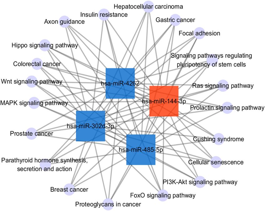
Role of exosomal miRNAs in brain metastasis 129
Carlsbad, CA, USA), followed by treatment with DNase to 2.6 Quantitative reverse transcription-
remove potential DNA contamination. The integrity and polymerase chain reaction validation of
concentration of the RNA were determined photometrically the differentially expressed miRNAs
at 260 nm and 280 nm using an Agilent 2100 Bioanalyzer
(Agilent Technologies, USA). To validate the miRNA expression results from sequen-
cing, total RNA from the exosomes was extracted using a
TaqMan® MicroRNA Reverse Transcription kit (Thermo
Fisher Scientific, Inc., Carlsbad, CA, USA) and analyzed
2.3 Small RNA library construction and
by quantitative reverse transcription-polymerase chain
sequencing reaction (RT-qPCR) as per manufacturer’s instructions.
Briefly, qPCR was carried out using SYBR‑Green (Thermo
Approximately 1 µg of total RNA per sample was used for
Fisher Scientific, Inc.) according to the manufacturer’s
the RNA sample preparations. Small RNA library prepara-
instructions in an ABI 7300 real‑time qPCR system
tion was conducted using TruSeq Small RNA Sample Prep
(Thermo Fisher Scientific, Inc., Carlsbad, CA, USA). The
Kits (Illumina, San Diego, USA). After ensuring sample
PCR conditions were as follows: 95℃ for 3 min, followed
purity with an Agilent 2100 Bioanalyzer, the purified
by 45 cycles at 95℃ for 10 s, 60℃ for 45 s, and 72℃ for 30 s.
libraries were sequenced on an Illumina HiSeq 2500.
The quantification of miRNAs was performed in relation to
the U6 housekeeping miRNA. The relative abundance of
miRNAs was calculated using the comparative Cq method
2.4 miRNA-sequencing data analysis (2−ΔΔCq) and assessed by t-test.
Quality control was performed on raw data using the
FASTX-Toolkit, which involved the removal of low-quality
reads and trimming adapters. Clean reads were aligned to 3 Results
the human reference transcriptome sequence. Reads per
kilobase of exon model per million mapped reads were
3.1 Brain metastasis miRNA signature
calculated to obtain normalized expression levels. Finally,
differential expression analysis was performed using edgeR. derived from plasma exosomes
Before differential expression analysis, for each sequenced
library, the read counts were adjusted using one scaling nor- For the global analysis of the miRNA signature derived
malized factor. The relative counts of miRNAs were compared from plasma exosomes of brain metastasis, ten clinical
with the corresponding values before and after radiotherapy. plasma exosomes were collected for sequence analysis,
The p-value was adjusted using the Benjamini–Hochberg including five brain metastasis samples before radiotherapy
method. The miRNAs with corrected p-values 2 and a p-value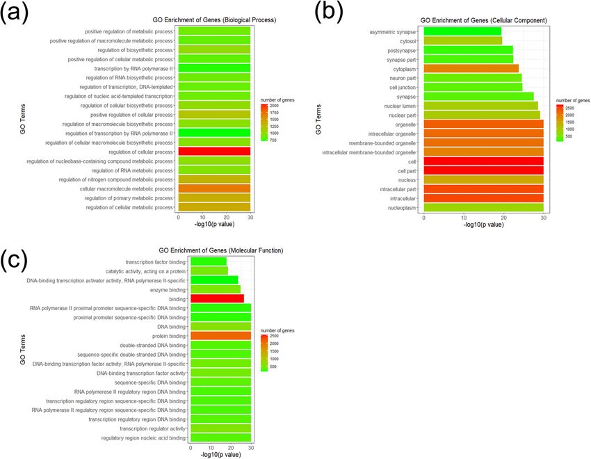
130 Zihuang Li et al.
3.2 GO enrichment analysis
Size of brain metastases (mm)
GO enrichment analysis was performed on significantly
differentially expressed miRNA target genes predicted by
TargetScan, and the related biological processes, cellular
components, molecular functions, and gene numbers
28 × 24 were analyzed. The most significantly enriched GO terms
25 × 20
28 × 16
14 × 11
15 × 15
for the biological process group were regulation of cel-
lular processes and cellular macromolecule metabolic
processes. In the cellular component category, many
target genes were found in the nucleoplasm as well as
Left frontal lobe and right temporal pole
intracellular components. In the molecular function cate-
Left parietal lobe and occipital bone
gory, the areas of regulation included nucleic acid bind-
ing, transcription regulator activity, and protein binding
(Figure 3). The result of GO enrichment analysis showed
Bilateral brain parenchyma
Intracranial supratentorial
that the significantly differentially expressed miRNAs in
plasma exosomes were associated with metabolic processes
Metastasis sites
Left frontal lobe
and transcription regulator activity, which were associated
with cancer-associated pathways, and may play an impor-
tant role in brain metastasis.
3.3 KEGG pathway analysis
Single/multiple
KEGG pathway analysis of the differentially expressed
Multiple
Multiple
Multiple
Multiple
Table 1: Sample pathological diagnosis information (M-BR, metastases before radiotherapy)
Single
miRNAs was used to identify target genes. Axon guidance
was the most significant pathway according to p-values.
In addition, the signaling pathways regulating pluripo-
tency of stem cells, proteoglycans in cancer, focal adhe-
Lung adenocarcinoma brain metastases
Lung adenocarcinoma brain metastases
Lung adenocarcinoma brain metastases
Lung adenocarcinoma brain metastases
sion, the microtubule associated protein kinase (MAPK)
signaling pathway, and the phosphatidylinositol 3-kinase-
protein kinase B (PI3K-Akt) signaling pathway were iden-
Melanoma brain metastases
tified as differentially regulated pathways (Figure 4). To
understand the potential pathway targets of the differen-
tially expressed miRNAs better, the interaction between the
top 20 significant KEGG pathways and the differentially
expressed miRNAs was studied (Figure 5). The results
Diagnosis
showed a significant interaction between top 20 KEGG path-
ways and the differentially expressed miR-144-3p, miR-
4262, miR-302d-3p, and miR-485-5p.
Age (years)
3.4 Validation by RT-qPCR
36
47
33
75
75
To confirm and validate the expression of the signifi-
cantly differentially expressed miRNAs identified in
Specimens
this study, we evaluated the expression of six miRNAs
M-BR4
M-BR2
M-BR3
M-BR5
M-BR1
(Table 2) in the plasma exosomes of patients with brain
metastasis using stem-loop RT-qPCR. The miRNAs were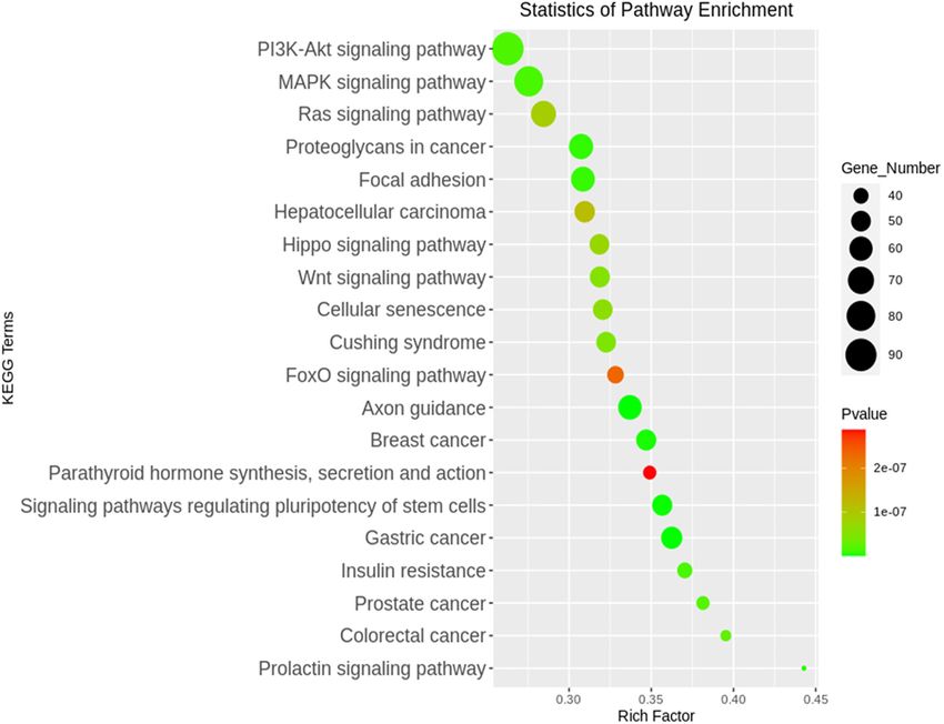
Role of exosomal miRNAs in brain metastasis 131
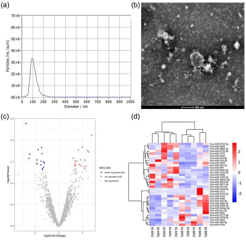
Figure 1: Brain metastases were diagnosed histologically and MRI in lung adenocarcinoma clinical samples. (a) The expression of tumor-
related indexes was detected by immunohistochemistry with 20× objective lens in lung adenocarcinoma. (b) Imaging examination of
multiple brain metastases in lung adenocarcinoma.
differentially expressed in the pre-and post-radiotherapy cells, may promote cancer cell colonization [17]. miRNAs
samples. The expression of three miRNAs (miR-4262, can regulate gene expression by degrading mRNA or
miR-6752-5p, and miR-302d-3p) decreased in the plasma repressing mRNA translation. The intercellular exchange
exosomes of patients after radiotherapy. The expression of miRNA through exosomes is a potential and effective
of miR-502-5p, miR-144-3p, and miR-609 was higher in method of intercellular communication that may have mul-
the plasma exosomes of pre-radiotherapy samples as tiple functions, especially in tumor survival and metastasis.
compared to the post-radiotherapy samples (Figure 6). Exosomal miRNAs can induce phosphatase and tensin
homolog (PTEN) loss in the microenvironment, thus pro-
moting brain metastasis [18]. Currently, because of the lim-
itations of our understanding of molecular mechanisms
4 Discussion underlying brain metastasis, the development of effective
treatments for brain metastasis is hindered. In this study,
Exosomes are considered intercellular communicators the changes in exosome miRNA expression resulting from
that can promote the development of cancer, and they radiotherapy performed for brain metastasis were defined
can be used potentially as biomarkers and in therapeutic for the first time.
methods. Exosomes play an important role in the tumor This study identified 35 miRNAs that were differen-
microenvironment interactions in primary and metastatic tially expressed in the five patients with brain metastasis
brain tumors [15]. Studies have reported that exosome before and after radiotherapy. Because of the limited
integrin in tumors can be used to predict organ-specific sample size collected clinically, the study should be
metastasis [16]. In addition, cell migration-inducing and referred to as a “pilot” study. Besides, there are still
hyaluronan-binding (CEMIP) protein in tumor exosomes many interesting results to be found, which could reflect
from brain metastasis, but not from lung or bone metastatic the overall characteristics to a certain extent. Homo sapiens132 Zihuang Li et al. Figure 2: Identification of exosomes and the expression of differentially expressed miRNAs in exosomes. (a and b) Measurement of diameter of vesicles to identify exosomes by nanoparticle tracking analysis and transmission electron microscopy. (c and d) Volcano plot and heatmap showed the differentially expressed miRNAs in each sample. M-AR, metastasis after radiotherapy; M-BR, metastasis before radiotherapy. (hsa)-miR-4262 has been reported to participate in pacli- cell proliferation and migration by targeting large tumor taxel resistance in non-small cell lung cancer by suppressor 1 in gliomas [22,23]. The expression of miR- regulating PTEN, and it promotes the proliferation and 302d-3p is increased by a low-Se environment in the invasion of human breast cancer cells directly by tar- Caco-2 cell line. Dysregulation of the cell cycle and of geting kruppel-like 6 (KLF6) and KLF15 [19,20]. hsa- the stress response pathways caused by low Se may influ- miR-4262 was also expressed in five human melanoma ence key genes involved in carcinogenesis. The effect of cell lines, and its expression pattern was opposite to low Se on biological pathways may be partly because of KLF6 expression. Bioinformatics analysis and KLF6-3′ the action of Se-sensitive miR-302d-3p [24]. It has been UTR luciferase reporter gene analysis indicated that found that miR-4262 can be sponged by circAGFG1, and it KLF6 was the direct target gene of miR-4262. The miR- can regulate the YY1/CTNNB1 axis to drive colorectal 4262 can significantly reduce the expression of KLF6 pro- cancer metastasis [25]. In this study, the expression of tein and promote the proliferation of melanoma cells [21]. miR-4262 and miR-302d-3p decreased in the pre-radio- Moreover, an abnormal increase in miR-4262 promotes therapy samples as compared to the pre-and post-
Role of exosomal miRNAs in brain metastasis 133 Figure 3: Top 20 functional GO terms of the differentially expressed miRNA target genes shown according to p-value. (a–c) Biological process, cellular component, and molecular function terms of the differentially expressed miRNA target genes. radiotherapy samples, indicating that radiotherapy may as a tumor suppressor in renal cell carcinoma and inhi- inhibit the proliferation of cancer cells by reducing the bits its invasion and metastasis by targeting MAP3K8, expression of miR-4262 and miR-302d-3p in exosomes of also a tumor suppressor, by targeting FZD7. Furthermore, patients with brain metastasis. Furthermore, hsa-miR- these outcomes can be used to predict the prognosis of 502-5p and hsa-miR-144-3p were upregulated after radio- human glioblastoma [28,29]. These results suggest that therapy. The expression of miR-502 is regulated by a miR-502 and miR-144-3p may be potential tumor sup- negative feedback action of p53, and the expression of pressors and may have some function in the role of radio- miR-502-5p was found to be downregulated in colon therapy in brain metastasis treatment. cancer patients as compared to that in matched normal GO and KEGG pathway enrichment analysis showed controls. Moreover, miR-502 inhibits the growth of colon the significantly enriched functions and pathways targeted cancer in mice. Studies have shown that miR-502 regu- by the differentially expressed miRNAs. In this study, the lates autophagy by inhibiting RAB1B, which is the key GO terms of regulation of cellular process, cellular macro- mediator of autophagy, and miR-502-5p inhibits auto- molecule metabolic process, regulatory region nucleic acid phagy, growth, and cell cycle progression in colon cancer binding, and transcription regulator activity were all enriched cells [26]. It has been reported that miR-144-3p plays by the significantly differentially expressed miRNA-targeted different roles in various diseases and promotes tumor genes. miRNAs can regulate mRNAs in different biological growth and metastasis of papillary thyroid carcinoma pathways, and miRNA expression may play a role in the by targeting the paired box gene 8 [27]; However, it serves modulation of anticancer effects via regulation of different
134 Zihuang Li et al. Figure 4: Top 20 KEGG pathways of the differentially expressed miRNA target genes shown according to p-value. Figure 5: Interaction between the top 20 KEGG pathways and the differentially expressed miRNAs. The upregulated miRNAs were repre- sented as red square nodes, the downregulated miRNAs were represented as blue square nodes, and the KEGG pathways were represented as violet circular nodes.
Role of exosomal miRNAs in brain metastasis 135 Table 2: miRNA forward primer sequences Accession number Gene name Target forward primer sequence (5′–3′) MIMAT0016894 miR-4262 GACATTCAGACTACCTG MIMAT0027404 miR-6752-5p GGGGGGTGTGGAGCCAGGGGGC MIMAT0000718 miR-302d-3p TAAGTGCTTCCATGTTTGAGTGT MIMAT0002873 miR-502-5p ATCCTTGCTATCTGGGTGCTA MIMAT0000436 miR-144-3p TACAGTATAGATGATGTACT MIMAT0003277 miR-609 AGGGTGTTTCTCTCATCTCT NR_004394.1 U6 GCGCGTCGTGAAGCGTTC cellular processes [30]. Metabolic changes are a common dynamics and cell motility [38]. These results suggest that feature of invasive cancer cells. Cancer cells reprogram the miRNAs identified in plasma exosomes affected by metabolic pathways to generate energy and macromole- radiotherapy may be involved in the regulation of genes cules required for cell growth [31]. Therefore, cellular related to neurodevelopment, proliferation, or apoptosis of macromolecule metabolic processes are of great signifi- tumor cells in brain metastasis. cance to cancer cells. Regulatory region nucleic acid binding modulates the tumor microenvironment in color- ectal cancer tumorigenesis [32]. Transcription regulator 5 Conclusion activity can regulate the proliferation or apoptosis of cancer cells by affecting cell proliferation-related genes and This study reported the first description of miRNAs in directly regulating the cell cycle [33,34]. Using the KEGG plasma exosomes from patients with brain metastasis pathway, axon guidance promoted perineural invasion and who had undergone radiotherapy. Although the sample metastasis of orthotopic pancreatic tumors in mice and size of the study was small, it provides a possible role of regulated the tumor microenvironment [35,36]. Further- plasma exosomes as a potential therapeutic target for more, the signaling pathways regulating the pluripotency brain metastasis. Besides, it is time to go a step further of stem cells and the MAPK and PI3K-Akt signaling path- to explore the discovery and application of plasma exo- ways are closely related to the efficient differentiation of somes as noninvasive biomarkers in the treatment of endothelial cells [37]. Proteoglycan regulates focal adhesion brain metastasis with large sample size. Figure 6: Validation of miRNA by RT-qPCR. (a) Downregulated miRNAs in the brain metastasis with radiotherapy group; (b) upregulated miRNAs in the brain metastasis with radiotherapy group. Compared with the control group, the expression of all miRNAs in the experimental group had significant changes. *p < 0.05.
136 Zihuang Li et al.
Acknowledgment: This work was supported by the Science, [9] Boire A, Zou Y, Shieh J, Macalinao DG, Pentsova E, Massagué J.
Technology and Innovation Commission of Shenzhen Complement component 3 adapts the cerebrospinal fluid for
Municipality [JCYJ20180305180540801]; a key scientific leptomeningeal metastasis. Cell. 2017 Mar;168(6):1101–13.
[10] Ramirez SH, Andrews AM, Paul D, Pachter JS. Extracellular
research project for young people in Shenzhen People’s
vesicles: mediators and biomarkers of pathology along CNS
Hospital [SYKYPY201931]. barriers. Fluids Barriers CNS. 2018 Jul;15(1):19.
[11] Amiri A, Pourhanifeh MH, Mirzaei HR, Nahand JS,
Author contributions: Zhihuang Li provided the project Moghoofei M, Sahebnasagh R, et al. Exosomes and lung
design and article writing of the whole work. Hongli Yang cancer: roles in pathophysiology, diagnosis and therapeutic
applications. Curr Med Chem. 2021;28(2):308–28.
was responsible for the collection and collation of samples
[12] Ghaemmaghami AB, Mahjoubin-Tehran M, Movahedpour A,
as well as completing the real‑time qPCR experiments. Ling Morshedi K, Sheida A, Taghavi SP, et al. Role of exosomes in
ye and Rencui Quan provided important help for sorting malignant glioma: microRNAs and proteins in pathogenesis
out the clinical information of the samples. Meili Chen and diagnosis. Cell Commun Signal. 2020 Aug;18(1):120.
helped to revise the language of the article. [13] Sadri Nahand J, Moghoofei M, Salmaninejad A, Bahmanpour Z,
Karimzadeh M, Nasiri M, et al. Pathogenic role of exosomes
and microRNAs in HPV-mediated inflammation and cervical
Conflict of interest: The authors state no conflict of
cancer: a review. Int J Cancer. 2020 Jan;146(2):305–20.
interest. [14] Eichler AF, Chung E, Kodack DP, Loeffler JS, Fukumura D,
Jain RK. The biology of brain metastases-translation to new
Data availability statement: The datasets generated during therapies. Nat Rev Clin Oncol. 2011 Jun;8(6):344–56.
and/or analyzed during the current study are available [15] Morad G, Moses MA. Brainwashed by extracellular vesicles:
the role of extracellular vesicles in primary and metastatic
from the corresponding author on reasonable request.
brain tumour microenvironment. J Extracell Vesicles.
2019 Jun;8(1):1627164.
[16] Hoshino A, Costa-Silva B, Shen TL, Rodrigues G, Hashimoto A,
Tesic Mark M, et al. Tumour exosome integrins determine
References organotropic metastasis. Nature. 2015 Nov;527(7578):329–35.
[17] Rodrigues G, Hoshino A, Kenific CM, Matei IR, Steiner L,
[1] Berghoff AS, Schur S, Füreder LM, Gatterbauer B, Freitas D, et al. Tumour exosomal CEMIP protein promotes
Dieckmann K, Widhalm G, et al. Descriptive statistical analysis cancer cell colonization in brain metastasis. Nat Cell Biol.
of a real life cohort of 2419 patients with brain metastases of 2019 Nov;21(11):1403–12.
solid cancers. ESMO Open. 2016 Mar;1(2):e000024. [18] Zhang L, Zhang S, Yao J, Lowery FJ, Zhang Q, Huang WC, et al.
[2] Sperduto PW, Chao ST, Sneed PK, Luo X, Suh J, Roberge D, Microenvironment-induced PTEN loss by exosomal microRNA
et al. Diagnosis-specific prognostic factors, indexes, and primes brain metastasis outgrowth. Nature. 2015
treatment outcomes for patients with newly diagnosed brain Nov;527(7576):100–4.
metastases: a multi-institutional analysis of 4,259 patients. [19] Sun H, Zhou X, Bao Y, Xiong G, Cui Y, Zhou H. Involvement of
Int J Radiat Oncol Biol Phys. 2010 Jul;77(3):655–61. miR-4262 in paclitaxel resistance through the regulation of
[3] Achrol AS, Rennert RC, Anders C, Soffietti R, Ahluwalia MS, PTEN in non-small cell lung cancer. Open Biol.
Nayak L, et al. Brain metastases. Nat Rev Dis Primers. 2019 Jul;9(7):180227.
2019 Jan;5(1):5. [20] Wang K, Ren Y, Liu Y, Zhang J, He JJ. miR-4262 promotes pro-
[4] Moraes FY, Taunk NK, Marta GN, Suh JH, Yamada Y. The liferation and invasion of human breast cancer cells through
Rationale for targeted therapies and stereotactic radiosurgery directly targeting KLF6 and KLF15. Oncol Res.
in the treatment of brain metastases. Oncologist. 2017 Jan;25(2):277–83.
2016 Feb;21(2):244–51. [21] Zhang D, Li Z, Zhang Y, Tu C, Huo J, Liu Y. miR-4262 promotes
[5] Bagherian A, Mardani R, Roudi B, Taghizadeh M, Banfshe HR, the proliferation of human cutaneous malignant melanoma
Ghaderi A, et al. Combination therapy with nanomicellar-cur- cells through KLF6-mediated EGFR inactivation and p21 upre-
cumin and temozolomide for in vitro therapy of glioblastoma gulation. Oncol Rep. 2016 Dec;36(6):3657–63.
multiforme via Wnt signaling pathways. J Mol Neurosci. [22] Liu C, Ma T, Jiang T, Jia G, Yang C, Peng Y, et al. Abnormal
2020 Oct;70(10):1471–83. increase of miR-4262 promotes cell proliferation and migra-
[6] Shabaninejad Z, Pourhanifeh MH, Movahedpour A, Mottaghi R, tion by targeting large tumor suppressor 1 in gliomas. Pathol
Nickdasti A, Mortezapour E, et al. Therapeutic potentials of Res Pract. 2020 Feb;216(2):152778.
curcumin in the treatment of glioblstoma. Eur J Med Chem. [23] Yang J, Yu D, Liu X, Changyong E, Yu S. LINC00641/miR-4262/
2020 Feb;188:112040. NRGN axis confines cell proliferation in glioma. Cancer Biol
[7] Shi C, Pamer EG. Monocyte recruitment during infection and Ther. 2020 Aug;21(8):758–66.
inflammation. Nat Rev Immunol. 2011 Oct;11(11):762–74. [24] McCann MJ, Rotjanapun K, Hesketh JE, Roy NC. Expression
[8] Johanson CE, Stopa EG, McMillan PN. The blood-cerebrospinal profiling indicating low selenium-sensitive microRNA levels
fluid barrier: structure and functional significance. Methods linked to cell cycle and cell stress response pathways in the
Mol Biol. 2011;686:101–31. CaCo-2 cell line. Br J Nutr. 2017 May;117(9):1212–21.Role of exosomal miRNAs in brain metastasis 137
[25] Zhang L, Dong X, Yan B, Yu W, Shan L. CircAGFG1 drives [32] Caiazza F, Oficjalska K, Tosetto M, Phelan JJ, Noonan S,
metastasis and stemness in colorectal cancer by modulating Martin P, et al. KH-type splicing regulatory protein controls
YY1/CTNNB1. Cell Death Dis. 2020 Jul;11(7):542. colorectal cancer cell growth and modulates the tumor
[26] Zhai H, Song B, Xu X, Zhu W, Ju J. Inhibition of autophagy and microenvironment. Am J Pathol. 2019 Oct;189(10):1916–32.
tumor growth in colon cancer by miR-502. Oncogene. [33] Hirte HW. Profile of erlotinib and its potential in the treatment
2013 Mar;32(12):1570–9. of advanced ovarian carcinoma. OncoTargets Ther.
[27] Liu C, Su C, Chen Y, Li G. miR-144-3p promotes the tumor 2013 Apr;6:427–35.
growth and metastasis of papillary thyroid carcinoma [34] Rozengurt E, Eibl G. Central role of Yes-associated protein and
by targeting paired box gene 8. Cancer Cell Int. WW-domain-containing transcriptional co-activator with PDZ-
2018 Apr;18(1):54. binding motif in pancreatic cancer development. World J
[28] Cheng ZX, Song YX, Wang ZY, Wang Y, Dong Y. miR-144-3p Gastroenterol. 2019 Apr;25(15):1797–816.
serves as a tumor suppressor by targeting FZD7 and predicts [35] Jurcak NR, Rucki AA, Muth S, Thompson E, Sharma R, Ding D,
the prognosis of human glioblastoma. Eur Rev Med Pharmacol et al. Axon guidance molecules promote perineural invasion
Sci. 2017 Sep;21(18):4079–86. and metastasis of orthotopic pancreatic tumors in mice.
[29] Liu F, Chen N, Xiao R, Wang W, Pan Z. miR-144-3p serves as a Gastroenterology. 2019 Sep;157(3):838–50.
tumor suppressor for renal cell carcinoma and inhibits its [36] Nakayama H, Higashiyama S. Novel function of axon guidance
invasion and metastasis by targeting MAP3K8. Biochem molecule as a regulator of tumor microenvironment. Nippon
Biophys Res Commun. 2016 Nov;480(1):87–93. Yakurigaku Zasshi. 2017;150(6):286–92.
[30] Seo J, Jin D, Choi CH, Lee H. Integration of microRNA, [37] Harding A, Cortez-Toledo E, Magner NL, Beegle JR, Coleal-
mRNA, and protein expression data for the identification Bergum DP, Hao D, et al. Highly efficient differentiation of
of cancer-related microRNAs. PLoS One. endothelial cells from pluripotent stem cells requires the
2017 Jan;12(1):e0168412. MAPK and the PI3K pathways. Stem Cells.
[31] Tataranni T, Agriesti F, Ruggieri V, Mazzoccoli C, Simeon V, 2017 Apr;35(4):909–19.
Laurenzana I, et al. Rewiring carbohydrate catabolism differ- [38] Feutlinske F, Browarski M, Ku MC, Trnka P, Waiczies S,
entially affects survival of pancreatic cancer cell lines with Niendorf T, et al. Stonin1 mediates endocytosis of the pro-
diverse metabolic profiles. Oncotarget. teoglycan NG2 and regulates focal adhesion dynamics and cell
2017 Jun;8(25):41265–81. motility. Nat Commun. 2015 Oct;6(1):8535.You can also read