Frequency of microsatellite instability (MSI) in upper tract urothelial carcinoma: comparison of the Bethesda panel and the Idylla MSI assay in a ...
←
→
Page content transcription
If your browser does not render page correctly, please read the page content below
Original research
J Clin Pathol: first published as 10.1136/jclinpath-2021-207855 on 28 September 2021. Downloaded from http://jcp.bmj.com/ on October 26, 2021 by guest. Protected by copyright.
Frequency of microsatellite instability (MSI) in upper
tract urothelial carcinoma: comparison of the
Bethesda panel and the Idylla MSI assay in a
consecutively collected, multi-institutional cohort
Friederike Kullmann,1 Pamela L Strissel,1,2 Reiner Strick,2,3 Robert Stoehr,1,3
Markus Eckstein,1,3 Simone Bertz,1,3 Bernd Wullich,3,4 Danijel Sikic,3,4 Sven Wach,3,4
Helge Taubert,3,4 Peter Olbert,5 Hendrik Heers,6 María Fernanda Lara,7,8
Maria Luisa Macias,8 Elisa Matas-Rico,8,9 Maria José Lozano,10,11 Daniel Prieto,12
Isabel Hierro,12 Thomas van Doeveren,13 Ivan Bieche,14 Julien Masliah-Planchon,14
Romane Beaurepere,14 Joost L Boormans,13 Yves Allory ,15
Bernardo Herrera-Imbroda,7,8 Arndt Hartmann,1,3 Veronika Weyerer 1,3
►► Additional supplemental ABSTRACT of advanced age, and three times more often in men
material is published online Aims Upper tract urothelial carcinoma (UTUC) is a than in women.2 3 In contrast to bladder cancer,
only. To view, please visit the
journal online (http://d x.doi. rare malignancy with a poor prognosis which occurs UTUCs present as an invasive disease at diagnosis
org/1 0.1136/jclinpath-2021- sporadically or in few cases results from a genetic in 60% of cases and have a poor prognosis with
207855). disorder called Lynch syndrome. Recently, examination of a 5-year survival of less than 50%.2 UTUC can be
microsatellite instability (MSI) has gained importance as sporadic and is significantly associated with expo-
For numbered affiliations see
end of article.
a biomarker: MSI tumours are associated with a better sure to tobacco and aromatics.2 4 On the other
response to immunomodulative therapies. Limited data hand, an autosomal- dominant inherited tumour
Correspondence to are known about the prevalence of MSI in UTUC. New syndrome called the Lynch syndrome caused by
Dr Veronika Weyerer, Friedrich- detection methods using the fully automated Idylla MSI germline mutations in genes of DNA mismatch
Alexander-Universität Erlangen- Assay facilitate analysis of increased patient numbers. repair (MMR), increases the risk for developing
Nürnberg, Erlangen, Germany; Methods We investigated the frequency of MSI in a different tumour types, especially colorectal cancer
veronika.weyerer@uk-erlangen.
multi-institutional cohort of 243 consecutively collected and endometrial cancer. UTUC related to the Lynch
de
UTUC samples using standard methodology (Bethesda syndrome is relatively rare, with an estimated risk
Received 27 July 2021 panel), along with immunohistochemistry of mismatch of 6%–15%.5 6
Accepted 6 September 2021 repair (MMR) proteins. The same tumour cohort was The sporadic as well as the hereditary forms of
retested using the Idylla MSI Assay by Biocartis. UTUC are associated with microsatellite instability
Results Using standard methodology, 230/243 (MSI). A deficient DNA MMR system caused by
tumours were detected as microsatellite stable (MSS), germline or sporadic mutations of MMR genes
4/243 tumours as MSI and 9/243 samples as invalid. lead to a nucleotide length variation of DNA repeat
In comparison, the Idylla MSI Assay identified four regions called microsatellites.7 Although previous
additional tumours as MSS, equalling 234/243 tumours; reports on small patient cohorts indicate that the
4/243 were classified as MSI and only 5/243 cases as frequency of MSI in UTUCs is approximately 20%,
invalid. At the immunohistochemical level, MSI results a uniform description has not been defined to
were supported in all available cases with a loss in MMR date.8 It is important to note that the MSI status
proteins. The overall concordance between the standard represents an important prognostic and predic-
and the Idylla MSI Assay was 98.35%. Time to result tive tumour marker.9 In many tumour types, MSI
differed between 3 hours for Idylla MSI Assay and 2 days is associated with a better outcome and improved
with the standard methodology. response to adjuvant chemotherapy and immu-
Conclusion Our data indicate a low incidence rate of notherapy regimes compared with tumours with
MSI tumours in patients with UTUC. Furthermore, our stable microsatellite DNA regions.10–14
findings highlight that Idylla MSI Assay can be applied as Currently, detection of MSI is mostly performed
© Author(s) (or their
employer(s)) 2021. Re-use an alternative method of MSI analysis for UTUC. using the National Cancer Institute (NCI) consensus
permitted under CC BY-NC. No marker panel accompanied by immunohistochem-
commercial re-use. See rights
and permissions. Published
istry analysis, which results in a time-to-diagnosis
by BMJ. INTRODUCTION period of approximately two working days.15
Upper tract urothelial carcinoma (UTUC), including With rising diagnostic numbers due to therapeutic
To cite: Kullmann F, options, procedures in terms of testing duration and
tumours in pyelocaliceal cavities and ureter, is
Strissel PL, Strick R, et al.
J Clin Pathol Epub ahead of a rare cancer with incidence rates close to 2/100 the specific detection method need to be improved.
print: [please include Day 000 inhabitants per year in Western countries.1 The Biocartis Idylla MSI Assay is a fully automated,
Month Year]. doi:10.1136/ Overall, UTUC accounts for only 5%–10% of all real-time PCR-based molecular test which imple-
jclinpath-2021-207855 urothelial tumours, is more often found in people ments a new set of seven markers for detection
Kullmann F, et al. J Clin Pathol 2021;0:1–7. doi:10.1136/jclinpath-2021-207855 1Original research
J Clin Pathol: first published as 10.1136/jclinpath-2021-207855 on 28 September 2021. Downloaded from http://jcp.bmj.com/ on October 26, 2021 by guest. Protected by copyright.
of MSI. The new marker set for MSI analysis by Biocartis was collaborating Institutes of Pathology: Málaga (Spain), Marburg
developed and reviewed in the work of Zhao et al.16 In previous and Erlangen (Germany). We excluded six cases due to insuffi-
reports, the new marker set has previously been approved by cient preserved tumour tissue. The UTUC cohort was histolog-
in vitro diagnostic regulation including Conformité Européenne ically re-evaluated and classified according to the most recent
(CE) marking as an alternative testing method for MSI analysis tumour, node, metastasis (TNM) classification (2017) and the18
in colorectal carcinoma.17 classification of genitourinary tumours by two uropathologists
The aim of this study was to assess the frequency of MSI among (VW and AH).18 Pathological as well as clinical characteristics
a large retrospective cohort of 243 consecutively collected, are shown in table 1.
multi-institutional UTUC samples using standard NCI consensus
methods along with immunohistochemistry compared with the Microdissection and DNA isolation
Idylla MSI Assay. DNA extractions of matched tumour and normal tissue from
formalin-fixed paraffin-embedded (FFPE) tissues were isolated
MATERIALS AND METHODS according to a standard protocol, described previously.19 Briefly,
Analysed UTUC cohort regions of UTUC tumour or normal tissue for microdissection
A consecutively collected, multi-institutional cohort of 249 were initially identified. Then 5–10 m histological tissue sections
primary tumours collected from 1995 to 2017 from the renal of tumour and normal tissues were fractionated via manual
pelvis or ureter were retrieved from the archives from three microdissection following deparaffinisation and staining with
Table 1 Clinicopathological characteristics of the analysed cohort
UTUC cohort
Málaga Marburg Erlangen All
n (%)
Total number of cases 48 75 126 249
Localisation UTUC
Renal pelvis 27 (58.6) 38 (50.6) 4 (3,2) 69 (27.9)
Ureter 19 (41.3) 31 (41.3) 0 50 (20.2)
Renal pelvis and ureter 0 6 (8.0) 122 (96.8) 128 (51.8)
Not available 2 0 0 2
pT stage
pTa 4 (8.9) 17 (23.2) 19 (15.1) 40 (16.5)
pT1 17 (37.8) 11 (15.1) 23 (18.2) 51 (20.9)
pT2 10 (22.2) 12 (16.4) 16 (12.6) 38 (15.6)
pT3 12 (26.7) 29 (39.7) 51 (40.5) 92 (37.7)
pT4 2 (4.4) 4 (5.5) 17 (13.5) 23 (9.4)
Not available 3 2 0 5
pN stage
pN0 30 (75.0) 20 (27.4) 0 50 (44.2)
pN+ 9 (22.5) 10 (13.7) 0 19 (16.8)
pNX 1 (2.5) 43 (58.9) 0 44 (38.9)
Not available 8 2 126 136
WHO grading 1973
G1 7 (15.2) 0 0 7 (5.9)
G2 20 (43.5) 36 (48.0) 0 56 (46.3)
G3 19 (41.3) 39 (52.0) 0 58 (47.9)
Not available 2 0 126 128
WHO grading 2016
Low-grade 12 (60.0) 22 (29.3) 0 34 (35.8)
High-grade 8 (40.0) 53 (70.7) 0 61 (64.2)
Not available 28 0 126 154
Gender
Female 12 (26.1) 27 (36.0) 40 (31.7) 79 (32.0)
Male 34 (73.9) 48 (64.0) 86 (68.3) 168 (68.0)
Not available 2 0 0 2
Age (years)
Median 67 71 71 70
Minimum 40 40 41 40
Maximum 87 89 94 94
Not available 2 2 0
pN, pathological nodal status; pT, pathological tumour stage; UTUC, upper tract urothelial carcinoma; WHO, World Health Organization.
2 Kullmann F, et al. J Clin Pathol 2021;0:1–7. doi:10.1136/jclinpath-2021-207855Original research
J Clin Pathol: first published as 10.1136/jclinpath-2021-207855 on 28 September 2021. Downloaded from http://jcp.bmj.com/ on October 26, 2021 by guest. Protected by copyright.
5% methylene blue. DNA isolation was performed using the
Promega Maxwell 16 LEV Blood DNA kit (Promega, Mann-
heim, Germany) according to the operator’s instructions.
Microsatellite analysis
Microsatellite analysis was performed using tumour cells as well
as corresponding normal tissue DNA. The Bethesda panel of
five markers, consisting of two mononucleotide repeats (MNRs)
(BAT25 and BAT26) and three dinucleotide repeats (D2S123,
D17S250 and D5S346), was used as previously recommended
by the NCI.15 Approximately 100 ng of DNA was used for PCR
amplifications; primer sequences and PCR conditions were
used as previously described.20 For samples with low amounts
of normal tissue DNA, a panel of five MNRs (BAT25, BAT26,
NR21, NR24 and NR27) was used for tumour DNA only and
called the MNR panel. Reproducibility of this MSI method has
been validated in a previous work.21 Both MSI analyses are
implemented in routine diagnostic workflow of the Institute of
Pathology, University Hospital Erlangen, with accreditation by
the German Accreditation Office (DAKKs) according to DIN
EN ISO/IEC 17020. Detailed information on primer sequences
and conditions were previously described. Briefly, the amplifica-
tion products were analysed by capillary electrophoresis using
the ABI Prism 3500 genetic analyser, and fragment analysis was Figure 1 (A) Representative capillary electrophoresis results of MSI
performed using GeneMapper software V.4.1 (Applied Biosys- tumour of (a) normal tissue and (b) tumour tissue. X-axis is the number
tems, Foster City, California, USA). As illustrated in figure 1A, at of bases; y-axis is the number of amplicons reflected in fluorescence
least two of five markers are required to be different with novel intensity, scaled to the variable amount of analysed DNA. Blue, green
peaks in order to classify a cancer as MSI.8 If this is not the case and black peaks are products of the amplifications of the different MSI
and length changes are seen with only one marker, as shown in markers: BAT25, D2S123, BAT26, D17S250 and D5S346. Comparing (a)
figure 1B, they are defined as microsatellite stable (MSS). For and (b), we saw a shift and extension of base amplification product
cases with invalid as well as MSI results, a second microsatellite in each marker of tumour tissue compared with normal tissue. (B)
analysis was performed to check for reproducibility. Representative capillary electrophoresis results of MSS tumour of (a)
normal tissue and (b) tumour tissue. X-axis is the number of bases;
Immunohistochemistry y-axis is the number of amplicons reflected in fluorescence intensity,
To validate microsatellite testing and to detect possible defi- scaled to the variable amount of analysed DNA. Blue, green and black
ciencies in MMR proteins (mutL homologue 1 (MLH1), mutS peaks are products of the amplifications of the different MSI markers:
homologue2 (MSH2), mutS homologue 6 (MSH6), PMS1 BAT25, D2S123, BAT26, D17S250 and D5S346. No shift and no
homologue 2 (PMS2)), an immunohistochemical staining was expansion of the base amplification product are seen in the markers of
performed. A tumour tissue microarray (TMA) of each paraffin tumour tissue (b) compared with normal tissue (a). MSI, microsatellite
block from cases derived from Germany (n=197) was produced instability; MSS, microsatellite stable.
to gain a high degree of standardisation. Digitally scanned H&E
slides (Panoramic P250; 3DHistech, Hungary) were marked for
tumour centres as well as invasion borders using the computer MSI Assay cartridges were loaded with FFPE tumour tissue
software CaseViewer V.2.0 (3DHistech). Then two cores (diam- sections (5–10 m, neoplastic cells 20%). After a 150 min auto-
eter 1 mm) of each region were punched by using the TMA- mated workflow, the Idylla MSI Assay system automatically eval-
Grandmaster (3DHistech). Immunohistochemistry staining uates test results with an interpretation including MSI status and
with anti-MLH1, anti-MSH2, anti-MSH6 and anti-PMS2 was separate results of MSI biomarkers. Possible sample results are
performed. Detailed information of the antibodies are displayed ‘microsatellite stable (MSS)’ or ‘microsatellite instability–High
in online supplemental table 1. The expressions of MMR (MSI-H)’. A sample is defined as MSI-H if a DNA mutation is
proteins MLH1, MSH2, MSH6 and PMS2 were assessed as detected in at least two biomarkers. A sample is defined as MSS
described previously.22 Nuclear staining of surrounding stromal if a mutation is detected in less than two biomarkers In cases
and immune cells was applied as internal positive control. A loss with invalid as well as MSI results, a second microsatellite anal-
of expression of the four MMR genes can lead to a deficient ysis was performed to check for reproducibility.
MMR system.23
Idylla MSI assay Statistical analysis
The Idylla MSI Assay (Biocartis NV, Mechelen, Belgium) is a new Descriptive statistical analysis and differences between altered
diagnostic tool which connects the steps of MSI analysis into a (MSI) and non-altered (MSS) were compared using Fisher’s exact
single automated process. PCR amplification followed by a high- test. Statistical analysis was performed using JMP SAS V.13.2. P
resolution melting curve analysis allows the detection of DNA values were two-sided, and a p value ofOriginal research
J Clin Pathol: first published as 10.1136/jclinpath-2021-207855 on 28 September 2021. Downloaded from http://jcp.bmj.com/ on October 26, 2021 by guest. Protected by copyright.
RESULTS loss were all identified as MSI tumours. One MSI tumour had
MSI frequency in UTUC using standard methodology and an invalid immunohistochemistry analysis. The other two cases
associations with clinicopathological characteristics with MSH6 loss were MSS.
Due to invalid results with the Bethesda panel, 15 cases were
additionally retested with the MNR panel using only tumour MSI frequency using the Biocartis Idylla MSI assay
DNA. In the first run, in 13 out of 243 tumours, no valid result All 243 samples were analysed with the Idylla MSI Assay. In the
could be assigned using both panels. MSI tumours were detected first run, 5 out of the 243 tumours were identified as MSI-H.
in four samples (two by Bethesda panel and two by MNR panel). Furthermore, 235 tumour samples were analysed as MSS and 3
In contrast, 226 cases were evaluated as MSS tumours. These cases were invalid. To validate the results and check for repro-
preliminary invalid results were cross-checked in a second run. ducibility, a second run was performed (for details, see further).
Thereby, four of the previously invalid results were detected as We obtained a final result of 234 (96.3%) MSS tumours, 4
MSS (for a detailed description, see further). The final results of (1.6%) MSI tumours and 5 (2.1%) invalid cases.
MSI testing with standard methodology showed 230 (94.7%)
MSS tumours, 4 (1.6%) MSI tumours and 9 (3.7%) invalid cases.
Reproducibility testing and comparison of the Bethesda panel
One MSI tumour was included in the Erlangen cohort, two cases
and Idylla MSI assay
from Marburg and one case from Málaga. The patients’ age
To prove reproducibility of Idylla MSI Assay testing, all seven
ranged from 52 years to 83 years. Three cases were male, and
cases with discordant results regarding MSS/MSI status as well as
one case was female. One pT1, one pT2 and two pT3 tumours
six concordant cases were selected. All seven mismatched cases,
with high-grade morphology were included. Localisation of
which presented with results from the NCI panel as ‘invalid’,
the primary tumour in two cases was found in the renal pelvis
and Idylla MSI Assay: ‘MSS or MSI’, were repeated with both
and the other two in the ureter. No differences of MSI tumours
methods. Table 2A summarises the repetition testing for both
compared with MSS samples in terms of clinicopathological
methods. In one case, no difference between the first and second
characteristics were observed (data not shown).
runs was found. Three samples were tested as ‘MSS’ with the
standard method (first run: NCI panel invalid) and MSS with
Immunohistochemical analysis of MMR protein expression Idylla MSI Assay. One case which was invalid using the NCI panel
and comparison with MSI status and MSS with Idylla MSI Assay in the first run was confirmed
For immunohistochemical analysis, a cohort of 197 patients as invalid in the second run for both methods. One tumour that
was available. Due to the lack of an internal positive control was initially identified as ‘MSI’ with Idylla was identified as MSS
or tumour cells of some TMA cores, no reliable results were with both NCI panel and Idylla confirmed again in the second
available in 41 cases. In 151 (96.8%) out of 156 tumours expres- run and in the third Idylla run. In addition, the sample also
sion of all MMR proteins could be detected. In five tumours, showed preserved protein expression of MSH2, MSH6, PMS2
loss of expression was observed: two cases showed an isolated and MLH1. The last differing case was initially tested as invalid
loss of MSH6 expression; in two tumours, a loss in MSH2 and using the NCI panel and MSS with Idylla. In the repeating run,
MSH6 expressions was detected, and one tumour showed a both the NCI panel and Idylla showed an MSI tumour. In a third
loss of PMS2 expression. Figure 2 illustrates the staining results repeat using Idylla, an invalid result was detected. Thus, no final
of MMR-deficient tumours. Compared with MSI analysis, we result could be assigned to this tumour and it is therefore consid-
identified 148 out of 150 MMR cases as MSS, the other two ered invalid. Table 2B summarises the concordant cases with the
cases were invalid using DNA-derived MSI analysis. The two result MSI (three samples) or invalid (three samples), which were
cases with MSH2 and MSH6 loss as well as one case with PMS2 all repeated only with Idylla. All these results were confirmed in
the second run.
Taken together, both the standard methodology and Idylla
MSI Assay could identify four tumours as MSI. 230 tumours
were classified as MSS by the standard method and 234 tumours
by Idylla, respectively. Nine tumours and five tumours could not
be classified by either standard method or Idylla, respectively, so
considered invalid (table 3). The concordance rate between the
standard methodology and Idylla for the detection of microsatel-
lite instable tumours is 100%. The overall concordance rate was
98.35%. At the immunohistochemical level, MSI results were
supported in three available cases with a loss in MMR proteins.
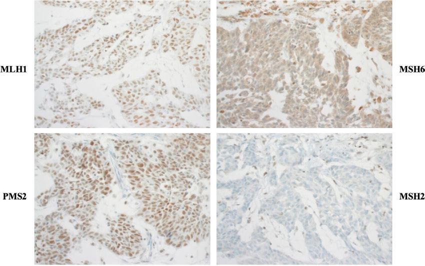
Figure 3 depicts the different workflows and detailed working
steps, including hands-on-time and time-to-result of Idylla versus
standard method. Time to results differed between 3 hours using
Idylla and 2 days for standard methodology.
Figure 2 Whole slides of immunohistochemical staining of MMR- DISCUSSION
proteins MLH1, MSH2, MSH6 and PMS2. Note the strong staining of In recent years, with the implementation of innovative thera-
inflammatory and stromal cells and additionally the strong nuclear peutic options, evaluation of MSI as a biomarker for immuno-
expression of tumour cells for MLH1 and PMS2. In contrast, immune modulative therapy has gained importance in many different
and stromal cells are strongly stained (internal positive control), but the tumours. Due to personalised treatment options and with rising
tumour cells are showing a nuclear loss of expression for MSH2 and numbers of diagnostically evaluated tumours, analysis and
MSH6. MLH1, mutL homologue 1; MMR, mismatch repair; MSH2, mutS processing times as well as the methodologies have to be reviewed
homologue2; MSH6, mutS homologue 6; PMS2; PMS1 homologue 2. with new options available.25 26 In this study, we evaluated the
4 Kullmann F, et al. J Clin Pathol 2021;0:1–7. doi:10.1136/jclinpath-2021-207855Original research
J Clin Pathol: first published as 10.1136/jclinpath-2021-207855 on 28 September 2021. Downloaded from http://jcp.bmj.com/ on October 26, 2021 by guest. Protected by copyright.
Table 2 Rerun of cases for reproducibility testing
(A) Cases with discordant results
Run 1 Run 2 Run 3
Standard method Idylla Standard method Idylla Idylla
Discordant cases Bethesda panel MNR panel MSI test Bethesda panel MNR panel MSI test MSI test
1 Invalid MSS Invalid MSS
2 Invalid MSS MSS MSS
3 Invalid MSS MSS MSS
4 Invalid MSS MSS MSS
5 Invalid MSS Invalid Invalid
6 Invalid MSI-H MSS MSS MSS
7 Invalid MSS MSI MSI-H Invalid
(B) Cases with concordant results
Concordant cases Run 1 Run 2
Standard method Idylla Idylla
Bethesda panel MNR panel MSI test MSI test
1 Invalid Invalid Invalid Invalid
2 Invalid Invalid Invalid
3 Invalid Invalid Invalid
4 MSI MSI-H MSI-H
5 MSI MSI-H MSI-H
6 MSI MSI-H MSI-H
MNR, mononucleotide repeat; MSI, microsatellite instable; MSI-H, microsatellite instability–high; MSS, microsatellite stable.
frequency of MSI in UTUC among a large cohort of 243 patients including more than 200 patients, observed a significantly lower
and identified a low frequency of MSI cases. Additionally, the number of MSI tested samples.30 31 Importantly, patient median
results using standard methods were compared with the Idylla age appears not to be a factor explaining the MSI differences
Assay in order to validate a new approach for MSI analysis. since among the aforementioned studies patients had a median
UTUC is a rare entity, which mostly arise sporadically or due age from 65 to 71 years. Additionally, tumour stage and grade
to the inherited Lynch syndrome.2 In the literature, variable characteristics between the different cohorts were also compa-
frequencies of MSI among this malignancy have been reported. rable.8 10 27 30 Furthermore, it cannot be ruled out that differ-
On the one hand, several studies have indicated a frequency of ences in results could arise due to diverse methodologies for
MSI between 13% and 28% and MMR deficiency of 30%–83% MSI analysis using another marker panel. Except Necchi et al,
in UTUC.7 8 10 27–29 Conversely, our findings present lower tested who performed MSI analysis in 114 homopolymer repeats via
frequencies of MSI (1.6%) and MMR deficiency (3.2%) in UTUC
comprehensive genomic profiling, all other studies used the
among a consecutively collected, multi- institutional cohort.
Bethesda panel for MSI analysis.8 10 27 30 In summary, our results
Importantly, our results are consistent with the recent work of
show that MSI has a low incidence rate in UTUC. More assess-
Necchi et al and Ericson et al, who estimated an MSI status in
ments of biomarker identification for immunotherapeutic ther-
3.4% and 4.6% of analysed cases, respectively.30 31 One possible
apies are needed.
interpretation for the discrepancies could be differences in tissue
sample size. In studies presenting with high MSI frequencies and/
or high numbers of MMR deficiency, analysis was performed
with sample sizes of ≤128 cases.7 8 27–29 For example, Hartmann
et al analysed 62 UTUC samples and identified a frequency of
21% MSI.8 Catto et al tested 71 UTUC samples and determined
a 27% MSI frequency.27 In contrast, studies with larger cohorts,
Table 3 Summary of MSI results
Idylla MSI assay
n=243 MSS MSI Invalid Total n (%)
Standard method MSS 230 0 0 230 (94.7)
MSI 0 4 0 4 (1.6)
Invalid 4 0 5 9 (3.7)
Total n 234 (96.3) 4 (1.6) 5 (2.1) 243
(%) Figure 3 Comparison of workflow of (A) standard method and
Overall concordance: 239/243×100=98.35% (B) Idylla MSI assay. FFPE, formalin-fixed paraffin-embedded; MSI,
MSI, microsatellite instable; MSS, microsatellite stable. microsatellite instability.
Kullmann F, et al. J Clin Pathol 2021;0:1–7. doi:10.1136/jclinpath-2021-207855 5Original research
J Clin Pathol: first published as 10.1136/jclinpath-2021-207855 on 28 September 2021. Downloaded from http://jcp.bmj.com/ on October 26, 2021 by guest. Protected by copyright.
In this investigation, with the Idylla MSI Assay, a new, inno-
vative easy to use and time-saving approach for MSI diagnostics
Take home messages
was tested for UTUCs. To date, the newly designed marker set,
►► We investigated the frequency of microsatellite instability
consisting of seven mononucleotides, is clinically validated and
(MSI) in upper urinary tract urothelial carcinoma with a large
applied for diagnostic use for colorectal cancer and is ongoing
retrospective cohort and performed a comparison between
for verification of endometrial carcinoma.32 In our study of 243
the standard MSI methodology and the new diagnostic tool
UTUCs, we observed a high concordance of 98.35% of MSI
Idylla MSI Assay from Biocartis.
testing between the two methods. These concordance rates are
►► We validated Idylla MSI Assay as an alternative diagnostic
comparable with those of colorectal cancer. Compared with
approach for MSI analysis in UTUC, opening the possibility to
standard diagnostics, we propose that the Idylla MSI Assay is a
simplify and fasten the diagnostic process in the future.
more robust methodology with a lower invalid rate. Nine vs five
►► The rate of microsatellite instable tumours is low with four
invalid results were detected using standard method compared
MSI cases out of 243 tested samples.
with the Idylla MSI Assay, respectively. Additional reasons for
implementing the Idylla MSI Assay into routine laboratory
testing are: (1) the turnaround time was shortened significantly 2
to approximately 3 hours compared with a workload of 2 days Laboratory for Molecular Medicine, Department of Gynecology and Obstetrics,
University Hospital Erlangen-Nürnberg, Friedrich-Alexander-Universität Erlangen-
using the NCI method and (2) implementing a fully automated Nürnberg, Erlangen, Germany
analysis system as well as a reduction of preanalytical steps 3
Comprehensive Cancer Center Erlangen-EMN (CCC ER-EMN), Erlangen, Germany
also significantly reduces human resources. Therefore, our 4
Department of Urology and Pediatric Urology, University Hospital Erlangen,
study could determine that the same marker panel, applied for Friedrich-Alexander Universität Erlangen-Nürnberg, Erlangen, Germany
5
colorectal cancer samples, is transferable to UTUC with a high Privatklinik Brixsana, Urology, Brixen, Italy
6
Department of Urology and Pediatric Urology, University Hospital Gießen and
concordance to current established testing methods. Marburg, Marburg, Germany
One case with preserved MMR protein expression was 7
Department of Urology, Virgen de la Victoria University Hospital Málaga, Instituto
initially detected as MSI-H by Idylla, while no valid result could de Investigación Biomédica de Málaga, Málaga, Spain
8
be obtained with the standard method. Thus, both assays were Genitourinary Cancer Translational Research Group, Instituto de Investigación
Biomédica de Málaga, Málaga, Spain
repeated twice and revealed an MSS tumour concordantly 9
Department of Cell Biology, Genetics and Physiology, Málaga University, Málaga,
matching with preserved protein expression. The initial discor- Spain
dant result could be caused by lo-quality DNA or a possible 10
Department of Pathology, Faculty of Medicine, Universidad de Málaga, Málaga,
heterogeneity in the tumour mass of the same tumour block, as Spain
11
it has been observed in colorectal cancer.33 Lastly, the isolated Instituto de Investigación Biomédica de Málaga (IBIMA), Hospital Universitario
Virgen de la Victoria, Málaga, Spain
loss of the MSH6 protein with MSS status in two tumours could 12
Department of Pathology, Unidad de Gestión Clínica de Patología, Instituto de
indicate a different way of activation among the MMR path- Investigación Biomédica de Málaga (IBIMA), Hospital Universitario Virgen de la
ways. In the study of Gayhart et al investigating MMR proteins Victoria, Málaga, Spain
13
in UTUC, an isolated loss of MSH6 was described in 3 of 74 Department of Urology, Erasmus MC Cancer Institute, University Medical Center,
Rotterdam, Netherlands
analysed cases. Unfortunately, there is no investigation about the 14
Department of Genetics, Institut Curie, PSL Research University, Paris, France
relation with MSI status.28 Nonetheless, there are a few studies 15
Department of Pathology, René Huguenin Curie Institute, Saint Cloud, Paris, France
describing a discordance between MSI analysis and immunohis-
tochemistry in colorectal cancer, where in some cases an isolated Handling editor Runjan Chetty.
MSH6 loss is associated with an MSS status. This is attributed to Acknowledgements The present work was performed in fulfilment of the
a partial activity of the MMR pathway in some tumour cells.34 requirements of Friedrich‐Alexander University Erlangen‐Nürnberg for Friederike
Limitations of our study include the use of a retrospective Kullmann for obtaining the degree ’Dr Med’. We are grateful to Verena Popp,
cohort. That is particularly reflected in the quality of the older Natascha Leicht, Christa Winkelmann and Claudia Schmied for their excellent
aged tumour material, which leads to a possible difficulty and technical support. EM-R is supported by the ’Ramón y Cajal’ Award (RYC2019-
inaccuracy in evaluation, especially with immunohistochem- 027950-I) from MICINN, Spain.
ical results. In addition, it should be noted that to implement Contributors AH, RS and VW conceived the study. ME, SB, BW, DS, SW, HT, PO,
the Idylla MSI Assay into routine diagnosis, the purchasing of HH, MFL, MLM, EM-R, MJL, DP, IH, BH-I, AH, JLB, TvD and VW collected samples and
the instrument is needed. Moreover, we were unable to obtain clinical data. FK, MFL, EMP, VW and RoS performed the experimental work. FK and
VW completed statistical analysis. VW, RoS, BH-I, MFL, EM-R and AH supervised the
patient characteristics, like family history, information about experimental work. PLS, ReS, ME, FK and VW wrote the manuscript. All the authors
Lynch syndrome and smoking history. reviewed and edited the article and approved the final version of the manuscript.
Funding This work was supported by the Interdisciplinary Centre for Clinical
Research at the University Hospital of the Friedrich-Alexander University Erlangen-
Nürnberg and by a grant of the ERA-NET TRANSCAN for ’Translational research on
CONCLUSION rare cancers’. Idylla MSI Assay cartridges were given free of charge by Biocartis NV
Taken together, our data support that MSI status has less influ- (Biocartis NV, Mechelen, Belgium).
ence and prevalence in UTUC compared with other tumour Competing interests None declared.
types, which are also part of the spectrum of Lynch syndrome-
Patient consent for publication Not required.
associated malignancies like colorectal or endometrial cancer.
Our study confirmed Idylla MSI Assay as a valid MSI analysis Ethics approval This study was conducted in accordance with the Declaration of
Helsinki, and the protocol was approved by the ethics committee of the Friedrich-
tool with an extremely low failure rate. With regard to UTUC, Alexander University Erlangen-Nürnberg (number 329_16B) and the ethics
other biomarkers, such as tumour mutational burden or PD-L1 committee of Biomedical Investigation of Andalucía (number 03619003 CCEIBA).
expression, have to be analysed in terms of therapy selection. Provenance and peer review Not commissioned; externally peer reviewed.
Author affiliations Data availability statement Data are available upon reasonable request.
1
Institute of Pathology, University Hospital Erlangen-Nürnberg, Friedrich-Alexander- Supplemental material This content has been supplied by the author(s). It
Universität Erlangen-Nürnberg, Erlangen, Germany has not been vetted by BMJ Publishing Group Limited (BMJ) and may not have
6 Kullmann F, et al. J Clin Pathol 2021;0:1–7. doi:10.1136/jclinpath-2021-207855Original research
J Clin Pathol: first published as 10.1136/jclinpath-2021-207855 on 28 September 2021. Downloaded from http://jcp.bmj.com/ on October 26, 2021 by guest. Protected by copyright.
been peer-reviewed. Any opinions or recommendations discussed are solely those 16 Zhao H, Thienpont B, Yesilyurt BT, et al. Mismatch repair deficiency endows tumors
of the author(s) and are not endorsed by BMJ. BMJ disclaims all liability and with a unique mutation signature and sensitivity to DNA double-strand breaks. Elife
responsibility arising from any reliance placed on the content. Where the content 2014;3:e02725.
includes any translated material, BMJ does not warrant the accuracy and reliability 17 De Craene B, Van de Velde J, Rondelez E, et al. Detection of microsatellite instability
of the translations (including but not limited to local regulations, clinical guidelines, (MSI) in colorectal cancer samples with a novel set of highly sensitive markers by
terminology, drug names and drug dosages), and is not responsible for any error means of the Idylla MSI test prototype. Journal of Clinical Oncology 2018;36:e15639.
and/or omissions arising from translation and adaptation or otherwise. 18 Humphrey PA, Moch H, Cubilla AL, et al. The 2016 who classification of tumours of
Open access This is an open access article distributed in accordance with the the urinary system and male genital Organs-Part B: prostate and bladder tumours. Eur
Creative Commons Attribution Non Commercial (CC BY-NC 4.0) license, which Urol 2016;70:106–19.
permits others to distribute, remix, adapt, build upon this work non-commercially, 19 Giedl J, Schneckenpointner R, Filbeck T, et al. Low frequency of HNPCC-associated
and license their derivative works on different terms, provided the original work is microsatellite instability and aberrant MMR protein expression in early-onset bladder
properly cited, appropriate credit is given, any changes made indicated, and the use cancer. Am J Clin Pathol 2014;142:634–9.
is non-commercial. See: http://creativecommons.org/licenses/by-nc/4.0/. 20 Burger M, Denzinger S, Hammerschmied CG, et al. Elevated microsatellite alterations
at selected tetranucleotides (EMAST) and mismatch repair gene expression in prostate
ORCID iDs cancer. J Mol Med 2006;84:833–41.
Yves Allory http://o rcid.org/0000-0002-5526-077X 21 Nardon E, Glavač D, Benhattar J, et al. A multicenter study to validate the
Veronika Weyerer http://orcid.org/0000-0001-5919-7651 reproducibility of MSI testing with a panel of 5 quasimonomorphic mononucleotide
repeats. Diagn Mol Pathol 2010;19:236–42.
22 Dietmaier W, Wallinger S, Bocker T, et al. Diagnostic microsatellite instability:
REFERENCES definition and correlation with mismatch repair protein expression. Cancer Res
1 Siegel RL, Miller KD, Jemal A. Cancer statistics, 2019. CA Cancer J Clin 2019;69:7–34. 1997;57:4749–56.
2 Rouprêt M, Babjuk M, Compérat E, et al. European association of urology guidelines 23 Fadhil W, Ilyas M. Immunostaining for mismatch repair (MMR) protein expression
on upper urinary tract urothelial carcinoma: 2017 update. Eur Urol 2018;73:111–22. in colorectal cancer is better and easier to interpret when performed on diagnostic
3 Shariat SF, Favaretto RL, Gupta A, et al. Gender differences in radical biopsies. Histopathology 2012;60:653–5.
nephroureterectomy for upper tract urothelial carcinoma. World J Urol
24 Zhao H, De Craene B, Sagaert X, et al. Association of a novel set of 7 homopolymer
2011;29:481–6.
indels for detection of MSI with tumor mutation burden and total indel load in
4 Colin P, Koenig P, Ouzzane A, et al. Environmental factors involved in carcinogenesis
endometrial and colorectal cancers. Journal of Clinical Oncology 2018;36:e15654.
of urothelial cell carcinomas of the upper urinary tract. BJU Int 2009;104:1436–40.
25 Luchini C, Bibeau F, Ligtenberg MJL, et al. ESMO recommendations on microsatellite
5 Kohlmann W, Gruber SB, Syndrome L, eds. GeneReviews((R)). Seattle (WA), 1993.
instability testing for immunotherapy in cancer, and its relationship with PD-1/PD-L1
6 Therkildsen C, Eriksson P, Höglund M, et al. Molecular subtype classification of
expression and tumour mutational burden: a systematic review-based approach. Ann
urothelial carcinoma in Lynch syndrome. Mol Oncol 2018;12:1286–95.
Oncol 2019;30:1232–43.
7 Patel N, Arya M, Muneer A, et al. Molecular aspects of upper tract urothelial
26 Li Q, Bagrodia A, Cha EK, et al. Prognostic genetic signatures in upper tract urothelial
carcinoma. Urol Oncol 2014;32:28.e11–28.e20.
carcinoma. Curr Urol Rep 2016;17:12.
8 Hartmann A, Zanardo L, Bocker-Edmonston T, et al. Frequent microsatellite instability
in sporadic tumors of the upper urinary tract. Cancer Res 2002;62:6796–802. 27 Catto JWF, Azzouzi A-R, Amira N, et al. Distinct patterns of microsatellite instability are
9 Boland CR, Goel A. Microsatellite instability in colorectal cancer. Gastroenterology seen in tumours of the urinary tract. Oncogene 2003;22:8699–706.
2010;138:2073–87. 28 Gayhart MG, Johnson N, Paul A, et al. Universal mismatch repair protein screening in
10 Rouprêt M, Fromont G, Azzouzi A-R, et al. Microsatellite instability as predictor of upper tract urothelial carcinoma. Am J Clin Pathol 2020;154:792–801.
survival in patients with invasive upper urinary tract transitional cell carcinoma. 29 Schneider B, Glass Änne, Jagdmann S, et al. Loss of mismatch-repair protein
Urology 2005;65:1233–7. expression and microsatellite instability in upper tract urothelial carcinoma and
11 Ribic CM, Sargent DJ, Moore MJ, et al. Tumor microsatellite-instability status as a clinicopathologic implications. Clin Genitourin Cancer 2020;18:e563–72.
predictor of benefit from fluorouracil-based adjuvant chemotherapy for colon cancer. 30 Necchi A, Pal SK, Ross JS, et al. Comprehensive genomic profiling (CGP) of upper-tract
N Engl J Med 2003;349:247–57. (UTUC) and bladder (Buc) urothelial carcinoma reveals opportunities for therapeutic
12 Sepulveda AR, Hamilton SR, Allegra CJ, et al. Molecular biomarkers for the evaluation and biomarker development. Journal of Clinical Oncology 2019;37:4581.
of colorectal cancer. Am J Clin Pathol 2017;147:221–60. 31 Ericson KM, Isinger AP, Isfoss BL, et al. Low frequency of defective mismatch repair in
13 Arabi H, Guan H, Kumar S, et al. Impact of microsatellite instability (MSI) on survival in a population-based series of upper urothelial carcinoma. BMC Cancer 2005;5:23.
high grade endometrial carcinoma. Gynecol Oncol 2009;113:153–8. 32 Li X, Xu J, Li L, et al. Evaluation of a fully automated Idylla test system for
14 Marcus L, Lemery SJ, Keegan P, et al. Fda approval summary: pembrolizumab microsatellite instability in colorectal cancer. Clin Colorectal Cancer 2019;18:e316–23.
for the treatment of microsatellite Instability-High solid tumors. Clin Cancer Res 33 Chapusot C, Martin L, Bouvier AM, et al. Microsatellite instability and intratumoural
2019;25:3753–8. heterogeneity in 100 right-sided sporadic colon carcinomas. Br J Cancer
15 Boland CR, Thibodeau SN, Hamilton SR, et al. A national cancer Institute workshop on 2002;87:400–4.
microsatellite instability for cancer detection and familial predisposition: development 34 Evrard C, Tachon G, Randrian V, et al. Microsatellite instability: diagnosis,
of international criteria for the determination of microsatellite instability in colorectal heterogeneity, discordance, and clinical impact in colorectal cancer. Cancers
cancer. Cancer Res 1998;58:5248–57. 2019;11:1567.
Kullmann F, et al. J Clin Pathol 2021;0:1–7. doi:10.1136/jclinpath-2021-207855 7You can also read