A single-center, retrospective study of COVID-19 features in children: a descriptive investigation - BMC Medicine
←
→
Page content transcription
If your browser does not render page correctly, please read the page content below
Ma et al. BMC Medicine      (2020) 18:123
https://doi.org/10.1186/s12916-020-01596-9

 RESEARCH ARTICLE                                                                                                                                   Open Access

A single-center, retrospective study of
COVID-19 features in children: a descriptive
investigation
Huijing Ma1†, Jiani Hu2†, Jie Tian3†, Xi Zhou4, Hui Li5, Maxwell Thomas Laws2, Luke David Wesemann2, Baiqi Zhu1,
Wei Chen6,7, Rafael Ramos2, Jun Xia4* and Jianbo Shao1*

  Abstract
  Background: Compared to adults, there are relatively few studies on COVID-19 infection in children, and even less
  focusing on the unique features of COVID-19 in children in terms of laboratory findings, locations of computerized
  tomography (CT) lesions, and the role of CT in evaluating clinical recovery. The objective of this study is to report
  the results from patients at Wuhan Children’s Hospital, located within the initial center of the outbreak.
  Methods: Clinical, imaging, and laboratory data of 76 children were collected retrospectively and analyzed with the
  Fisher exact test and Cox regression statistical methods.
  Results: Among 50 children with a positive COVID-19 real-time reverse-transcriptase polymerase chain reaction
  (PCR), five had negative PCR results initially but showed positive results in subsequent tests. Eight (16%) patients
  had lymphopenia, seven (14%) with thrombocytopenia, four (8%) with lymphocytosis, two (4%) with
  thrombocytosis, ten (20%) with elevated C-reactive protein, four (8%) with hemoglobin above, and six (12%) with
  below standard reference values. Seven (14%) of the 50 had no radiologic evidence of disease on chest CT. For the
  43 patients who had abnormal CT findings, in addition to previously reported patterns of ground-glass opacity
  (67%), local patchy shadowing (37%), local bilateral patchy shadowing (21%), and lesion location of lower lobes
  (65%), other CT features include that an overwhelming number of pediatric patients had lesions in the subpleural
  area (95%) and 22 of the 28 lower lobe lesions were in the posterior segment (78%). Lesions in most of the 15
  patients (67%) who received chest CT at discharge were not completely absorbed, and 26% of these pediatric
  patients had CT lesions that were either unchanged or worse.
  (Continued on next page)

* Correspondence: xiajun@email.szu.edu.cn; drshaojb@sina.com
†
 Huijing Ma, Jiani Hu, and Jie Tian are joint first authors.
4
 Department of Radiology, Shenzhen Second People’s Hospital, The First
Affiliated Hospital of Shenzhen University Health Science Center, 3002
SunGang Xi Road West, Shenzhen 518035, China
1
 Imaging Center, Wuhan Children’s Hospital (Wuhan Maternal and Child
Healthcare Hospital), Tongji Medical College, Huazhong University of Science
& Technology, No.100 Hongkong Road, Wuhan 430016, China
Full list of author information is available at the end of the article

                                        © The Author(s). 2020 Open Access This article is licensed under a Creative Commons Attribution 4.0 International License,
                                        which permits use, sharing, adaptation, distribution and reproduction in any medium or format, as long as you give
                                        appropriate credit to the original author(s) and the source, provide a link to the Creative Commons licence, and indicate if
                                        changes were made. The images or other third party material in this article are included in the article's Creative Commons
                                        licence, unless indicated otherwise in a credit line to the material. If material is not included in the article's Creative Commons
                                        licence and your intended use is not permitted by statutory regulation or exceeds the permitted use, you will need to obtain
                                        permission directly from the copyright holder. To view a copy of this licence, visit http://creativecommons.org/licenses/by/4.0/.
                                        The Creative Commons Public Domain Dedication waiver (http://creativecommons.org/publicdomain/zero/1.0/) applies to the
                                        data made available in this article, unless otherwise stated in a credit line to the data.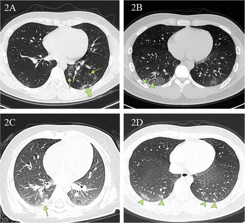
Ma et al. BMC Medicine    (2020) 18:123                                                                        Page 2 of 11

 (Continued from previous page)
 Conclusions: There were a few differences between COVID-19 children and COVID-19 adults in terms of laboratory
 findings and CT characteristics. CT is a powerful tool to detect and characterize COVID-19 pneumonia but has little
 utility in evaluating clinical recovery for children. These results oppose current COVID-19 hospital discharge criteria
 in China, as one requirement is that pulmonary imaging must show significant lesion absorption prior to discharge.
 These differences between pediatric and adult cases of COVID-19 may necessitate pediatric-specific discharge
 criteria.
 Keywords: Children, Pediatric, Coronavirus, COVID-19, SARS-CoV-2, Epidemiology, Clinical features, Computerized
 tomography

Background                                                     values than a 9-year-old. Confounding these results is
Since initially identified in Wuhan city of China’s Hubei      the fact that the reference values used among the studies
province in December 2019, the coronavirus disease             lack consistency and appear to be hospital-self-defined
2019 (COVID-19) has resulted in 466,836 confirmed              values [15, 22–24]. This inconsistency of reference
cases and 21,152 deaths as of March 25, 2020. Two              values makes any systemic review of the published data
months prior, on January 23, 2020, there were only 581         less meaningful [23].
reported cases. COVID-19 can rapidly spread from                  There is also no research on the role of CT in moni-
human-to-human and is more contagious than other               toring clinical recovery in children. CT has been widely
notable members of the coronavirus family, such as se-         used in the clinical management of adult patients due to
vere acute respiratory syndrome (SARS) and Middle              its ability to reveal detailed features of pneumonia [25–
Eastern respiratory syndrome (MERS) [1, 2]. The World          28]. Because of how many unknowns there were about
Health Organization recently declared COVID-19 a glo-          the disease, particularly at the beginning of the COVID-
bal pandemic, and the USA has declared a national              19 outbreak, CT was frequently used in the clinical man-
emergency. Even though the incidence of COVID-19 in-           agement and diagnosis of children in China. Notably, re-
fection in children is less than it is in adults, the total    peated use of CT can be harmful, particularly for
number of pediatric cases is expected to increase rapidly      children [29, 30].
in the coming weeks.                                              The objective of this study is to report relevant find-
  Compared to adults [3–7], there are a few studies on         ings from the COVID-19-positive patients treated at
the COVID-19 in children. Although mortality in chil-          Wuhan Children’s Hospital. Specifically, we attempt to
dren has been reported [8], studies have demonstrated          answer three questions based on the patient’s clinical, la-
that COVID-19 is generally less severe compared to             boratory, diagnostic, and treatment outcome data. The
adults in terms of both symptoms and computerized              questions are, in hospitalized COVID-19 children, (i)
tomography (CT) manifestations [9–18]. The common              what are the typical laboratory findings, (ii) is there any
chest CT patterns are ground-glass opacities (GGO)             unique CT feature, and (iii) is CT necessary for evaluat-
followed by local bilateral shadowing (LPS), in contrast       ing clinical recovery?
to a large percentage of bilateral patchy shadowing
(BPS) pattern in adults [19, 20]. However, there are no
studies that quantitatively examine the location of lung       Methods
lesions in COVID-19-positive pediatric patients [21].          Study design and patient selection
Most of the pediatric patients are at the early stages of      For this retrospective, single-center study, patients were
the disease when admitted to hospitals. Thus, a detailed       recruited from January 21 to February 14, 2020, at Wu-
localization study is meaningful both clinically and sci-      han Children’s Hospital in Wuhan, China. Real-time
entifically, as it could help pinpoint lung regions that are   reverse-transcriptase polymerase chain reaction (PCR)
particularly susceptible to COVID-19 infection.                was performed on children 16 years of age and under
  Several studies have reported on the laboratory find-        who had a family or social history of COVID-19 expos-
ings of children infected with COVID-19. However, the          ure. Subsequently, these patients received a chest CT
interpretations of these results vary substantially [15,       examination to evaluate lung pathology. Based on the
22–24]. The discrepancy in laboratory interpretations          PCR and CT results, these patients were stratified into
could be attributed to the studies each referring to a dif-    groups A–C (Fig. 1). This study was approved by the
ferent set of reference values. Of note, the range of nor-     Ethics Committee of Wuhan Children’s Hospital
mal lab values changes depending upon the age of the           (Wuhan Maternal and Child Health Care Hospital #
child, i.e., a 1-year-old has a different set of reference     WHCH 2020005). Written informed parental/guardian
Ma et al. BMC Medicine      (2020) 18:123                                                                                             Page 3 of 11

 Fig. 1 Flow chart for patient selection. Group A: 43 children with COVID-19 exposure history, positive CT, and positive PCR. Group B: seven
 children with COVID-19 exposure history, negative CT, and positive PCR. Group C: 26 children with COVID-19 exposure history, positive CT, and
 persistently negative PCR results

consent and child assent (where appropriate) were ob-                      CT features were inconsistent, the two radiologists dis-
tained prior to enrollment in the study.                                   cussed and decided together. Only final decisions
                                                                           reached by consensus are reported. No negative control
Procedures                                                                 cases were examined.
We obtained demographic information, clinical symp-                          PCR confirmation of COVID-19 was performed at
toms, laboratory results, management, and outcome data                     two different institutions: Hubei Center for Disease
from each patient’s electronic medical records. Clinical                   Control and Prevention and Wuhan Children’s
outcomes were followed up to February 17, 2020.                            Hospital.
  Chest CT without intravenous contrast was per-
formed on all patients using a Siemens SOMATOM                             Patient discharge
Definition AS128 or GE Optima CT 660 with a 1-                             Criteria for discharging pediatric patients in this hospital
mm or 0.625-mm slice thickness, respectively.                              were normal body temperature for 3 days, two negative
Children under 5 years old, as well as uncooperative                       PCR results at 24-h intervals, and resolution of all clin-
children, received oral chloral hydrate sedation (0.5 ml/kg)               ical symptoms.
prior to CT. Cooperative children above 5 years old were
trained with breathing exercises prior to CT.                              Statistical analysis
  All CT images were reviewed by at least two radiolo-                     The Fisher exact test method was used to determine
gists with more than 10 years of experience. Imaging                       whether there is a significant difference in CT image
was reviewed independently. When the opinions on the                       characteristics and lesion locations between group A and
Ma et al. BMC Medicine (2020) 18:123 Page 4 of 11
group C. The Cox regression analysis was used to deter- Results
mine whether changes in CT images during treatment From January 21 to February 14, 2020, 158 children at
were associated with clinical outcomes for children with Wuhan Children’s Hospital were radiologically examined
COVID-19 infection. All analyses were performed using with chest CT, and respiratory secretions were obtained
EmpowerStats (http://www.empowerstats.com) and the and subsequently tested for COVID-19 with PCR. A CT
statistical package R (version 3.2.3). p value of less than scan was considered positive when at least one lesion
0.05 was considered to indicate a statistically significant was identified. Among them, 43 had a positive CT and
difference. positive PCR (group A), 7 had a negative CT and positive
Table 1 Demographics and characteristics of patients
Characteristics Group A Group B Group A + B Group C
Age, years 3.0 (0.9–7.5) 1.0 (1.0–4.5) 2.5 (0.9–7.0) 2.5 (1.2–9.8)
> 2.5 22/43 (51%) 3/7 (43%) 25/50 (50%) 13/26 (50%)
≤ 2.5 21/43 (49%) 4/7 (57%) 25/50 (50%) 13/26 (50%)
Sex
Male 23/43 (53%) 5/7 (71%) 28/50 (56%) 14/26 (54%)
Female 20/43 (47%) 2/7 (29%) 22/50 (44%) 12/26 (46%)
Type of care
Discharged patients 33/43 (77%) 5/7 (71%) 38/50 (76%) 26/26 (100%)
Hospitalization duration 11.0 (9.0~13.0) 13.0 (7.0~14.0) 11.0 (8.2~13.8) 10.5 (8.0~12.0)
Clinical status
Asymptomatica 0/43 (0%) 2/7 (29%) 2/50 (4%) 0/26 (0%)
Very mild 0/43 (0%) 5/7 (71%) 5/50 (10%) 0/26 (0%)
Mild 41/43 (95%) 0/7 (0%) 41/50 (82%) 26/26 (100%)
Severe 0/43 (0%) 0/7 (0%) 0/50 (0%) 0/26 (0%)
Critically ill 2/43 (5%) 0/7 (0%) 2/50 (4%) 0/26 (0%)
Symptoms
Fever 29/43 (67%) 3/7 (43%) 32/50 (64%) 21/26 (81%)
Cough 21/43 (49%) 1/7 (14%) 22/50 (44%) 19/26 (73%)
Myalgia or fatigue 2/43 (5%) 0/7 (0%) 2/50 (4%) 0/26 (0%)
Sore throat (pharyngalgia) 1/43 (2%) 0/7 (0%) 1/50 (2%) 0/26 (0%)
Diarrhea 3/43 (7%) 0/7 (0%) 3/50 (6%) 0/26 (0%)
Abdominal pain 2/43 (5%) 0/7 (0%) 2/50 (4%) 0/26 (0%)
Rhinorrhea 7/43 (16%) 1/7 (14%) 8/50 (16%) 2/26 (8%)
Loss of appetite 2/43 (5%) 0/7 (0%) 2/50 (4%) 0/26 (0%)
Chest pain 0/43 (0%) 0/7 (0%) 0/50(0%) 1/26 (4%)
Intussusception 1/43 (2%) 0/7 (0%) 1/50 (2%) 0/26 (0%)
No symptoms 4/43 (9%) 2/7 (29%) 6/50 (12%) 0/26 (0%)
Mortality 0/43 (0%) 0/7 (0%) 0/50 (0%) 0/26 (0%)
Comorbidity
Cardiac damage 4/43 (9%) 1/7 (14%) 5/50 (10%) 3/26 (12%)
Appendicitis 1/43 (2%) 0/7 (0%) 1/50 (2%) 0/26 (0%)
Foreign body in bronchus 1/43 (2%) 0/7 (0%) 1/50 (2%) 0/26 (0%)
Mycoplasma infection 5/43 (12%) 0/7 (0%) 5/50 (10%) 11/26 (42%)
Respiratory syncytial virus infection 1/43 (2%) 0/7 (0%) 1/50 (2%) 0/26 (0%)
Renal failure 1/43 (2%) 0/7 (0%) 1/50 (2%) 0/26 (0%)
Intestinal necrosis in MODS 1/43 (2%) 0/7 (0%) 1/50 (2%) 0/26 (0%)
a
No clinical symptoms and no abnormal CT findingsMa et al. BMC Medicine (2020) 18:123 Page 5 of 11
PCR (group B), and 26 had a positive CT and at least two Laboratory reference normal ranges were age- and
negative consecutive PCR results (group C, Fig. 1). gender-adjusted according to values in Reference Range
PCR-positive groups A and B (n = 50) were chosen to Values for Pediatric Care 2nd edition, pages 92–98 [31].
interpret clinical and chest CT features because group C On laboratory assessment, eight (16%) and seven (14%)
patients were not deemed COVID-19 positive by PCR. patients had lymphopenia and thrombocytopenia, re-
Over half of the patients were males (56%, Table 1). The spectively. In contrast, four (8%) were noted to have
most common symptoms at the onset of illness (Table 1) lymphocytosis, and two (4%) had thrombocytosis.
were fever (64%) and cough (44%); less common symp- Overall, leukopenia was observed in 19 (38%) patients
toms were rhinorrhea (16%), abdominal pain (4%), diar- and elevated C-reactive protein in ten (20%) patients. A
rhea (6%), fatigue (4%), and pharyngalgia (2%). Six small set of patients had hemoglobin abnormalities, four
children (12%) were asymptomatic. After treatment, 38 (8%) with elevated hemoglobin, and six (12%) with
(76%) children were discharged. anemia (Table 2).
Table 2 Laboratory examination and CT radiographic characteristics
Characteristics Group A Group B Group A + B Group C
Laboratory examinationa
Hemoglobin count increase (↑) 2/43 (5%) 2/7 (29%) 4/50 (8%) 6/26 (23%)
Hemoglobin count normal 36/43 (83%) 4/7 (57%) 40/50 (80%) 18/26 (69%)
Hemoglobin count decrease (↓) 5/43 (12%) 1/7 (14%) 6/50 (12%) 2/26 (8%)
C-reactive protein level increase (↑) 10/43 (23%) 0/7 (0%) 10/50 (20%) 8/26 (31%)
C-reactive protein level normal 33/43 (77%) 7/7 (100%) 40/50 (80%) 18/26 (69%)
Platelet count increase (↑) 1/43 (2%) 1/7 (14%) 2/50 (4%) 1/26 (4%)
Platelet count normal 35/43 (82%) 6/7 (86%) 41/50 (82%) 18/26 (69%)
Platelet count decrease (↓) 7/43 (16%) 0/7 (0%) 7/50 (14%) 7/26 (27%)
Lymphocyte count increase (↑) 2/43 (5%) 2/7 (29%) 4/50 (8%) 2/26 (8%)
Lymphocyte count normal 34/43(79%) 4/7 (57%) 38/50 (76%) 18/26 (69%)
Lymphocyte count decrease (↓) 7/43 (16%) 1/7 (14%) 8/50 (16%) 6/26 (23%)
Blood leukocyte count increase (↑) 2/43 (5%) 0/7 (0%) 2/50 (4%) 1/26 (4%)
Blood leukocyte count normal 23/43 (53%) 6/7 (86%) 29/50 (58%) 13/26 (50%)
Blood leukocyte count decrease (↓) 18/43 (42%) 1/7 (14%) 19/50 (38%) 12/26 (46%)
CT radiographic characteristics
Overall patients with CT abnormalities 43/43 (100%) 0/7 (0%) 43/50 (86%) 26/26 (100%)
Ground-glass opacity 29/43 (67%) 0/7 (0%) 29/50 (58%) 21/26 (81%)
Local patchy shadowing 16/43 (37%) 0/7 (0%) 16/50 (32%) 7/26 (27%)
Bilateral patchy shadowing 9/43 (21%) 0/7 (0%) 9/50 (18%) 5/26 (19%)
Interstitial abnormalities 3/43 (7%) 0/7 (0%) 3/50 (6%) 0/26 (0%)
Pleural fluid 1/43 (2%) 0/7 (0%) 1/50 (2%) 0/26 (0%)
Lymphadenopathy 0/43 (0%) 0/7 (0%) 0/50 (0%) 0/26 (0%)
Lesion characteristics and locationb
Subpleural 41/43 (95%) 0/7 (0%) 41/50 (82%) 21/26 (81%)
Parallel to the pleura 21/43 (49%) 0/7 (0%) 21/50 (42%) 10/26 (38%)
Vascular thickening shadowing 10/43 (23%) 0/7 (0%) 10/50 (20%) 10/26 (38%)
Upper lobe of the lung 22/43 (51%) 0/7 (0%) 22/50 (44%) 14/26 (54%)
Middle lobe of the lung 9/43 (21%) 0/7 (0%) 9/50 (18%) 6/26 (23%)
Lower lobe of the lung 28/43 (65%) 0/7 (0%) 28/50 (56%) 19/26 (73%)
Posterior segment of lower lung lobes 22/43 (51%) 0/7 (0%) 22/50 (44%) 12/26 (46%)
a
Data from the first laboratory examination of the patient admission. The normal range of laboratory examination is the standard of Reference Range Values for
Pediatric Care 2nd ed [31] released by the American Academy of Pediatrics
b
How many patients have the following lesion locationMa et al. BMC Medicine (2020) 18:123 Page 6 of 11 Of the 26 patients in group C, all had more than Among the 50 children with positive PCR results, five two negative consecutive PCR results. However, they of them (10%) had negative initial PCR results but all had a history of exposure to COVID-19 infection showed positive results in subsequent tests. Two of the (or strongly suspected infection), and their chest CT 50 (4%) had no clinical symptoms and no radiologic had similar patterns to confirmed patients in group A findings. Seven of the 50 (14%) were negative for any ab- (Table 2). Fisher exact test results indicated that there normal CT findings. The spectrum of COVID-19 sever- was no significant difference in CT characteristics ity was two (4%) had no symptoms or radiologic signs, (ground-glass opacity [p > 0.05], local patchy shadow- five (10%) very mild, 41 (82%) mild, and two (4%) critic- ing [p > 0.05], bilateral patchy shadowing [p > 0.05], ally ill with one having multiple organ dysfunction syn- interstitial abnormalities [p > 0.05]) and lesion location drome (MODS) and another with renal failure (Table 1). (parallel pleura [p > 0.05], visible vascular thickening There were no patient mortalities in this study. The crit- [p > 0.05], subpleural [p > 0.05], lower lobe of the lung ically ill patient with renal failure has since fully [p > 0.05], middle lobe of the lung [p > 0.05], upper recovered. lobe of the lung [p > 0.05]) between groups A and C Among the 43 children with positive PCR results and (Table 3). abnormal CT findings, 41 patients had lesions present in Table 3 Differences in CT image characteristics between Groups A and C Characteristics Group A Group C Standardize diff. p value Ground-glass opacity – – 0.31 (− 0.18, 0.80) 0.230 No 14 (32.56%) 5 (19.23%) – – Yes 29 (67.44%) 21 (80.77%) – – Local patchy shadowing – – 0.22 (− 0.27, 0.71) 0.380 No 27 (62.79%) 19 (73.08%) – – Yes 16 (37.21%) 7 (26.92%) – – Bilateral patchy shadowing – – 0.04 (− 0.44, 0.53) 0.865 No 34 (79.07%) 21 (80.77%) – – Yes 9 (20.93%) 5 (19.23%) – – Interstitial abnormalities – – 0.39 (− 0.10, 0.88) 0.168 No 40 (93.02%) 26 (100.00%) – – Yes 3 (6.98%) 0 (0.00%) – – Subpleural – – 0.46 (− 0.03, 0.95) 0.095 No 2 (4.65%) 5 (19.23%) – – Yes 41 (95.35%) 21 (80.77%) – – Upper lobe of the lung – – 0.05 (− 0.43, 0.54) 0.829 No 21 (48.84%) 12 (46.15%) – – Yes 22 (51.16%) 14 (53.85%) – – Middle lobe of the lung – – 0.05 (− 0.44, 0.54) 0.834 No 34 (79.07%) 20 (76.92%) – – Yes 9 (20.93%) 6 (23.08%) – – Upper lobe of the lung – – 0.17 (− 0.31, 0.66) 0.492 No 15 (34.88%) 7 (26.92%) – – Yes 28 (65.12%) 19 (73.08%) – – Parallel to the pleura – – 0.21 (− 0.28, 0.70) 0.401 No 22 (51.16%) 16 (61.54%) – – Yes 21 (48.84%) 10 (38.46%) – – Vascular thickening shadowing – – 0.33 (− 0.16, 0.82) 0.177 No 33 (76.74%) 16 (61.54%) – – Yes 10 (23.26%) 10 (38.46%) – – Using the Fisher exact test method, p < 0.05 was considered to indicate a statistically significant difference
Ma et al. BMC Medicine      (2020) 18:123                                                                                               Page 7 of 11

the subpleural area (95%) and lower lung lobes (65%),                         Figure 2 illustrates typical radiographic features of
especially in the posterior segment of the lower lung                       COVID-19 pneumonia in children. Figure 3 shows
lobes (22 [78%] of 28). Ground-glass opacities (GGO)                        chest CT before and after treatment from three
were the most common radiologic lesion identified on                        COVID-19 children. Cox regression results (Table 4)
chest CT (67%). Local patchy shadowing (37%) was the                        indicated that there is no association between changes
second most common radiologic lesion, followed by local                     in CT lesions (completely absorbed [p > 0.05], partially
bilateral patchy shadowing (21%, Table 2). Interstitial le-                 absorbed [p > 0.05], worse [p > 0.05]). Table 5 lists
sions were rare (7%). Pleural fluid was observed in one                     changes in CT lesions during treatment. Table 6 lists
case, and no lymphadenopathy was noted (Table 2). Ap-                       the normal ranges for children of different ages based
pearances of lesions were irregular shaped, flaky, wedge-                   on Reference Range Values for Pediatric Care 2nd edi-
shaped, or strip-shaped. The long axis of some lesions                      tion pages 92–98 [31].
(49%) was parallel to the pleura. However, lesions did not
follow the segment of the lung lobe, single or multiple,                    Discussion
and diffuse consolidation was rare. Bilateral lesions can be                The symptoms in children with COVID-19 infection
seen radiating around the bronchial blood vessels or                        have been well described in the literature [9–18]. Our
showing large areas of consolidation, which can be traced                   results are consistent with these previous reports. For
by the lung segment into the bronchial tube.                                example, the clinical symptoms from our study versus
  Among the 50 confirmed children (groups A and B),                         the recent study with the most pediatric patients are
29 patients (including 23 discharged children) had more                     similar [10]: fever, 64% versus 41.5%; cough, 44% versus
than one chest CT. Nineteen of the 29 patients (65%)                        48.5%; diarrhea, 6% versus 8.8%; and fatigue, 4% versus
had improved CT presentations after treatment, and le-                      7.6%. Results including ours indicate that COVID-19
sions in two of the 19 patients completely disappeared.                     symptoms in children follow a similar pattern in adults,
Two of the 29 patients (7%) showed no change in CT le-                      albeit much less severe.
sions, and 8 of the 29 patients (28%) had more CT le-                         Our results of abnormal laboratory findings for chil-
sions after treatment.                                                      dren infected with COVID-19 contrast with recently

 Fig. 2 Chest CT images depicting typical radiographic findings of COVID-19 pneumonia in children. 2A A unilateral chest CT from a 14-year-old
 boy with a cough. Ground-glass opacities under and parallel to the pleura (thick green arrow) in the inferior lobes of the left lungs. Ground-glass
 opacities distributed along the bronchovascular bundle (thin green arrow). 2B Bilateral ground-glass opacities with vascular thickening
 (arrowheads) in the subpleural area from a 13-year-old boy with a fever and a cough. 2C Local patchy shadowing (green arrow) image from a 6
 month-old girl with a fever and a cough. 2D Lesions in the lower lobe of both lungs (green arrows) on chest CT obtained from a 15-year-old boy
 with a fever and a cough
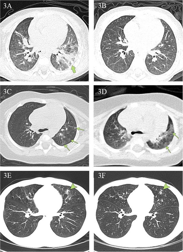
Ma et al. BMC Medicine      (2020) 18:123                                                                                                Page 8 of 11

 Fig. 3 Chest CT findings at initial presentation and at discharge. 3A, 3B Chest CT scans obtained from a 1-year-old boy, presenting with fever
 and diarrhea, at arrival (3A) and after (3B) treatment. The first CT scan shows a large, patchy shadow in the left inferior lobe (green arrow). The
 second CT scan shows no lesions. The patient was hospitalized for 17 days prior to discharge. 3C, 3D Chest CT scans from a 4-month-old girl,
 who presented with a fever and a cough at arrival. The first CT scan reveals multiple ground-glass opacities under the pleura in the left superior
 lobe (green arrows). The second CT scan reveals that the range of original lesions was enlarged and extended to the center. The girl was
 hospitalized for 13 days and subsequently discharged. 3E, 3F Chest CT scans from a 14-year-old boy, presenting with rhinorrhea and a cough, at
 arrival and discharge. The first CT scan reveals a patchy shadow in the left middle lobe (arrowhead). There were no obvious changes in the areas
 of pulmonary consolidation on the second CT scan. The boy was hospitalized for 11 days and then discharged

published ones [15, 22–24]. For example, our results                           Like clinical symptoms, the laboratory findings in
for lymphopenia compared to Zheng et al. are 16%                             COVID-19-positive pediatric patients can vary from
versus 40% [22]. Their normal reference values for                           adult patients. Guan et al. [25] noted that 731 (82%) of
lymphocytes were (2.1–5.7) × 109/L (< 3 years), (1.4–                        890 adult patients had lymphopenia, whereas only eight
4.2) × 109/L (4–6 years), and (1.1–3.2) × 109/L (≥ 6                         (16%) children had lymphopenia in this study. Similarly,
years). To date, this is the only paper that has expli-                      481 (61%) of 793 adult patients were found to have an
citly listed the normal ranges for children of different                     elevated C-reactive protein. In contrast, only ten (20%)
age ranges [22]. Thus, the differences between ours                          children in this study had elevated C-reactive protein.
and those in the literature are most likely due to dif-                      Some laboratory findings were consistent between chil-
ferent normal ranges used for children of different                          dren and adult groups: leukopenia 38% versus 36% and
ages or the small number of children who partici-                            thrombocytopenia 14% versus 18%. The mechanism be-
pated in their studies.                                                      hind the observations is unknown and might provide an
Ma et al. BMC Medicine         (2020) 18:123                                                                                                       Page 9 of 11

Table 4 Association between CT imaging changes and clinical                        and cough (73%). Although they received a negative
outcomea                                                                           PCR result at least twice, all 26 patients had similar CT
Exposure                                               Adjustb                     patterns to the PCR-positive COVID-19 patients in
Changes in CT images during treatment                                              group A. Twenty-one (81%) had ground-glass opacities
    Completely absorbed                                1.0                         (GGO). Seven (27%) had local patchy shadowing. Five
                                                                                   (19%) had bilateral patchy shadowing. Furthermore, our
    Partially absorbed                                 0.56 (0.25, 1.28), 0.168
                                                                                   Fisher exact analysis indicated that there was no signifi-
    No change                                          0.11 (0.01, Inf)c
                                                                                   cant difference in CT image characteristics and lesion
    Worse                                              0.34 (0.10, 1.13), 0.08     location between groups A and C. Although a positive
a
  Using the time-vary Cox regression method, p < 0.05 was considered to            CT alone cannot rule out the possibility of other causes
indicate statistically significant difference
b
  Adjusted for gender, age, PCR positive and CT positive                           of virus-induced pneumonia [11, 26], all 26 children
c
 This model failed when analyzing “no change in CT image” due to the               were hospitalized and given immediate antiviral and sup-
small sample
                                                                                   portive therapy. Whether or not a child presents with
                                                                                   pneumonia is one of the key considerations for clinical
explanation for the differences between pediatric and                              management, and it is crucial to start treatment as early
adult patients.                                                                    as possible, considering that many deaths in the adult
   The most common pattern of chest CT is ground-                                  population are due to complications resulting from se-
glass opacities, followed by local patchy shadowing and                            vere pneumonia [3–7].
then local bilateral patchy shadowing, which is consist-                             It has been well documented that chest CT is a
ent with published data [9–18]. Our study indicates that                           powerful tool to identify and characterize pneumonia
chest CT manifested with a predominance of lesions in                              for COVID-19 adult patients [25–28]. However, there
the subpleural area (41 [95%] of 43) and in lower lung                             is no publication to study its usefulness in evaluating
lobes (28 [65%] of 43), especially in the posterior seg-                           clinical recovery for children with COVID-19 infec-
ment (22 [78%] of 28), an area with a relatively dense                             tion. To determine whether CT is necessary, we in-
amount of bronchioles, blood vessels, and alveoli. To the                          vestigated the data of 23 patients who had been
best of our knowledge, these are the first quantitative re-                        discharged after effective treatment and had at least
sults on the locations of chest CT lesions for COVID-19                            two CT scans. All patients had normal body tempera-
children [21]. COVID-19 is less severe in children than                            tures for more than 3 days at the time of discharge,
in adults, and the children infected with COVID-19 were                            clinical symptoms disappeared, and PCR tests all
at the early stages of the disease when admitted to the                            returned negative twice at 24-h intervals. Of the 23
hospital. The fact that an overwhelming percentage of                              children, eight patients did not receive CT scans
pediatric patients had lesions in the subpleural area sug-                         within the 2 days before their discharge. However, in
gests this site is the first target for the COVID-19 virus.                        their most recent CT scan performed in the hospital,
   The current gold standard for the diagnosis of                                  most children either still had lesions (50%), or more
COVID-19 is PCR. However, it has been documented                                   developed lesions since the previous scan (37%). The
that patients with a negative PCR result cannot be de-                             remaining 15 discharged children had a CT obtained
finitively ruled out for COVID-19 infection [11, 26]. Our                          within 2 days of discharge. Again, ten patients had le-
results are consistent with the literature. Among the 50                           sions that were not completely absorbed (67%), two
hospitalized children with positive PCR results, five of                           were the same (13%), and lesions in another two be-
them (10%) had negative initial PCR results but showed                             came worse (13%). These results indicate that CT
positive results in subsequent tests. Moreover, 26 pa-                             may not be better than symptoms in evaluating
tients in group C never had a positive PCR result but                              recovery. Our Cox regression analysis further showed
had histories of contact with COVID-19 patients. Most                              that there was no association between changes in CT
of them exhibited clinical symptoms such as fever (81%)                            lesions and clinical outcomes. The results are

Table 5 Changes in CT presentation after treatment
Result                                                   Completely absorbed                 Partially absorbed              No change                Worse
                                        a
Between the first and discharge CT                       1/15 (7%)                           10/15 (67%)                     2/15 (13%)               2/15 (13%)
Between the first and the nearest CTb                    1/8 (13%)                           4/8 (50%)                       0/8 (0%)                 3/8 (37%)
                                    c
Between the first and latest CT                          2/29 (7%)                           17/29 (58%)                     2/29 (7%)                8/29 (28%)
a
  Only for those discharged patients with at least two CT; discharge CT here means CT within 2 days of discharging; total 15 patients out of group A and group B
b
  Only for those discharged patients with at least two CT but no CT within 2 days of discharging; the nearest CT means that the CT taken closest to the date of
discharging; total 8 patients out of group A and group B
c
 For all patients with at least two CT; total 29 patients out of group A and group B
Ma et al. BMC Medicine (2020) 18:123 Page 10 of 11
Table 6 Laboratory examination reference ranges
Age Sex Hemoglobin (g/dL) C-reactive protein (mg/L) Platelets (× 109/L) Lymphocytes (× 109/L) Leukocytes (× 109/L)
1 to < 2 months M 102–127 0.09–10.41 221–471 2.22–5.63 8.36–13.66
F 111–137 184–430 2.49–6.26 7.34–12.32
2 to < 6 months M 105–130 215–448 2.57–7.54 7.91–13.41
F 107–134 147–423 2.22–7.11 6.85–12.84
6 months to 2 years M 104–125 185–399 2.47–6.41 7.73–13.12
F 108–126 211–408 2.34–6.44 7.05–12.98
3–5 years M 114–143 187–444 1.60–5.30 4.40–12.9
F 114–143 187–444 1.60–5.30 4.40–12.9
6–8 years M 115–143 186–400 1.40–3.90 3.80–10.4
F 115–143 186–400 1.40–3.90 3.80–10.4
9–10 years M 118–147 186–400 1.40–3.90 3.80–10.4
F 118–147 186–400 1.40–3.90 3.80–10.4
11–14 years M 124–157 176–381 1.00–3.20 3.80–10.4
F 119–148 176–381 1.00–3.20 3.80–10.4
15–19 years M 133–169 138–319 1.00–3.20 3.80–10.4
F 119–148 158–361 1.00–3.20 3.80–10.4
From Reference Range Values for Pediatric Care 2nd edition pages 92–98 [31] released by the American Academy of Pediatrics
consistent with the knowledge that clinical improve- Conclusions
ment predates radiographic improvement by weeks The severity of COVID-19 infection in children is less
for children with community-acquired pneumonia. than it is in adults in terms of symptoms, lung consoli-
When deciding whether to use CT on children, the dation as visualized by CT, and laboratory abnormalities.
harmful effects that radiation may have on a growing COVID-19 has a preference for subpleural areas of the
body must be considered. Hong et al., in a study of lung in pediatric patients. Chest CT is an excellent tool
12,068,821 children aged 0 to 19 years, found a statis- to detect and characterize COVID-19 pneumonia but
tically significant increase in cancer in children ex- not to evaluate the resolution of illness for children.
posed at least once to diagnostic low-dose ionizing
radiation after adjusting for age and sex [29, 30]. Abbreviations
Based on our data, we do not recommend using CT BPS: Bilateral patchy shadowing; COVID-19: Coronavirus disease 2019;
CT: Computerized tomography; GGO: Ground-glass opacities; LPS: Local
for determining clinical recovery unless it is necessary bilateral shadowing; MERS: Middle Eastern respiratory syndrome;
to evaluate the status of pneumonia. For comparison, MODS: Multiple organ dysfunction syndrome; PCR: Polymerase chain
the current criteria for discharging adult patients in- reaction; SARS: Severe acute respiratory syndrome
fected with COVID-19 in China are (1) normal body
temperature for 3 days, (2) two negative PCR tests at Acknowledgements
We would like to thank Pin He for her help in image processing.
24-h intervals, (3) resolution of clinical symptoms
(these three are the current criteria for discharging
Authors’ contributions
pediatric patients in this hospital), plus (4) a chest HM, JH, JT, JX, and JS had roles in the study design, data analysis, data
imaging requirement: pulmonary imaging must show interpretation, literature search, and writing of the manuscript. XZ, HL, MTL,
significant absorption of lesions. To date, there are no LDW, BZ, WC, and RR had roles in the data analysis, data interpretation,
literature search, and writing of the manuscript. JS, HM, HL, and BZ had roles
child-specific discharge criteria for COVID-19 in in clinical management, patient recruitment, and data collection and had full
China. access to all of the data in the study and take responsibility for the integrity
Our study had a few limitations. First, this study has a of the data. HM, JH, and JT contributed equally. The authors read and
approved the final manuscript.
small sample size and was conducted at a single-center
in Wuhan, China, located at the center of the outbreak.
The clinical severity of pediatric patients outside Wuhan Funding
The authors received no specific funding for this work.
may be less severe. Indeed, it is reported that there is a
lower death rate of adult patients outside Wuhan areas.
Availability of data and materials
Second, long-term follow-up was not done because of The datasets used and/or analyzed during the current study are available
the short time for data collection. from the corresponding author on reasonable request.Ma et al. BMC Medicine (2020) 18:123 Page 11 of 11
Ethics approval and consent to participate 13. Dong Y, Mo X, Hu Y, Qi X, Jiang F, Jiang Z, Tong S. Epidemiology of COVID-
This study was approved by the Ethics Committee of Wuhan Children’s 19 among children in China. Pediatrics. 2020. https://doi.org/10.1542/peds.
Hospital (Wuhan Maternal and Child Health Care Hospital # WHCH 2020005). 2020-0702.
Written informed parental/guardian consent and child assent (where 14. Li W, Cui H, Li K, Fang Y, Li S. Chest computed tomography in children with
appropriate) were obtained prior to enrollment in the study. COVID-19 respiratory infection. Pediatr Radiol. 2020. https://doi.org/10.1007/
s00247-020-04656-7.
15. Qiu H, Wu J, Hong L, Luo Y, Song Q, Chen D. Clinical and epidemiological
Consent for publication
features of 36 children with coronavirus disease 2019 (COVID-19) in
Not applicable.
Zhejiang, China: an observational cohort study. Lancet Infect Dis. 2020.
https://doi.org/10.1016/S1473-3099(20)30198-5.
Competing interests 16. Kelvin AA, Halperin S. COVID-19 in children: the link in the transmission
The authors declare that they have no competing interests. chain. Lancet Infect Dis. 2020. https://doi.org/10.1016/S1473-3099(20)30236-
X.
Author details 17. Liu H, Liu F, Li J, Zhang T, Wang D, Lan W. Clinical and CT imaging features
1 of the COVID-19 pneumonia: focus on pregnant women and children. J Inf
Imaging Center, Wuhan Children’s Hospital (Wuhan Maternal and Child
Healthcare Hospital), Tongji Medical College, Huazhong University of Science Secur. 2020. https://doi.org/10.1016/j.jinf.2020.03.007.
& Technology, No.100 Hongkong Road, Wuhan 430016, China. 2Department 18. Shen Q, Guo W, Guo T, Li J, He W, Ni S, Ouyang X, Liu J, Xie Y, Tan X, et al.
of Radiology, School of Medicine, Wayne State University, Detroit, MI 48201, Novel coronavirus infection in children outside of Wuhan, China. Pediatr
USA. 3Beijing Advanced Innovation Center for Big Data-Based Precision Pulmonol. 2020. https://doi.org/10.1002/ppul.24762.
Medicine, School of Medicine and Engineering, Beihang University, Beijing 19. Chung M, Bernheim A, Mei X, Zhang N, Huang M, Zeng X, et al. CT imaging
100191, China. 4Department of Radiology, Shenzhen Second People’s features of 2019 novel coronavirus (2019-nCoV). Radiology. 2020;2020.
Hospital, The First Affiliated Hospital of Shenzhen University Health Science https://doi.org/10.1148/radiol.2020200230.
Center, 3002 SunGang Xi Road West, Shenzhen 518035, China. 5Medical 20. Bernheim A, Mei X, Huang M, Yang Y, Fayad ZA, Zhang N, et al. Chest CT
department, Wuhan Children’s Hospital (Wuhan Maternal and Child findings in coronavirus disease-19 (COVID-19): relationship to duration of
Healthcare Hospital), Tongji Medical College, Huazhong University of Science infection. Radiology. 2020;2020. https://doi.org/10.1148/radiol.2020200463.
& Technology, No.100 Hongkong Road, Wuhan 430016, China. 6Department 21. Pan Y, Guan H, Zhou S, Wang Y, Li Q, Zhu T, et al. Initial CT findings and
of Radiology, Tongji Hospital, Tongji University School of Medicine, Shanghai temporal changes in patients with the novel coronavirus pneumonia (2019-
200065, China. 7Pingshan District People’s Hospital, Pingshan General nCoV): a study of 63 patients in Wuhan, China. Eur Radiol. 2020. https://doi.
Hospital of Southern Medical University, Shenzhen 518118, Guangdong, org/10.1007/s00330-020-06731-x.
China. 22. Zheng F, Liao C, Fan QH, Chen HB, Zhao XG, Xie ZG, Li XL, Chen CX, Lu XX,
Liu ZS, et al. Clinical characteristics of children with coronavirus disease
Received: 26 March 2020 Accepted: 16 April 2020 2019 in Hubei, China. Curr Med Sci. 2020. https://doi.org/10.1007/s11596-
020-2172-6.
23. Henry BM, Lippi G, Plebani M. Laboratory abnormalities in children with
novel coronavirus disease 2019. Clin Chem Lab Med. 2020. https://doi.org/
References 10.1515/cclm-2020-0272.
1. Cao Q, Chen YC, Chen CL, Chiu CH. SARS-CoV-2 infection in children: 24. Li Y, Guo F, Cao Y, Li L, Guo Y. Insight into COVID-2019 for pediatricians.
transmission dynamics and clinical characteristics. J Formos Med Assoc. Pediatr Pulmonol. 2020. https://doi.org/10.1002/ppul.24734.
2020;119:670–3. 25. Guan WJ, Ni ZY, Hu Y, Liang WH, Ou CQ, He JX, et al. Clinical characteristics
2. Lee PI, Hu YL, Chen PY, Huang YC, Hsueh PR. Are children less susceptible of 2019 novel coronavirus infection in China. NEJM. 2020. https://doi.org/10.
to COVID-19? J Microbiol Immunol. 2020. https://doi.org/10.1016/ j.jmii.2020. 1056/NEJMoa2002032.
02.011. 26. Xie X, Zhong Z, Zhao W, Zheng C, Wang F, Liu J. Chest CT for typical 2019-
3. Lu H, Stratton CW, Tang YW. Outbreak of pneumonia of unknown etiology nCoV pneumonia: relationship to negative RT-PCR testing. Radiology. 2020.
in Wuhan China: the mystery and the miracle. J Med Virol. 2020. https://doi. https://doi.org/10.1148/radiol.2020200343.
org/10.1002/jmv.25678. 27. Fang Y, Zhang H, Xu Y, et al. CT manifestations of two cases of 2019 novel
4. Paules CI, Marston HD, Fauci AS. Coronavirus infections—more than just the coronavirus (2019-nCoV) pneumonia. Radiology. 2020;2020. https://doi.org/
common cold. JAMA. 2020. https://doi.org/10.1001/jama.2020.0757. 10.1148/radiol.2020200343.
5. Zhu N, Zhang D, Wang W, Li X, Yang B, Song J, et al. A novel coronavirus 28. Pan F, Ye T, Sun P, Gui S, Liang B, Li L, et al. Time course of lung changes
from patients with pneumonia in China, 2019. N Engl J Med. 2020. https:// on chest CT during recovery from 2019 novel coronavirus (COVID-19)
doi.org/10.1056/NEJMoa2001017. pneumonia. Radiology. 2020. https://doi.org/10.1148/radiol.2020200370.
6. Chen N, Zhou M, Dong X, Qu J, Han Y, Qiu Y, et al. Epidemiological and 29. Hong JY, Han K, Jung JH, Kim JS. Association of exposure to diagnostic low-
clinical characteristics of 99 cases of 2019 novel coronavirus pneumonia in dose ionizing radiation with risk of cancer among youths in South Korea.
Wuhan, China: a descriptive study. Lancet. 2020. https://doi.org/10.1016/ JAMA Netw Open. 2019. https://doi.org/10.1001/jamanetworkopen.2019.
S0140-6736(20)30211-7. 10584.
7. Wang D, Hu B, Hu C, Zhu F, Liu X, Zhang J, et al. Clinical characteristics of 30. Kalra MK, Maher MM, Rizzo S, Kanarek D, Shephard JO. Radiation exposure
138 hospitalized patients with 2019 novel coronavirus–infected pneumonia from chest CT: issues and strategies. J Korean Med Sci. 2004;19(2):159–66.
in Wuhan. JAMA. 2020;323(11):1061–9. 31. Soghier LM, Fratantoni K, Reyes C. Reference range values for pediatric care.
8. WHO-China Joint Mission. Report of the WHO-China Joint Mission on 2nd ed. Itasca: American Academy of Pediatrics; 2019. p. 92–8.
Coronavirus Disease 2019 (COVID-19). Geneva 2020. https://www.who.int/
docs/default-source/coronaviruse/who-china-jointmission-on-covid-19-final-
report.pdf. Accessed 1 Mar 2020. Publisher’s Note
9. Cai J, Xu J, Lin D, Yang Z, Xu L, Qu Z, et al. A case series of children with Springer Nature remains neutral with regard to jurisdictional claims in
2019 novel coronavirus infection: clinical and epidemiological features. Clin published maps and institutional affiliations.
Infect Dis. 2020. https://doi.org/10.1093/cid/ciaa198.
10. Lu X, Zhang L, Du H, Zhang J, Li YY, Qu J, et al. SARS-CoV-2 infection in
children. N Engl J Med. 2020. https://doi.org/10.1056/NEJMc2005073.
11. Xia W, Shao J, Guo Y, Peng X, Li Z, Hu D. Clinical and CT features in
pediatric patients with COVID-19 infection: different points from adults.
Pediatr Pulmonol. 2020. https://doi.org/10.1002/ppul.24718.
12. Ji LN, Chao S, Wang YJ, Li XJ, Mu XD, Lin MG, et al. Clinical features of
pediatric patients with COVID-19: a report of two-family cluster cases. World
J Pediatr. 2020. https://doi.org/10.1007/s12519-020-00356-2.You can also read