Diagnosis and Evaluation of Thyroid Nodules-the Clinician's Perspective
←
→
Page content transcription
If your browser does not render page correctly, please read the page content below
Diagnosis and Evaluation
o f T h y ro i d N o d u l e s - t h e
C l i n i c i a n ’s P e r s p e c t i v e
Rajasree Nambron, MDa,1, Richard Rosenthal, MDb, Deepti Bahl, MDc,1,*

 KEYWORDS
 ! Thyroid nodules ! Thyroid incidentaloma ! Fine needle aspiration ! Thyroid cancer
 ! Thyroid ultrasound ! Thyroid imaging

 KEY POINTS
 ! Thyroid nodules are a common clinical problem, and the increased use of imaging techniques has
   led to increased diagnosis of thyroid incidentalomas and low-risk thyroid cancers.
 ! Greater emphasis is being placed on risk assessment and sonographic features to avoid unneces-
   sary evaluation and therapy.
 ! Ultrasound-guided fine need aspiration cytology remains the gold standard test to evaluate thyroid
   nodules.
 ! Molecular diagnostics are being widely used for further risk assessment and characterization of
   indeterminate thyroid nodules.

INTRODUCTION                                              assessment and outcome prediction to minimize
                                                          morbidity and unnecessary therapy. This strategy
Epidemiologic studies show that about 5%                  led to the changing paradigm in thyroid cancer man-
women and 1% men have palpable nodules in                 agement from the traditional model of one size fits all
the iodine-sufficient areas of the world.1 With the       to a risk adapted paradigm that involves manage-
advent of high-quality imaging techniques and             ment based on individualized risk assessment.
more patients undergoing radiological imaging
for a myriad of clinical problems, thyroid nodules
have become a common clinical issue. Most of              THYROID NODULES
these nodules are benign; however, the clinical           A thyroid nodule is a discrete lesion within the thy-
importance lies in the need to exclude cancer.            roid gland that is radiologically and histologically
  As per the Surveillance, Epidemiology and End           different from the surrounding thyroid paren-
Results (SEER) data, thyroid cancer constitutes           chyma. Both benign and malignant thyroid disease
3.0% of all newly diagnosed cancers, and there            can cause thyroid nodules. Thyroid cancer occurs
were an estimated 52,070 new cases diagnosed              in 7% to 15% of any thyroid nodule.4,5
in 2019. However, the prognosis is excellent,
with an overall 5-year survival of 98.2%.2
                                                          PALPABLE AND NONPALPABLE NODULES
  Greater use of thyroid ultrasound has led to an
increased diagnosis of low-risk thyroid cancer.3          The estimated annual incidence rate of thyroid
Thus, there is a greater emphasis on risk                 nodules is 0.1% in the United States.6 The
                                                                                                                    radiologic.theclinics.com

 a
   University of Alabama at Birmingham, UAB Multispecialty Clinic, 2119 East South Boulevard, Montgomery,
 AL 36116, USA; b University of Alabama at Birmingham, The Kirklin Clinic of UAB Hospital, 2000 6th Avenue
 South, Birmingham, AL 35233, USA; c University of Alabama at Birmingham, 510 20th Street South, FOT 702,
 Birmingham, AL 35294-3407, USA
 1
   Both of them are first authors.
 * Corresponding author. 510, 20th street south, FOT 502, Birmingham, AL 35294.
 E-mail address: dbahl@uabmc.edu

 Radiol Clin N Am 58 (2020) 1009–1018
 https://doi.org/10.1016/j.rcl.2020.07.007
 0033-8389/20/! 2020 Elsevier Inc. All rights reserved.
1010       Nambron et al

       frequency of thyroid nodules, about half of which     complete review of systems for any signs and
       are solitary on physical examination, increases       symptoms of hypothyroidism and hyperthyroidism
       throughout life.7 Thyroid nodules are more com-       should also be performed (Table 1).
       mon in elderly persons, females, people from
       iodine-deficient geographic areas, and in those
                                                             DIAGNOSIS
       with a history of radiation exposure. Single nod-
                                                             Laboratory Studies
       ules are about 4 times more common in women
       than in men. Nodules are 10 times more frequent       The initial laboratory step in the work-up to eval-
       when the gland is examined at autopsy, during         uate a thyroid nodule is obtaining a TSH (thyroid-
       surgery, or by ultrasonography. Clinically unrecog-   stimulating hormone) level. A suppressed or low
       nized thyroid nodules are common and can be           TSH, which signifies a hyperthyroid state, is asso-
       found in up to 50% to 60% of patients at autopsy.6    ciated with a decreased probability of malig-
                                                             nancy.12 The management of patients with a low
       DETECTION                                             serum TSH is described later in this article. On
                                                             the other hand, an increased level of serum TSH,
       Thyroid nodules can be detected during palpation      even when the level is still within reference limits,
       by the patient or on physical examination by a        is statistically associated with an increased risk
       physician. They are often diagnosed during            of cancer in thyroid nodular disease.13 Routine
       work-up for hypothyroidism or hyperthyroidism.        measurement of serum thyroglobulin and serum
       They are also commonly noted incidentally on im-      calcitonin is not recommended in the initial evalu-
       aging studies performed for an unrelated condi-       ation of thyroid nodules.14
       tion. A thyroid nodule discovered during either
       imaging study or surgery performed because of         Imaging
       an unrelated thyroid gland pathology is known as
       a thyroid incidentaloma. The prevalence rate of a     Thyroid ultrasound
       thyroid incidentaloma is 67% with thyroid ultra-      All patients with a suspected thyroid nodule, a
       sound imaging,8 16% with computed tomography          known nodular goiter, or a thyroid nodule inciden-
       (CT) or MR imaging,9 9.4% by carotid duplex ultra-    tally diagnosed on any other imaging study
       sound,10 and 2% to 3% with fluorodeoxyglucose         should undergo a diagnostic thyroid ultrasound.
       (FDG) positron emission tomography (PET).11 Thy-      High-resolution ultrasound is the most sensitive
       roid incidentalomas, thus, represents a large pro-    imaging technique to detect thyroid nodules,
       portion of patients seen for evaluation of thyroid    and it is well-suited to evaluate the gland archi-
       nodules in an endocrine practice.                     tecture.15 Thyroid ultrasound should be used to
                                                             determine the size and number of nodules and
                                                             provide a description of any abnormal lymphade-
       INITIAL EVALUATION
                                                             nopathy in the neck. The size and sonographic
       History and Physical Examination
                                                             features of the nodules (eg, composition, echoge-
       The initial evaluation for thyroid nodule(s) is       nicity, shape, margins, and echogenic foci) are
       comprised of a thorough history and physical ex-      taken into consideration while deciding the need
       amination. Any personal or family history of          for fine needle aspiration (FNA) as described later
       benign or malignant thyroid disease should be         in this article.16,17
       obtained. The patient should be evaluated for
       symptoms of hypothyroidism or hyperthyroidism.
       Pertinent history that increases the risk of malig-    Table 1
       nancy includes a history of head or neck radia-        Shows the symptoms associated with a
                                                              hypothyroid and hyperthyroid state
       tion, presentation at extremes of age (less than
       14 or more than 70 years), history of rapid            Hyperthyroidism              Hypothyroidism
       growth of the nodule, persistent dysphonia,
       male gender,6 and significant family history of        Palpitations                 Dry skin and hair
       differentiated thyroid cancer, medullary thyroid       Heat intolerance             Cold intolerance
       cancer, or multiple endocrine neoplasia (MEN),         Weight loss                  Weight gain
       Type 2.                                                Frequent bowel               Constipation
          A complete thyroid examination with palpation         movements
       of the thyroid gland should be performed. The          Anxiety                      Fatigue
       location, size, and consistency of any palpable        Oligomenorrhea               Menorrhagia
       nodules should be assessed. Any neck tenderness
                                                              Increased appetite           Decreased appetite
       or cervical adenopathy should also be noted. A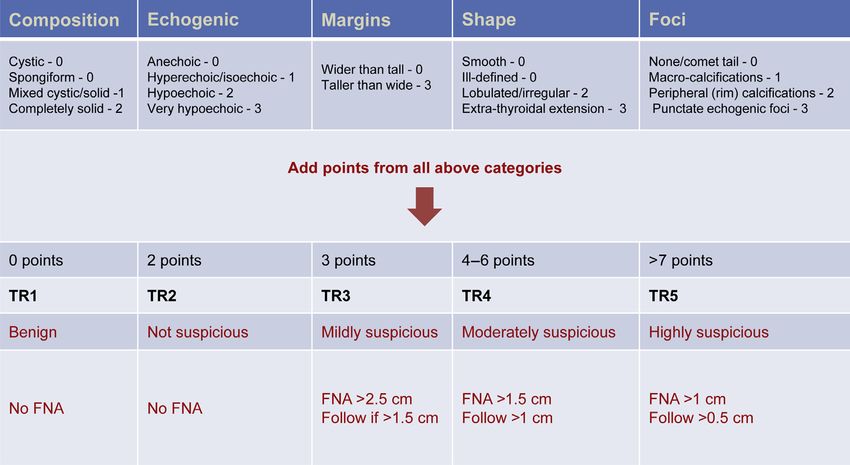
Diagnosis and Evaluation of Thyroid Nodules                1011

   Thyroid ultrasound is not indicated in patients           adenoma with suppressed uptake in the surround-
with medical thyroid disease if the gland is normal          ing and contralateral thyroid tissue (Fig. 1).
in size without evidence of a palpable nodule on                No further cytology evaluation is generally rec-
physical examination. It is also not indicated as a          ommended for a hyperfunctioning nodule, as
screening test except in patients with high genetic          these nodules rarely harbor malignancy. However,
risk or possibly in those with history of radiation to       the prevalence of thyroid cancer in hyperfunction-
the head or neck region.                                     ing nodules is approximately 3%.22 Therefore, in
   Ultrasound elastography, which uses both so-              clinical practice, FNA of a hyperfunctioning nodule
nography and a computational module to measure               should be considered in patients with risk factors
tissue stiffness, has been used in some institutions         for thyroid cancer, any nodules with suspicious
to assess cancer risk. Recently, larger clinical trials      sonographic features, and those that show growth
show that ultrasound elastography has been infe-             on surveillance.
rior to gray scale sonography, especially with                  Patients with a suppressed TSH who are noted
partially cystic or cystic nodules.18 Patients with          to have one or more hyperfunctioning nodules on
multinodular goiter, coalescence of nodules,                 uptake and scan should also undergo thyroid ul-
obese patients, or those with nodules that are infe-         trasound to evaluate the presence of nodules
rior or posterior are not candidates for ultrasound          concordant with the hyperfunctioning areas on
elastography.19                                              the scan and other nonfunctioning nodules that
                                                             might be present (Fig. 2).
Other imaging modalities                                        A multicenter study looked at association of thy-
A chest radiograph is not useful and therefore not           roid cancer in patients with nodular Graves’ disease
recommended for evaluation of the thyroid gland,             and found the rate of carcinoma in a cold nodule
although it may indicate a substernal goiter, which          was 15% (n 5 140 patients). In patients with
typically presents as a mass associated with                 nodular Graves’ disease, ultrasound-guided FNA
tracheal narrowing, tracheal stenosis, and medias-           is useful before radioiodine therapy or surgery.23
tinal widening causing shortness of breath.                     The risk of thyroid cancer in patients with a mul-
   Cross-sectional imaging with CT scan is recom-            tinodular goiter is the same as in those with a sol-
mended to evaluate and confirm substernal exten-             itary nodule.24,25 Therefore, all nodules within a
sion and tracheal compression. It is also                    multinodular goiter that meet sonographic and
recommended in suspected advanced thyroid                    size criteria concerning for malignancy should un-
cancer to assess for nodal disease and wide-                 dergo biopsy.14
spread metastases.14 Iodinated contrast adminis-
tration should be avoided in patients with
suspected thyroid cancer, as it will delay therapy           Fine Needle Aspiration
with radioactive iodine (RAI).                               FNA is the procedure of choice in the histologic
   FDG-PET imaging is not recommended for the                evaluation of thyroid nodules. The nodule size at
evaluation of patients with newly detected thyroid           initial ultrasound, the ultrasound characteristics,
nodules or thyroid disease. Incidental FDG-PET               and definite increase in size during follow-up are
uptake in the thyroid gland is seen in 2% to 3% pa-          generally considered as reasonable criteria for
tients and can be either focal or diffuse.11 Focal           deciding whether to proceed with FNA. FNA
FDG-PET uptake in the thyroid is associated with             should be performed under ultrasound guidance
an approximately 35% risk of being cancerous.20              to ensure optimal placement of the needle tip for
For PET-positive nodules greater than 1 cm diam-             nodule sampling.
eter, a dedicated ultrasound and FNA are recom-                 In the United States, the two commonly used
mended. However, diffuse FDG uptake in                       guidelines to estimate risk of malignancy, and
conjunction with sonographic and clinical evi-               thus assess a need for FNA, are the ATA (American
dence of chronic lymphocytic thyroiditis does not            Thyroid Association) guidelines14 and the ACR TI-
require further imaging or FNA.                              RADS (American College of Radiology Thyroid Im-
                                                             aging Reporting and Data System).16,26
MULTINODULAR GOITER AND TOXIC                                   Table 2 shows the ATA ultrasound features and
NODULES                                                      criteria for biopsy as per the guidelines.14 In com-
                                                             parison, Fig. 3 shows thyroid nodule imaging fea-
A radioactive iodine uptake and scan should be               tures and guidelines for biopsy as recommended
obtained if the TSH is low to assess whether the             by the ACR TI-RADS reporting system.16
nodule is hyperfunctioning.21 The pattern of up-                Both guidelines recommend biopsy if size of the
take in a patient with a single hyperfunctioning             thyroid nodule is over 1 cm and there are high-
nodule generally shows focal uptake in the                   suspicion sonographic features. For intermediate-
1012 Nambron et al
Fig. 1. Thyroid scan with I-131 of a solitary functioning nodule in the right thyroid lobe in a 22-year-old woman
noted to have a suppressed TSH. A functioning nodule is nearly always benign, whereas, a nonfunctioning
nodule (approximately 90% of nodules) has a 5% to 15% risk of being malignant.
Fig. 2. A 62-year-old man presented
with thyrotoxicosis from Graves’ dis-
ease. Thyroid ultrasound showed a
left thyroid lobe solid nodule
2.3 " 1.9 cm (ACR TI-RADS 4). A nu-
clear uptake scan reported this
nodule to be cold, and biopsy proved
to be medullary thyroid cancer.Diagnosis and Evaluation of Thyroid Nodules 1013 Table 2 Ultrasound features and criteria for fine need aspiration High Risk of Intermediate Risk Low Risk of Malignancy of Malignancy Malignancy Very Low Risk of (70%–90%) (10%–20%) (5%–10%) Malignancy (
1014        Nambron et al

         Category 1 - nondiagnostic; the estimated risk         Table 3
           of malignancy is 1% to 4%                            The common mutations seen in papillary and
         Category 2 - benign cytology implies a less than       follicular thyroid cancer
           3% likelihood of cancer
         Category 3 – follicular lesion of undetermined         Papillary                 Follicular
           significance (FLUS) or atypia of undetermined        BRAF (40%–50%)            RAS (40%–50%)
           significance (AUS); the risk for malignancy is
                                                                RAS (7%–20%)              PAX8/PPAR(30%–35%)
           5% to 15%
         Category 4 – follicular neoplasm/suspicious for        RET/PTC(10%–20%)          TP53(21%)
           follicular neoplasm (FN/SFN), Hurthle cell           EGFR (5%)                 PTEN(8%)
           neoplasm or suspicious for Hurthle cell              TRK(4 cm),
       specificity (86%–100%) and positive predictive          continue to grow and/or are causing compressive
       value (84%–100%). Table 3 shows a panel of              symptoms including dysphonia, dysphagia, or
       common mutations in differentiated thyroid              dyspnea.36,37 (Fig. 4).
       cancer.                                                   A single-center study evaluated the FNA results
          The Cancer Genome Atlas project mapped mu-           and ultrasound features in patients with nodules
       tations to various forms of differentiated thyroid      over 4 cm. Of the subset of 125 nodules that
       cancer. This subsequently led to development of         were identified as benign in preoperative FNA,
       Next Generation Sequencing (ThyroSeq) to                10.4% were malignant on postoperative pathol-
       improve the sensitivity and negative predictive         ogy.38 Another retrospective study confirmed
Diagnosis and Evaluation of Thyroid Nodules 1015
Fig. 4. A 50-year-old man followed
for an incidentally found multinodu-
lar goiter. Image A shows solid left
thyroid nodule 5 " 3.5 cm with
some cystic content. Biopsy at that
time was benign. Images B and C
show follow-up demonstrating more
solid sonographic features 2 years
later. Considering the size of the
nodule and patient and clinician pref-
erence, a partial thyroidectomy was
performed. Surgical pathology
showed a 4 cm papillary thyroid can-
cer. Patient subsequently underwent
a completion thyroidectomy.
that patients with initially benign FNA had a low distant metastasis. For patients with tumors
mortality risk during long-term follow-up even greater than 1 cm but less than 4 cm with no evi-
though they had a low, but real risk of false dence of extrathyroidal spread or metastatic dis-
negatives.39 ease, the initial procedure of choice can be a
Surgical excision of indeterminate thyroid nod- lobectomy versus total thyroidectomy based on
ules (AUS/FLUS-Bethesda Category 3) should be clinical concern and patient preference. Thyroid
considered if repeat biopsy or molecular testing lobectomy is the procedure of choice for patients
or both is either not performed or is conclusive or with tumors less than 1 cm and no evidence of
suspicious. extrathyroidal extension, nodal, or distant meta-
Surgical excision is also recommended for follic- static disease.42
ular neoplasms (FN/SFN-Bethesda Category 4). In For patients with a nondiagnostic cytology
these patients, thyroid lobectomy is the initial pro- (Bethesda Category 1), FNA should be repeated,
cedure of choice. Total thyroidectomy, however, preferably with onsite cytologic evaluation.43 Sur-
should be considered in patients with high clinical gery should be considered in nondiagnostic nod-
suspicion, presence of high-risk sonographic fea- ules with high-suspicion sonographic features,
tures, suspicious molecular diagnostics, presence growth during surveillance, or presence of clinical
of risk factors, presence of multiple nodules, and risk factors for malignancy.
patient preference.
Surgical management is similar to that of a ma-
Toxic Nodules
lignant nodule for lesions classified Suspicious for
Malignancy (Bethesda Category 5). Historically, Radioactive iodine therapy and surgery are the
total thyroidectomy has been the recommended recommended treatment modalities for patients
approach to a patient with biopsy-proven malig- with toxic multinodular goiter and toxic adenomas.
nant disease (Bethesda Category 6). However, The size and number of nodules, sonographic fea-
recent data have suggested similar outcomes in tures, other comorbidities, and patient preference
patients with unilateral or bilateral thyroid surgery should be taken into consideration when deciding
in appropriately chosen patients with low-risk dis- on the treatment. For patients with hyperfunction,
ease.40,41 Total thyroidectomy is recommended surgery is usually recommended for patients with
for patients with large tumors (>4 cm), gross extra- large multiple nodules, presence of compressive
thyroidal extension, and either regional nodal or symptoms, substernal or retrosternal extension,1016 Nambron et al
concern for thyroid cancer, or need for rapid SUMMARY
correction of hyperthyroid state. RAI therapy
should be considered for patients with contraindi- Thyroid nodules are a common clinical problem
cations to surgery, advanced age, and prior sur- encountered in an endocrine practice. Increas-
gery or other comorbidities. FNA biopsy should ingly, thyroid nodules are being detected inciden-
be performed for any nodules with suspicious tally, leading to an increased diagnosis of low-risk
sonographic features before radioactive iodine thyroid cancers. There is, therefore, a greater
therapy, if chosen. Antithyroid drugs may be emphasis on risk assessment based on clinical
considered for patients with small nodules and and sonographic features to avoid morbidity sec-
mild hyperthyroidism, in those with advanced ondary to unnecessary therapy. Molecular diag-
age, and patients with contraindications to both nostics are also being widely used to further
surgery and radioactive iodine therapy. characterize indeterminate nodules. The ATA and
ACR TI-RADS guidelines are the most used in clin-
Nodules in Pregnancy ical practice for risk assessment. Ultimately, it is
important to take into consideration a patient’s
Clinically relevant nodules in a pregnant patient are risk factors, clinical findings, comorbidities, life ex-
evaluated the same as in nonpregnant adults. pectancy, and preference prior to making man-
However, radioactive diagnostic scanning is con- agement decisions.
traindicated in pregnancy. Patients with nodules
diagnosed as differentiated thyroid cancer during DISCLOSURE
the course of pregnancy should be monitored
sonographically; delaying surgery until after deliv- The authors have nothing to disclose.
ery has not been shown to affect outcome.44 Sur-
gery should be considered in the second trimester REFERENCES
if there is evidence of growth of the nodule, cervi-
cal lymphadenopathy, or distant metastasis. 1. Vander JB, Gaston EA, Dawber TR. The significance
of nontoxic thyroid nodules. Final report of a 15-year
FOLLOW-UP study of the incidence of thyroid malignancy. Ann
Intern Med 1968;69(3):537–40.
Thyroid ultrasound is used for follow-up over time 2. SEER-Database. 2019. Available at: https://seer.
to assess for changes in nodule size and charac- cancer.gov/statfacts/html/thyro.html. Accessed February
teristics. A significant increase in nodule size, 2, 2020.
defined as increase in size of nodule by 20% in 2 3. Haymart MR, Banerjee M, Reyes-Gastelum D, et al.
dimensions or a 50% increase in volume is an indi- Thyroid ultrasound and the increase in diagnosis of
cation for repeat sampling/FNA.14 As described low-risk thyroid cancer. J Clin Endocrinol Metab
elsewhere, the possibility of malignancy is best 2019;104(3):785–92.
judged by the ultrasound characteristics, rather 4. Hegedüs L. The thyroid nodule. N Engl J Med 2004;
than growth. As per the ATA 2015 guidelines, any 351(17):1764–71.
thyroid nodule with high-suspicion sonographic 5. Mandel SJ. A 64-year-old woman with a thyroid
features and benign FNA cytology should be fol- nodule. JAMA 2004;292(21):2632–42.
lowed with a repeat ultrasound or FNA in 6. Dean DS, Gharib H. Epidemiology of thyroid nod-
12 months. For the nodules with intermediate sus- ules. Best Pract Res Clin Endocrinol Metab 2008;
picion, the recommended interval of follow-up is 22(6):901–11.
typically 12 to 24 months. For a low-suspicion 7. Mazzaferri EL. Management of a solitary thyroid
nodule, ultrasound can be repeated in more than nodule. N Engl J Med 1993;328(8):553–9.
24 months. During the course of surveillance, if 8. Ezzat S, Sarti DA, Cain DR, et al. Thyroid incidenta-
the nodule shows any growth or change in charac- lomas: prevalence by palpation and ultrasonogra-
teristic or development of high-risk sonographic phy. Arch Intern Med 1994;154(16):1838–40.
features, a repeat FNA should be performed. 9. Youserm D, Huang T, Loevner LA, et al. Clinical and
The ACR TI-RADS system guidelines have economic impact of incidental thyroid lesions found
similar recommendations as the ATA 2015 with with CT and MR. AJNR Am J Neuroradiol 1997;18(8):
regards to long-term follow-up of benign thyroid 1423–8.
nodules. ACR TI-RADS recommends following 10. Steele SR, Martin MJ, Mullenix PS, et al. The signifi-
nodules up to 5 years and discontinuing surveil- cance of incidental thyroid abnormalities identified
lance if stable. However, repeat imaging or during carotid duplex ultrasonography. Arch Surg
continued surveillance should be done if there is 2005;140(10):981–5.
increase in ACR TI-RADS score or increase in 11. Cohen MS, Arslan N, Dehdashti F, et al. Risk of ma-
nodule size. lignancy in thyroid incidentalomas identified byDiagnosis and Evaluation of Thyroid Nodules 1017
fluorodeoxyglucose-positron emission tomography. value of ultrasound and color-doppler features.
Surgery 2001;130(6):941–6. J Clin Endocrinol Metab 2002;87(5):1941–6.
12. Boelaert K, Horacek J, Holder RL, et al. Serum 26. Grant EG, Tessler FN, Hoang JK, et al. Thyroid ultra-
thyrotropin concentration as a novel predictor of sound reporting lexicon: white paper of the ACR thy-
malignancy in thyroid nodules investigated by roid imaging, reporting and data system (TIRADS)
fine-needle aspiration. J Clin Endocrinol Metab Committee. J Am Coll Radiol 2015;12(12 Pt A):1272–9.
2006;91(11):4295–301. 27. Baloch ZW, LiVolsi VA, Asa SL, et al. Diagnostic ter-
13. Gerschpacher M, Göbl C, Anderwald C, et al. Thyro- minology and morphologic criteria for cytologic
tropin serum concentrations in patients with papillary diagnosis of thyroid lesions: a synopsis of the na-
thyroid microcancers. Thyroid 2010;20(4):389–92. tional cancer institute thyroid fine-needle aspiration
14. Haugen BR, Alexander EK, Bible KC, et al. 2015 state of the science conference. Diagn Cytopathol
American Thyroid Association Management guide- 2008;36(6):425–37.
lines for adult patients with thyroid nodules and 28. Cibas ES, Ali SZ. The 2017 Bethesda system for re-
differentiated thyroid cancer: The American Thyroid porting thyroid cytopathology. Thyroid 2017;27(11):
Association Guidelines Task Force on Thyroid Nod- 1341–6.
ules and Differentiated Thyroid Cancer. Thyroid 29. Cibas ES, Baloch ZW, Fellegara G, et al.
2016;26(1):1–133. A prospective assessment defining the limitations
15. Radecki PD, Arger PH, Arenson RL, et al. Thyroid of thyroid nodule pathologic evaluation. Ann Intern
imaging: comparison of high-resolution real-time ul- Med 2013;159(5):325–32.
trasound and computed tomography. Radiology 30. Roth MY, Witt RL, Steward DL. Molecular testing for
1984;153(1):145–7. thyroid nodules: Review and current state. Cancer
16. Tessler FN, Middleton WD, Grant EG, et al. ACR Thy- 2018;124(5):888–98.
roid Imaging, Reporting and Data System (TI- 31. Patel KN, Angell TE, Babiarz J, et al. Performance of
RADS): white paper of the ACR TI-RADS committee. a genomic sequencing classifier for the preopera-
J Am Coll Radiol 2017;14(5):587–95. tive diagnosis of cytologically indeterminate thyroid
17. Fish SA, Langer JE, Mandel SJ. Sonographic imag- nodules. JAMA Surg 2018;153(9):817–24.
ing of thyroid nodules and cervical lymph nodes. En- 32. Nikiforova MN, Mercurio S, Wald AI, et al. Analytical
docrinol Metab Clin North Am 2008;37(2):401–17. performance of the ThyroSeq v3 genomic classifier
18. Moon HJ, Sung JM, Kim E-K, et al. Diagnostic per- for cancer diagnosis in thyroid nodules. Cancer
formance of gray-scale US and elastography in 2018;124(8):1682–90.
solid thyroid nodules. Radiology 2012;262(3): 33. Sdano MT, Falciglia M, Welge JA, et al. Efficacy of
1002–13. thyroid hormone suppression for benign thyroid
19. Azizi G, Keller J, Lewis M, et al. Performance of elas- nodules: meta-analysis of randomized trials. Otolar-
tography for the evaluation of thyroid nodules: a pro- yngol Head Neck Surg 2005;133(3):391–6.
spective study. Thyroid 2013;23(6):734–40. 34. Yousef A, Clark J, Doi SAR. Thyroxine suppression
20. Soelberg KK, Bonnema SJ, Brix TH, et al. Risk of therapy for benign, non-functioning solitary thyroid
malignancy in thyroid incidentalomas detected by nodules: a quality-effects meta-analysis. Clin Med
18f-fluorodeoxyglucose positron emission tomogra- Res 2010;8(3–4):150–8.
phy: a systematic review. Thyroid 2012;22(9): 35. Grussendorf M, Reiners C, Paschke R, et al. Reduc-
918–25. tion of thyroid nodule volume by levothyroxine and
21. Ross DS, Burch HB, Cooper DS, et al. 2016 American iodine alone and in combination: a randomized,
Thyroid Association guidelines for diagnosis and placebo-controlled trial. J Clin Endocrinol Metab
management of hyperthyroidism and other causes 2011;96(9):2786–95.
of thyrotoxicosis. Thyroid 2016;26(10):1343–421. 36. Shin JJ, Caragacianu D, Randolph GW. Impact of
22. Mirfakhraee S, Mathews D, Peng L, et al. A solitary thyroid nodule size on prevalence and post-test
hyperfunctioning thyroid nodule harboring thyroid probability of malignancy: a systematic review.
carcinoma: review of the literature. Thyroid Res Laryngoscope 2015;125(1):263–72.
2013;6(1):7. 37. Aydog ! an B_I, S‚ahin M, Ceyhan K, et al. The influ-
23. Kraimps JL, Bouin-Pineau MH, Mathonnet M, et al. ence of thyroid nodule size on the diagnostic
Multicentre study of thyroid nodules in patients efficacy and accuracy of ultrasound guided fine-
with Graves’ disease. Br J Surg 2000;87(8):1111–3. needle aspiration cytology. Diagn Cytopathol
24. Marqusee E, Benson CB, Frates MC, et al. Useful- 2019;47(7):682–7.
ness of ultrasonography in the management of 38. Wharry LI, McCoy KL, Stang MT, et al. Thyroid nod-
nodular thyroid disease. Ann Intern Med 2000; ules (#4 cm): can ultrasound and cytology reliably
133(9):696–700. exclude cancer? World J Surg 2014;38(3):614–21.
25. Papini E, Guglielmi R, Bianchini A, et al. Risk of ma- 39. Nou E, Kwong N, Alexander LK, et al. Determination
lignancy in nonpalpable thyroid nodules: predictive of the optimal time interval for repeat evaluation after1018 Nambron et al
a benign thyroid nodule aspiration. J Clin Endocrinol 42. Nixon IJ, Ganly I, Patel SG, et al. Thyroid lobectomy
Metab 2014;99(2):510–6. for treatment of well differentiated intrathyroid malig-
40. Matsuzu K, Sugino K, Masudo K, et al. Thyroid lo- nancy. Surgery 2012;151(4):571–9.
bectomy for papillary thyroid cancer: long-term 43. Lin DM, Tracht J, Rosenblum F, et al. Rapid on-site
follow-up study of 1,088 cases. World J Surg 2014; evaluation with telecytology significantly reduced
38(1):68–79. unsatisfactory rates of thyroid fine-needle aspiration:
41. Barney B, Hitchcock Y, Sharma P, et al. Overall and a case-control study. Am J Clin Pathol 2020;153(3):
cause-specific survival for patients undergoing lo- 342–5.
bectomy, near-total, or total thyroidectomy for 44. Moosa M, Mazzaferri EL. Outcome of differentiated
differentiated thyroid cancer. Head Neck 2011;33: thyroid cancer diagnosed in pregnant women.
645–9. J Clin Endocrinol Metab 1997;82(9):2862–6.You can also read