Analysis of RHOA mutations and their significance in the proliferation and transcriptome of digestive tract cancer cells
←
→
Page content transcription
If your browser does not render page correctly, please read the page content below
ONCOLOGY LETTERS 22: 735, 2021

        Analysis of RHOA mutations and their significance in the
      proliferation and transcriptome of digestive tract cancer cells
 NAOKI IKARI1,2, AKIKO SERIZAWA1, ETSUKO TANJI2, MASAKAZU YAMAMOTO1 and TORU FURUKAWA1‑3

  1
   Department of Surgery, Institute of Gastroenterology; 2Institute for Integrated Medical Sciences, Tokyo Women's Medical
                    University, Shinjuku-ku, Tokyo 162‑8666; 3Department of Investigative Pathology,
                    Tohoku University Graduate School of Medicine, Aoba‑ku, Sendai 980‑8575, Japan

                                         Received April 23, 2021; Accepted July 14, 2021

                                                       DOI: 10.3892/ol.2021.12996

Abstract. The ras homolog family member A (RHOA) gene                showed that the genes associated with small molecule metabolic
encodes a member of the Rho family of small GTPases and is           process, oxidation‑reduction processes, protein kinase activity,
known to function in reorganization of the actin cytoskeleton,       transport, and cell junction were commonly downregulated in
which is associated with regulation of cell shape, attachment        cells whose proliferation was attenuated by the knockdown of
and motility. RHOA has been found to be recurrently mutated          RHOA. These results suggested that certain RHOA mutations
in gastrointestinal cancer; however, the functional signifi‑         may result in upregulation of lnc‑DERA‑1 and genes associated
cance of the mutated RHOA protein in digestive tract cancers         with cellular metabolism and proliferation in digestive tract
remains to be uncovered. The aim of the present study was            cancers.
to understand the role of mutant RHOA in the proliferation
and transcriptome of digestive tract cancer cells. Mutations of      Introduction
RHOA in one esophageal cancer cell line, OE19, eight gastric
cancer cell lines, namely, AGS, GCIY, HGC‑27, KATO III,              The ras homolog family member A (RHOA) gene encodes a
MKN1, MKN45, SNU16 and SNU719, as well as two colon                  member of the Rho family of small GTPases and is known
cancer cell lines, CCK‑81 and SW948, were determined using           to function in reorganization of the actin cytoskeleton, which
Sanger sequencing. The results uncovered several mutations,          is associated with regulation of cell shape, attachment and
including p.Arg5Gln and p.Tyr42Cys in CCK‑81, p.Arg5Trp              motility. RHOA has been found to be recurrently mutated
and p.Phe39Leu in SNU16, p.Gly17Glu in SW948, p.Tyr42Ser             in gastrointestinal cancer, especially in diffuse‑type gastric
in OE19, p.Ala61Val in SNU719, p.Glu64del in AGS.                    cancer cases (1‑3). In this cancer, residues p.Arg5, p.Gly17,
Wild‑type RHOA was identified in GCIY, HGC‑27, KATO                  p.Tyr42 and p.Leu57 of RHOA are considered hotspot
III, MKN1 and MKN45. Knockdown of RHOA using small                   missense mutations (1,2). However, the functional significance
interfering RNA attenuated the in vitro proliferation in the         of these mutations has not been consistently demonstrated.
three‑dimensional culture systems of GCIY, MKN1, OE19 and            Kakiuchi et al (1) suggested that the hotspot mutations were
SW948, whereas no apparent changes were seen in CCK‑81,              gain‑of‑function mutations because inhibiting the expression
HGC‑27 and SNU719. Transcriptome analysis revealed that              of the mutant RHOA induced the suppression of proliferation
downregulation of the long non‑coding RNA (lnc)‑DERA‑1               of gastric cancer cells. In addition, Zhang et al (4) showed
was observed in all tested cell lines following RHOA knock‑          that RHOAp.Tyr42Cys was a gain‑of‑function mutation that could
down in the RHOA‑mutated cell lines. Gene Ontology analysis          sufficiently induce diffuse‑type gastric cancer in a mouse
                                                                     model. On the other hand, Wang et al (2) indicated that these
                                                                     were loss‑of‑function mutations because the mutant RHOA
                                                                     protein showed reduced small GTPase activity and lost
                                                                     the ability to mediate anoikis. Sakata‑Yanagimoto et al (5)
Correspondence to: Dr Toru Furukawa, Department of                   also reported that the RHOA p.Gly17Val mutation was a
Investigative Pathology, Tohoku University Graduate School of        loss‑of‑function mutation because it showed loss of GTP
Medicine, 2‑1 Seiryomachi, Aoba‑ku, Sendai 980‑8575, Japan           binding activity and inhibition of wild‑type RHOA function.
E‑mail: toru.furukawa@med.tohoku.ac.jp
                                                                     Interestingly, knockdown of RHOA in gastric cancer cells
Abbreviations: RHOA, ras homolog family member A; YAP,               with intrinsic abundant expression of RHOA, irrespective
Yes‑associated protein 1; TAZ, WW‑domain‑containing transcription    of its mutational status, results in inhibition of proliferation
regulator 1                                                          in vitro (6). Downregulation of RHOA via miR‑31 inhibits
                                                                     cell proliferation and invasiveness (7). Moreover, overexpres‑
Key words: RHOA, gastric cancer, colon cancer, mutation,             sion of wild‑type RHOA induces immortalization of human
proliferation, transcriptome, lnc‑DERA‑1, metabolism                 mammary epithelial cells. However, these immortalized cells
                                                                     were anchorage‑dependent and were unable to form tumors
                                                                     when implanted in nude mice (8). Although these pieces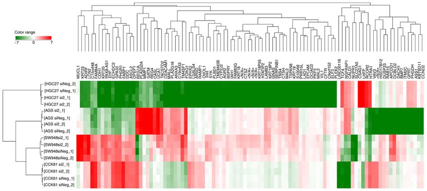
2 IKARI et al: RHOA IN DIGESTIVE TRACT CANCER CELLS
of evidence have highlighted the different aspects of the antisense, 5'‑UACGAAUGACGUG CGGUACGU‑3'. For the
molecular functions of RHOA, the functional role of RHOA cellular proliferation assay, 1.0x10 4 cells/well were seeded
mutations in the digestive tract cancers are yet to be deter‑ into a 96‑well clear flat bottom ultra‑low attachment plate
mined. In the present study, to understand the functional role (Corning, Inc.) with 100 µl growth medium containing 1 nM
of RHOA mutations in digestive tract cancers, genotyping, of siRNA and 0.16% (vol/vol) RNAiMAX (Thermo Fisher
transcriptome analysis and proliferation assays were carried Scientific, Inc.) according to the manufacture's instruction.
out in cell lines expressing the mutant or wild‑type RHOA, as The cells were incubated at 37˚C with 5% CO2 in humidified
well as in cells where RHOA has been knocked down. conditions, except for SW948 cells that were incubated at 37˚C
with 100% air. Cells were assayed 24 h later (Day 1) and then
Materials and methods every 48 h (Day 3 and 5) until Day 7. For immunoblotting,
2.5x10⁵ cells/well were seeded into a 6‑well clear flat‑bottom
Cell culture. The AGS cell line was obtained from American ultra‑low attachment plate with 1 ml medium containing 1 nM
Type Culture Collection. The GCIY, KATO III, HGC‑27, siRNA and 0.16% (vol/vol) of RNAiMAX. The transfected
MKN1 and MKN45 cell lines were obtained from RIKEN cells were incubated as aforementioned, and collected 48 h
BioResource Center. The OE19 and SW948 cell lines were later. The low attachment plates were used to allow prolif‑
obtained from Public Health England (Salisbury, UK). SNU16 eration in three‑dimensional spheroid conditions, a method
and SNU719 cell lines were obtained from Korean Cell that is more suitable for in vitro bioassays than conventional
Line Bank. CCK‑81 cell line was obtained from Japanese two‑dimensional assays (14).
Collection of Research Bioresources Cell Bank. All cell lines
were cultured according to recommendations from suppliers. Three‑dimensional cell proliferation assay. Following
AGS cells were cultured in F‑12 Ham, Kaighn's Modification RHOA‑knockdown, cell viability was assessed using the
(Sigma‑Aldrich; Merck KGaA) supplemented with 10% CellTiter‑Glo® 3D Cell Viability Assay (Promega Corporation)
FBS (Immuno‑Biological Laboratories Co., Ltd.). Minimum according to the manufacturer's instruction. The viability of
Essential Medium (MEM; Sigma‑Aldrich; Merck KGaA) the cells transfected with NC siRNA was used as the control.
supplemented with 15% FBS was used for GCIY cells. Cell viability was calculated after subtraction of background
RPMI‑1640 (Sigma‑Aldrich; Merck KGaA) supplemented absorbance as follows: Cell viability (%)=(absorbance of the
with 10% FBS was used for KATO III, MKN1, MKN45, OE19, sample/absorbance of the control) x100.
SNU16 and SNU719 cell culture. MEM supplemented with
10% FBS was used for HGC‑27 and CCK‑81 cell maintenance. Immunoblotting. Cells were harvested and lysed in modified
Leibovitz's L‑15 (Thermo Fisher Scientific, Inc.) supplemented RIPA buffer containing 1X complete mini protease inhibitor
with 2 mM Glutamine (Thermo Fisher Scientific, Inc.) and cocktail (Sigma‑Aldrich; Merck KGaA) and 1X PhosSTOP
10% FBS was used for SW948 cell culture. SW948 cells were phosphatase inhibitor cocktail (Sigma‑Aldrich; Merck KGaA).
maintained at 37˚C with 100% air in a humidified atmosphere; Protein concentration was determined by using the Bradford
all other cell lines were cultured at 37˚C with 5% CO2 in a Protein Assay Kit (Bio‑Rad Laboratories, Inc.) according
humidified atmosphere. These cell lines were selected because to the manufacture's instruction. Cell extracts containing
of availability and of being characterized previously as origi‑ 40 µg protein were separated by electrophoresis on a 10‑20%
nating from digestive tract tumors (1,9‑12). gradient polyacrylamide gel and blotted onto a polyvinylidene
difluoride membrane (ATTO Corporation) using the XV
Mutational analysis of the cell lines. DNA was extracted from Pantera MP System (DRC Co., Ltd.), according to the manu‑
the cultured cells using GenElute™ Mammalian Genomic facturer's instructions. Blocking was performed for 1 h using
DNA Miniprep Kit (Sigma‑Aldrich; Merck KGaA) according the ECL Blocking Agent (Amersham Biosciences; Cytiva) at
to the manufacturer's instructions. All coding exons and splice room temperature (RT), and the membrane was incubated with
sites of RHOA were amplified by using AccuPrime™ Taq DNA primary antibodies overnight at 4˚C. Primary antibodies used
Polymerase (Thermo Fisher Scientific, Inc.) and paired primers were the rabbit monoclonal anti‑RHOA antibody (clone 67B9;
shown in Table SI. The amplified products were analyzed using 1:1,000 dilution; cat. no. 2117; Cell Signaling Technology, Inc.)
Sanger sequencing, as described previously (13). To investigate and the mouse monoclonal anti‑β‑actin antibody (clone AC‑15;
whether RHOAp.Arg5Gln and RHOAp.Tyr42Cys in the CCK‑81 cell 1:1,000 dilution; cat. no. A5441; Sigma‑Aldrich; Merck KGaA).
line were cis‑ or trans‑compound heterozygous mutations, The membrane was incubated with a corresponding secondary
mutation‑specific primers, wild‑type specific primers and antibody for 1 h at RT. The secondary antibodies used were
intron primers were designed as shown in Table SII. horseradish peroxidase‑conjugated anti‑mouse and anti‑rabbit
immunoglobulin antibody (1:10,000 dilution) (cat. nos. NA931
Small interfering RNA (siRNA) targeting RHOA. Knockdown and NA934; GE Healthcare). The signals were visualized using
of RHOA using siRNA was conducted, as previously the ECL Prime Western Blotting Detection Reagent (Cytiva)
reported (1). The validated RHOA siRNA used included: and LAS 4000 Mini system (Fujifilm Wako Pure Chemical
i) RHOA siRNA2 sense, 5'‑CUAUGAU UAU UAACGAUG Corporation).
UTT‑3' and antisense, 5'‑ACAUCGU UAAUAAUCAUA
GTT‑3'; and ii) RHOA siRNA3 sense, 5'‑GGCUUUACUCCG Microarray analysis. Total RNA was isolated from the
UAACAGATT‑3' and antisense, 5'‑UCUGUUACGGAGUAA cultured cells using the RNeasy Mini kit (Qiagen GmbH)
AGCCCT‑3'. The negative control (NC) siRNA sequences and was subjected to microarray analysis for transcriptome.
were: sense, 5'‑GUACCGCACGUCAUUCGUAUC‑3' and The microarray analysis was performed by RIKEN Genesis,ONCOLOGY LETTERS 22: 735, 2021 3
using Agilent SurePrint G3 Human GE Microarray 8x60k
Ver3.0 (G4851C) (Agilent Technologies, Inc.). Gene Ontology
analysis was performed (http://geneontology.org) using the
PANTHER Classification System (http://pantherdb.org/) (15).
Statistical analysis. The cell growth rate was represented in
terms of mean and standard error and was compared using
one‑way ANOVA and Tukey's test. In microarray analysis,
only genes whose expression levels were detected were
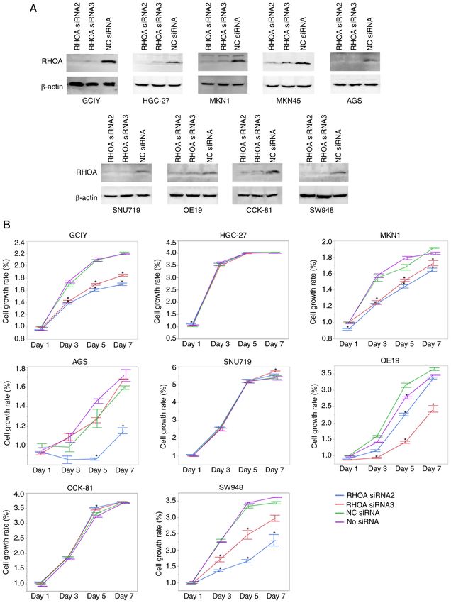
Figure 1. RHOA expression in digestive tract cancer cell lines. The expression
considered for further analysis. Changes in gene expression of RHOA in digestive tract cancer cell lines was detected by immunoblot‑
levels were compared using unpaired two‑tailed Student's ting using a rabbit monoclonal anti‑RHOA antibody. β‑actin was used as a
t‑tests. Hierarchical clustering analysis was performed using loading control. The pictures were cropped from the same blot probed with
absolute values of fold changes of genes by the following the anti‑RHOA antibody firstly (upper) and the anti‑β‑actin antibody subse‑
quently (lower). The experiments were performed twice and similar results
conditions: Clustering Algorithm, Hierarchical; Clustered By, were obtained. GCIY, HGC‑27, KATO III, MKN1 and MKN45 harbored
Normalized intensity values; Similarity Measure, Euclidean; wild‑type RHOA. RHOA mutations were detected as p.Glu64del in AGS,
Linkage Rule, Wards. P4 IKARI et al: RHOA IN DIGESTIVE TRACT CANCER CELLS Figure 2. RHOA knockdown efficiency and cell proliferation after RHOA knockdown. (A) Knockdown of RHOA using RHOA siRNA2 or RHOA siRNA3 confirmed by immunoblotting. The pictures were cropped from the same blot for each cell line probed with the anti‑RHOA antibody firstly (upper) and the anti‑β‑actin antibody subsequently (lower). The experiments were performed twice and similar results were obtained. (B) Cell proliferation rates after RHOA knockdown using the RHOA siRNA2 or RHOA siRNA3. Data were normalized using the cell viability data of mock transfectants using negative control siRNA (NC) in Day 1. The data has been represented as the means ± standard error. The experiments were performed twice in triplicate wells and similar results were obtained. *P
ONCOLOGY LETTERS 22: 735, 2021                                                     5

Table I. Genes of significantly altered expression following RHOA knockdown.

Cell line               Downregulated gene                                              Upregulated gene

AGS          CLDN18, CYP26C1, KRT28, LGSN,                    GATS, KDR, KRT39, LINC00113, lnc‑DHX34‑1, lnc‑EIF2B5‑2,
             LINC00909, LINC00933, lnc‑ARRDC3‑1,              lnc‑GABARAPL3‑4, lnc‑RIC3‑1, LOC399900, LOC643339,
             lnc‑DERA‑1, LOC155060, RHOA,                     OR4C15, SMIM24, SZT2
             SLC26A1, STK31
CCK81        FOXQ1, lnc‑DERA‑1, lnc‑FAM189A1‑3,               CDK15, CYSLTR1, KLF2, lnc‑C5orf38‑3, lnc‑NTRK2‑3, MXRA7,
             lnc‑OXNAD1‑2, lnc‑RP11, 181C3.1.1‑1,             RHOB, ZG16
             METTL6, RHOA, SP5
HGC27        EGFR, IGFBP3, lnc‑AL020996.1‑2,                  CDK19, COL5A1, CSRNP3, LINC01529, lnc‑ANLN‑4,
             lnc‑CPSF7‑1, lnc‑ZNF730‑1, MEIS1,                LOC102724301, PABPC1L2B, SLC36A1, SLC4A4, SWAP70,
             OPN1SW, RHOA, SPIN3, TRIAP1                      ZDHHC20
SW948        ACSL6, AIFM3, CAMKK1, CERKL,                     ADM, AMOTL2, ANO1, ARL14, ATP2B4, ATP8B3, CACNB4, CAV1,
             CMKLR1, GPR128, KCNMB4,                          CDRT1, CITED2, CPE, CRYGC, CTGF, CXCL1, CYR61, DOCK4,
             lnc‑C9orf80‑1, lnc‑CILP‑1, lnc‑DERA‑1,           DOK7, EDN1, EPHA2, GALNT5, GJB3, GNGT2, GPR37L1, GRPR,
             lnc‑RNF219‑3, LOC102724484,                      GULP1, HDAC5, IL1RN, KCNK9, KRT34, KRTAP1‑5, KRTAP3‑1,
             LOC729732, NCKAP5, PNLIPRP2,                     LAMA3, LIMCH1, LIMS2, LINC00520, LINC00592, LINC00704,
             PTPN20B, PTPRO, RHOA, RIIAD1,                    LINC01468, LMO1, lnc‑ACTBL2‑1, lnc‑ANKRD10‑1,
             SEMA3C, SMPX, SNX22, TAS2R45,                    lnc‑ARFGEF2‑2, lnc‑CEP44‑1, lnc‑COL1A1‑4, lnc‑COX4NB‑1,
             XLOC_l2_010029                                   lnc‑MRP63‑6, lnc‑MYO1D‑1, lnc‑OR10H5‑2, lnc‑PAX4‑1,
                                                              lnc‑RP11‑582J16.5.1‑3, lnc‑RP11‑817J15.3.1‑2, lnc‑SNURF‑3,
                                                              lnc‑YPEL5‑3, LOC101927260, LOC101928620, LOC101928666,
                                                              MAFF, MYL9, NT5DC4, OR1S2, PAG1, PDGFB, PLK2, PPP1R15A,
                                                              PTPRR, PXDN, RGCC, S100A2, SCARA3, SH2D5, SH3RF1,
                                                              SLC1A3, SLC26A9, SLC2A14, SLC2A3, SLC6A20, SPANXA1,
                                                              SPTSSB, SSUH2, TAGLN, TCTEX1D4, TM4SF1, TM4SF1‑AS1,
                                                              TMCC3, TNNC1, UCA1, WBSCR28, WFDC2, WWTR1,
                                                              XLOC_l2_009441

Discussion                                                           cell line showed the conflicting result of decreased proliferation
                                                                     with one siRNA but no change with the other siRNA, although
The present study identified RHOA mutations in digestive tract       both siRNAs resulted in the same level of RHOA knockdown,
cancer cell lines and showed that the protein was evidently but      which is different from the result of a similar experiment using
varyingly expressed in these cells regardless of the genotype.       the same siRNAs, performed by Kakiuchi et al (1) (showing no
The mutations included missense mutations and one in‑frame           significant change by either siRNA). Knockdown of RHOA in
deletion (p.Arg5Gln, p.Arg5Trp, p.Gly17Glu, p.Phe39Leu,              AGS cells was shown to inhibit cell proliferation in a previously
p.Tyr42Cys, p.Tyr42Ser, p.Ala61Val and p.Glu64del). According        published study by Liu et al (18), which is partially consistent
to the COSMIC database, p.Arg5Gln, p.Arg5Trp, p.Gly17Glu,            with the findings of the current study. The biological reason for
p.Tyr42Cys and p.Tyr42Ser are common hotspot mutations while         these conflicting results is obscure, and requires further inves‑
p.Phe39Leu, p.Ala61Val and p.Glu64del are rare mutations. It         tigation. The knockdown of RHOA in the current experiments
is indicated that the frequencies of the p.Arg5Gln, p.Arg5Trp,       were not specific to mutated transcripts, but specific to both the
p.Gly17Glu, p.Tyr42Cys and p.Tyr42Ser represented 4, 10, 7, 23       mutated and the wild‑type transcripts in cells with heterologous
and 4% of 99 nonsynonymous mutations detected in 1,854 gastric       alleles. The cell cycle and apoptosis of RHOA knockdown cells
cancer samples, respectively (COSMIC database; accessed on           were not examined; therefore, it is unclear whether the inhibition
2019.1.15). However, p.Phe39Leu, and Ala61Val have not been          of proliferation was due to attenuation of cell cycle or increase
identified in the gastric cancer samples, but in the hematopoietic   of apoptosis.
system (p.Phe39Leu) and large intestine (p.Ala61Val), in the             Furthermore, the present study also evaluated the change
COSMIC database. In the present study, knockdown of RHOA             in the expression profile of other genes associated with
inhibited the proliferation of some cell lines. The inhibition was   RHOA. Hence, the transcriptome of RHOA knockdown
observed in two of the three cell lines expressing wild‑type         cells was analyzed. It was hypothesized that genes that
RHOA and three of the five cell lines with mutant RHOA (AGS          were down‑ and upregulated following RHOA knockdown
with p.Glu64del, OE19 with p.Tyr42Cys and SW948 with                 would represent genes promoted and inhibited by RHOA
p.Gly17Glu). This suggested that RHOA promoted cell prolifera‑       expression, respectively. lnc‑DERA‑1 was commonly
tion depending on some intrinsic nature of the cells. The AGS        downregulated in examined cells with RHOA mutation.
6 IKARI et al: RHOA IN DIGESTIVE TRACT CANCER CELLS Figure 3. Hierarchical cluster analysis of transcriptome data of cells with RHOA knockdown and those without the knockdown. The hierarchical clustering analysis was performed using absolute values of fold changes of genes normalized by the percentile shift (75%) and baseline to median of all samples. Top 100 genes of absolute altered values (log2) among 16 samples were chosen to be clustered. There were two experimental replicates. Data from the siRNA NC in each cell line were used as controls. NC, negative control; RHOA, ras homolog family member A; siRNA, small interfering RNA. Color range indicates the log2 value of the fold changes. In sample names, si2 and siNeg indicates siRNA2 and NC siRNA, respectively. The microarray analysis was performed for 2 independent experimental sets denoted as _1 and _2 for every sample. According to LNCipedia (https://hg19.lncipedia.org; accessed with oxidation‑reduction included CYP26C1 and AIFM3 in 2019.1.21), lnc‑DERA‑1 is a non‑coding RNA encoded by a the AGS and SW948 cell line, respectively. AIFM3 encodes gene at chr12:16573561‑16573994, whose function has not apoptosis inducing factor mitochondria associated 3 that has been uncovered yet. In the gene ontological analysis, small a pyridine nucleotide‑disulfide oxidoreductase domain and molecule metabolic process and oxidation‑reduction process mediates apoptosis (21). Downregulation of these genes may were commonly downregulated biological processes in cells induce metabolic stress. However, the mechanistic relationship with the attenuated proliferation, which could be associ‑ between the inhibition of RHOA and the altered expression of ated with in vitro cell proliferation. Protein kinases play these genes was not evaluated in the present study. Recently, a critical role in cell proliferation. Downregulated genes one clue potentially associated with the transcriptional encoding protein kinases in cells with attenuated proliferation regulation by RHOA has emerged. Regulation of the actin cyto‑ were STK31 in AGS and CAMKK1 in SW948. STK31 is a skeleton by RHOA is associated with nuclear translocation of cancer‑associated gene that encodes a serine/threonine protein Yes‑associated protein 1 (YAP) and WW‑domain‑containing kinase known to play a role in microtubule assembly that is transcription regulator 1 (WWTR1/TAZ) that are known to be necessary for cell cycle progression (19). CAMKK1 encodes an important transcriptional regulator (22). Interestingly, the calcium/calmodulin dependent protein kinase kinase 1 that acti‑ transcription analysis in the present study demonstrated that vates calcium/calmodulin dependent protein kinase (CAMK). WWTR1/TAZ was upregulated after RHOA knockdown in CAMK plays a central role in calcium/calmodulin‑dependent SW948, which potentially indicates some negative feedback signaling cascades implicated in cell survival and carcinogen‑ regulations. esis (20). The genes associated with the metabolic process of One limitation of the present study was that the transcrip‑ small molecules which were downregulated include CYP26C1 tome analyses were not performed for all the cell lines. The and SLC26A1 in AGS and ACSL6 and PNLIPRP2 in SW948 functional significance of RHOA mutations was not evaluated. cell lines. CYP26C1 encodes a member of the cytochrome Further study on the regulation of transcription by RHOA P450 superfamily of enzymes, which is involved in several including the upregulation of lnc‑DERA‑1 may be needed processes, including drug metabolism and lipid synthesis for improved understanding of the mechanism of association (Entrez Gene; https://www.ncbi.nlm.nih.gov/gene). SLC26A1 between RHOA and cell proliferation. encodes a sulfate/anion transporter that functions in trans‑ porting of glucose and other sugars, bile salts and organic Acknowledgements acids, metal ions and cytochrome P450‑arranged by substrate type (GeneCards; https://www.genecards.org). ACSL6 encodes The authors would like to thank the depositors of the cell lines Acyl‑CoA synthase that catalyzes the formation of acyl‑CoA used in this study, namely, Dr Mutsumi Nozue, University of from fatty acids, ATP and CoA (Entrez Gene: https://www. Tsukuba, Ibaraki, Japan, for depositing GCIY cells to RIKEN ncbi.nlm.nih.gov/gene). PNLIPRP2 encodes pancreatic lipase BRC; Dr Masuo Obinata, Tohoku University, Sendai, Japan, for that hydrolyzes galactolipids (Entrez Gene; https://www. depositing KATO III cells to RIKEN BRC; Dr Tetsuo Kimoto, ncbi.nlm.nih.gov/gene). The downregulated genes associated Okayama University, Okayama, Japan, for depositing HGC‑27
ONCOLOGY LETTERS 22: 735, 2021                                                        7

cells to RIKEN BRC; Dr Teiichi Motoyama, Yamagata                      4. Zhang H, Schaefer A, Wang Y, Hodge RG, Blake DR,
                                                                          Diehl JN, Papageorge AG, Stachler MD, Liao J, Zhou J, et al:
University School of Medicine, Yamagata, Japan, depositing                Gain‑of‑function RHOA mutations promote focal adhesion
MKN1 and MKN45 cells to RIKEN BRC; Drs J.C. Rockett and                   kinase activation and dependency in diffuse gastric cancer.
A. Morriss, University of Warwick, Coventry, UK, and Dr S.J.              Cancer Discov 10: 288‑305, 2020.
                                                                       5. Sakata‑Yanagimoto M, Enami T, Yoshida K, Shiraishi Y, Ishii R,
Darnton, Birmingham Heartlands Hospital, Birmingham,                      Miyake Y, Muto H, Tsuyama N, Sato‑Otsubo A, Okuno Y, et al:
UK, for depositing OE19 to the European Collection of                     Somatic RHOA mutation in angioimmunoblastic T cell
Authenticated Cell Cultures, Public Health England; Dr.                   lymphoma. Nat Genet 46: 171‑175, 2014.
                                                                       6. Chang HR, Nam S, Lee J, Kim JH, Jung HR, Park HS, Park S,
Jae‑Gahb Park, Cancer Research Institute, Korean Cell Line                Ahn YZ, Huh I, Balch C, et al: Systematic approach identifies
Bank, Korean Cell Line Research Foundation, Seoul, Republic               RHOA as a potential biomarker therapeutic target for Asian
of Korea, for depositing SNU16 and SNU719 cells to the                    gastric cancer. Oncotarget 7: 81435‑81451, 2016.
                                                                       7. Korourian A, Roudi R, Shariftabrizi A and Madjd Z:
Korean Cell Line Bank; and Dr Isaka, Hidehiko, Kagoshima                  MicroRNA‑31 inhibits RhoA‑mediated tumor invasion and
University, Kagoshima, Japan, for depositing CCK‑81 cells to              chemotherapy resistance in MKN‑45 gastric adenocarcinoma
the Japanese Collection of Research Bioresources Cell Bank.               cells. Exp Biol Med (Maywood) 242: 1842‑1847, 2017.
                                                                       8. Zhao X, Lu L, Pokhriyal N, Ma H, Duan L, Lin S, Jafari N,
                                                                          Band H and Band V: Overexpression of RhoA induces preneo‑
Funding                                                                   plastic transformation of primary mammary epithelial cells.
                                                                          Cancer Res 69: 483‑491, 2009.
                                                                       9. Ideo H, Seko A and Yamashita K: Galectin‑4 binds to sulfated
The present study was supported by the Japan Society for the              glycosphingolipids and carcinoembryonic antigen in patches on
Promotion of Science‑KAKENHI (grant no. JP16K10518).                      the cell surface of human colon adenocarcinoma cells. J Biol
                                                                          Chem 280: 4730‑4737, 2005.
                                                                      10. Ma C, Xie J, Luo C, Yin H, Li R, Wang X, Xiong W, Zhang T,
Availability of data and materials                                        Jiang P, Qi W, et al: OxLDL promotes lymphangiogenesis and
                                                                          lymphatic metastasis in gastric cancer by upregulating VEGF‑C
The microarray data are available in the Gene Expression                  expression and secretion. Int J Oncol 54: 572‑584, 2019.
                                                                      11. Ohashi N, Kodera Y, Nakanishi H, Yokoyama H, Fujiwara M,
Om n ibus repositor y under the accession number                          Koike M, Hibi K, Nakao A and Tatematsu M: Efficacy of intra‑
GSE110237 (http://www.ncbi.nlm.nih.gov/geo/query/acc.                     peritoneal chemotherapy with paclitaxel targeting peritoneal
cgi?acc=GSE110237). All data generated or analyzed during                 micrometastasis as revealed by GFP‑tagged human gastric
                                                                          cancer cell lines in nude mice. Int J Oncol 27: 637‑644, 2005.
this study are included in this published article.                    12. Kim YI, Lee HJ, Khang I, Cho BN and Lee HK: Selective
                                                                          inhibition of cell growth by activin in SNU‑16 cells. World J
Authors' contributions                                                    Gastroenterol 12: 3000‑3005, 2006.
                                                                      13. Kuboki Y, Shimizu K, Hatori T, Yamamoto M, Shibata N,
                                                                          Shiratori K and Furukawa T: Molecular biomarkers for progres‑
NI, AS and TF conceived the study and designed the experi‑                sion of intraductal papillary mucinous neoplasm of the pancreas.
ments. NI and ET performed the experiments. NI, AS, MY                    Pancreas 44: 227‑235, 2015.
                                                                      14. Fennema E, Rivron N, Rouwkema J, van Blitterswijk C and
and TF performed the bioinformatics data analysis. NI, AS,                de Boer J: Spheroid culture as a tool for creating 3D complex
MY and TF contributed to drafting and critical review of                  tissues. Trends Biotechnol 31: 108‑115, 2013.
manuscript. NI and TF confirm the authenticity of all the raw         15. Mi H, Huang X, Muruganujan A, Tang H, Mills C, Kang D and
                                                                          Thomas PD: PANTHER version 11: Expanded annotation data
data. All authors have read and approved the final manuscript.            from gene ontology and reactome pathways, and data analysis
                                                                          tool enhancements. Nucleic Acids Res 45: D183‑D189, 2017.
Ethics approval and consent to participate                            16. Liu J, McCleland M, Stawiski EW, Gnad F, Mayba O, Haverty PM,
                                                                          Durinck S, Chen YJ, Klijn C, Jhunjhunwala S, et al: Integrated
                                                                          exome and transcriptome sequencing reveals ZAK isoform usage
Not applicable.                                                           in gastric cancer. Nat Commun 5: 3830, 2014.
                                                                      17. Mouradov D, Sloggett C, Jorissen RN, Love CG, Li S, Burgess AW,
                                                                          Arango D, Strausberg RL, Buchanan D, Wormald S, et al:
Patient consent for publication                                           Colorectal cancer cell lines are representative models of the
                                                                          main molecular subtypes of primary cancer. Cancer Res 74:
Not applicable.                                                           3238‑3247, 2014.
                                                                      18. Liu N, Bi F, Pan Y, Sun L, Xue Y, Shi Y, Yao X, Zheng Y and
                                                                          Fan D: Reversal of the malignant phenotype of gastric cancer
Competing interests                                                       cells by inhibition of RhoA expression and activity. Clin Cancer
                                                                          Res 10: 6239‑6247, 2004.
                                                                      19. Kuo PL, Huang YL, Hsieh CC, Lee JC, Lin BW and Hung LY:
The authors declare that they have no competing interests.                STK31 is a cell‑cycle regulated protein that contributes to the
                                                                          tumorigenicity of epithelial cancer cells. PLoS One 9: e93303,
                                                                          2014.
References                                                            20. Hsu WC, Le HN, Lin YJ, Chen MC, Wang TF, Li CC, Kuo WW,
                                                                          Mahalakshmi B, Singh CH, Chen MC and Huang CY:
 1. Kakiuchi M, Nishizawa T, Ueda H, Gotoh K, Tanaka A,                   Calmodulin/CaMKII‑ γ mediates prosurvival capability
    Hayashi A, Yamamoto S, Tatsuno K, Katoh H, Watanabe Y, et al:         in apicidin‑persistent hepatocellular carcinoma cells via
    Recurrent gain‑of‑function mutations of RHOA in diffuse‑type          ERK1/2/CREB/c‑fos signaling pathway. J Cell Biochem 122:
    gastric carcinoma. Nat Genet 46: 583‑587, 2014.                       612‑625, 2021.
                                                                      21. Xie Q, Lin T, Zhang Y, Zheng J and Bonanno JA: Molecular
 2. Wang K, Yuen ST, Xu J, Lee SP, Yan HH, Shi ST, Siu HC,                cloning and characterization of a human AIF‑like gene with
    Deng S, Chu KM, Law S, et al: Whole‑genome sequencing and             ability to induce apoptosis. J Biol Chem 280: 19673‑19681, 2005.
    comprehensive molecular profiling identify new driver mutations   22. Kofler M, Speight P, Little D, Di Ciano‑Oliveira C, Szászi K and
    in gastric cancer. Nat Genet 46: 573‑582, 2014.                       Kapus A: Mediated nuclear import and export of TAZ and the
 3. Ushiku T, Ishikawa S, Kakiuchi M, Tanaka A, Katoh H,                  underlying molecular requirements. Nat Commun 9: 4966, 2018.
    Aburatani H, Lauwers GY and Fukayama M: RHOA mutation
    in diffuse‑type gastric cancer: A comparative clinicopathology                      This work is licensed under a Creative Commons
    analysis of 87 cases. Gastric Cancer 19: 403‑411, 2016.                             Attribution-NonCommercial-NoDerivatives 4.0
                                                                                        International (CC BY-NC-ND 4.0) License.
You can also read