Visualization of flow-induced ATP release and triggering of Ca2+ waves at caveolae in vascular endothelial cells
←
→
Page content transcription
If your browser does not render page correctly, please read the page content below

Research Article                                                                                                                        3477

                          Visualization of flow-induced ATP release and
                          triggering of Ca2+ waves at caveolae in vascular
                          endothelial cells
                          Kimiko Yamamoto1, Kishio Furuya2, Makiko Nakamura3, Eiry Kobatake3, Masahiro Sokabe4 and Joji Ando5,*
                          1
                           Laboratory of System Physiology, Department of Biomedical Engineering, Graduate School of Medicine, University of Tokyo, Tokyo 113-0033, Japan
                          2
                           FIRST Research Center for Innovative Nanobiodevice, Nagoya University, Nagoya 466-8550, Japan
                          3
                           Department of Biological Information, Graduate School of Bioscience and Biotechnology, Tokyo Institute of Technology, Yokohama 226-8501, Japan
                          4
                           Department of Physiology, Nagoya University School of Medicine, Nagoya 466-8550, Japan
                          5
                           Laboratory of Biomedical Engineering, School of Medicine, Dokkyo Medical University, 880 Kita-kobayashi, Mibu, Tochigi 321-0293, Japan
                          *Author for correspondence (jo-ji@umin.ac.jp)

                          Accepted 23 May 2011
                          Journal of Cell Science 124, 3477–3483
                          ß 2011. Published by The Company of Biologists Ltd
                          doi: 10.1242/jcs.087221

                          Summary
                          Endothelial cells (ECs) release ATP in response to shear stress, a fluid mechanical force generated by flowing blood but, although its
                          release has a crucial role in controlling a variety of vascular functions by activating purinergic receptors, the mechanism of ATP release
                          has never been established. To analyze the dynamics of ATP release, we developed a novel chemiluminescence imaging method by
Journal of Cell Science

                          using cell-surface-attached firefly luciferase and a CCD camera. Upon stimulation of shear stress, cultured human pulmonary artery ECs
                          simultaneously released ATP in two different manners, a highly concentrated, localized manner and a less concentrated, diffuse manner.
                          The localized ATP release occurred at caveolin-1-rich regions of the cell membrane, and was blocked by caveolin-1 knockdown with
                          siRNA and the depletion of plasma membrane cholesterol with methyl-b-cyclodexrin, indicating involvement of caveolae in localized
                          ATP release. Ca2+ imaging with Fluo-4 combined with ATP imaging revealed that shear stress evoked an increase in intracellular Ca2+
                          concentration and the subsequent Ca2+ wave that originated from the same sites as the localized ATP release. These findings suggest that
                          localized ATP release at caveolae triggers shear-stress-dependent Ca2+ signaling in ECs.

                          Key words: Endothelial cells, Shear stress, ATP release, Calcium (Ca2+) signalling, Caveolae

                          Introduction                                                              previous studies have shown that ATP-mediated Ca2+ signaling
                          Adenosine 59-triphosphate (ATP) is the source of the energy that          plays an important role in shear stress mechanotransduction.
                          drives virtually all cell functions; it also functions as an autocrine    Shear stress increases intracellular Ca2+ concentration dose-
                          and paracrine regulatory signaling molecule. It is generally              dependently by causing an influx of extracellular Ca2+ through a
                          acknowledged that many different cell types release ATP in                subtype of P2X purinoceptors, the P2X4 receptors (Yamamoto
                          response to mechanical or biochemical stimulation, and that the           et al., 2000a; Yamamoto et al., 2000b). Activation of P2X4
                          released ATP modulates cell function by activating nearby                 receptors requires ATP, which is supplied in the form of
                          purinoceptors, such as ion channel P2X receptors and G-protein-           endogenous ATP released by ECs (Yamamoto et al., 2003). In
                          coupled P2Y receptors (Khakh and Burnstock, 2009; Milner et               addition, our recent study in P2X4-knockout mice revealed that
                          al., 1990; Yegutkin, 2008). However, it remains unclear how               the Ca2+ signaling triggered by shear stress has a crucial role in
                          cells release endogenous ATP into the extracellular space and, so         vascular physiology and pathophysiology, because P2X4-
                          far, a main hurdle in ATP research has been the difficulty of             knockout mice were found to exhibit impaired vasodilator
                          directly visualizing ATP release by living cells.                         responses to acute increases in blood flow and to have higher
                             The vascular endothelial cells (ECs) that line the inner surface       blood pressure than wild-type mice, both of which were
                          of blood vessels are exposed to shear stress – a biomechanical            attributable to reduced endothelial nitric oxide (NO) production
                          force generated by flowing blood – and they alter their                   (Yamamoto et al., 2006). Adaptive vascular structural remodeling
                          morphology, function and gene expression in response to                   in response to a chronic decrease in blood flow was also impaired
                          changes in shear stress (Ando and Yamamoto, 2009). These EC               in the knockout mice (Yamamoto et al., 2006). The mechanism
                          responses to shear stress play important roles not only in the            of ATP release as an early response to shear stress, however, is
                          homeostasis of the circulatory system but in blood-flow-                  still unknown, and whether ATP release occurs at a specific
                          dependent phenomena, such as angiogenesis, vascular                       cellular location and whether it is sufficient to activate nearby
                          remodeling, aneurysm formation and atherosclerosis. Numerous              purinoceptors remain unclear.
                          studies have been undertaken to clarify how ECs sense shear                  To analyze the dynamics of ATP release, we developed a novel
                          stress and transmit the signal into the cell interior, but the            chemiluminescence imaging method by utilizing a biotin–
                          mechanism has only partially been revealed (Davies, 1995). Our            luciferase chimera protein that can be stably immobilized on a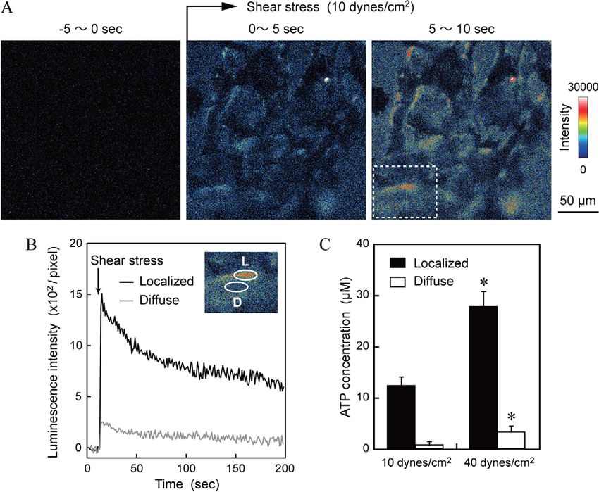
3478      Journal of Cell Science 124 (20)

                          biotinylated cell surface with streptavidin, and an intensified
                          charge-coupled device (CCD) camera. This method allowed us to
                          visualize ATP release at the cell surface in real time and at high
                          resolution. Since caveolae have been implicated as plasma
                          membrane microdomains that sense or transduce altered shear
                          stress into biochemical signals, thereby regulating EC function
                          (Yu et al., 2006), we investigated the relationship between ATP
                          release and caveolae by immunostaining with an antibody against
                          caveolin-1, the primary structural protein of caveolae. In
                          addition, to investigate the role of ATP release in shear stress
                          Ca2+ signaling we performed Ca2+ imaging by using a fluorescent
                          probe Fluo-4 in the same cells as examined by ATP imaging.

                          Results
                          Distribution and ATP sensitivity of cell-surface-
                          attached luciferase
                          The distribution of cell-surface-attached luciferase on cells
                          immunostained with a FITC-labeled antibody against luciferase
                          was examined with a confocal laser scanning microscope. En-
                          face images showed an even and dense distribution of biotin–
                          luciferase over the entire cell surface, and longitudinally cut
                          images showed that the biotin–luciferase was localized uniformly
                          on the apical cell membrane (Fig. 1A). Application of shear
Journal of Cell Science

                          stress caused no significant change in the distribution of cell-
                          surface-attached luciferase, indicating that this method can be
                          used even under flow conditions.
                                                                                                Fig. 1. Distribution and ATP sensitivity of luciferase attached to the cell
                             To assess the ability of cell-surface-attached luciferase to       surface. (A) Confocal laser scanning photomicrographs of luciferase-labeled
                          detect ATP, various concentrations of ATP were added to               HPAECs immunostained with a FITC-labeled antibody to luciferase. En-face
                          luciferase-labeled human pulmonary artery ECs (HPAECs), and           images of the apical cell membrane show an even and dense distribution of
                          luminescence emitted as a result of the ATP-triggered luciferase–     biotin–luciferase over the entire cell surface under static conditions (Static),
                          luciferin reaction was measured with a CCD camera. The                and longitudinal images cut at the dashed lines depict the biotin–lucifease
                          intensity of the luminescence in response to the addition of ATP      localized to the apical cell membrane. Application of shear stress (15 dynes/
                          increased in a concentration-dependent manner, and there was a        cm2) for 10 minutes caused no change in the distribution of luciferase.
                                                                                                (B) ATP sensitivity of cell-surface-attached luciferase. Increasing
                          clear correlation between the luminescence intensity and ATP
                                                                                                concentrations of ATP were added to luciferase-labeled HPAECs and the
                          concentration (Fig. 1B). These findings indicate that this method     luminescence emitted was measured with a CCD camera. Luminescence
                          of ATP detection allows to determine quantitatively the               increased as the ATP concentration increased. The inset shows a correlation
                          extracellular ATP concentrations at the cell surface.                 between luminescence intensity and the ATP concentration. Values are
                                                                                                means¡s.d. of data obtained from three separate HPAEC cultures.
                          Visualization of shear-stress-induced ATP release by ECs
                          Luminescent ATP signals at the cell surface were monitored with       ATP release that was almost spatiotemporally identical to that of
                          a CCD camera before and after shear stress application, and the       the initial stimulation (data not shown).
                          signals were transformed into pseudo-color images. As soon as
                          the cells were exposed to shear stress, ATP was released from the     Localized ATP release occurs in caveolin-1-rich regions of
                          entire surface of the cell membrane that was monitored, and           the cell edge
                          the ATP signals were particularly strong at localized regions at      To examine the relationship between the regions of localized
                          the edge of the cell (Fig. 2A), thereby indicating the existence of   ATP release and caveolae (cholesterol-rich plasma membrane
                          two distinct manners of ATP release, a diffuse manner and a           microdomains) after visualizing ATP release, cells were
                          highly concentrated, localized manner.                                immunostained with an antibody to caveolin-1, a marker
                             The temporal changes in ATP signals were quantified in the         protein for caveolae. Caveolin-1 was unevenly distributed over
                          regions of diffuse ATP release and the regions of localized ATP       the cell surface and was concentrated at specific parts of the cell
                          release (Fig. 2B). ATP release began simultaneously in both           edge. Comparison between the sites of ATP release and caveolin-
                          regions, but the peak of the ATP signal in both regions was           1 distribution revealed that the localized ATP release occurred in
                          markedly different. The ATP concentration estimated from the          the caveolin-1-rich cell edge regions (Fig. 3).
                          luminescence intensity reached more than 10 mM in the regions            To investigate the role of caveolae in shear-stress-induced ATP
                          of localized ATP release, but it remained below 1 mM in the           release, we used small interfering RNA (siRNA) in order to
                          regions of diffuse ATP release (Fig. 2C). The ATP signal in both      specifically knockdown the expression of caveolin-1. In clear
                          regions increased further when the shear stress was raised from       contrast to the control cells, which had been subjected to
                          10 dynes/cm2 to 40 dynes/cm2, indicating that the amount of           transfection conditions alone, marked suppression of localized
                          ATP release is shear-stress dependent (Note: 1 dyne510 mN).           ATP release was observed in the HPAECs transfected with
                          Secondary application of shear stress to the same cells induced an    caveolin-1 siRNA (Fig. 4A,B). Caveolin-1 siRNA had no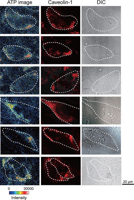
Visualization of endothelial ATP release                          3479
Journal of Cell Science

                          Fig. 2. Visualization of ATP release in response to shear stress. (A) Sequential pseudo-color images of shear-stress-induced ATP release. Luciferase-labeled
                          HPAECs were exposed to shear stress (10 dynes/cm2) for 3 seconds and changes in the ATP signal at the cell surface were recorded in real time. Each pseudo-
                          color image was created by integrating the luminescence for 5 seconds. Relatively low concentrations of ATP were released diffusely from the entire surface of
                          the cells; at the same time highly concentrated ATP release occurred at localized regions at the edge of the cell. (B) Comparison between localized ATP release
                          and diffuse ATP release. The temporal changes in ATP signal were quantified by placing regions of interest on a region of localized ATP release (L) and a region
                          of diffuse ATP release (D). The start of ATP release after shear stress application was identical in both regions, but the peak ATP signal in the localized release
                          region was substantially larger than in the diffuse release region. It should be noted that luminescence decays slowly because the PicaGene reagent contains
                          coenzyme A as a substrate of luciferase in addition to luciferin. (C) Shear-stress-dependency of ATP release. The amount of ATP that was released increased
                          further when shear stress was increased from 10 dynes/cm2 to 40 dynes/cm2. The ATP concentration was determined from the correlation with the intensity of
                          luminescence as shown in Fig.1B. Values are means¡s.d. of 30 cells in three separate experiments. *P,0.01 10 dynes/cm2 vs 40 dynes/cm2.

                          significant effect on diffuse ATP release. Next, we treated                         localized ATP release coincided exactly with the initiation sites
                          HPAECs with methyl-b-cyclodextrin (MbCD), which disrupts                            of Ca2+ waves. Comparison between the start of localized ATP
                          caveolae and lipid rafts by depleting plasma-membrane                               release and increase in [Ca2+]i showed that ATP release always
                          cholesterol. Treatment with MbCD significantly inhibited                            preceded the increase in [Ca2+]i. Treatment of HPAECs with
                          shear-stress-induced localized ATP release but did not have a                       angiostatin, a known blocker of ATP release, almost completely
                          significant effect on diffuse ATP release (Fig. 4A,B). The                          abolished both the shear-stress-induced ATP release and the
                          inhibitory effect of MbCD on localized ATP release was                              increase in [Ca2+]i (Fig. 5B). These results suggest that the
                          partially prevented by pretreatment with cholesterol. These                         localized release of ATP at caveolae triggers the increase in
                          findings suggest that caveolae are involved in shear-stress-                        [Ca2+]i by activating nearby purinoceptors.
                          induced, localized ATP release.
                                                                                                              Discussion
                          Colocalization of localized ATP release and the                                     The novel ATP imaging method described here clearly
                          subsequent initiation of Ca2+ waves                                                 demonstrated that HPAECs release ATP in response to shear
                          Luciferase-labeled HPAECs were exposed to shear stress and                          stress in two distinct manners; i.e. a highly concentrated,
                          examined for changes in intracellular Ca2+ concentration                            localized manner and a diffuse manner. A variety of methods
                          ([Ca2+]i) by using the Ca2+ indicator Fluo-4 and a fluorescence                     can be used to detect ATP that is released at the surface of living
                          microscope. Shear stress evoked a rapid increase in [Ca2+]i that                    cells, including biosensor techniques (Bell et al., 2003; De Proost
                          started at a single site in the cell and propagated throughout the                  et al., 2009; Hayashi et al., 2004; Hazama et al., 1998; Llaudet et
                          entire cell in the form of a Ca2+ wave (Fig. 5A). The Ca2+ wave                     al., 2005; Schneider et al., 1999) and methods that induce
                          also propagated into the cell nucleus. After Ca2+ imaging, ATP                      luminescence or measure fluorescence by using ATP-sensitive
                          imaging was performed on the same cells. The regions of                             proteins added to the extracellular space (Arcuino et al., 2002;
3480        Journal of Cell Science 124 (20)

                                                                                                             the protein marker caveolin-1 and are known to play crucial roles
                                                                                                             in multiple signal transduction events at the surface of various
                                                                                                             cell types (Shaul and Anderson, 1998). Recent studies have
                                                                                                             demonstrated that caveolae sense or transduce shear stress into
                                                                                                             biochemical signals that regulate EC functions. Shear stress has
                                                                                                             been found to activate extracellular-signal-regulated kinase
                                                                                                             (ERK) in bovine aortic ECs that was blocked by the delivery
                                                                                                             of polyclonal caveolin-1 antibody into the cells (Park et al.,
                                                                                                             2000). The production of a potent vasodilator, nitric oxide (NO),
                                                                                                             by ECs increases in response to shear stress, and one of the
                                                                                                             mechanisms of shear-stress-induced NO production is
                                                                                                             dissociation of endothelial NO synthase (eNOS) from caveolae
                                                                                                             followed by activation of eNOS (Rizzo et al., 1998). A more
                                                                                                             recent study comparing caveolin-1 knockout mice with wild-type
                                                                                                             mice showed that lack of caveolin-1 impaired blood-flow-
                                                                                                             dependent eNOS activation, vasodilation, and vascular
                                                                                                             remodeling and that these abnormalities were rescued by
                                                                                                             reconstituting caveolin-1 into the vascular endothelium of the
                                                                                                             knockout mice (Yu et al., 2006). The present study revealed that
                                                                                                             localized ATP release induced by exposing HPAECs to shear
                                                                                                             stress occurs in caveolin-1-rich regions of the plasma membrane.
                                                                                                             The localized ATP release was markedly suppressed by
                                                                                                             knockdown of caveolin-1 expression with siRNA and by
                                                                                                             depletion of membrane cholesterol with MbCD, which disrupts
Journal of Cell Science

                                                                                                             caveolae/lipid rafts. Although the possibility remains that the
                                                                                                             inhibition of the localized ATP release by caveolin-1 siRNA and
                                                                                                             MbCD is attributable to secondary rather than primary effects,
                                                                                                             these findings suggest that caveolae are involved in the localized
                                                                                                             ATP release that occurs in response to shear stress.
                                                                                                                It remains unclear why the localized ATP release occurs
                          Fig. 3. Comparison between the regions of localized ATP release and
                          caveolin-1 distribution. ATP image, pseudo-color images of shear-stress-
                                                                                                             preferentially at caveolin-1-rich regions of the cell membrane.
                          induced ATP release. Each image was created by integrating luminescence            Since shear stress causes microscale deformation/displacement of
                          for 5 seconds. Caveolin-1, fluorescent photomicrographs of caveolin-1. DIC,        cell surface membrane proteins, the lipid bilayer itself, and the
                          differential interference contrast (DIC) images. Broken lines represent the cell   cytoskeleton and connected proteins, it seems likely that the sites
                          outlines obtained from the DIC images. Six pairs of images demonstrate that        of shear-stress-induced deformation are linked to the localization
                          the regions of localized ATP release coincide with the caveolin-1-rich regions     of ATP release. Caveolae/lipid raft microdomains are
                          at the cell edge. These six sets of images were selected because they are          characterized by a unique lipid composition that contains high
                          representative of dozens of images, all of which showed similar results.           concentrations of cholesterol and sphingolipids in places where
                                                                                                             the lipid bilayer is more rigid and lipid movement is more
                          Corriden et al., 2007; Wang et al., 2000) or targeted to the plasma                restricted than in other parts of the membrane (Gaus et al., 2003).
                          membrane (Beigi et al., 1999; Joseph et al., 2003; Okada et al.,                   These differences in the physical properties of the membrane
                          2006; Pellegatti et al., 2005). However, some of these methods                     may be responsible for the localization of ATP release. On the
                          provide only semi-quantitative information on ATP release,                         other hand, caveolae/lipid rafts are closely associated with
                          others are unsuitable for the visualization of ATP release, mainly                 cytoskeleton filaments, and shear stress may affect the caveolae
                          because of weak signals. In our study, however, we were able to                    and associated molecules through cytoskeleton networks. Helmke
                          visualize ATP release of ECs by using a cell-surface-targeting                     et al. showed that strain in the intermediate filament was
                          luciferase and a high-resolution CCD camera. To obtain stronger                    heterogeneous, with small regions of high-strain concentrations
                          signals, we generated a biotin–luciferase fusion protein that can                  located at the cell periphery and at several sites in the cell
                          attach to biotinylated cell surfaces through interaction with                      interior, and that the hot spots of strain concentration were
                          streptavidin (Nakamura et al., 2006). Since various plasma                         repositioned by shear stress (Helmke et al., 2003). These hot
                          membrane proteins can bind to many biotins and one streptavidin                    spots of cytoskeletal strain may coincide with the locations of
                          molecule has four biotin-binding sites, a large amount of                          ATP release. It requires further research to clarify whether the
                          luciferase can be bound to the cell surface. In addition, by                       localization of ATP release is a plasma membrane domain
                          using the PicaGene reagent, which emits several times more                         phenomenon or influenced by interference with cytoskeleton
                          luminescence than other luciferase–luciferin reaction reagents, it                 function or both.
                          became possible to obtain luminescence strong enough for high-                        Depending on the cell type and extrinsic stimulus, ATP is
                          resolution imaging of ATP release. This imaging method should                      released into the extracellular space by ATP-permeable
                          prove useful for studying ATP release mechanisms and the                           membrane channels, including connexin hemichannels (Stout et
                          functional roles of ATP in various cell types.                                     al., 2002) and volume-regulated anion channels (Sabirov and
                             Caveolae are small flask-shaped invaginations of cholesterol-                   Okada, 2004), by diffusion facilitated by ATP-binding cassette
                          rich cell membranes. They are characterized by the presence of                     transporters, such as the cystic fibrosis transmembrane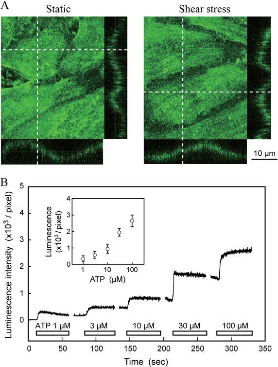
Visualization of endothelial ATP release                              3481

                          Fig. 4. Involvement of caveolae in shear-stress-induced ATP release. (A) Pseudo-color images of ATP release obtained 5–10 seconds after application of shear
                          stress (10 dynes/cm2 for 3 seconds). Each image was created by integrating luminescence for 5 seconds. Control, control luciferase-labeled HPAECs. Caveolin-1
                          siRNA, cells transfected with caveolin-1 siRNA. MbCD, the cells treated with 10 mM methyl-b-cyclodextrin. MbCD+Cholesterol, the cells pretreated with
                          1.3 mM cholesterol and then with 10 mM MbCD. Caveolin-1 knockdown with siRNA and disruption of caveolae and lipid rafts with MbCD markedly suppressed
Journal of Cell Science

                          the shear-stress-induced localized ATP release. Pretreatment with cholesterol partially prevented the effect of MbCD. (B) Quantitative analysis of shear-stress-
                          induced ATP release. Luminescence intensity was measured separately in the region of diffuse ATP release and the region of localized ATP release by setting an
                          region of interest over the cell nucleus and over the site in the cell periphery where the intensity was highest. The data are expressed as means¡s.d. (n530).
                          *P,0.01 vs control. Localized ATP release was significantly suppressed by caveolin-1 siRNA and MbCD, but diffuse ATP release was not, suggesting that
                          caveolae are involved in the shear-stress-induced localized ATP release.

                          conductance regulator (Schwiebert et al., 1999), or by vesicular                   adhesion molecule-1, the cytoskeleton, the glycocalyx, and primary
                          transport and exocytotic secretion (Bodin and Burnstock, 2001).                    cilia (Ando and Yamamoto, 2009; Davies, 1995). However, it is
                          Our previous study revealed that ATP synthase is localized in                      unclear how these molecules and microdomains enable ECs to sense
                          caveolae and involved in shear-stress-induced ATP release by                       shear stress and transmit the signal to downstream effectors to allow
                          HPAECs (Yamamoto et al., 2007). However, it remained unclear                       cells to respond. Our previous studies in ECs demonstrated that the
                          whether caveolar ATP synthase is involved in any of the above-                     P2X4 receptor contributes to shear stress mechanotransduction
                          mentioned ATP-releasing pathways or in an unknown pathway.                         through Ca2+ signaling, which plays a crucial role in the control of
                          In view of the discovery of the two different manners of                           vascular function in vivo (Yamamoto et al., 2000a; Yamamoto et al.,
                          ATP release in this study, HPAECs may use dual pathways to                         2003). Clarification of the mechanisms of ATP release as an early
                          release ATP in response to shear stress. Further study will be                     response to shear stress should lead to a better understanding of the
                          needed to identify the pathway responsible for each manner of                      mechanotransduction of shear stress.
                          ATP release.
                             In the present study we performed intracellular Ca2+ imaging and                Materials and Methods
                                                                                                             Cell culture
                          ATP imaging of the same cells. Shear stress evoked an increase in                  Human pulmonary artery endothelial cells (HPAECs) were obtained from
                          [Ca2+]i that originated at a specific site and propagated throughout               Clonetics and grown on a 1% gelatin-coated tissue culture flask in M199
                          the entire cell in the form of a Ca2+ wave in a manner that resembled              supplemented with 15% FBS, 2 mM L-glutamine (Gibco), 50 mg/ml heparin, and
                          to the Ca2+ responses previously observed in bovine fetal aortic ECs               30 mg/ml EC growth factor (Becton Dickinson). The cells used in the present
                                                                                                             experiments were in the 7th and 10th passage.
                          (Isshiki et al., 1998). The sites where the increase in [Ca2+]i
                          originated coincided with the sites of localized ATP release.                      Production and purification of biotin–luciferase
                          Comparisons between the start of the [Ca2+]i increase and ATP                      Biotin–luciferase protein was produced and purified as previously reported
                          release revealed that ATP release always preceded the Ca2+                         (Nakamura et al., 2006). Briefly, biotin acceptor peptide (BAP) was fused to
                                                                                                             thermostabilized firefly luciferase, and the biotin–luciferase gene fusion plasmid,
                          increase. The ATP concentration at the sites of localized ATP                      pET-NHis-BAP-Luc, was constructed. For purification, six repeats of the histidine
                          release reached more than 10 mM, which is sufficient to activate                   sequence (His-tag) were coded to the N terminus of the fusion gene. The pET-
                          purinoceptors. Thus, it seems that shear stress first triggers ATP                 NHis-BAP-Luc plasmid was transduced into Escherichia coli BL21 (DE3)
                                                                                                             competent cells and the cells were grown and lyzed by sonication. The soluble
                          release, which activates nearby P2X and/or P2Y receptors, and, in                  fraction of the bacterial lysates was subjected to metal-ion affinity chromatography
                          turn, leads to Ca2+ responses. Many studies have been devoted to                   in order to purify biotin–luciferase protein by using the His-tag.
                          investigation of shear stress mechanotransduction and have
                          demonstrated the involvement of various membrane molecules                         Cell-surface labeling with biotin–luciferase
                          and cellular microdomains in its mechanisms, including ion                         HPAECs cultured on coverslips were incubated with 250 mg/ml EZ-Link2 Sulfo-
                                                                                                             NHS-Biotin (Thermo Scientific Pierce, Rockford, IL) for 10 minutes at room
                          channels, growth factor receptors, G proteins, caveolae, adhesion                  temperature. After washing with HBSS, cells were treated with 2 mM streptavidin
                          proteins such as integrin, VE-cadherin, and platelet endothelial cell              (Wako) for 30 minutes at 37 ˚C and then, after another HBSS wash, incubated with
3482 Journal of Cell Science 124 (20)
Journal of Cell Science
Fig. 5. Colocalization of sites of localized ATP release and the subsequent initiation of Ca2+ waves. (A) Pseudo-color images of shear-stress-induced ATP
release and Ca2+ responses. Ca2+ imaging with Fluo-4 showed that shear stress evoked an increase in intracellular Ca2+ concentrations ([Ca2+]i) that started at a single
site and propagated throughout the entire cell in the form of a Ca2+ wave. The first Ca2+ image was obtained 1 second after application of shear stress, the rest of the
images shown were captured at intervals of 140 mseconds. ATP imaging was performed after the Ca2+ imaging of the same cells. Each ATP image was created by
integrating luminescence for 1 second after the application of shear stress. Broken lines represent the cell outlines obtained from the DIC images. Five pairs of images
demonstrate colocalization of the localized ATP release and sites of initiation of the Ca2+ waves. Quantitative analysis of the rising phase of ATP release and [Ca2+]i
increase showed that ATP release always preceded the [Ca2+]i increase. Values of [Ca2+]i are expressed as a ratio of Fluo-4 fluorescence (DF:F0) to the control before
application of shear stress. Open rectangles indicate the duration of shear stress (10 dynes/cm2 for 3 seconds), and the left end of each rectangle corresponds to time
zero. These five sets of images were selected because they are representative of dozens of images, all of which showed similar results. (B) Role of ATP release in the
shear-stress-induced [Ca2+]i increase. Treatment of cells with angiostatin, a known blocker of ATP release, abolished the ATP release and [Ca2+]i increase, and both
were restored by removing the angiostatin (by flushing). This suggests that the ATP release triggered the Ca2+ increase. Values are means¡s.d. of data obtained from
24 cells in three separate experiments. *P,0.01 angiostatin vs after flushing.
1 mg/ml biotin–luciferase for 30 minutes at room temperature. To observe the Shear-stress stimulation and ATP imaging
distribution of biotin–luciferase on the cell surface, cells were immunostained with A coverslip on which luciferase-labeled cells had been cultured was placed in a
FITC-conjugated anti-luciferase antibody (10 mg/ml, Rockland). parallel-plate flow chamber whose temperature can be controlled (FCS2,Visualization of endothelial ATP release 3483
Bioptechs). The flow chamber was then placed on the stage of an inverted Corriden, R., Insel, P. A. and Junger, W. G. (2007). A novel method using
microscope (Diophot 300, Nikon) and connected to a syringe pump (PHD2000; fluorescence microscopy for real-time assessment of ATP release from individual
Harvard Apparatus, Holliston, MA) with a Tygon2 tube. The ATP-free PicaGene cells. Am. J. Physiol. Cell Physiol. 293, C1420-C1425.
reagent (PGL5500; Toyo B-Net, Tokyo, Japan) was perfused at constant flow rate Davies, P. F. (1995). Flow-mediated endothelial mechanotransduction. Physiol. Rev. 75,
through the chamber as a luciferase substrate solution at 37 ˚C. The PicaGene 519-560.
contains 470 mM luciferin, 270 mM coenzyme A, 33.3 mM DTT, 2.67 mM De Proost, I., Pintelon, I., Wilkinson, W. J., Goethals, S., Brouns, I., Van Nassauw,
MgSO4, 1.07 mM (MgCO3)4Mg(OH)2/5H2O, 20 mM Tricine, 1.4 mM KH2PO4, L., Riccardi, D., Timmermans, J. P., Kemp, P. J. and Adriaensen, D. (2009).
Purinergic signaling in the pulmonary neuroepithelial body microenvironment
4.3 mM Na2HPO4, 2.7 mM KCl, and 137 mM NaCl. The intensity of shear stress
unraveled by live cell imaging. Faseb. J. 23, 1153-1160.
(t, dynes/cm2) was calculated by using the equation t56 mQ/a2b, where m is the
Gaus, K., Gratton, E., Kable, E. P., Jones, A. S., Gelissen, I., Kritharides, L. and
viscosity of the perfusate (poise), Q is the flow volume (ml/second), and a and b Jessup, W. (2003). Visualizing lipid structure and raft domains in living cells with
are the cross-sectional dimensions of the flow path. The shear stress used in this two-photon microscopy. Proc. Natl. Acad. Sci. USA. 100, 15554-15559.
study ranged from 10–40 dynes/cm2. Hayashi, S., Hazama, A., Dutta, A. K., Sabirov, R. Z. and Okada, Y. (2004).
Luminescence emitted as a result of the ATP-triggered, luciferase-aided Detecting ATP release by a biosensor method. Sci. STKE. 2004, pl14.
breakdown of luciferin was detected through a water immersion objective (Fluor Hazama, A., Hayashi, S. and Okada, Y. (1998). Cell surface measurements of ATP
40, 1.15 NA, Nikon) with a water-cooling electron multiplier CCD camera release from single pancreatic beta cells using a novel biosensor technique. Pflugers
(ImagEM C9100-13, Hamamatsu), extracellular ATP levels at the cell surface Arch. 437, 31-35.
were visualized by using the Aquacosmos software program (Hamamatsu). ATP Helmke, B. P., Rosen, A. B. and Davies, P. F. (2003). Mapping mechanical strain of an
images were acquired sequentially with an exposure period of 100 mseconds as endogenous cytoskeletal network in living endothelial cells. Biophys. J. 84, 2691-2699.
full-frame images (5126512 pixels). Isshiki, M., Ando, J., Korenaga, R., Kogo, H., Fujimoto, T., Fujita, T. and Kamiya,
A. (1998). Endothelial Ca2+ waves preferentially originate at specific loci in caveolin-
rich cell edges. Proc. Natl. Acad. Sci. USA. 95, 5009-5014.
Immunohistochemistry
Joseph, S. M., Buchakjian, M. R. and Dubyak, G. R. (2003). Colocalization of ATP
Cells were fixed with 4% paraformaldehyde (Sigma) and maintained in 1% normal
release sites and ecto-ATPase activity at the extracellular surface of human astrocytes.
bovine serum albumin (Sigma) to block nonspecific protein-binding sites. The J. Biol. Chem. 278, 23331-23342.
cells were incubated with rabbit anti-caveolin-1 polyclonal antibody (Transduction Khakh, B. S. and Burnstock, G. (2009). The double life of ATP. Sci. Am. 301, 84-90, 92.
Laboratories) and, after washing, they were incubated with Alexa-Fluor-594- Llaudet, E., Hatz, S., Droniou, M. and Dale, N. (2005). Microelectrode biosensor for
labeled goat anti-rabbit IgG (Molecular Probes) at a 1:500 dilution. Stained cells real-time measurement of ATP in biological tissue. Anal. Chem. 77, 3267-3273.
were photographed through a confocal fluorescence microscope (Leica), and all Milner, P., Kirkpatrick, K. A., Ralevic, V., Toothill, V., Pearson, J. and Burnstock, G.
images were imported into Adobe Photoshop as TIF files for figure assembly. (1990). Endothelial cells cultured from human umbilical vein release ATP, substance P
and acetylcholine in response to increased flow. Proc. Biol. Sci. 241, 245-248.
Ca2+ measurement Nakamura, M., Mie, M., Funabashi, H., Yamamoto, K., Ando, J. and Kobatake, E.
Journal of Cell Science
The same cells were subjected to ATP imaging and Ca2+ imaging. Cells that had (2006). Cell-surface-localized ATP detection with immobilized firefly luciferase.
been labeled with biotin–luciferase were loaded with the Ca2+-sensitive dye Fluo- Anal. Biochem. 352, 61-67.
4-acetoxymethyl ester (5 mM; Dojindo, Kumamoto, Japan) and placed in the FCS3 Okada, S. F., Nicholas, R. A., Kreda, S. M., Lazarowski, E. R. and Boucher, R. C.
(2006). Physiological regulation of ATP release at the apical surface of human airway
flow chamber (Bioptechs) on the stage of an upright microscope system (BX51WI,
epithelia. J. Biol. Chem. 281, 22992-23002.
Olympus) equipped with a water immersion objective (Olympus XL Plan N25X,
Park, H., Go, Y. M., Darji, R., Choi, J. W., Lisanti, M. P., Maland, M. C. and Jo, H.
1.05 NA). Fluo-4 was excited by light that passed through a 490 nm band-pass (2000). Caveolin-1 regulates shear stress-dependent activation of extracellular signal-
filter (Lambda DG-4), the emitted light was guided through a 510 nm band-pass regulated kinase. Am. J. Physiol. Heart Circ. Physiol. 278, H1285-H1293.
filter to an image intensifier (C8600-04, Hamamatsu) connected to a water-cooling Pellegatti, P., Falzoni, S., Pinton, P., Rizzuto, R. and Di Virgilio, F. (2005). A novel
electron multiplier CCD camera (Cascade 512, Roper). Fluorescence intensity is a recombinant plasma membrane-targeted luciferase reveals a new pathway for ATP
reflection of the intracellular Ca2+ concentration ([Ca2+]i). Ca2+ images were secretion. Mol. Biol. Cell. 16, 3659-3665.
acquired sequentially with an exposure period of 30 mseconds as full-frame Rizzo, V., McIntosh, D. P., Oh, P. and Schnitzer, J. E. (1998). In situ flow activates
images (5126512 pixels). The ATP and Ca2+ images were analyzed by using endothelial nitric oxide synthase in luminal caveolae of endothelium with rapid
MetaMorpf software program version 7.7 (Molecular Devices). caveolin dissociation and calmodulin association. J. Biol. Chem. 273, 34724-34729.
Sabirov, R. Z. and Okada, Y. (2004). Wide nanoscopic pore of maxi-anion channel
Statistical analysis suits its function as an ATP-conductive pathway. Biophys. J. 87, 1672-1685.
All results are expressed as the mean¡s.d. Statistical significance was evaluated Schneider, S. W., Egan, M. E., Jena, B. P., Guggino, W. B., Oberleithner, H. and
Geibel, J. P. (1999). Continuous detection of extracellular ATP on living cells by
by ANOVA and Bonferonni adjustments applied to the results of Student’s t-test
using atomic force microscopy. Proc. Natl. Acad. Sci. USA. 96, 12180-12185.
performed with SPSS software (SPSS Inc). Values of P,0.01 were regarded as
Schwiebert, E. M., Benos, D. J., Egan, M. E., Stutts, M. J. and Guggino, W. B.
statistically significant. (1999). CFTR is a conductance regulator as well as a chloride channel. Physiol. Rev.
79, S145-S166.
Acknowledgements Shaul, P. W. and Anderson, R. G. (1998). Role of plasmalemmal caveolae in signal
transduction. Am. J. Physiol. 275, L843-L851.
We acknowledge Akira Kamiya for his invaluable support in our
Stout, C. E., Costantin, J. L., Naus, C. C. and Charles, A. C. (2002). Intercellular
work and thank Yuko Sawada for technical assistance. calcium signaling in astrocytes via ATP release through connexin hemichannels. J.
Biol. Chem. 277, 10482-10488.
Funding Wang, Z., Haydon, P. G. and Yeung, E. S. (2000). Direct observation of calcium-
independent intercellular ATP signaling in astrocytes. Anal. Chem. 72, 2001-2007.
This work was partly supported by Grants-in-Aid for Scientific Yamamoto, K., Korenaga, R., Kamiya, A. and Ando, J. (2000a). Fluid shear stress
Research (Grant numbers S21220011 and B22300150) from the activates Ca2+ influx into human endothelial cells via P2X4 purinoceptors. Circ. Res.
Ministry of Education, Culture, Sports, Science and Technology to 87, 385-391.
J.A. and K.Y, and Grant-in-Aid from the Japan Science and Yamamoto, K., Korenaga, R., Kamiya, A., Qi, Z., Sokabe, M. and Ando, J. (2000b).
P2X4 receptors mediate ATP-induced calcium influx in human vascular endothelial
Technology Agency to K.Y. (Grant number 7815). cells. Am. J. Physiol. Heart Circ. Physiol. 279, H285-H292.
Yamamoto, K., Sokabe, T., Ohura, N., Nakatsuka, H., Kamiya, A. and Ando, J.
References (2003). Endogenously released ATP mediates shear stress-induced Ca2+ influx into
Ando, J. and Yamamoto, K. (2009). Vascular mechanobiology: endothelial cell pulmonary artery endothelial cells. Am. J. Physiol. Heart Circ. Physiol. 285, H793-
responses to fluid shear stress. Circ. J. 73, 1983-1992. H803.
Arcuino, G., Lin, J. H., Takano, T., Liu, C., Jiang, L., Gao, Q., Kang, J. and Yamamoto, K., Sokabe, T., Matsumoto, T., Yoshimura, K., Shibata, M., Ohura, N.,
Nedergaard, M. (2002). Intercellular calcium signaling mediated by point-source Fukuda, T., Sato, T., Sekine, K., Kato, S. et al. (2006). Impaired flow-dependent control
burst release of ATP. Proc. Natl. Acad. Sci. USA. 99, 9840-9845. of vascular tone and remodeling in P2X4-deficient mice. Nat. Med. 12, 133-137.
Beigi, R., Kobatake, E., Aizawa, M. and Dubyak, G. R. (1999). Detection of local Yamamoto, K., Shimizu, N., Obi, S., Kumagaya, S., Taketani, Y., Kamiya, A. and Ando,
ATP release from activated platelets using cell surface-attached firefly luciferase. Am. J. (2007). Involvement of cell surface ATP synthase in flow-induced ATP release by
J. Physiol. 276, C267-C278. vascular endothelial cells. Am. J. Physiol. Heart Circ. Physiol. 293, H1646-H1653.
Bell, P. D., Lapointe, J. Y., Sabirov, R., Hayashi, S., Peti-Peterdi, J., Manabe, K., Yegutkin, G. G. (2008). Nucleotide- and nucleoside-converting ectoenzymes: Important
Kovacs, G. and Okada, Y. (2003). Macula densa cell signaling involves ATP release modulators of purinergic signalling cascade. Biochem. Biophys. Acta. 1783, 673-694.
through a maxi anion channel. Proc. Natl. Acad. Sci. USA. 100, 4322-4327. Yu, J., Bergaya, S., Murata, T., Alp, I. F., Bauer, M. P., Lin, M. I., Drab, M.,
Bodin, P. and Burnstock, G. (2001). Evidence that release of adenosine triphosphate Kurzchalia, T. V., Stan, R. V. and Sessa, W. C. (2006). Direct evidence for the role
from endothelial cells during increased shear stress is vesicular. J. Cardiovasc. of caveolin-1 and caveolae in mechanotransduction and remodeling of blood vessels.
Pharmacol. 38, 900-908. J. Clin. Invest. 116, 1284-1291.You can also read