MIR-103A PROMOTES TUMOUR GROWTH AND INFLUENCES GLUCOSE METABOLISM IN HEPATOCELLULAR CARCINOMA - NATURE
←
→
Page content transcription
If your browser does not render page correctly, please read the page content below
www.nature.com/cddis

ARTICLE                 OPEN

MiR-103a promotes tumour growth and influences glucose
metabolism in hepatocellular carcinoma
Yuling Liu1,3, Yuanzhou Zhang1,3, Bowen Xiao1,3, Ning Tang1,3, Jingying Hu1, Shunshun Liang1, Yechun Pang1, Huili Xu1, Junping Ao1,
                                                     ✉                    ✉
Juan Yang1, Xiaofei Liang1, Lin Wei1, Yunfeng Wang2 and Xiaoying Luo1

© The Author(s) 2021

    Hepatocellular carcinoma (HCC) is a common and high-mortality cancer worldwide. Numerous microRNAs have crucial roles in the
    progression of different cancers. However, identifying the important microRNAs and the target biological function of the
    microRNA in HCC progression is difficult. In this study, we selected highly expressed microRNAs with different read counts as
    candidate microRNAs and then tested whether the microRNAs were differentially expressed in HCC tumour tissues, and we found
    that their expression was related to the HCC prognosis. Then, we investigated the effects of microRNAs on the cell growth and
    mobility of HCC using a real-time cell analyser (RTCA), colony formation assay and subcutaneous xenograft models. We further
    used deep-sequencing technology and bioinformatic analyses to evaluate the main functions of the microRNAs. We found that
    miR-103a was one of the most highly expressed microRNAs in HCC tissues and that it was upregulated in HCC tissue compared
    with the controls. In addition, high miR-103a expression was associated with poor patient prognosis, and its overexpression
    promoted HCC cell growth and mobility. A functional enrichment analysis showed that miR-103a mainly promoted glucose
    metabolism and inhibited cell death. We validated this analysis, and the data showed that miR-103a promoted glucose
    metabolism-likely function and directly inhibited cell death via ATP11A and EIF5. Therefore, our study revealed that miR-103a may
    act as a key mediator in HCC progression.

    Cell Death and Disease (2021)12:618 ; https://doi.org/10.1038/s41419-021-03905-3

INTRODUCTION                                                                             signalling pathway [21]. MiR-541 potentiates the response of
Hepatocellular carcinoma (HCC) is one of the most common                                 human HCC to sorafenib treatment by inhibiting autophagy [22].
cancers worldwide [1] and remains the third leading cause of                             A recent study also indicated that miR-1269b downregulated
cancer mortality [2–5]. Although great improvements have been                            SVEP1 expression and promoted HCC proliferation and metastasis,
made in diagnostic and surgical techniques and the development                           likely through the PI3K/Akt signalling pathway [23]. In addition,
of new molecularly targeted drugs, the 5-year overall survival rate                      previous studies have shown that many abnormally expressed
is still low [4]. In addition, some patients progress to advanced                        microRNAs are closely associated with the prognosis of HCC
stages of the disease due to recurrence and metastasis [6, 7].                           patients [6, 24, 25]. However, these analyses are mainly based on
Therefore, it is necessary to gain a full understanding of the                           the results of the differential expression between tumour tissues
molecular mechanisms underlying the tumorigenesis and tumour                             and normal tissues, whereas the expression level of microRNA is
progression of HCC.                                                                      less frequently considered.
   MicroRNAs (miRNAs/miRs) are a class of small noncoding RNA                               The rapid development of high-throughput sequencing tech-
molecules that are ~22 nucleotides in length [8–10] and regulate                         nologies and comprehensive bioinformatics analyses allows for
gene expression and cellular processes by targeting messenger                            the analysis of important differentially regulated molecules in HCC
RNA (mRNA) transcripts [11]. MicroRNAs take part in many                                 progression and their major functions [26]. Although many studies
important biological processes, including early embryonic develop-                       have focused on differential molecular regulation in HCC
ment [12] and fat metabolism [13], and they even regulate the                            progression and its function [27, 28], the main regulatory factors
differentiation of stem cells [14]. However, the abnormal expression                     or the main nodes in the regulatory network remain issues that
of miRNAs is closely related to the occurrence and development of                        need to be considered. To address this challenge, we analysed
human cancer (cell proliferation [15], apoptosis [16], and cell death                    HCC microRNA expression data acquired from The Cancer
[17]). MicroRNAs have been shown to play crucial roles in the                            Genome Atlas (TCGA) database. We screened for differentially
development and progression of different kinds of cancer, including                      expressed microRNAs (DEMs) in tumours vs adjacent normal
HCC [18–20]. For example, miR-29c-3p acts as a tumour suppressor                         tissues based on the expression level of microRNAs. We
in HCC by targeting DNMT3B and the LATS1-associated Hippo                                hypothesize that these high-expression DEMs have significant

1
 State Key Laboratory of Oncogenes and Related Genes, Shanghai Cancer Institute, Renji Hospital, Shanghai Jiao Tong University School of Medicine, Shanghai 200032, People’s
Republic of China. 2Department of General Surgery, Pudong New Area People’s Hospital, Shanghai University of Medicine & Health Sciences, Shanghai, China. 3These authors
contributed equally: Yuling Liu, Yuanzhou Zhang, Bowen Xiao, Ning Tang. Edited by E. Candi ✉email: wangyunfeng197911@163.com; luoxy@shsci.org

Received: 28 February 2021 Revised: 3 June 2021 Accepted: 7 June 2021

Official journal of CDDpress
Y. Liu et al.
                  2
                      functions in HCC progression, and investigations of these high-           promoted cell proliferation (Fig. 3A, p < 0.0001). Furthermore, the
                      expression DEMS may obtain information about the main factors             colony formation assay results suggested that miR-103a over-
                      or nodes in the regulatory network for HCC progression.                   expression remarkably promoted cell colony formation abilities
                         In the present study, we found that miR-103a is a highly               compared with the negative control groups (Fig. 3B–E), and
                      expressed and upregulated microRNA in HCC tissues and                     downregulated miR-103 expression in SMMC-7721 inhibited cell
                      showed that its high expression is associated with poor                   proliferation (Fig. S1B).
                      prognosis in patients with HCC. We further found that miR-103               These data showed that miR-103a promotes cell growth in vitro.
                      promotes cell proliferation, migration, invasion and glucose
                      metabolism in vitro and that miR-103a promotes tumour growth              miR-103a promotes HCC cell migration and invasion in vitro
                      in vivo. Glucose metabolism is the main function of miR-103a.             Next, we performed cell migration assays and invasion assays to
                      Our study suggested that high expression and upregulation of              study the influence of miR-103a on HCC mobility. The data
                      miR-103a are the main differentiation factors in the regulatory           showed that miR-103a overexpression obviously promoted HCC
                      network and indicated that the ability of miR-103a to promote             cell migration and invasion relative to the negative control
                      glucose metabolism function might represent the main change               groups (Fig. 4A, B). And downregulated miR-103 expression in
                      in HCC progression.                                                       SMMC-7721 inhibited cell invasion (Fig. S1C). These data
                                                                                                showed that miR-103a overexpression promotes the mobility
                                                                                                of HCC cells.
                      RESULTS
                      miR-103a is a highly expressed and upregulated microRNA in                miR-103a influences glucose metabolism in HCC cells
                      HCC tissues                                                               To obtain a more comprehensive functional analysis of miR-103,
                      We performed an expression analysis of publicly available                 we checked the microRNA expression profile of miR-103a-
                      microRNA sequencing data from HCC patients in the TCGA                    overexpressing MHCC97L cells by next-generation sequencing.
                      database. Then, microRNAs were sorted from high to low                    We analyzed the sequencing data by STAR-htseq workflows and
                      according to the read counts in HCC tissues. We identified 15              then obtained the gene functional enrichment information by the
                      top-ranked microRNAs (Fig. 1A), and 6 were upregulated while 9            EGSEA r package. In the biological function analysis, we found that
1234567890();,:

                      were downregulated (Fig. 1A, according to the read counts, not            the glucose metabolic process, cell death and glycolysis were the
                      normalized). Furthermore, we detected the microRNA proportion             top three biological functions (Fig. 5A), with 14 genes participating
                      compared to the total microRNA sequencing read counts in HCC              in these biological functions (Fig. 5B).
                      tissues, and the data showed that the miR-103a read counts were              To validate the gene functional enrichment information, we
                      1.458% of the total microRNA read counts (Fig. 1B). The above             assessed the effect of miR-103a overexpression on glycolytic
                      data showed that miR-103a was a high-expression microRNA. miR-            metabolism via a seahorse assay. The results showed that the
                      103a plays a vital role in physiological [29] and pathological            overexpression of miR-103a enhanced the extracellular acidifica-
                      processes [30]. Because of the limited studies of liver cancer, we        tion rate (ECAR), which reflects the overall glycolytic flux, and
                      chose this microRNA as a candidate.                                       decreased the OCR, which is an indicator of mitochondrial
                         High-expression/differentiated expression microRNAs might              respiration (Fig. 5C, D). And downregulated miR-103 expression
                      play an important role in HCC progression. To validate that miR-          in SMMC-7721 decreased the ECAR and enhanced the OCR
                      103a is a DEM in HCC, we checked the miR-103a expression in               (Fig. S1D/E).
                      TCGA datasets after normalizing the read counts. The data showed             These results revealed that miR-103a mainly influences glucose
                      that miR-103a expression was 1.6-fold upregulated in HCC tissues          metabolism in HCC.
                      (n = 372) compared with normal adjacent tissues (n = 50) (Fig. 1C,
                      p < 0.0001, unpaired). High expression of miR-103a was associated         ATP11A and EIF5 are direct targets of miR-103a
                      with poor prognosis of HCC patients in TCGA (Fig. 1D, follow-up           MicroRNAs can repress transcription by binding to complementary
                      cut-off is 36 months).                                                    sequences in the 3’-UTRs. A target prediction analysis (miRDB) was
                         Alternatively, miR-103a levels in 89 primary HCC and paired            performed to explore the potential direct targets of miR-103a, and
                      adjacent tissue samples were evaluated by real-time PCR (Table            the findings were combined with the results for the down-
                      S1)Then, we checked the expression of miR-103a in our cohort,             regulated genes in miR-103a-overexpressing MHCC97L cells. Of
                      and the data showed that miR-103a was 1.94-fold upregulated in            the 6 candidate genes, ATP11A and EIF5 (Fig. 6A) could interact
                      HCC tissues compared with adjacent tissues (Fig. 2A, p < 0.0001,          with the gene enrichment functional network (STRINGdb).
                      paired t test, n = 89). To evaluate the potential prognostic value of     Furthermore, the 3′-UTR of ATP11A contains five putative
                      miR-103a in HCC, we performed an overall survival analysis of our         complementary binding sites for miR-103a, and the 3′-UTR of
                      cohort, and the data showed that the upregulation of miR-103a in          EIF5 contains two putative complementary binding sites for miR-
                      HCC patients was correlated with poorer survival (Fig. 2B,                103a (Fig. 6B). To verify that ATP11A and EIF5 are targets of miR-
                      p = 0.0447). As expected, we obtained similar results in our cohort       103a, HEK293T cells were transfected with miR-103a agomiR and
                      as the TCGA database analysis.                                            the luciferase reporter vector harbouring the wild-type or mutant
                         These results showed that miR-103a is a highly expressed and           3’-UTR of ATP11A and EIF5, respectively. The results demonstrated
                      upregulated microRNA in HCC tissues, and its high expression is           that overexpression of miR-103a significantly decreased the
                      associated with poor prognosis.                                           luciferase activity of cells expressing the wild-type but not the
                                                                                                mutant 3′-UTR of ATP11A and EIF5 (Fig. 6C). In addition, miR-103a
                      miR-103a promotes the proliferation of HCC cells in vitro                 antagomiRs enhanced the mRNA expression of ATP11A and EIF5
                      To further investigate the biological function of miR-103a in HCC,        in SMMC-7721 cells. In contrast, miR-103a agomiR transfection had
                      four types of HCC cells (SMMC-7721, MHCC97L, MHCC97H and                  the opposite effect on the mRNA of ATP11A and EIF5 (Fig. 6D).
                      HCCLM3), which are widely used in the study of HCC, were                  Moreover, an analysis using the TCGA database demonstrated
                      transfected with lentivirus-miR-103a or lentivirus-control, and the       that ATP11A and EIF5 had significant negative correlations with
                      transfection efficiency was determined via qRT-PCR analysis                miR-103a (ATP11A: p = 0.0348, r = −0.1149; EIF5: p = 0.0256,
                      (Fig. 2C–E).                                                              r = −0.1229; Fig. 6E).
                         To identify the potential function of miR-103a, a real-time cell          Together, these findings revealed that miR-103a could regulate
                      analyzer (RTCA) was performed to measure the proliferation rates          glucose metabolic processes, cell death and glycolysis by directly
                      of miR-103a-overexpressing HCC cells. miR-103a overexpression             targeting ATP11A and EIF5.

                                                                                                                     Cell Death and Disease (2021)12:618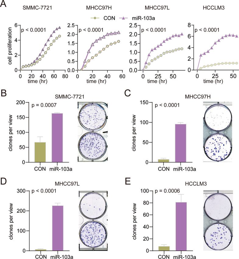
Y. Liu et al.
3
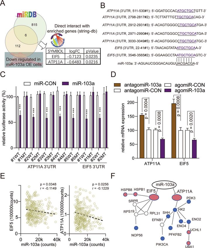
Fig. 1 miR-103a is a highly expressed/upregulated microRNA in HCC, and its high expression is related to poor prognosis in TCGA
datasets. A Fifteen highest microRNA read counts in hepatocellular carcinoma tissues (n = 372) and adjacent normal tissues (n = 50). B MiR-
103a reads in next-generation sequencing accounted for 1.458% of the total reads, and miR-103a was the 13th most highly expressed
microRNA in HCC tissues. C miR-103a expression levels (normalized reads) were upregulated in HCC tissues (n = 372) compared with adjacent
normal tissues (n = 50). D Kaplan–Meier analysis showing that the level of miR-103a was negatively correlated with the overall survival rate of
HCC patients (follow-up threshold was 36 months). NT adjacent tumour tissues, TP tumour tissues. miR-103a microRNA-103a, HCC
hepatocellular carcinoma cells. These data were obtained from TCGA database.
Cell Death and Disease (2021)12:618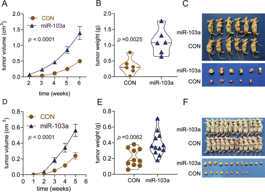
Y. Liu et al.
4

    Fig. 2 miR-103a expression was upregulated in HCC and related to poor prognosis in the Renji cohort. A Reverse transcription-
    quantitative PCR analysis was performed to assess miR-103a expression in 89 paired HCC tissues and adjacent normal tissues and two healthy
    volunteer liver tissues. B Survival curves suggested that increased miR-103a expression was significantly associated with unfavourable
    prognosis of patients with HCC. C–F Reverse transcription-quantitative PCR analysis was performed to validate miR-103a expression in SMMC-
    7721 (p < 0.001), MHCC97H (p < 0.001), MHCC97L (p < 0.01) and HCCLM3 (p < 0.001) cells transfected with miR-103a lentivirus. All results are
    representative of at least three independent experiments.

    Overexpression of miR-103a promotes HCC growth in a                      methods and dual-luciferase reporter assays showed that miR-103a
    xenograft model                                                          mainly affected glucose metabolism and cell death by targeting
    To further confirm the in vitro findings, we observed the biological       ATP11A and EIF5 directly, thus contributing to HCC cell growth and
    roles of miR-103a in vivo. miR-103a overexpression in SMMC-7721          mobility.
    cells increased the tumour volume 2.79-fold relative to that of its         Previous studies have shown that miR-103a plays a critical role in
    control (Fig. 7A) and increased the tumour weight 3.36-fold              the progression of cancer metastasis. miR-103/107 attenuates
    relative to its control (Fig. 7B), and the representative images are     miRNA biosynthesis by targeting Dicer, induces the epithelial-to-
    shown in Fig. 7C. MiR-103a overexpression in MHCC97L cells               mesenchymal transition (EMT), and promotes breast cancer cell
    increased the tumour volume 2.35-fold relative to that of the            metastasis [30]. miR-103/107-mediated repression of DAPK and
    control (Fig. 7D) and increased the tumour weight 1.82-fold              KLF4 promoted metastasis in CRC [31]. miR-103a plays both an
    relative to that of the control (Fig. 7E), and the representative        oncogenic and tumour-suppressive role in various types of cancers.
    images are shown in Fig. 7F.                                             For example, miR-103 promotes proliferation and metastasis by
       The results demonstrated that after implantation of miR-103a-         targeting KLF4 in gastric cancer [32] and regulates triple-negative
    overexpressing cells, tumour xenografts grew significantly faster         breast cancer cell migration and invasion by targeting olfactomdin4
    than those in the control group.                                         [33], while miR-103a functions as a tumour suppressor by directly
                                                                             targeting programmed cell death 10 in NSCLC [34]. A recent study
                                                                             showed that upregulation of serum miR-103 predicts an unfavour-
    DISCUSSION                                                               able prognosis in patients with colorectal cancer [35]. Taken
    In this study, we found that miR-103a was a highly expressed and         together, these results suggest that promoting cancer metastasis
    upregulated microRNA in HCC tissues from TCGA. In addition, the          might be the main function of miR-103a.
    high expression of miR-103a was correlated with poor prognosis in           miR-103a is also involved in cell growth and metastasis in liver
    HCC patients. Functional experiments revealed that miR-103a              cancer. Previous reports indicated that miR-103a is upregulated in
    overexpression promoted HCC cell growth, migration, invasion and         HCC. Moreover, miR-103 promotes HCC cell proliferation and
    glucose metabolism. Furthermore, the results of bioinformatics           migration in the simulation transition zone of RFA through the

                                                                                                  Cell Death and Disease (2021)12:618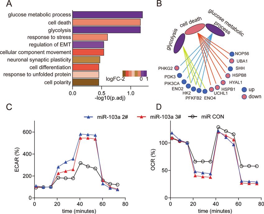
Y. Liu et al.
                                                                                                                                            5

Fig. 3 miR-103a overexpression promoted hepatocellular carcinoma cell growth. A RTCA was performed to determine the proliferation of
SMMC-7721, MHCC97H, MHCC97L and HCCLM3 cells overexpressing miR-103a. B–E Colony formation assay was performed to examine the
colony formation abilities of SMMC-7721, MHCC97H, MHCC97L and HCCLM3 cells overexpressing miR-103a. *p < 0.5, **p < 0.01, ***p < 0.001.
All results are representative of at least three independent experiments.

PI3K/Akt signalling pathway by targeting PTEN [36], promotes the       The liver is a major metabolic organ and a major site of glucose
metastasis and EMT of HCC by directly inhibiting LATS2 [37], and     metabolism [40, 41], which is an important process in cancer cell
regulates HCC growth by targeting AKAP12 [38]. Moreover,             growth [42]. Changes in glucose metabolism have been listed as
hepatoma cell-secreted exosomal microRNA-103 has been shown          an important factor that directly contributes to carcinogenesis
to increase vascular permeability and promote metastasis by          [43, 44]. However, to the best of our knowledge, no causal
targeting junction proteins [39]. However, the above studies         association has been established between miR-103a and glucose
mainly focused on exploring the effects and mechanisms of miR-       metabolism in HCC.
103a on HCC cell growth without addressing the role of miR-103a        In summary, we identified miR-103a as one of the most highly
in glucose metabolism in HCC or determining its main function.       expressed microRNAs in HCC tissues, found that it was upregulated in
   In our study, the bioinformatics analysis showed that the EMT,    HCC tissues compared with the controls, and showed that high
which is the main malignant biological behaviour of miR-103a in      expression of miR-103a was associated with poor patient prognosis.
the literature, was the fifth most important biological process.      Moreover, miR-103a overexpression promoted HCC cell growth
Interestingly, miR-103a was mainly involved in the regulation of     and mobility, and the functional enrichment analysis showed that
glucose metabolism and cell death. In the current study, we          miR-103a mainly promoted glucose metabolism and inhibited cell
revealed a novel function of miR-103a in regulating the glucose      death. We validated this analysis, and the results showed that miR-
metabolism phenotypes of HCC.                                        103a directly influenced glucose metabolism and inhibited cell death

Cell Death and Disease (2021)12:618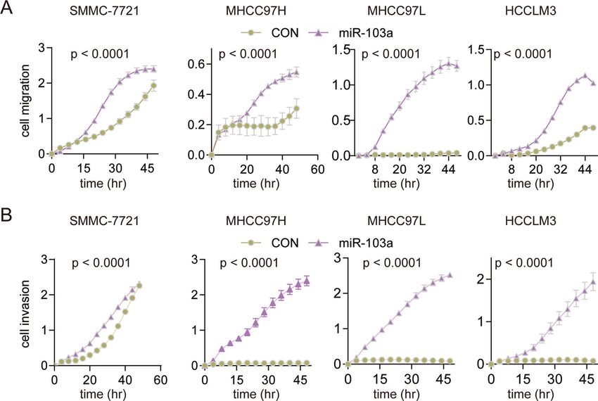
Y. Liu et al.
6

    Fig. 4 miR-103a promoted the mobility of hepatocellular carcinoma cells. A Overexpression of miR-103a promotes HCC cell migration. The
    migration ability of SMMC-7721, MHCC97H, MHCC97L and HCCLM3 cells overexpressing miR-103a was detected by RTCA. B Overexpression of
    miR-103a promoted HCC cell invasion. The invasion ability of SMMC-7721, MHCC97H, MHCC97L and HCCLM3 cells overexpressing miR-103a
    was detected by RTCA. All results are representative of at least three independent experiments.

    via ATP11A and EIF5. Therefore, our study revealed that miR-103a                following the manufacturer’s instructions. qRT-PCR was carried out with
    may act as a key mediator in HCC progression.                                   SYBR Premix EX Tag (Takara) on an ABI Prism 7500 fast RT-PCR instrument
                                                                                    (Applied Biosystems, Foster City, CA). Each experiment was performed in
                                                                                    triplicate. MicroRNA qRT-PCR primers were obtained from RiboBio
                                                                                    (Guangzhou, China). The microRNA expression levels were calculated
    MATERIALS AND METHODS
                                                                                    using the delta-delta Ct method, with RNU6B as an internal control. A Ct
    Cell lines and culture                                                          value of 35 was set as the cut-off value for a nondetected classification.
    Four human HCC cell lines (SMMC-7721, MHCC97H, MHCC97L and
                                                                                    β-actin was used as the internal reference gene for gene expression.
    HCCLM3) and HEK293T cells were purchased from The Cell Bank of Type             Objective CT values were normalized to β-actin, and the 2−ΔΔCt method
    Culture Collection of the Chinese Academy of Sciences (Shanghai, China).        was used to calculate relative mRNA levels of gene expression. The primer
    All cell lines were cultured in Dulbecco’s modified Eagle’s medium (DMEM;        sequences contained in this study were as follows (http://pga.mgh.harvard.
    Gibco, USA) supplemented with 10% foetal bovine serum (FBS; Gibco,              edu/primerbank/):
    USA), 100 units/mL penicillin and 100 μg/mL streptomycin (Gibco, USA)              ATP11A: F, 5′-TACCCAGACAACAGGATCGTC-3′ and
    and incubated at 37 °C in humidified incubators containing 5 % CO2.
                                                                                       R, 5′- AGCCGTCACAGTAATGACAAAG-3′;
                                                                                       EIF5: F, 5′- AGCGTGTCAGACCAGTTCTAT-3′ and
    Clinical samples and TCGA data analysis                                            R, 5′- CTGTCTTGATTCCATTGCCTTTG-3′;
    Eighty-nine paired samples of tumour tissues (TPs) and their corresponding         β-actin: F, 5′-TTGTTACAGGAAGTCCCTTGCC-3′ and
    nontumour tissues (NTs) from patients with HCC and two normal liver                R, 5′- ATGCTATCACCTCCCCTGTGTG-3′.
    tissues were obtained from the surgical specimen archives of Guangxi
    Medical University, Guangxi Province, China. One of the two normal liver        Lentiviral production and transduction
    tissue samples was collected from a person who died because of an
                                                                                    Particles carrying the hsa-pri-miR-103a precursor and its control were
    accident; the other was purchased from Clontech (Palo Alto, CA). The
                                                                                    purchased from GENECHEM (Shanghai, China). Lentiviral transduction was
    whole transcriptome sequencing (RNA-seq) data of 374 liver TPs and 50           carried out according to advice from GENECHEM. The high expression was
    adjacent NTs were obtained from TCGA liver cancer dataset (LIHC) (http://       validated by qRT-PCR.
    cancergenome.nih.gov). Informed consent was obtained for all human
    materials, and the institutional ethics review committee of the Shanghai
    Cancer Institute approved the protocols used in this study.                     Proliferation, colony formation, migration and invasion
                                                                                    analysis
    RNA extraction and quantitative real-time polymerase chain                      Cell proliferation, invasion and migration assays were measured with the
    reaction analysis                                                               xCELLigence System’s Real-Time Cell Analyzer (RTCA, Roche/ACEA Bios-
    Total RNA was extracted from cell lines and tissue samples using a TRIzol       ciences) placed in a humidified incubator and maintained at 37 °C with 95%
    kit (Invitrogen, Carlsbad, CA, USA). RNA (1 μg) was reverse-transcribed into    air/5% CO2. This system continuously monitored electrical impedance
    cDNA immediately using a Prime-Script RT kit (Takara, Shiga, Japan)             created by cell adhesion and proliferation in a microelectrode-integrated

                                                                                                          Cell Death and Disease (2021)12:618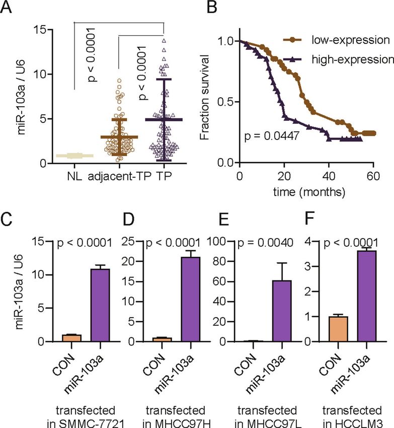
Y. Liu et al.
                                                                                                                                                              7

Fig. 5 miR-103a influenced glucose metabolism in HCC cells. A Functional enrichment analysis revealed that miR-103a participated in
glucose metabolic processes, cell death and glycolysis biological processes. B Total of 14 genes were matched with these biological functions.
C, D, ECAR and OCR of HCC cells were analysed by a Seahorse XFe 96 Extracellular Flux Analyzer. All results are representative of at least three
independent experiments.

membrane, and the output was a unitless parameter (cell index). For            phenylhydrazone (FCCP) and 2 mM antimycin A and rotenone (Sigma-
proliferation assays, 1 × 10⁴ to 3 × 10⁴ cells were seeded into E-plate 16     Aldrich, Saint Louis, Missouri, USA). Both the ECAR and OCR measurements
(ACEA Biosciences) with 200 μL DMEM containing 10% FBS (n = 3). The cell       were normalized to the total protein content.
index was normalized to the baseline reading at time point 0 and measured
every 30 min for 72 h. Migration and invasion assays were performed in 16-
well CIM plates (ACEA Biosciences). For migration assays, 1.5 × 10⁵ cells      Target gene prediction of miR-103a and functional
were seeded in triplicate in the upper chamber in a serum-free medium.         enrichment analysis
The upper chamber was then placed on the lower part of the CIM device,         The bioinformatic online database miRDB was used to predict potential
which contained DMEM with 10% FBS as a chemoattractant. The cell index         target genes of miR-103a. The consensus results of the miRDB database
was measured every 30 min for 48 h. For the invasion assays, the upper         and RNA-seq were selected for further analysis. Total RNA was isolated
chamber of the CIM-16 plate was initially coated with Matrigel (BD             from MHCC97L cells using TRIzol reagent (Invitrogen) and purified by an
Biosciences, Bedford, MA, USA) diluted in serum-free medium at a ratio of      RNeasy Mini Kit (Qiagen). Transcriptome sequencing was conducted using
1:20. The next steps were consistent with those for the migration assay.       Illumina HiSeq™ 2000 by BerryGenomics (Beijing Biotech, China). Gene
   For the colony formation assay, 8 × 10² cells were seeded in six-well       functional enrichment analyses were performed using EGSEA r-package
plates and maintained in DMEM with 5% FBS. After 14 days, the cells were       software, which is a widely used tool for gene functional enrichment. To
washed with PBS and stained with 0.5% crystal violet (Sigma, USA).             construct the interaction network, protein-protein interaction data from
                                                                               the STRING database were used. All procedures were conducted according
                                                                               to official protocols and default parameters.
ECAR and OCR
A Seahorse XF96 Flux Analyzer (Seahorse Bioscience, Billerica, Massachu-
setts, USA) was used to measure the oxygen consumption rate (OCR) and          Transfection of microRNA agomiR and antagomiR
extracellular acidification rate (ECAR) in lung cancer cells according to the   An agomiR is a type of specially labelled and chemically modified double-
manufacturer’s instructions. Approximately 1 × 10⁵ SMMC-7721 cells per         stranded microRNA that can regulate the biological function of a target gene
well were seeded into an XF96-well plate and attached overnight. For the       by mimicking endogenous microRNA. An antagomiR is a type of specifically
assessment of ECAR, cells were incubated with nonbuffered RPMI 1640            labelled and chemically modified single-stranded microRNA designed based
under basal conditions, followed by a sequential injection of 10 mM            on the mature microRNA sequence that can inhibit the expression of
glucose, 1 mM mitochondrial poison (oligomycin, Sigma-Aldrich, Saint           endogenous microRNA. AgomiR-103a, antagomiR-103a and their respective
Louis, Missouri, USA) and 80 mM glycolysis inhibitor (2-deoxyglucose, 2-       control materials were procured from RiboBio (Guangzhou, China) and
DG, Sigma-Aldrich). OCR was assessed under basal conditions and after          transfected into HCCs and HEK293T cells according to the manufacturer’s
sequential injection of 1 μM oligomycin, 1 μM fluoro-carbonyl cyanide           protocol. The medium was changed once after 24 h of transfection.

Cell Death and Disease (2021)12:618
Y. Liu et al.
8
Fig. 6 ATP11A and EIF5 were direct targets of miR-103a. A Identification of potential miR-103a target genes by microRNA prediction
databases and downregulated genes in miR-103a-overexpressing HCC cells. B Predicted miR-103a target sequences in the 3′-UTR of ATP11A
and EIF5. In addition, the target sequences were mutated. C Luciferase reporter activity of WT or MT ATP11A and EIF5 promoters in SMMC-
7721 cells transfected with miR-103a mimics and related constructs. D MRNA levels of ATP11A and EIF5 in SMMC-7721 cells treated with miR-
103a agomiRs or antagomiRs. E Correlations between the mRNA expression of miR-103a and ATP11A or EIF5 in TCGA datasets analysed by
Pearson’s correlation analysis. F miR-protein network analysis using STRINGdb showing that ATP11A and EIF5 were glycolysis-associated target
genes of miR-103a. **p < 0.01, ***p < 0.001. All results are representative of at least three independent experiments.
Luciferase reporter assay Tumour xenograft models
The ATP11A/EIF5 3′-UTR (untranslated region) and mutants were A subcutaneous xenograft mouse model was used to assess tumour
obtained from RiboBio (Guangzhou, China). HEK293T cells were growth. Animal experiments were approved by the Ethics Committee of
cotransfected with pmiR-103a agomiR or negative controls (NCs). Renji Hospital, Shanghai Jiao Tong University School of Medicine. Female
Luciferase activity was measured 24 h after transfection using the Dual- nude mice (age, 4–5 weeks; weight, 15–20 g; Institute of Zoology, Chinese
Glo® Luciferase Assay System (Promega) according to the manufacturer’s Academy of Sciences) were randomly divided into two groups: the miR-
instructions. 103a overexpression group and the CON group (at least 6 per group).
Cell Death and Disease (2021)12:618Y. Liu et al.
9
Fig. 7 miR-103a promoted cell growth in vivo. miR-103a overexpression promoted SMMC-7721/MHCC97L cell growth in vivo. A, D
Significantly higher tumour volumes were observed in mouse xenografts with HCC cells transfected with the miR-103a mimic than in control
xenografts. B, E Tumour weights were significantly higher in mouse xenografts. C, F Representative images of xenograft tumours are shown.
**p < 0.01, ***p < 0.001.
A total of 2 × 10⁶ SMMC-7721 cells in 100 µL of DMED without FBS were 6. Cao MQ, You AB, Zhu XD, Zhang W, Zhang YY, Zhang SZ, et al. miR-182-5p
injected into nude mice. A total of 2 × 10⁶ MHCC97L cells in 100 µL of promotes hepatocellular carcinoma progression by repressing FOXO3a. J.
DMED without FBS were injected into nude mice. The tumour volume was Hematol. Oncol. 2018;11:12.
measured by calliper measurements every week and calculated with the 7. Ma XL, Shen MN, Hu B, Wang BL, Yang WJ, Lv LH, et al. CD73 promotes hepa-
formula (length × width2)/2. tocellular carcinoma progression and metastasis via activating PI3K/AKT signaling
by inducing Rap1-mediated membrane localization of P110beta and predicts
poor prognosis. J. Hematol. Oncol. 2019;12:37.
Statistical analysis 8. Ambros V. The functions of animal microRNAs. Nature. 2004;431:350–5.
Data are expressed as the mean ± standard deviation (SD) from at least 9. Bartel DP. MicroRNAs: genomics, biogenesis, mechanism, and function. Cell.
three independent experiments. Statistical differences between groups
2004;116:281–97.
were evaluated by Student’s t test (paired/unpaired). Two-way analysis of 10. Fabian MR, Sonenberg N, Filipowicz W. Regulation of mRNA translation and
variance (ANOVA) followed by Tukey’s multiple comparisons test was stability by microRNAs. Annu. Rev. Biochem. 2010;79:351–79.
performed to compare significant differences and calculate the P-value 11. Bartel DP. MicroRNAs: target recognition and regulatory functions. Cell.
between the different groups. Pearson correlation tests were performed 2009;136:215–33.
for the correlation analyses. A survival analysis was performed with the 12. Prodromidou K, Vlachos IS, Gaitanou M, Kouroupi G, Hatzigeorgiou AG, Matsas R.
Kaplan–Meier method, and the log-rank test was used for comparisons.
MicroRNA-934 is a novel primate-specific small non-coding RNA with neurogenic
Statistical results were analysed using Prism software (GraphPad Software).
function during early development. eLife. 2020;9:e50561.
A probability of 0.05 or less was considered statistically significant. 13. Meerson A, Traurig M, Ossowski V, Fleming JM, Mullins M, Baier LJ. Human
adipose microRNA-221 is upregulated in obesity and affects fat metabolism
downstream of leptin and TNF-alpha. Diabetologia. 2013;56:1971–9.
REFERENCES 14. Arderiu G, Peña E, Aledo R, Juan-Babot O, Crespo J, Vilahur G, et al. MicroRNA-145
1. Miller KD, Nogueira L, Mariotto AB, Rowland JH, Yabroff KR, Alfano CM, et al. regulates the differentiation of adipose stem cells toward microvascular endo-
Cancer treatment and survivorship statistics, 2019. CA. 2019;69:363–85. thelial cells and promotes angiogenesis. Circulation Res. 2019;125:74–89.
2. Hu W, Zheng S, Guo H, Dai B, Ni J, Shi Y, et al. PLAGL2-EGFR-HIF-1/2alpha sig- 15. He T, Shen H, Wang S, Wang Y, He Z, Zhu L, et al. MicroRNA-3613-5p promotes
naling loop promotes HCC progression and erlotinib insensitivity. Hepatology. lung adenocarcinoma cell proliferation through a RELA and AKT/MAPK positive
2021;73:674–91. feedback loop. Mol. Ther. Nucleic Acids. 2020;22:572–83.
3. Dai H, Jia G, Wang H, Yang J, Jiang H, Chu M. Epidermal growth factor receptor 16. Xiao Y, Sun Y, Ma X, Wang C, Zhang L, Wang J, et al. MicroRNA-22 inhibits the
transactivation is involved in the induction of human hepatoma SMMC7721 cell apoptosis of vascular smooth muscle cell by targeting p38MAPKalpha in vascular
proliferation by insufficient radiofrequency ablation. Oncol. Lett. 2017;14:2463–7. remodeling of aortic dissection. Mol. Ther. Nucleic Acids. 2020;22:1051–62.
4. Siegel RL, Miller KD, Jemal A. Cancer statistics, 2019. CA. 2019;69:7–34. 17. Dhingra R, Lin J, Kirshenbaum LA. Disruption of RIP1-FADD complexes by Micro-
5. de Martel C, Ferlay J, Franceschi S, Vignat J, Bray F, Forman D, et al. Global burden RNA-103/107 provokes necrotic cardiac cell death. Circulation Res. 2015;117:314–6.
of cancers attributable to infections in 2008: a review and synthetic analysis. 18. Wang X, He Y, Mackowiak B, Gao B. MicroRNAs as regulators, biomarkers and
Lancet Oncol. 2012;13:607–15. therapeutic targets in liver diseases. Gut. 2021;70:784–95.
Cell Death and Disease (2021)12:618Y. Liu et al.
10
19. Wei X, Zhao L, Ren R, Ji F, Xue S, Zhang J, et al. MiR-125b loss activated HIF1alpha/ 42. Hoxhaj G, Manning BD. The PI3K-AKT network at the interface of oncogenic
pAKT loop, leading to transarterial chemoembolization resistance in hepatocel- signalling and cancer metabolism. Nat Rev Cancer. 2020;20:74–88.
lular carcinoma. Hepatology. 2021;73:1381–98. 43. Ganapathy-Kanniappan S, Geschwind JF. Tumor glycolysis as a target for cancer
20. Komoll RM, Hu Q, Olarewaju O, von Döhlen L, Yuan Q, Xie Y, et al. MicroRNA-342- therapy: progress and prospects. Mol Cancer. 2013;12:152.
3p is a potent tumour suppressor in hepatocellular carcinoma. J. Hepatol. 44. Vander Heiden MG, Cantley LC, Thompson CB. Understanding the Warburg effect:
2021;74:122–34. the metabolic requirements of cell proliferation. Science. 2009;324:1029–33.
21. Wu H, Zhang W, Wu Z, Liu Y, Shi Y, Gong J, et al. miR-29c-3p regulates DNMT3B
and LATS1 methylation to inhibit tumor progression in hepatocellular carcinoma.
Cell Death Dis. 2019;10:48. AUTHOR CONTRIBUTIONS
22. Xu WP, Liu JP, Feng JF, Zhu CP, Yang Y, Zhou WP, et al. miR-541 potentiates the Y.W. and X.L. designed the experiments, revised the manuscript, and supervised the
response of human hepatocellular carcinoma to sorafenib treatment by inhibit- progress throughout this study. Y.L, Y.Z., B.X. and N.T. carried out the experiments
ing autophagy. Gut. 2020;69:1309–21. and wrote the manuscript and analysed the data. J.H., S.L., Y.P., H.X., J.A., J.Y., X.L. and
23. Chen L, Liu D, Yi X, Qi L, Tian X, Sun B, et al. The novel miR-1269b-regulated L.W. participated in the experiments. All authors reviewed and approved the final
protein SVEP1 induces hepatocellular carcinoma proliferation and metastasis manuscript.
likely through the PI3K/Akt pathway. Cell death Dis. 2020;11:320.
24. Li S, Shao J, Lou G, Wu C, Liu Y, Zheng M. MiR-144-3p-mediated dysregulation of
EIF4G2 contributes to the development of hepatocellular carcinoma through the
ERK pathway. J Exp Clin Cancer Res. 2021;40:53. FUNDING
25. Liu Y, Lu LL, Wen D, Liu DL, Dong LL, Gao DM, et al. MiR-612 regulates invado- This study was supported by grants from the National Natural Science Foundation of
podia of hepatocellular carcinoma by HADHA-mediated lipid reprogramming. J China (No. 81572454 and 81773173) and the Research Project of Science and
Hematol Oncol. 2020;13:12. Technology Development Fund of Shanghai Pudong New Area (PKJ2019-Y30).Ethics
26. Fang F, Chang RM, Yu L, Lei X, Xiao S, Yang H, et al. MicroRNA-188-5p suppresses All clinical studies were approved by the ethics committees of the Shanghai Cancer
tumor cell proliferation and metastasis by directly targeting FGF5 in hepatocel- Institute. All animal experimental procedures acquired official approval of the
lular carcinoma. J. Hepatol. 2015;63:874–85. Institutional Animal Care and Use Committee of Model Animal Research Center of
27. Kudo Y, Sugimoto M, Arias E, Kasashima H, Cordes T, Linares JF, et al. PKClambda/ Shanghai Cancer Institute and the Institutional Animal Care and Use Committee of
iota loss induces autophagy, oxidative phosphorylation, and NRF2 to promote Shanghai Institute of Materia Medica, Chinese Academy of Sciences.
liver cancer progression. Cancer Cell. 2020;38:247–62. e11.
28. Xu D, Wang Z, Xia Y, Shao F, Xia W, Wei Y, et al. The gluconeogenic enzyme PCK1
phosphorylates INSIG1/2 for lipogenesis. Nature. 2020;580:530–5. CONFLICT OF INTEREST
29. Trajkovski M, Hausser J, Soutschek J, Bhat B, Akin A, Zavolan M, et al. MicroRNAs The authors declare no competing interests.
103 and 107 regulate insulin sensitivity. Nature. 2011;474:649–53.
30. Martello G, Rosato A, Ferrari F, Manfrin A, Cordenonsi M, Dupont S, et al. A
MicroRNA targeting dicer for metastasis control. Cell. 2010;141:1195–207. ADDITIONAL INFORMATION
31. Chen HY, Lin YM, Chung HC, Lang YD, Lin CJ, Huang J, et al. miR-103/107 pro- Supplementary information The online version contains supplementary material
mote metastasis of colorectal cancer by targeting the metastasis suppressors available at https://doi.org/10.1038/s41419-021-03905-3.
DAPK and KLF4. Cancer Res. 2012;72:3631–41.
32. Zheng J, Liu Y, Qiao Y, Zhang L, Lu S. miR-103 promotes proliferation and Correspondence and requests for materials should be addressed to Y.W. or X.L.
metastasis by targeting KLF4 in gastric cancer. Int J Mol Sci. 2017;18:1–13.
33. Xiong B, Lei X, Zhang L, Fu J. miR-103 regulates triple negative breast cancer Reprints and permission information is available at http://www.nature.com/
cells migration and invasion through targeting olfactomedin 4. Biomed Phar- reprints
macother. = Biomedecine pharmacotherapie. 2017;89:1401–8.
34. Yang D, Wang JJ, Li JS, Xu QY. miR-103 functions as a tumor suppressor by directly Publisher’s note Springer Nature remains neutral with regard to jurisdictional claims
targeting programmed cell death 10 in NSCLC. Oncol. Res. 2018;26:519–28. in published maps and institutional affiliations.
35. Wang DS, Zhong B, Zhang MS, Gao Y. Upregulation of serum miR-103 predicts
unfavorable prognosis in patients with colorectal cancer. Eur Rev Med Pharmacol
Sci. 2018;22:4518–23.
36. Tan Y, Zhao L. miR-103 promotes hepatocellular carcinoma cell proliferation and
migration in the simulation transition zone of RFA through PI3K/Akt signaling Open Access This article is licensed under a Creative Commons
pathway by targeting PTEN. Int J Clin Exp Pathol. 2020;13:473–9. Attribution 4.0 International License, which permits use, sharing,
37. Han LL, Yin XR, Zhang SQ. miR-103 promotes the metastasis and EMT of adaptation, distribution and reproduction in any medium or format, as long as you give
hepatocellular carcinoma by directly inhibiting LATS2. Int J Oncol. appropriate credit to the original author(s) and the source, provide a link to the Creative
2018;53:2433–44. Commons license, and indicate if changes were made. The images or other third party
38. Xia W, Ni J, Zhuang J, Qian L, Wang P, Wang J. MiR-103 regulates hepatocellular material in this article are included in the article’s Creative Commons license, unless
carcinoma growth by targeting AKAP12. Int J Biochem Cell Biol. 2016;71:1–11. indicated otherwise in a credit line to the material. If material is not included in the
39. Fang JH, Zhang ZJ, Shang LR, Luo YW, Lin YF, Yuan Y, et al. Hepatoma cell- article’s Creative Commons license and your intended use is not permitted by statutory
secreted exosomal microRNA-103 increases vascular permeability and promotes regulation or exceeds the permitted use, you will need to obtain permission directly
metastasis by targeting junction proteins. Hepatology. 2018;68:1459–75. from the copyright holder. To view a copy of this license, visit http://creativecommons.
40. Zhang L, Yao W, Xia J, Wang T, Huang F. Glucagon-induced acetylation of energy- org/licenses/by/4.0/.
sensing factors in control of hepatic metabolism. Int J Mol Sci. 2019;20:1–20.
41. Cohen JC, Horton JD, Hobbs HH. Human fatty liver disease: old questions and
new insights. Science. 2011;332:1519–23. © The Author(s) 2021
Cell Death and Disease (2021)12:618You can also read