Membrane Repair Mechanisms against Permeabilization by Pore-Forming Toxins - MDPIhttps://www.mdpi.com2072-6651/10/6/234/pdf
←
→
Page content transcription
If your browser does not render page correctly, please read the page content below
toxins
Review
Membrane Repair Mechanisms against
Permeabilization by Pore-Forming Toxins
Asier Etxaniz, David González-Bullón, César Martín and Helena Ostolaza *           ID

 Biofisika Institute (UPV/EHU, CSIC) and University of the Basque Country (UPV/EHU) Parque Científico s/n,
 48940 Leioa, Spain; aetxaniz7@gmail.com (A.E.); david_go88@hotmail.com (D.G.-B.);
 cesar.martin@ehu.eus (C.M.)
 * Correspondence: elenaamaya.ostolaza@ehu.es; Tel.: +34-946-018-164

 Received: 24 May 2018; Accepted: 7 June 2018; Published: 9 June 2018

 Abstract: Permeabilization of the plasma membrane represents an important threat for any cell, since
 it compromises its viability by disrupting cell homeostasis. Numerous pathogenic bacteria produce
 pore-forming toxins that break plasma membrane integrity and cause cell death by colloid-osmotic
 lysis. Eukaryotic cells, in turn, have developed different ways to cope with the effects of such
 membrane piercing. Here, we provide a short overview of the general mechanisms currently
 proposed for plasma membrane repair, focusing more specifically on the cellular responses to
 membrane permeabilization by pore-forming toxins and presenting new data on the effects and
 cellular responses to the permeabilization by an RTX (repeats in toxin) toxin, the adenylate cyclase
 toxin-hemolysin secreted by the whooping cough bacterium Bordetella pertussis, which we have
 studied in the laboratory.

 Keywords: membrane permeabilization; membrane repair; pore-forming toxins; RTX toxins;
 adenylate cyclase toxin; Bordetella pertussis

 Key Contribution: Cells are prepared to cope with membrane disruption, triggering a more or less
 generalized response using all the available resources to resist damage and ensure cell homeostasis.

1. Introduction
     The plasma membrane, a thin layer of only ≈40 Å thickness, regulates the necessary flow of
matter, energy, and information between the cell interior and the external medium for cells to live, and
thus, its structural integrity is essential for the proper functioning of cells. Membrane permeabilization,
independently of the means by which it is caused, may constitute a life or death risk for any cell, and
hence, it is expectable that cells possess evolutionarily conserved mechanisms to rapidly repair the
injured membranes and ensure survival.
     Despite it was reported previously that wounded eukaryotic cells (sea urchin eggs) repair more
or less large wounds in their plasma membrane in a few seconds, by a mechanism strictly dependent
on extracellular Ca2+ [1,2], a systematic study of plasma membrane repair was not developed till
the 1990s, with the works of McNeil and Steinhardt [3], who demonstrated that Ca2+ entry into
wounded cells triggers exocytosis of intracellular vesicles close to the wound site [4,5]. From then,
it was clear that plasma membrane repair represents an active, energy-dependent process that is
orchestrated by specific membrane traffic events [6]. Here, we provide a short overview of the more
general mechanisms currently proposed for plasma membrane repair, focusing more specifically on
the cellular responses to membrane permeabilization by pore-forming toxins, and presenting new
data on the cellular responses to the pores formed by an RTX (repeats in toxin) toxin, the adenylate

Toxins 2018, 10, 234; doi:10.3390/toxins10060234                               www.mdpi.com/journal/toxins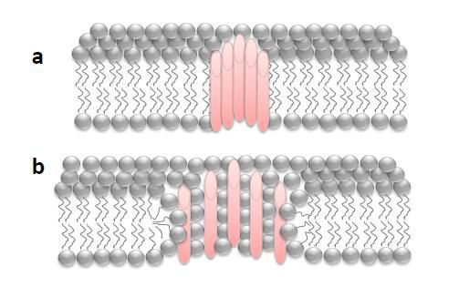
Toxins 2018, 10, 234                                                                                                 2 of 13

Toxins 2018, 10, x FOR PEER REVIEW                                                                                        2 of 13
   cyclase toxin-hemolysin secreted by the whooping cough bacterium Bordetella pertussis, which we have
2. studied
   “Holes”ininthe
                the Membrane: Some General Aspects
                  laboratory.
      The permeability
   2. “Holes”               barrier Some
               in the Membrane:     of theGeneral
                                             plasmaAspects
                                                    membrane can be frequently breached during the
lifetime of most cells by different means: external mechanical forces [7], internal forces generated by
        The permeability
contraction                barrier[8],
              and/or migration     of the
                                       or plasma membrane
                                          pore-forming      cansecreted
                                                       toxins    be frequently   breached [9–11],
                                                                          by pathogens    during the
                                                                                                  andlifetime
                                                                                                       thus, the
   of most cells by different means: external mechanical forces [7], internal forces generated by contraction
size and nature of the “holes” that can be formed are heterogeneous, and so the repair mechanisms
   and/or migration [8], or pore-forming toxins secreted by pathogens [9–11], and thus, the size and
will also vary. Wounds due to mechanical scratching are not delimited by protein boundaries and
   nature of the “holes” that can be formed are heterogeneous, and so the repair mechanisms will also
have an exclusively lipidic lumen (“lipid pores”), while membrane damage by pore-forming
   vary. Wounds due to mechanical scratching are not delimited by protein boundaries and have an
proteins give rise to holes with defined boundaries (“proteinaceous or proteolipidic pores”) (Figure
   exclusively lipidic lumen (“lipid pores”), while membrane damage by pore-forming proteins give rise
1).
   to holes with defined boundaries (“proteinaceous or proteolipidic pores”) (Figure 1).

                    Schematicdepiction
         Figure1.1.Schematic
      Figure                  depictionof
                                        ofdifferent
                                          different pores
                                                    pores with
                                                          with aa defined
                                                                  definedboundary
                                                                          boundaryinserted  into
                                                                                     inserted    a biological
                                                                                              into a biological
         phospholipid membrane: (a) pure proteinaceous pore in which the pore lumen is formed exclusively
      phospholipid membrane: (a) pure proteinaceous pore in which the pore lumen is formed exclusively
         by protein segments; (b) proteolipidic or toroidal pore in which the pore lumen is formed by both
      by protein segments; (b) proteolipidic or toroidal pore in which the pore lumen is formed by both
         lipids and protein segments.
      lipids and protein segments.

         Whena asimple
       When         simple“lipid
                             “lipidpore”
                                     pore” forms
                                              forms in
                                                     in the
                                                         the membrane,
                                                               membrane,an   anenergetically
                                                                                 energeticallyunfavorable
                                                                                                   unfavorable  situation   is is
                                                                                                                    situation
   created,and
created,     and most
                   most likely
                          likelythethe
                                     hole   will will
                                          hole   tend to
                                                       tendsealto
                                                                spontaneously     [12]. The anchoring
                                                                   seal spontaneously        [12]. The ofanchoring
                                                                                                             the membrane of the
   to  the cytoskeleton    and   the presence    of transmembrane        proteins  make
membrane to the cytoskeleton and the presence of transmembrane proteins make the membrane the  membrane      more   resistant
   and favor the spontaneous closure of this kind of hole, although this resistance is within damage
more resistant and favor the spontaneous closure of this kind of hole, although this resistance is
   limits [13] and will in turn be influenced by the lipid composition of the membrane [14]. By contrast,
within damage limits [13] and will in turn be influenced by the lipid composition of the membrane
   pore-forming proteins create stable holes that do not close spontaneously, and an energy cost paid by
[14]. By contrast, pore-forming proteins create stable holes that do not close spontaneously, and an
   cellular machinery will be necessary to close or eliminate the pore [13]. Even in the case of the lipid
energy cost paid by cellular machinery will be necessary to close or eliminate the pore [13]. Even in
   holes, a drop in membrane tension may not be sufficient to fully close them, and it is likely that active
the case of the lipid holes, a drop in membrane tension may not be sufficient to fully close them, and
   cellular mechanisms are also involved in the closure of most wounds [13].
it is likely
         When thatstudying
                    active cellular
                              the cellmechanisms
                                        responses to are     also involved by
                                                        permeabilization      in the  closure of toxins,
                                                                                  pore-forming     most wounds        [13].
                                                                                                            two important
       When    studying     the  cell  responses    to  permeabilization       by  pore-forming      toxins,
   aspects to consider are: the pore size (small pores of less than 1.0 nm in diameter to large oligomeric     two   important
aspects
   pores toof consider
              more than  are:
                            30 the
                                nm)pore
                                      and size   (small pores
                                            the secondary         of less of
                                                               structure  than
                                                                             the1.0  nmformed
                                                                                  pore   in diameter
                                                                                                  in the to  large
                                                                                                          lipid     oligomeric
                                                                                                                 membrane
pores    of more    than  30   nm)   and   the  secondary      structure   of the  pore  formed     in the
   (α-helix/β-barrel), which can determine the repair mechanism that will be activated, or its efficiency,   lipid  membrane
(α-helix/β-barrel),
   in terms of speed.which       can determine
                         Furthermore,               theyears,
                                            in the last   repairaccumulated
                                                                   mechanism evidence
                                                                                 that will indicates
                                                                                            be activated,
                                                                                                       that or   its efficiency,
                                                                                                              certain  toxins
in form
    termstheofso-called
               speed. Furthermore,         in  the last years,    accumulated     evidence    indicates
                         “toroidal pores” [15] in membranes, in which the pore lumen is composed of both  that  certain   toxins
form    the so-called
   proteins             “toroidal
              (or protein  segments) pores”    [15] inofmembranes,
                                         and lipids                     in which
                                                         different curvature    thatthe pore lumen
                                                                                      interact         is composed
                                                                                                with each                of both
                                                                                                            other. Toroidal
proteins    (or   protein    segments)      and   lipids    of  different   curvature    that   interact
   pores differ from the more classical purely proteinaceous pores in that: (i) the pore characteristics   with    each   other.
Toroidal pores differ from the more classical purely proteinaceous pores in that: (i) the pore
characteristics depend on the membrane lipid composition, (ii) they have lower stability than pure
proteinaceous pores, and (iii) the pore size may vary with the protein concentration and the
incubation time [15,16].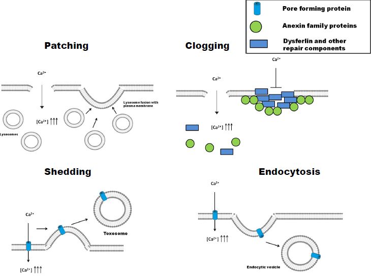
Toxins 2018, 10, 234                                                                              3 of 13

depend on the membrane lipid composition, (ii) they have lower stability than pure proteinaceous
pores, and (iii) the pore size may vary with the protein concentration and the incubation time [15,16].

3. Different Strategies to the Same Problem: Patching, Clogging, Shedding, or Endocytosis
     After plasma membrane damage, cells may activate different repair mechanisms, which have
been classified in four general strategies, namely: patching, clogging, shedding, and endocytosis [17].
The first two are involved in the repair of damage of mechanical/physical origin, while the last two
have been described in the elimination of pore-forming toxins (PFT). In some cell types, different
pathways may be complementary and might operate simultaneously [17].

3.1. Patching
     Membrane repair is achieved by fusion of intracellular vesicles with the injured plasma membrane
at the wound site, which closes or “patches” the lesion (Figure 2) [7]. Early studies in the 1990s had
already noted that membrane damage was repaired with membranes coming from cytoplasmic
vesicles [5,18]. The vesicles were mainly lysosomes [19], and the influx of extracellular Ca2+ was
essential for the activation of this process [4]. As a variant of this mechanism, the so-called “tension
release hypothesis” was also proposed [20], according to which, a direct fusion of the internal
membranes with the lesion site would not be necessary, but rather the fusion itself would decrease the
membrane tension, that would in turn facilitate the closing of the wound. The mechanism of membrane
patching seems more appropriate for repairing large “lipid pores” created in the cell membranes by
mechanical stresses, but may not be effective against pure “proteinaceous pores” generated by PFTs,
since these can be very resistant to changes in membrane tension [3].

3.2. Clogging
     Plasma membrane repair by this mechanism involves an accumulation of proteins around the
lesion, forming a barrier (clog) that prevents the loss of cytoplasmic contents to the extracellular
medium and isolates the area of membrane damage [21,22] (Figure 2). Clogging has been implicated
in the repair of medium-size membrane lesions (from one to a few microns) [23–25]. Although a
large variety of proteins putatively involved in this mechanism have been described, the family of
annexins seems to be the most important and main regulator of the process. Annexin A1, A2, A4,
A5, A6, A7, and A11 have been detected in this process, though annexin A1, A5, and A6 seem to
be the most important [17,24,26]. Annexins are activated in response to increases in the intracellular
concentration of Ca2+ and specifically bind to phosphatidylserines (PS) present in the inner side of the
plasma membrane [22]. The different annexins have different calcium sensitivities for their activation
and subsequent membrane binding, which confers to the cell the capacity to “sense” the severity and
localization of the membrane damage and the possibility of regulating the type of response that will be
activated in each particular case [27,28]. As an example, annexin A6 will bind to the membrane upon
minor damage, since it is activated at very low calcium concentrations (free [Ca2+ ] ≤ 5 µM), while
annexin A1 will only bind when the membrane lesion is more important and involves larger Ca2+
entry (free [Ca2+ ] >10–20 µM) [24,29]. Besides annexins, in different cell types, membrane clogging
would involve additional protein partners, such as dysferlin [30], or other repair components such as
EHD1, EHD2, MG53, and BIN1, which would establish a complex network of interactions as recruiting
platforms [23].

3.3. Shedding (Ectocytosis)
     By this mechanism, it is possible to isolate and expel the damaged membrane area in the form of
vesicles called “toxosomes” or “ectosomes” [31] (Figure 2). Shedding is one of the two mechanisms
by which small pores (
Membrane repair is achieved by fusion of intracellular vesicles with the injured plasma
membrane at the wound site, which closes or “patches” the lesion (Figure 2) [7]. Early studies in the
1990s had already noted that membrane damage was repaired with membranes coming from
cytoplasmic vesicles [5,18]. The vesicles were mainly lysosomes [19], and the influx of extracellular
  Toxins 2018, 10, 234                                                                                           4 of 13
Ca2+ was essential for the activation of this process [4]. As a variant of this mechanism, the so-called
“tension release hypothesis” was also proposed [20], according to which, a direct fusion of the
  For themembranes
internal    pores formedwith     theStaphylococcus
                             by the                aureus not
                                      lesion site would   alpha-hemolysin,
                                                               be necessary,itbut
                                                                               has rather
                                                                                   been proposed
                                                                                           the fusion thatitself
                                                                                                            shedding
                                                                                                                  would
decrease the membrane tension, that would in turn facilitate the closing of the wound.toThe
  occurs   after   the  toxin has been  endocytosed   and  it has not been  possible  to degrade    it, leading       it
  being expelled
mechanism              from the patching
                of membrane      cell [34]. Proteins of the
                                            seems more      ESCRT (endosomal
                                                         appropriate              sorting
                                                                       for repairing   largecomplex     required
                                                                                              “lipid pores”         for
                                                                                                                created
  transport)    machinery    have been  directly implicated  in the budding  and vesicular   fission
in the cell membranes by mechanical stresses, but may not be effective against pure “proteinaceous   steps   required
  for shedding
pores”   generated  [17,35,36].
                        by PFTs, since these can be very resistant to changes in membrane tension [3].

     Figure
       Figure2.2.Schematic
                   Schematicdepiction
                                depictionofofdifferent
                                              different membrane      repair pathways:
                                                         membrane repair        pathways:patching,
                                                                                             patching,clogging,
                                                                                                           clogging,shedding,
                                                                                                                       shedding,
     and
       andendocytosis.
            endocytosis. In repair
                                repairbybypatching,
                                             patching,    membrane
                                                     membrane     repairrepair    is achieved
                                                                          is achieved    by fusionbyoffusion    of intracellular
                                                                                                        intracellular   vesicles
     vesicles with
       with the        the injured
                   injured   plasmaplasma
                                      membranemembrane      at the wound
                                                    at the wound               site, seals
                                                                     site, which     whichorseals   or “patches”
                                                                                               “patches”              the lesion.
                                                                                                             the lesion.     The
     The  mechanism
       mechanism           of clogging
                      of clogging         involves
                                   involves           an accumulation
                                             an accumulation     of proteins ofaround
                                                                                 proteins
                                                                                        the around     the lesion,
                                                                                            lesion, forming           forming
                                                                                                                a barrier  (clog) a
       that prevents
     barrier (clog) that theprevents
                             loss of cytoplasmic    contents to the
                                      the loss of cytoplasmic         extracellular
                                                                   contents            medium, andmedium,
                                                                               to the extracellular       isolates the
                                                                                                                    andarea    of
                                                                                                                          isolates
       membrane      damage.     By the mechanism      of shedding,   it is possible   to isolate  and
     the area of membrane damage. By the mechanism of shedding, it is possible to isolate and expel the  expel   the damaged
       membranemembrane
     damaged         area in the form
                                   areaof in
                                           vesicles
                                               the called
                                                      form“toxosomes”
                                                              of vesicles   or “ectosomes”.    Endocytosis-mediated
                                                                                called “toxosomes”                          pore
                                                                                                              or “ectosomes”.
       removal    involves   sequential  steps  of exocytosis   and  endocytosis.     In a first step,
     Endocytosis-mediated pore removal involves sequential steps of exocytosis and endocytosis. In a    lysosomes    fuse   with
       the plasma membrane, releasing the lipid hydrolytic enzyme acid sphingomyelinase (ASM) into the
       extracellular medium, which converts membrane sphingomyelin into ceramide. This lipid seems to
       create a ceramide platform, which in a second step, induces an invagination of the membrane that
       promotes its engulfment.

  3.4. Endocytosis
        This is the second mechanism by which small pores formed by PFT are removed from the
  membrane. In this case, the cell responds by quickly internalizing the damaged area, including the
  pore [37]. Several PFT and some pore-forming proteins such as perforin [38], Staphylococcus aureus
  α-toxin [34], streptolysin-O [39], and Vibrio cholerae cytolysin [40] have been reported to be removed by
  this repair mechanism. Endocytosis-mediated pore removal involves sequential steps of exocytosis
Toxins 2018, 10, 234 5 of 13
and endocytosis (Figure 2). In a first step, lysosomes fuse with the plasma membrane, releasing their
contents to the extracellular medium [39]. Among the released material is the lipid hydrolytic enzyme
acid sphingomyelinase (ASM), which converts membrane sphingomyelin into ceramide. This lipid
seems to create a ceramide platform, which in a further, second step, induces an invagination of the
membrane that promotes its engulfment [39,41]. In fibroblasts, the endocytosis-mediated membrane
repair has been localized to caveolae [42], though it is possible that in cells that do not express caveolin,
such as certain immune cells, endocytosis may be coupled to clathrin [43]. Cells appear to have a
“pool” of lysosomes in the vicinity of the membrane that can fuse quickly with it [44]. The process
is triggered by the influx of extracellular Ca2+ [19], and it has been postulated that calcium-sensitive
proteins in the lysosome membrane might “guide” these vesicles to the injured area [45]. Other
released calcium-dependent cysteine proteases such as cathepsins B, D, and L have been implicated
in autoregulation of the process [46], avoiding excessive damage. The ESCRT machinery is also
involved in the endocytosis-mediated pore removal, together with several RAB proteins (Rab-5 and
Rab-11) [9,47]. This exocytosis/endocytosis-based repair mechanism is energy-dependent and requires
ATP, besides Ca2+ , to restore the membrane integrity [37].
4. Repair Mechanisms Activated by Pore-Forming Toxins
From numerous studies, it has been concluded that the size of the pore (lumen diameter) is one of
the most crucial factors determining the repair mechanism that will be activated by a PFT to restore
homeostasis [48,49]. To have an idea of the size of the pores formed by the different PFTs, they have
been classified as “large pores” with diameters above 3.0 nm and that can be as large as 30–40 nm,
and “small pores” which show pore diameters below 2.0–3.0 nm. We will go over several examples of
repair mechanisms activated by these two groups of pore-forming toxins.
4.1. Repair of Large Pores
Toxins of the CDC (cholesterol-dependent cytolysins) family are known to form big
transmembrane holes that can exceed 30–40 nm in diameter [50]. The consequences of opening such
big “holes” in the membrane could be so deleterious that cells will expectedly activate rapid repair
responses that may be similar to the processes followed for membrane ruptures due to mechanical
damage. In the last years, the repair mechanisms activated by several of these toxins have been reported:
listeriolysin [51–53], perfringolysin, and intermedilysin [54], though streptolysin-O has been the most
studied [37]. Both shedding- and endocytosis-mediated pore removal have been described in cells
attacked by these toxins, though it seems that depending on the cell type, the toxin concentration, and
the incubation time, one or the other mechanism might predominate [17]. The extent of the Ca2+ influx
induced by the toxin seems to make the difference, and so, when the intracellular [Ca2+ ] concentration
increase is small, the mechanism of vesicle shedding via annexins would predominate, while at high
cation concentrations, toxin endocytosis is detected by the presence of ceramide platforms [24], though
some studies have also noted caveolae-mediated entry of the toxins [42]. From the different studies
with cholesterol-dependent cytolysins (CDC), it is concluded that repair of these large pores by either
of the two mechanisms, shedding or endocytosis, is a rapid process (time scale of seconds to a few
minutes) that requires Ca2+ and ATP.
4.2. Repair of Small Pores
The best known two representatives of toxins forming small pores are α-hemolysin from
Staphylococcus aureus [33] and the Aeromonas hydrophila aerolysin [48]. Both toxins form heptameric
transmembrane β-barrel pores of small size (Toxins 2018, 10, 234 6 of 13
since a considerable amount of the toxin remains in the membrane hours after incubation [45,56].
Extracellular Ca2+ is not likely required for repair of small pores formed by S. aureus α toxin, as these
are Ca2+ -impermeable [33,57]; however, intracellular Ca2+ stores may still play a role. Some authors
have proposed that after the toxin has been endocytosed, cells tend to expel them as toxosomes, due
to their incapability to be degraded intracellullarly [34]. Cry5B, another toxin that forms small pores
of about 1 to 2 nm, was found to activate a repair response in which RAB-5 and -11 and the ESCRT
machinery are required [9]. Recent studies with other two toxins, Photobacterium damselae Phobalysin
(PhPly) and Vibrio cholerae cytolysin (VCC), have revealed that even subtle differences in the pore width
can be determinant for the type of response that will be activated [49]: the PhPly pore is ≈1.2–3 nm [58],
while the VCC pore is slightly smaller ≈1.2 nm [59], but the repair mechanisms activated in each
case are different. PhPly activates a lysosome-mediated fast endocytosis, while VCC repair has slow
kinetics, similar to those described for α-toxin and aerolysin [48], most likely because the VCC pore is
not Ca2+ -permeable. Mutations in the VCC protein that enlarge the pore size lead to the activation of
the lysosome-mediated endocytosis-based repair mechanism [49].
Besides the direct physical elimination of the toxin pores from the permeabilized membranes,
several signaling routes are also activated in the perforated cells [60]. K+ efflux, more than that of
Ca2+ , seems to be the key signal for activation of these pathways, and the MAPK p38 seems to be
the main regulator [61]. Although the effector proteins of the routes are not known, it seems that
their participation is crucial for the survival of the cells permeabilized by the Ca2+ -impermeable
pores [48]. In fibroblasts, activation of caspase-1 has been reported to help in cell survival upon the
action of small-pore PFTs [62], which highlights the importance of the cell type in the study of signaling
pathways, since activation of caspase-1 in immune cells leads to rapid cell death by pyroptosis.
5. ACT Toxin: An Adenylate Cyclase Enzyme Fused to an RTX Hemolysin
Adenylate cyclase toxin (ACT or CyaA) secreted by Bordetella pertussis, the bacterium causative
of whooping cough (pertussis), has a critical role in the early stages of respiratory tract colonization
by this pathogenic bacterium [63]. ACT is a prototypic toxin of the so-called RTX family of
proteins characterized by possessing, in their C-terminal sequence, a variable number of glycine-
and aspartate-rich repeats of nine amino acids (aa), mostly exhibiting cytotoxic/cytolytic pore-forming
activity [64]. ACT is a 1706-residue-long protein, in which an adenylate cyclase (AC) domain of
~400 N-terminal residues is fused to a ~1300-residue C-terminal RTX hemolysin domain (Figure 3).
The RTX domain consists of a hydrophobic pore-forming domain (aa ≈ 500–700); an acylation
domain, where the post-translational palmitoylation of lysine residues 860 and 983 occurs [65]; a
CD11b/CD18 receptor-binding domain (aa 1166–1281) [66]; and an RTX calcium-binding domain
harboring the nonapeptide repeats of a consensus sequence X–(L/I/F)–X–G–G–X–G–(N7D)–D, which
form numerous (~40) calcium-binding sites. The toxin segment extending approximately from residue
400 to 500, that connects the catalytic AC domain to the pore-forming RTX hemolysin domain, has
been recently revealed as possibly being involved in translocation of the AC domain across the cell
membrane, assisting membrane insertion [67,68]. All ACT activities depend on the covalent fatty
acylation of pro-ACT and on the binding of Ca2+ ions to the numerous sites formed in the RTX domain
by the glycine- and aspartate-rich repetitions [69]. The toxin targets CD11b/CD18-expressing (αM
β2 integrin or CR3) myeloid phagocytes and translocates its adenylate cyclase domain directly across
their cytoplasmic membrane without the participation of a receptor-mediated endocytosis step. In the
target cytosol, the AC domain is activated by calmodulin [70], acquiring high catalytic activity in the
conversion of ATP to cAMP, a key second messenger. The supraphysiological levels of cAMP subvert
the signaling of protein kinase A (PKA) and ablate the bactericidal functions of phagocytes, such as
oxidative burst and phagocytosis [71]. In parallel, ACT acts as a cytolysin, forming cation-selective
pores, which permeabilize the cell membrane and eventually provoke cell lysis. Translocation of the
AC domain and oligomerization into cation-selective pores appear to represent two independent and
parallel/competing activities of the membrane-inserted form of ACT [72].Toxins 2018, 10, 234 7 of 13
Toxins 2018, 10, x FOR PEER REVIEW 7 of 13
FigureFigure 3. Structural
3. Structural organizationof
organization of the ACT
ACTtoxin.
toxin. TheThe
adenylate cyclase
adenylate domain
cyclase (AC domain)
domain (in
(AC domain)
green)
(in green) extendsapproximately
extends approximately fromfromresidues
residues1 1toto400.
400.The RTX
The RTXhemolysin
hemolysin domain (residues
domain from from
(residues
≈500 ≈500 to 1706)
to 1706) contains
contains thethe hydrophobicdomain
hydrophobic domain (HD)
(HD) constituted
constitutedbybyfive hydrophobic/amphipathic
five hydrophobic/amphipathic
alpha-helices (in red); two conserved acylation sites, K860 and K983, required for activation by
alpha-helices (in red); two conserved acylation sites, K860 and K983, required for activation
palmitoylation mediated by CyaC acyltransferase; and five blocks formed by low-affinity
by palmitoylation mediated by CyaC acyltransferase; and five blocks formed by low-affinity
calcium-binding repeats. The Ca2+-binding region (residues 1006–1612) is denoted by multiple lines,
calcium-binding repeats. The Ca2+ -binding region (residues 1006–1612) is denoted by multiple lines,
with each line corresponding to single nonapeptide repeats with the consensus sequence
with each line corresponding
GGXGXDXLX. The segmentto single
betweennonapeptide repeats with
residues 1638–1706 the consensus
corresponds sequence GGXGXDXLX.
to the C-terminal secretion
The segment between residues 1638–1706 corresponds to the C-terminal secretion signal. Location
signal. Location of the catalytic residue serine 606 (S ), involved in the intrinsic phospholipase
606 A of
the catalytic residue 606
(PLA) activity of serine 606which
the toxin, (S has), involved in the
been recently intrinsic
reported by phospholipase
González-BullónA et (PLA)
al. [73],activity
has beenof the
toxin,included.
which has been recently reported by González-Bullón et al. [73], has been included.
5.1. Pore-Forming Activity of ACT
5.1. Pore-Forming Activity of ACT
Pore formation by ACT has been attributed to the insertion of the hydrophobic/amphipathic
Pore formation by ACT has been attributed to the insertion of the hydrophobic/amphipathic
α-helical segments located between residues 500 and 700 of the ACT hydrophobic domain (Figure 3)
α-helical segments
[74,75]. By analogy located between
with known residues 500
pore-forming and
toxins, 700 be
it may of assumed
the ACT thathydrophobic
the process thatdomain
(Figure 3) [74,75]. By analogy with known pore-forming toxins, it may be assumed
ultimately leads to osmotic cell lysis by ACT pores consists of three stages: binding, insertion, and that the process
that ultimately leads of
oligomerization to the
osmotic cell lysis
toxin within theby ACT pores
membrane. consistsbecause
However, of three of stages:
the lackbinding, insertion, and
of high-resolution
structures for
oligomerization full-length
of the ACT, or
toxin within theindeed for any
membrane. of the RTX
However, toxins,of knowledge
because the lack of of the lytic
high-resolution
mechanism
structures and pore ACT,
for full-length characteristics
or indeed of these proteins
for any of theisRTX
verytoxins,
limited.knowledge of the lytic mechanism
The fact that ACT
and pore characteristics of forms
these pores in membranes
proteins was concluded from conductance measurements
is very limited.
in black lipid membranes and hemolysis assays with osmotic protectants [76–78]. ACT pores in lipid
The fact that ACT forms pores in membranes was concluded from conductance measurements
bilayers were described as transient cation-selective “channels” with an extremely small
in black lipid membranes and hemolysis assays with osmotic protectants [76–78]. ACT pores in lipid
single-channel conductance of 27 pS in 1 M KCl and a half-life of ~2 s [76]. From those studies, it was
bilayers were described
estimated that ACT as transient
forms cation-selective
a 0.6–0.8-nm pore. More “channels” with blue
recently, using an extremely small single-channel
native polyacrylamide gel
conductance of 27 pS in 1 M KCl and a half-life of ~2 s [76]. From those
electrophoresis (BN-PAGE) and immunolabelling, putative ACT oligomers with apparent studies, it was molecular
estimated that
ACT forms
massesaof 0.6–0.8-nm
200, 300, 410,pore.andMore
470recently,
kDa have using
beenblue nativeinpolyacrylamide
described gel electrophoresis
erythrocyte membranes, being
attributed
(BN-PAGE) andtoimmunolabelling,
cleaved monomers putativelacking theACTAColigomers
domain (~200withkDa), full-length
apparent monomers
molecular masses(~300of 200,
kDa),
300, 410, and470
and ACT oligomers
kDa (~410 described
have been and 470 kDa), in respectively
erythrocyte[79].
membranes, being attributed to cleaved
monomersAgainst
lackingthetheview
AC that RTX (~200
domain toxins kDa),
form discrete-size pure proteinaceous
full-length monomers poresand
(~300 kDa), in membranes,
ACT oligomers
additional studies, including our own data, have revealed that the RTX-induced perturbation in
(~410 and 470 kDa), respectively [79].
membranes depends on a number of factors, including membrane lipid composition, temperature,
Against the view that RTX toxins form discrete-size pure proteinaceous pores in membranes,
time, and toxin concentration [80,81]. This suggests that rather than being a static process,
additional studies, including
permeabilization by RTX our own
toxins may data,
be have revealed
a complex, that the
dynamic RTX-induced
process involving perturbation
membrane in
membranes depends on a number of factors, including membrane lipid
remodeling processes, accompanied by the transient formation of nonlamellar proteolipidic composition, temperature,
time, structures
and toxin concentration
in the membrane [80–83]. [80,81]. This suggests that rather than being a static process,
permeabilization by RTX toxins may be a complex, dynamic process involving membrane remodeling
processes, accompanied by the transient formation of nonlamellar proteolipidic structures in the
membrane [80–83].
5.2. Irreversible Membrane Permeabilization by ACT
Recently, we explored the permeabilization elicited by sublytic doses of ACT on target
macrophages and evaluated whether the permeabilized cell membrane was resealed or not, andToxins 2018, 10, x FOR PEER REVIEW 8 of 13
5.2. Irreversible Membrane Permeabilization by ACT
Recently,
Toxins 2018, 10, 234 we explored the permeabilization elicited by sublytic doses of ACT8 of on13target
macrophages and evaluated whether the permeabilized cell membrane was resealed or not, and we
found that the full-length toxin induces an irreversible membrane permeabilization that eventually
we found
leadsthat
to the
cellfull-length toxin induces
death [84](Etxaniz an manuscript
et al., irreversible membrane
in revision), permeabilization
which suggeststhat thateventually
cellular repair
leads mechanisms
to cell death [84] (Etxaniz et al., manuscript in revision), which
are not operative in the ACT-treated macrophages. Interestingly, an ACT suggests that cellular repairmutant
mechanisms are not operative in the ACT-treated macrophages.
extending from amino acids 483 to 1706 (ACT-∆N482 hemolysin), which contains the entire ACT Interestingly, an ACT mutant
extending
RTX from aminodomain,
hemolysin acids 483 to 1706
induces by(ACT-∆N482 hemolysin),
contrast a transient membranewhichpermeabilization
contains the entire thatACTis rapidly
RTX hemolysin domain, induces by contrast a transient membrane permeabilization
reverted in a time scale of minutes (≈30 min) by a repair pathway activated by extracellular Ca2+ that is rapidly
2+ influx
reverted in a and
influx timethat
scalerequires
of minutes ATP(≈[84](Etxaniz
30 min) by aetrepair pathway activated
al., manuscript by extracellular
in revision). The increase Caof intracellular
and that
Ca2+requires
induced ATP by[84]
the(Etxaniz
sublyticetdoses al., manuscript
of ACT was in revision).
found to The beincrease of intracellular
very limited [84](Etxaniz Ca2+et al.,
induced by the sublytic
manuscript doses of
in revision), and ACT was foundof
translocation to the
be very limited
adenylate [84] (Etxaniz
cyclase domainetinto al., the
manuscript
cytosol of the
in revision), and translocation of the adenylate cyclase domain into
macrophages consumes cellular ATP; both factors might contribute to the incapability of thethe cytosol of the macrophages
consumes cellular ATP;
ACT-treated cells both factorsthe
to reseal might contribute
injured membrane to the[84](Etxaniz
incapability of et the
al., ACT-treated
manuscript cells to
in revision).
reseal Furthermore,
the injured membrane
we have[84] found (Etxaniz et al.,
that the manuscript
repair pathway inacting
revision).
in theFurthermore,
ACT-∆N482 wehemolysin-treated
have found
that the repair pathway acting in the ACT-∆N482 hemolysin-treated cells
cells involves consecutive steps of exocytosis and endocytosis, likely initiated by lysosomal involves consecutive stepsfusion
of exocytosis and endocytosis, likely initiated by lysosomal fusion
with the damaged cell membrane, subsequent secretion to the extracellular medium of acidwith the damaged cell membrane,
subsequent secretion to the
sphingomyelinase, andextracellular
concomitant medium of acid sphingomyelinase,
local generation of ceramide, all and concomitant
of which culminateslocalin the
generation of ceramide, all of which culminates in the endocytosis of
endocytosis of the pore-ridden membrane (Figure 4) [84](Etxaniz et al., manuscript in revision). the pore-ridden membrane
(Figure 4) [84]
Given the(Etxaniz et al., manuscript
high similarity among the ACT in revision).
RTX-hemolysinGiven the highand
moiety similarity
the other among
toxinsthefrom ACTthe RTX
RTX-hemolysin moiety and the other toxins from the RTX family, it is enticing
family, it is enticing to surmise that a similar, Ca -dependent, membrane repair mechanism might
2+ to surmise that a similar,
Ca2+ -dependent,
also operatemembrane repair mechanism
for those pore-forming might
toxins. Up also operate
to now, suchfor those pore-forming
a membrane toxins. Uphad
repair mechanism to only
now, such
beenareported
membrane forrepair
toxinsmechanism
forming large had only been reported
beta-barrel pores informembranes,
toxins forming for large beta-barrel
instance, streptolysin,
pores in membranes, for instance, streptolysin,
perfringolysin, pneumolysin, and listeriolysin [51–53]. perfringolysin, pneumolysin, and listeriolysin [51–53].
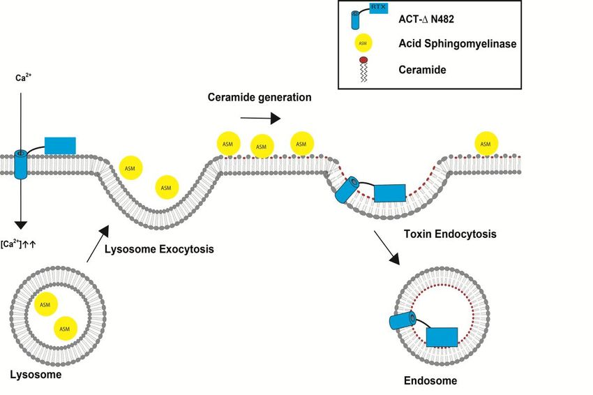
4. Scheme
FigureFigure 4. proposed for the membrane
Scheme proposed for the repair pathway
membrane activated
repair by theactivated
pathway ACT-∆N482 by hemolysin
the ACT-∆N482
pore inhemolysin
macrophages. The
pore in ACT-∆N482 The
macrophages. hemolysin pore (red)
ACT-∆N482 allowspore
hemolysin for (red)
the influx Ca2+
of for
allows theinto theof Ca2+
influx
cytosol,into
which
the is followed
cytosol, by the
which fusion of by
is followed lysosomes with
the fusion of the plasma membrane.
lysosomes Fusedmembrane.
with the plasma lysosomes Fused
releaselysosomes
acid sphingomyelinase
release acid(ASM) into the extracellular
sphingomyelinase (ASM)space.
into ASM converts sphingomyelin
the extracellular space. ASM in the
converts
membrane to ceramide, generating a ceramide-rich platform which may favor membrane
sphingomyelin in the membrane to ceramide, generating a ceramide-rich platform which may favor deformation
for invagination and subsequent endocytosis of the toxin-ridden membrane into the cell.Toxins 2018, 10, 234 9 of 13
6. Conclusions
Membrane repair mechanisms are among the most essential mechanisms to guarantee cell
survival. Although the variability of possible repair responses for a given type of lesion may seem
striking, a conclusion that can be drawn from all the known examples is that cells are prepared
to cope with membrane disruption, triggering a more or less generalized response using all the
available resources to resist damage and ensure cell homeostasis. Understanding the mechanism of
action of PFT as well as the host responses to toxin action would provide ways to deal with these
pathogens or with emerging pathogens, and more importantly, to improve the action of toxins that
have biotechnological applications.
Acknowledgments: This study was supported by grants from the Basque Government (Grupos Consolidados
IT849-13) and grant from the Spanish Ministerio de Economía y Competitividad (BFU2017-82758-P
AEI/FEDER, UE).
Conflicts of Interest: The author declares to have no conflict of interests.
References
1. Heilbrunn, L. The Dynamics of Living Protoplasm; Academic Press: New York, NY, USA, 1956; p. 634.
2. Chambers, R.; Chambers, E. Explorations into the Nature of the Living Cell; Harvard University Press:
Cambridge, MA, USA, 1961; p. 352.
3. Andrews, N.W.; Almeida, P.E.; Corrotte, M. Damage Control: Cellular Mechanisms of Plasma Membrane
Repair. Trends Cell Biol. 2014, 24, 734–742. [CrossRef] [PubMed]
4. Bi, G.Q.; Alderton, J.M.; Steinhardt, R.A. Calcium-Regulated Exocytosis Is Required for Cell Membrane
Resealing. J. Cell Biol. 1995, 131, 1747–1758. [CrossRef] [PubMed]
5. Miyake, K.; McNeil, P.L. Vesicle Accumulation and Exocytosis at Sites of Plasma Membrane Disruption.
J. Cell Biol. 1995, 131, 1737–1745. [CrossRef] [PubMed]
6. Steinhardt, R.A.; Bi, G.; Alderton, J.M. Cell Membrane Resealing by a Vesicular Mechanism Similar to
Neurotransmitter Release. Science 1994, 263, 390–393. [CrossRef] [PubMed]
7. McNeil, P.L.; Vogel, S.S.; Miyake, K.; Terasaki, M. Patching Plasma Membrane Disruptions with Cytoplasmic
Membrane. J. Cell Sci. 2000, 113, 1891–1902. [PubMed]
8. McNeil, P.L.; Khakee, R. Disruptions of Muscle Fiber Plasma Membranes: Role in Exercise-Induced Damage.
Am. J. Pathol. 1992, 140, 1097–1109. [PubMed]
9. Los, F.C.O.; Kao, C.-Y.; Smitham, J.; McDonald, K.L.; Ha, C.; Peixoto, C.A.; Aroian, R.V. RAB-5-and
RAB-11-Dependent Vesicle-Trafficking Pathways are Required for Plasma Membrane Repair after Attack by
Bacterial Pore-Forming Toxin. Cell Host Microbe 2011, 9, 147–151. [CrossRef] [PubMed]
10. Kao, C.Y.; Los, F.C.O.; Huffman, D.L.; Wachi, S.; Kloft, N.; Husmann, M.; Karabrahimi, V.; Schwartz, J.-L.;
Bellier, A.; Ha, C.; et al. Global Functional Analyses of Cellular Responses to Pore-Forming Toxins.
PLoS Pathog. 2011, 7, e1001314. [CrossRef] [PubMed]
11. Bischofberger, M.; Gonzalez, M.R.; van der Goot, F.G. Membrane Injury by Pore-Forming Proteins. Curr. Opin.
Cell Biol. 2009, 21, 589–595. [CrossRef] [PubMed]
12. Diz-Muñoz, A.; Fletcher, D.A.; Weiner, O.D. Use the Force: Membrane Tension as an Organizer of Cell Shape
and Motility. Trends Cell Biol. 2013, 23, 47–53. [CrossRef] [PubMed]
13. Jimenez, A.J.; Perez, F. Physico-Chemical and Biological Considerations for Membrane Wound Evolution
and Repair in Animal Cells. Semin. Cell Dev. Biol. 2015, 45, 2–9. [CrossRef] [PubMed]
14. Zhelev, D.V.; Needham, D. Tension-Stabilized Pores in Giant Vesicles: Determination of Pore Size and Pore
Line Tension. Biochim. Biophys. Acta Biomembr. 1993, 1147, 89–104. [CrossRef]
15. Gilbert, R.J.C. Protein–lipid Interactions and Non-Lamellar Lipidic Structures in Membrane Pore Formation
and Membrane Fusion. Biochim. Biophys. Acta (BBA)-Biomembr. 2016, 1858, 487–499. [CrossRef] [PubMed]
16. Bischofberger, M.; Iacovache, I.; Gisou Van Der Goot, F. Pathogenic Pore-Forming Proteins: Function and
Host Response. Cell Host Microbe 2012, 12, 266–275. [CrossRef] [PubMed]
17. Jimenez, A.J.; Perez, F. Plasma Membrane Repair: The Adaptable Cell Life-Insurance. Curr. Opin. Cell Biol.
2017, 47, 99–107. [CrossRef] [PubMed]Toxins 2018, 10, 234 10 of 13
18. Terasaki, M.; Miyake, K.; McNeil, P.L. Large Plasma Membrane Disruptions are Rapidly Resealed by Ca2+ -
Dependent Vesicle-Vesicle Fusion Events. J. Cell Biol. 1997, 139, 63–74. [CrossRef] [PubMed]
19. Rodríguez, A.; Webster, P.; Ortego, J.; Andrews, N.W. Lysosomes Behave as Ca2+ -Regulated Exocytic Vesicles
in Fibroblasts and Epithelial Cells. J. Cell Biol. 1997, 137, 93–104. [CrossRef] [PubMed]
20. Togo, T.; Krasieva, T.B.; Steinhardt, R.A. A Decrease in Membrane Tension Precedes Successful
Cell-Membrane Repair. Mol. Biol. Cell. 2000, 11, 4339–4346. [CrossRef] [PubMed]
21. Babiychuk, E.B.; Draeger, A. Defying Death: Cellular Survival Strategies Following Plasmalemmal Injury by
Bacterial Toxins. Semin. Cell Dev. Biol. 2015, 45, 39–47. [CrossRef] [PubMed]
22. McNeil, A.K.; Rescher, U.; Gerke, V.; McNeil, P.L. Requirement for Annexin A1 in Plasma Membrane Repair.
J. Biol. Chem. 2006, 281, 35202–35207. [CrossRef] [PubMed]
23. Demonbreun, A.R.; McNally, E.M. Plasma Membrane Repair in Health and Disease. Curr. Top. Membr. 2016,
77, 67–96. [PubMed]
24. Babiychuk, E.B.; Monastyrskaya, K.; Potez, S.; Draeger, A. Intracellular Ca2+ Operates a Switch between
Repair and Lysis of Streptolysin O-Perforated Cells. Cell Death Differ. 2009, 16, 1126–1134. [CrossRef]
[PubMed]
25. Demonbreun, A.R.; Quattrocelli, M.; Barefield, D.Y.; Allen, M.V.; Swanson, K.E.; McNally, E.M. An
Actin-Dependent Annexin Complex Mediates Plasma Membrane Repair in Muscle. J. Cell Biol. 2016,
213, 705–718. [CrossRef] [PubMed]
26. Bouter, A.; Gounou, C.; Bérat, R.; Tan, S.; Gallois, B.; Granier, T.; D’Estaintot, B.L.; Pöschl, E.; Brachvogel, B.;
Brisson, A.R. Annexin-A5 Assembled into Two-Dimensional Arrays Promotes Cell Membrane Repair.
Nat. Commun. 2011, 2. [CrossRef] [PubMed]
27. Potez, S.; Luginbühl, M.; Monastyrskaya, K.; Hostettler, A.; Draeger, A.; Babiychuk, E.B. Tailored Protection
against Plasmalemmal Injury by Annexins with Different Ca2+ Sensitivities. J. Biol. Chem. 2011, 286,
17982–17991. [CrossRef] [PubMed]
28. Monastyrskaya, K.; Babiychuk, E.B.; Draeger, A. The Annexins: Spatial and Temporal Coordination of
Signaling Events during Cellular Stress. Cell Mol. Life Sci. 2009, 66, 2623–2642. [CrossRef] [PubMed]
29. Monastyrskaya, K.; Babiychuk, E.B.; Hostettler, A.; Rescher, U.; Draeger, A. Annexins as Intracellular Calcium
Sensors. Cell Calcium. 2007, 41, 207–219. [CrossRef] [PubMed]
30. Han, R.; Campbell, K.P. Dysferlin and Muscle Membrane Repair. Curr. Opin. Cell Biol. 2007, 19, 409–416.
[CrossRef] [PubMed]
31. Atanassoff, A.P.; Wolfmeier, H.; Schoenauer, R.; Hostettler, A.; Ring, A.; Draeger, A.; Babiychuk, E.B.
Microvesicle Shedding and Lysosomal Repair Fulfill Divergent Cellular Needs during the Repair of
Streptolysin O-Induced Plasmalemmal Damage. PLoS ONE 2014, 9, e89743. [CrossRef] [PubMed]
32. Keyel, P.A.; Loultcheva, L.; Roth, R.; Salter, R.D.; Watkins, S.C.; Yokoyama, W.M.; Heuser, J.E. Streptolysin O
Clearance through Sequestration into Blebs that Bud Passively from the Plasma Membrane. J. Cell Sci. 2011,
124, 2414–2423. [CrossRef] [PubMed]
33. Walev, I.; Palmer, M.; Martin, E.; Jonas, D.; Weller, U.; Höhn-Bentz, H.; Husmann, M.; Bhakdi, S. Recovery of
Human Fibroblasts from Attack by the Pore-Forming a-Toxin of Staphylococcus aureus. Microb. Pathog. 1994,
17, 187–201. [CrossRef] [PubMed]
34. Husmann, M.; Beckmann, E.; Boller, K.; Kloft, N.; Tenzer, S.; Bobkiewicz, W.; Neukirch, C.; Bayley, H.;
Bhakdi, S. Elimination of a Bacterial Pore-Forming Toxin by Sequential Endocytosis and Exocytosis. FEBS Lett.
2009, 583, 337–344. [CrossRef] [PubMed]
35. Jimenez, A.J.; Maiuri, P.; Lafaurie-Janvore, J.; Divoux, S.; Piel, M.; Perez, F. ESCRT Machinery Is Required for
Plasma Membrane Repair. Science 2014, 343, 1247136. [CrossRef] [PubMed]
36. Scheffer, L.L.; Sreetama, S.C.; Sharma, N.; Medikayala, S.; Brown, K.J.; Defour, A.; Jaiswal, J.K. Mechanism
of Ca2 -Triggered ESCRT Assembly and Regulation of Cell Membrane Repair. Nat. Commun. 2014, 5, 5646.
[CrossRef] [PubMed]
37. Idone, V.; Tam, C.; Goss, J.W.; Toomre, D.; Pypaert, M.; Andrews, N.W. Repair of Injured Plasma Membrane
by Rapid Ca2+ -Dependent Endocytosis. J. Cell Biol. 2008, 180, 905–914. [CrossRef] [PubMed]
38. Thiery, J.; Keefe, D.; Saffarian, S.; Martinvalet, D.; Walch, M.; Boucrot, E.; Kirchhausen, T.; Lieberman, J.
Perforin Activates Clathrin- and Dynamin-Dependent Endocytosis, which is Required for Plasma Membrane
Repair and Delivery of Granzyme B for Granzyme-Mediated Apoptosis. Blood 2010, 115, 1582–1593.
[CrossRef] [PubMed]Toxins 2018, 10, 234 11 of 13
39. Tam, C.; Idone, V.; Devlin, C.; Fernandes, M.C.; Flannery, A.; He, X.; Schuchman, E.; Tabas, I.; Andrews, N.W.
Exocytosis of Acid Sphingomyelinase by Wounded Cells Promotes Endocytosis and Plasma Membrane
Repair. J. Cell Biol. 2010, 189, 1027–1038. [CrossRef] [PubMed]
40. Gutierrez, M.G.; Saka, H.A.; Chinen, I.; Zoppino, F.C.M.; Yoshimori, T.; Bocco, J.L.; Colombo, M.I. Protective
Role of Autophagy against Vibrio cholerae Cytolysin, a Pore-Forming Toxin from V. cholerae. Proc. Natl. Acad.
Sci. USA 2007, 104, 1829–1834. [CrossRef] [PubMed]
41. Tam, C.; Flannery, A.R.; Andrews, N. Live Imaging Assay for Assessing the Roles of Ca2+ and
Sphingomyelinase in the Repair of Pore-Forming Toxin Wounds. J. Vis. Exp. 2013. [CrossRef] [PubMed]
42. Corrotte, M.; Almeida, P.E.; Tam, C.; Castro-Gomes, T.; Fernandes, M.C.; Millis, B.A.; Cortez, M.; Miller, H.;
Song, W.; Maugel, T.K.; et al. Caveolae Internalization Repairs Wounded Cells and Muscle Fibers. Elife 2013.
[CrossRef] [PubMed]
43. Corrotte, M.; Fernandes, M.C.; Tam, C.; Andrews, N.W. Toxin Pores Endocytosed during Plasma Membrane
Repair Traffic into the Lumen of MVBs for Degradation. Traffic 2012, 13, 483–494. [CrossRef] [PubMed]
44. Jaiswal, J.K.; Andrews, N.W.; Simon, S.M. Membrane Proximal Lysosomes are the Major Vesicles Responsible
for Calcium-Dependent Exocytosis in Nonsecretory Cells. J. Cell Biol. 2002, 159, 625–635. [CrossRef]
[PubMed]
45. Reddy, A.; Caler, E.V.; Andrews, N.W. Plasma Membrane Repair is Mediated by Ca2+ -Regulated Exocytosis
of Lysosomes. Cell 2001, 106, 157–169. [CrossRef]
46. Castro-Gomes, T.; Corrotte, M.; Tam, C.; Andrews, N.W. Plasma Membrane Repair is Regulated
Extracellularly by Proteases Released from Lysosomes. PLoS ONE 2016, 11, e0152583. [CrossRef] [PubMed]
47. Vieira, O.V. Rab3a and Rab10 are Regulators of Lysosome Exocytosis and Plasma Membrane Repair.
Small GTPases. 2016, 1–3. [CrossRef] [PubMed]
48. Gonzalez, M.R.; Bischofberger, M.; Frêche, B.; Ho, S.; Parton, R.G.; Van der Goot, F.G. Pore-Forming Toxins
Induce Multiple Cellular Responses Promoting Survival. Cell. Microbiol. 2011, 13, 1026–1043. [CrossRef]
[PubMed]
49. Von Hoven, G.; Rivas, A.J.; Neukirch, C.; Meyenburg, M.; Qin, Q.; Parekh, S.; Hellmann, N.; Husmann, M.
Repair of a Bacterial Small ß-Barrel Toxin Pore Depends on Channel Width. mBio 2017, 8, e02083-16.
[CrossRef] [PubMed]
50. Hotze, E.M.; Tweten, R.K. Membrane Assembly of the Cholesterol-Dependent Cytolysin Pore Complex.
Biochim. Biophys. Acta Biomembr. 2012, 1818, 1028–1038. [CrossRef] [PubMed]
51. Pathak-Sharma, S.; Zhang, X.; Lam, J.G.T.; Weisleder, N.; Seveau, S.M. High-Throughput Microplate-Based
Assay to Monitor Plasma Membrane Wounding and Repair. Front. Cell. Infect. Microbiol. 2017, 7. [CrossRef]
[PubMed]
52. Wolfmeier, H.; Schoenauer, R.; Atanassoff, A.P.; Neill, D.R.; Kadioglu, A.; Draeger, A.; Babiychuk, E.B.
Ca2 -Dependent Repair of Pneumolysin Pores: A New Paradigm for Host Cellular Defense Against Bacterial
Pore-Forming Toxins. Biochim. Biophys. Acta 2015, 1853, 2045–2054. [CrossRef] [PubMed]
53. Wolfmeier, H.; Radecke, J.; Schoenauer, R.; Koeffel, R.; Babiychuk, V.S.; Drücker, P.; Hathaway, L.J.;
Mitchell, T.J.; Zuber, B.; Draeger, A.; et al. Active Release of Pneumolysin Prepores and Pores by Mammalian
Cells Undergoing a Streptococcus pneumoniae Attack. Biochim. Biophys. Acta Gen. Subj. 2016, 1860, 2498–2509.
[CrossRef] [PubMed]
54. Romero, M.; Keyel, M.; Shi, G.; Bhattacharjee, P.; Roth, R.; Heuser, J.E.; Keyel, P.A. Intrinsic Repair Protects
Cells from Pore-Forming Toxins by Microvesicle Shedding. Cell Death Differ. 2017, 24, 798–808. [CrossRef]
[PubMed]
55. Iacovache, I.; De Carlo, S.; Cirauqui, N.; Dal Peraro, M.; Van Der Goot, F.G.; Zuber, B. Cryo-EM Structure of
Aerolysin Variants Reveals a Novel Protein Fold and the Pore-Formation Process. Nat. Commun. 2016, 7, 7.
[CrossRef] [PubMed]
56. Valeva, A.; Walev, I.; Gerber, A.; Klein, J.; Palmer, M.; Bhakdi, S. Staphylococcal a-Toxin: Repair of a
Calcium-Impermeable Pore in the Target Cell Membrane. Mol. Microbiol. 2000, 36, 467–476. [CrossRef]
[PubMed]
57. Zitzer, A.; Wassenaar, T.M.; Walev, I.; Bhakdi, S. Potent Membrane-Permeabilizing and Cytocidal Action of
Vibrio cholerae Cytolysin on Human Intestinal Cells. Infect. Immun. 1997, 65, 1293–1298. [PubMed]Toxins 2018, 10, 234 12 of 13
58. Rivas, A.J.; Hoven, G.; Neukirch, C.; Meyenburg, M.; Qin, Q.; Füser, S.; Boller, K.; Lemos, M.L.;
Osorio, C.R.; Husmanna, M. Phobalysin, a Small ß-Pore-Forming Toxin of Photobacterium damselae Subsp.
Damselae Infect. Immun. 2015, 83, 4335–4348. [CrossRef] [PubMed]
59. Moschioni, M.; Tombola, F.; De Bernard, M.; Coelho, A.; Zitzer, A.; Zoratti, M.; Montecucco, C. The
Vibrio cholerae Haemolysin Anion Channel Is Required for Cell Vacuolation and Death. Cell. Microbiol. 2002,
4, 397–409. [CrossRef] [PubMed]
60. Husmann, M.; Dersch, K.; Bobkiewicz, W.; Beckmann, E.; Veerachato, G.; Bhakdi, S. Differential Role of p38
Mitogen Activated Protein Kinase for Cellular Recovery from Attack by Pore-Forming S. aureus α-Toxin or
Streptolysin O. Biochem. Biophys. Res. Commun. 2006, 344, 1128–1134. [CrossRef] [PubMed]
61. Kloft, N.; Busch, T.; Neukirch, C.; Weis, S.; Boukhallouk, F.; Bobkiewicz, W.; Cibis, I.; Bhakdi, S.; Husmann, M.
Pore-Forming Toxins Activate MAPK p38 by Causing Loss of Cellular Potassium. Biochem. Biophys.
Res. Commun. 2009, 385, 503–506. [CrossRef] [PubMed]
62. Gurcel, L.; Abrami, L.; Girardin, S.; Tschopp, J.; van der Goot, F.G. Caspase-1 Activation of Lipid Metabolic
Pathways in Response to Bacterial Pore-Forming Toxins Promotes Cell Survival. Cell 2006, 126, 1135–1145.
[CrossRef] [PubMed]
63. Carbonetti, N.H. Pertussis Toxin and Adenylate Cyclase Toxin: Key Virulence Factors of Bordetella pertussis
and Cell Biology Tools. Future Microbiol. 2010, 5, 455–469. [CrossRef] [PubMed]
64. Welch, R.A. RTX Toxin Structure and Function: A Story of Numerous Anomalies and Few Analogies in
Toxin Biology. Curr. Top. Microbiol. Immunol. 2000, 257, 85–111.
65. Hackett, M.; Guo, L.; Shabanowitz, J.; Hunt, D.F.; Hewlett, E.L. Internal Lysine Palmitoylation in Adenylate
Cyclase Toxin from Bordetella pertussis. Science 1994, 266, 433–435. [CrossRef] [PubMed]
66. El-Azami-El-Idrissi, M.; Bauche, C.; Loucka, J.; Osicka, R.; Sebo, P.; Ladant, D.; Leclerc, C. Interaction of
Bordetella pertussis Adenylate Cyclase with CD11b/CD18. Role of Toxin Acylation and Identification of the
Main Integrin Interaction Domain. J. Biol. Chem. 2003, 278, 38514–38521. [CrossRef] [PubMed]
67. Masin, J.; Osickova, A.; Sukova, A.; Fiser, R.; Halada, P.; Bumba, L.; Linhartova, I.; Osicka, R.; Sebo, P.
Negatively Charged Residues of the Segment Linking the Enzyme and Cytolysin Moieties Restrict the
Membrane-Permeabilizing Capacity of Adenylate Cyclase Toxin. Sci. Rep. 2016, 6. [CrossRef] [PubMed]
68. Subrini, O.; Sotomayor-Pérez, A.-C.; Hessel, A.; Spiaczka-Karst, J.; Selwa, E.; Sapay, N.; Veneziano, R.;
Pansieri, J.; Chopineau, J.; Ladant, D.; et al. Characterization of a Membrane-Active Peptide from the
Bordetella pertussis CyaA Toxin. J. Biol. Chem. 2013, 288, 32585–32598. [CrossRef] [PubMed]
69. Cannella, S.E.; Ntsogo Enguéné, V.Y.; Davi, M.; Malosse, C.; Sotomayor Pérez, A.C.; Chamot-Rooke, J.;
Vachette, P.; Durand, D.; Ladant, D.; Chenal, A. Stability, Structural and Functional Properties of a Monomeric,
Calcium-Loaded Adenylate Cyclase Toxin, CyaA, from Bordetella pertussis. Sci. Rep. 2017, 7. [CrossRef]
[PubMed]
70. Wolff, J.; Cook, G.H.; Goldhammer, A.R.; Londos, C.; Hewlett, E.L. Bordetella pertussis: Multiple Attacks
on Host Cell Cyclic AMP Regulation. Adv. Cyclic Nucleotide Protein Phosphorylation Res. 1984, 17, 161–172.
[PubMed]
71. Vojtova, J.; Kamanova, J.; Sebo, P. Bordetella Adenylate Cyclase Toxin: A Swift Saboteur of Host Defense.
Curr. Opin. Microbiol. 2006, 9, 69–75. [CrossRef] [PubMed]
72. Basler, M.; Masin, J.; Osicka, R.; Sebo, P. Pore-Forming and Enzymatic Activities of Bordetella pertussis
Adenylate Cyclase Toxin Synergize in Promoting Lysis of Monocytes. Infect. Immun. 2006, 74, 2207–2214.
[CrossRef] [PubMed]
73. González-Bullón, D.; Uribe, K.B.; Martín, C.; Ostolaza, H. Phospholipase A Activity of Adenylate Cyclase
Toxin Mediates Translocation of Its Adenylate Cyclase Domain. Proc. Natl. Acad. Sci. USA 2017, 114,
E6784–E6793. [CrossRef] [PubMed]
74. Osicková, A.; Osicka, R.; Maier, E.; Benz, R.; Šebo, P. An Amphipathic a-Helix Including Glutamates 509
and 516 is Crucial for Membrane Translocation of Adenylate Cyclase Toxin and Modulates Formation and
Cation Selectivity of its Membrane Channels. J. Biol. Chem. 1999, 274, 37644–37650. [PubMed]
75. Basler, M.; Knapp, O.; Masin, J.; Fiser, R.; Maier, E.; Benz, R.; Sebo, P.; Osicka, R. Segments Crucial for
Membrane Translocation and Pore-Forming Activity of Bordetella Adenylate Cyclase Toxin. J. Biol. Chem.
2007, 282, 12419–12429. [CrossRef] [PubMed]Toxins 2018, 10, 234 13 of 13
76. Benz, R.; Maier, E.; Ladant, D.; Ullmann, A.; Sebo, P. Adenylate Cyclase Toxin (CyaA) of Bordetella pertussis.
Evidence for the Formation of Small Ion-Permeable Channels and Comparison with HlyA of Escherichia coli.
J. Biol. Chem. 1994, 269, 27231–27239. [PubMed]
77. Szabo, G.; Gray, M.C.; Hewlett, E.L. Adenylate Cyclase Toxin from Bordetella pertussis Produces Ion
Conductance across Artificial Lipid Bilayers in a Calcium- and Polarity-Dependent Manner. J. Biol. Chem.
1994, 269, 22496–22499. [PubMed]
78. Ehrmann, I.E.; Gray, M.C.; Gordon, V.M.; Gray, L.S.; Hewlett, E.L. Hemolytic Activity of Adenylate Cyclase
Toxin from Bordetella pertussis. FEBS Lett. 1991, 278, 79–83. [PubMed]
79. Vojtova-Vodolanova, J.; Basler, M.; Osicka, R.; Knapp, O.; Maier, E.; Cerny, J.; Benada, O.; Benz, R.; Sebo, P.
Oligomerization is Involved in Pore Formation by Bordetella Adenylate Cyclase Toxin. FASEB J. 2009, 23,
2831–2843. [CrossRef] [PubMed]
80. Moayeri, M.; Welch, R.A. Effects of Temperature, Time, and Toxin Concentration on Lesion Formation by the
Escherichia coli Hemolysin. Infect. Immun. 1994, 62, 4124–4134. [PubMed]
81. Martin, C.; Requero, M.A.; Masin, J.; Konopasek, I.; Goni, F.M.; Sebo, P.; Ostolaza, H. Membrane Restructuring
by Bordetella pertussis Adenylate Cyclase Toxin, a Member of the RTX Toxin Family. J. Bacteriol. 2004, 186,
3760–3765. [CrossRef] [PubMed]
82. Brown, A.C.; Boesze-Battaglia, K.; Du, Y.; Stefano, F.P.; Kieba, I.R.; Epand, R.F.; Kakalis, L.; Yeagle, P.L.;
Epand, R.M.; Lally, E.T. Aggregatibacter actinomycetemcomitans Leukotoxin Cytotoxicity Occurs through
Bilayer Destabilization. Cell. Microbiol. 2012, 14, 869–881. [CrossRef] [PubMed]
83. Bakás, L.; Chanturiya, A.; Herlax, V.; Zimmerberg, J. Paradoxical Lipid Dependence of Pores Formed by
the Escherichia coli a-Hemolysin in Planar Phospholipid Bilayer Membranes. Biophys. J. 2006, 91, 3748–3755.
[CrossRef] [PubMed]
84. Etxaniz, A.; González-Bullón, D.; Alonso, M.; Martín, C.; Ostolaza, H. Irreversible vs. Reparairable Membrane
Poration: Differences in Permeabilization elicited by Bordetella Adenylate Cyclase Toxin and its Hemolysin
Domain in Macrophages. Manuscript under review.
© 2018 by the authors. Licensee MDPI, Basel, Switzerland. This article is an open access
article distributed under the terms and conditions of the Creative Commons Attribution
(CC BY) license (http://creativecommons.org/licenses/by/4.0/).You can also read