STIM1 promotes angiogenesis by reducing exosomal miR-145 in breast cancer MDA-MB-231 cells - Nature
←
→
Page content transcription
If your browser does not render page correctly, please read the page content below
Pan et al. Cell Death and Disease (2021)12:38
                                    https://doi.org/10.1038/s41419-020-03304-0                                                                                                             Cell Death & Disease

                                     ARTICLE                                                                                                                                                    Open Access

                                    STIM1 promotes angiogenesis by reducing
                                    exosomal miR-145 in breast cancer
                                    MDA-MB-231 cells
                                    Shunli Pan1, Xiaoxia Zhao1, Chen Shao1, Bingjie Fu1, Yingying Huang1, Ning Zhang1, Xiaojing Dou1, Zhe Zhang1,
                                    Yuling Qiu1, Ran Wang1, Meihua Jin1 and Dexin Kong1,2

                                      Abstract
                                      Cancer cells secrete abundant exosomes, and the secretion can be promoted by an increase of intracellular Ca2+.
                                      Stromal interaction molecule 1 (STIM1) plays a key role in shaping Ca2+ signals. MicroRNAs (miRNAs) have been
                                      reported to be potential therapeutic targets for many diseases, including breast cancer. Recently, we investigated the
                                      effect of exosomes from STIM1-knockout breast cancer MDA-MB-231 cells (Exo-STIM1-KO), and from SKF96365-treated
                                      MDA-MB-231 cells (Exo-SKF) on angiogenesis in human umbilical vein endothelial cells (HUVECs) and nude mice. The
                                      exosomes Exo-STIM1-KO and Exo-SKF inhibited tube formation by HUVECs remarkably. The miR-145 was increased in
                                      SKF96365 treated or STIM1-knockout MDA-MB-231 cells, Exo-SKF and Exo-STIM1-KO, and HUVECs treated with Exo-SKF
                                      or Exo-STIM1-KO. Moreover, the expressions of insulin receptor substrate 1 (IRS1), which is the target of miR-145, and
                                      the downstream proteins such as Akt/mammalian target of rapamycin (mTOR), Raf/extracellular signal regulated-
                                      protein kinase (ERK), and p38 were markedly inhibited in HUVECs treated with Exo-SKF or Exo-STIM1-KO. Matrigel plug
                  1234567890():,;
1234567890():,;
                  1234567890():,;
1234567890():,;

                                      assay in vivo showed that tumor angiogenesis was suppressed in Exo-STIM1-KO, but promoted when miR-145
                                      antagomir was added. Taken together, our findings suggest that STIM1 promotes angiogenesis by reducing exosomal
                                      miR-145 in breast cancer MDA-MB-231 cells.

                                    Introduction                                                                                        proliferation, migration, sprouting, branching, and tube
                                      Breast cancer is the most frequently diagnosed cancer                                             formation, therefore providing oxygen and nutrition to
                                    and the leading cause of cancer death among females                                                 tumor cells for proliferation and metastasis.
                                    worldwide1. Among all breast cancer subtypes, 10–24% of                                               Exosomes are membrane-derived vesicles of endocytic
                                    invasive breast cancers are triple-negative showing poor                                            origin ranging in size from 30 to 100 nm4. Exosomes are
                                    prognosis2. Angiogenesis, defined as the formation of new                                            secreted by most cells such as epithelial cells, endothelial
                                    blood vessels from a preexisting vascular network, has                                              cells, mast cells, stem cells, T cells, B cells, dendritic cells,
                                    been known to play pivotal roles in tumor progression3.                                             and cancer cells5. Accumulating evidence suggests that
                                    Tumor angiogenesis sustains enzymatic degradation of                                                exosomes secreted from cancer cells are involved in
                                    the vessel’s basement membrane, endothelial cell                                                    tumor growth, tumorigenesis, angiogenesis, tumor
                                                                                                                                        immune escape, drug resistance, and metastasis6. Func-
                                                                                                                                        tional contents of exosomes such as lipids (ceramide,
                                    Correspondence: Meihua Jin (jinmeihua@tmu.edu.cn) or
                                                                                                                                        cholesterol, phosphatidylserine, etc.), proteins (adhesion
                                    Dexin Kong (kongdexin@tmu.edu.cn)
                                    1
                                     Tianjin Key Laboratory on Technologies Enabling Development of Clinical                            molecules, MHC class II, tetraspanin, etc.), and nucleic
                                    Therapeutics and Diagnostics, School of Pharmacy, Tianjin Medical University,                       acids (DNA, mRNA, miRNA, etc.), could trigger specific
                                    300070 Tianjin, China
                                    2                                                                                                   intracellular cascades and affect the gene expression of the
                                     School of Medicine, Tianjin Tianshi College, Tianyuan University, 301700
                                    Tianjin, China                                                                                      recipient cells6. miRNAs are a type of endogenous and
                                    Edited by Y. Shi

                                    © The Author(s) 2021
                                                        Open Access This article is licensed under a Creative Commons Attribution 4.0 International License, which permits use, sharing, adaptation, distribution and reproduction
                                                        in any medium or format, as long as you give appropriate credit to the original author(s) and the source, provide a link to the Creative Commons license, and indicate if
                                    changes were made. The images or other third party material in this article are included in the article’s Creative Commons license, unless indicated otherwise in a credit line to the material. If
                                    material is not included in the article’s Creative Commons license and your intended use is not permitted by statutory regulation or exceeds the permitted use, you will need to obtain
                                    permission directly from the copyright holder. To view a copy of this license, visit http://creativecommons.org/licenses/by/4.0/.

                                    Official journal of the Cell Death Differentiation Association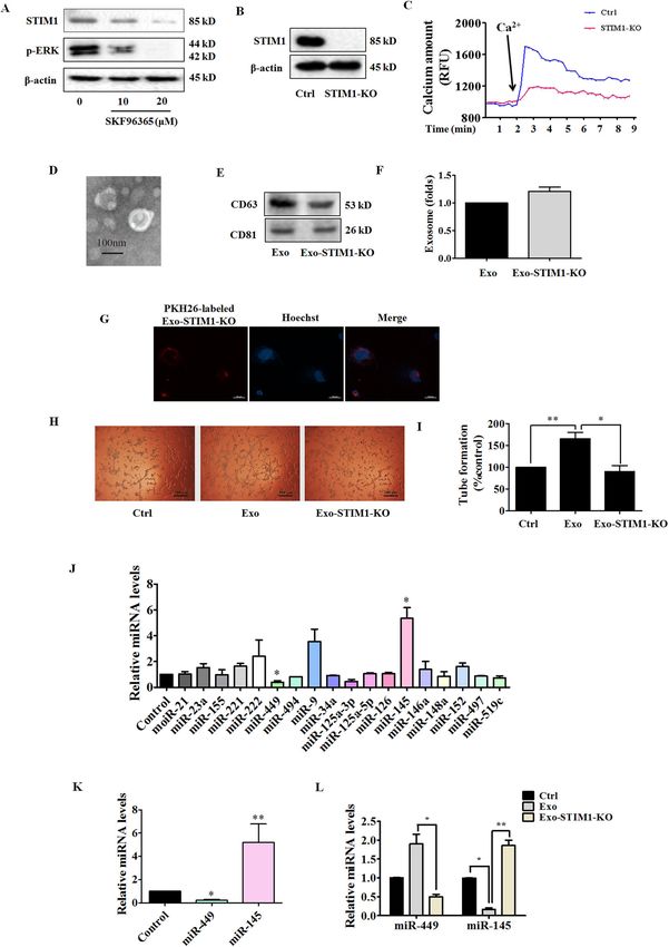
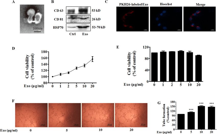
Pan et al. Cell Death and Disease (2021)12:38                                                                                       Page 2 of 15

small noncoding RNAs (~20–25 nt) that play important                       ionophore) or SKF96365 (a pharmacological store-
roles in cancer development as oncogenes or tumor                          operated Ca2+ influx inhibitor) to change the level of
suppressors7.                                                              intracellular Ca2+, and examined the effect of exosomes
  Intracellular calcium ions (Ca2+) are universal second                   from the treated MDA-MB-231 cells (Exo-A23187 or
messengers that are intimately related to a number of                      Exo-SKF) on angiogenesis of HUVECs. Furthermore, we
diverse cellular processes, including cell differentiation,                investigated the effect of exosomes from STIM1-knockout
proliferation, and apoptosis8. Store-operated Ca2+ entry                   MDA-MB-231 cells (Exo-STIM1-KO) on angiogenesis of
(SOCE) is the predominant Ca2+ entry mechanism                             HUVECs in vitro and Matrigel plug assay in vivo.
ubiquitous in different cell types, and stromal interac-
tion molecule 1 (STIM1) and Orai1 (also named                              Results
CRACM1) are responsible for SOCE. STIM1, a trans-                          Exosomes from MDA-MB-231 breast cancer cells promote
membrane protein located in the endoplasmic reticulum                      angiogenesis
(ER), activates Ca2+ influx through plasma membrane                           The exosome is a major player in cell–cell commu-
Ca2+ channels under stimuli, triggering a transient                        nication, in which many materials such as miRNA, RNA,
depletion of the intraluminal Ca2+ 9. Ca2+ store deple-                    and proteins can be effectively transferred from the donor
tion leads to a rapid translocation of STIM1 into puncta                   cells to the recipient cells11. We selected the triple-
that accumulate near the plasma membrane, and STIM1                        negative breast cancer MDA-MB-231 cells to study the
operates via interaction with Orai1 and regulates the                      effect of exosomes from cancer cells on angiogenesis in
SOCE10.                                                                    HUVECs. The typical cup-shaped morphology with a size
  The effect of Ca2+ level change on tumor angiogenesis                    range of 30–100 nm was observed under a transmission
through regulating exosome secretion has not been                          electron microscope (Fig. 1A). CD63, CD81, and HSP70,
reported. In the present study, we used A23187 (calcium                    all of which are known as exosome markers12, were

  Fig. 1 Exo promotes human umbilical vein endothelial cells (HUVEC) tube formation. A A representative transmission electron microscopic
  image of exosomes derived from MDA-MB-231 cells. B The protein markers of exosomes in Exo as determined by western blot. C The uptake of Exo
  labeled with PKH26 in HUVECs. D Effect of Exo on HUVEC viability. E Effect of Exo on MDA-MB-231 cell viability. F Exo promotes HUVEC tube
  formation. G Quantification of the results in (F), Data represent mean ± SEM from three independent experiments (n = 3). ***P < 0.001, compared
  with control (0 μg/ml).

Official journal of the Cell Death Differentiation Association
Pan et al. Cell Death and Disease (2021)12:38                                                                    Page 3 of 15

observed in MDA-MB-231 cells, and these MDA-MB-231                A23187 with a uniformly cup-shaped morphology within
cells-derived exosomes (Exo) (Fig. 1B).                           30–100 nm as diameter (Fig. 2B). A23187-treated MDA-
   To study the biological function of exosomes from              MB-231 cells showed a significant increase in the number
breast cancer on endothelial cells, we treated HUVECs             of exosomes compared to normal MDA-MB-231 cells
with Exo. When Exo labeled with PKH26 (red) was                   (Fig. 2C, D), and HUVECs also exhibited an efficient
incubated with HUVECs, the effective uptake of Exo by             uptake of Exo-A23187 (Fig. 2E). Next, we determined the
HUVECs was observed by electron microscopy (Fig. 1C).             effects of Exo and Exo-A23187 on in vitro angiogenesis of
Next, we determined the effect of Exo on cell proliferation       HUVECs. In the tube-formation assay, HUVECs were
of MDA-MB-231 and HUVECs. Figure 1D, E showed that                incubated with equivalent concentration (10 μg/ml) of
Exo increased proliferation of HUVECs in a                        Exo and Exo-A23187 for 24 h and seeded onto matrigel.
concentration-dependent manner, but without effect on             The results showed that the Exo-A23187 significantly
that of MDA-MB-231, after co-incubation for 24 h. These           increased tube formation by HUVECs compared to the
results revealed that MDA-MB-231-derived self exosomes            Exo-treated group, with the tube length elevated by 197 ±
(Exo) did not affect the proliferation of MDA-MB-231              8% (Fig. 2F, G). Cell migration was assessed by the scratch
cells, but increased the proliferation of endothelial cells. It   wound-healing assay. The migration ability of HUVECs
is known that tumor growth and metastasis need glorious           was enhanced in the Exo group compared with the con-
angiogenesis for nutrition provision. Therefore, we               trol, and the Exo-A23187 group was further enhanced
investigated the effects of Exo on HUVECs angiogenesis            compared with Exo by 28 ± 4% (Fig. 2H, I). These results
by measuring tube formation on Matrigel, after treatment          demonstrated that A23187 elevated intracellular calcium
for 24 h. Compared to the control group, Exo obviously            and therefore increased the release of exosomes from
increased HUVECs tube formation (Fig. 1F, G). The                 MDA-MB-231 cells, resulting in an increase of HUVEC
optimal Exo concentration of 10 μg/ml for angiogenesis            tube formation and migration.
was then used in subsequent experiments.
                                                                  Exo-SKF suppresses HUVEC tube formation and migration
Exo-A23187 promotes HUVEC proliferation, tube                       Inhibition of tumor angiogenesis might halt tumor
formation, and migration                                          progression. The above results showed that exosomes
  Tumorigenic pathways are associated with abnormal               from breast cancer cells with higher intracellular Ca2+
activation of Ca2+ channels13, and the increase of intra-         promoted HUVEC angiogenesis. We hypothesized that
cellular Ca2+ stimulates exosome secretion14. A23187 is a         exosomes from cancer cells with lower intracellular Ca2+
calcium ionophore which can be used to increase intra-            might exhibit an anti-angiogenic effect. Then we used
cellular Ca2+ level in tumor cells. Because prolonged             SKF96365, a pharmacological store-operated Ca2+ influx
intracellular elevation of Ca2+ might result in cell death,       inhibitor, to reduce intracellular Ca2+ level. We found
we first examined the effect of A23187 on cell viability,          that SKF96365 reduced the cell viability and augmented
apoptosis, and cell cycle. As a result, A23187 reduced cell       cell apoptosis at concentrations of 20 μM and 50 μM
viability, induced apoptosis, and G0/G1 cell cycle arrest at      (Supplementary Fig. S2a–c). Therefore, we chose the non-
high concentrations, and elevated intracellular Ca2+ level        cytotoxic but effective concentration of 10 μM for the
in a concentration-dependent manner (Supplementary                following experiments. We first examined the inhibitory
Fig. S1a–f). We also found that A23187 promoted the               effect of SKF96365 on Ca2+ entry in MDA-MB-231 cells.
migration of MDA-MB-231 cells at non-cytotoxic con-               MDA-MB-231 were stained with FluoForteTM dye-
centrations of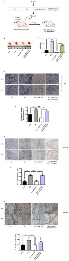
Pan et al. Cell Death and Disease (2021)12:38                                                                                       Page 4 of 15

  Fig. 2 Exo-A23187 promotes human umbilical vein endothelial cells (HUVEC) proliferation, tube formation, and migration. A Effect of
  A23187 on intracellular Ca2+ level in MDA-MB-231 cells was determined with FluoForte Calcium Assay Kit. B Representative electron microscopic
  image of Exo-A23187. C The markers of exosomes in Exo or Exo-A23187 as determined by western blot. D A23187 increased the production of
  exosomes in MDA-MB-231 cells. E Images of intercellular trafficking of exosomes by isolated Exo-A23187 labeled with PKH26 in HUVECs. F Tube
  formation was increased in the Exo-A23187 group compared with the control and Exo group. G Quantification of the tube formation results in (F).
  H The migration of HUVECs was increased in the Exo-A23187 group compared with the control and Exo group. I Quantification of the HUVEC
  migration results in (h). Data represent mean ± SEM from three independent experiments (n = 3). *P < 0.05, **P < 0.01.

the uptake of Exo-SKF by HUVECs was efficient (Fig. 3E).                    that intracellular Ca2+ regulated the release of exosomes
And the treatment of Exo-SKF significantly suppressed                       and affected tumor angiogenesis.
the tube-formation ability of HUVECs compared with Exo
(Fig. 3F, G). In addition, the migration ability of HUVECs                 Exo-SKF affects the levels of miR-145 and miR-449
was suppressed in the Exo-SKF group compared with Exo                       Exosomes released from tumor cells contain miRNAs
treatment only (Fig. 3H, I). These results demonstrated                    which could even affect the environment surrounding the

Official journal of the Cell Death Differentiation Association
Pan et al. Cell Death and Disease (2021)12:38                                                                Page 5 of 15

tumor. miRNAs work via translational inhibition or              generate the Exo-STIM-KO, and examined the anti-
degradation of their target mRNAs to downregulate gene          angiogenic effects on HUVECs. A schematic diagram of
expression. Thus, we determined the levels of miRNAs            gRNA-targeting exon 4 of the STIM1 gene is shown in
relating to angiogenesis in SKF96365-treated MDA-MB-            Supplementary Fig. S3a. We first confirmed the sequence
231 cells. We measured expression of 17 human miRNAs            of exon 4 in STIM1 in MDA-MB-231 cells with Sanger
including miR-21, miR-23a, miR-155, miR-221, miR-222,           sequencing, which revealed that MDA-MB-231 cells
miR-449, miR-494, miR-9, miR-34a, miR-125a-3p, miR-             carried 1-bp deletion at the gRNA-targeting region
125a-5p, miR-126, miR-145, miR-146a, miR-148a, miR-             (Supplementary Fig. S3b). After Sanger sequencing vali-
497, and miR-519c, which act as pro-angiogenic or anti-         dation, we obtained STIM1-knockout MDA-MB-231 cells
angiogenic factors16–21 or are dysregulated in breast           (STIM1-KO). To further determine the effectiveness of
cancer22–25. MDA-MB-231 cells were treated with                 CRISPR/Cas9, a western blot was carried out to detect
SKF96365 for 24 h. We found a significantly higher               STIM1 expression. STIM1-CRISPR/Cas9 gene-edited
expression of miR-145 and lower expression of miR-449           MDA-MB-231 cells showed no expression of STIM1
in SKF96365-treated MDA-MB-231 cells (Fig. 3J). Fur-            (Fig. 4B). The lack of STIM1 led to a significant drop in
thermore, we isolated Exo from the SKF96365-treated             calcium amount, which was assessed in the presence of
MDA-MB-231 cells and found that miR-145 was highly              additional Ca2+ (Fig. 4C). The characteristic morphology
expressed and miR-449 barely detected (Fig. 3K). Similar        of Exo-STIM1-KO was observed under an electron
results were also found in Exo-SKF-treated HUVECs. The          microscope (Fig. 4D), and both CD63 and CD81 were
miR-449 level was elevated in Exo-treated group com-            found in Exo-STIM1-KO (Fig. 4E). An equal number of
pared to the control, and Exo-SKF alleviated such effect.       MDA-MB-231 and STIM1-deficient MDA-MB-231 cells
In contrast, the miR-145 level was reduced in Exo-treated       were incubated in culture dishes, and then the exosomes
group, and Exo-SKF enhanced such effect (Fig. 3L). Since        were isolated respectively. The amount of Exo-STIM1-
miR-145 affected more obviously than miR-449, we                KO did not significantly change compared with Exo (Fig.
focused on miR-145 in the subsequent experiments.               4F). Exo-STIM1-KO was also labeled with PKH26 red
                                                                dye, and the uptake by HUVECs was observed (Fig. 4G).
Exo-SKF suppresses insulin receptor substrate 1 (IRS1)          We then examined whether Exo-STIM1-KO could affect
signaling pathway                                               angiogenesis. HUVECs were incubated with Exo or Exo-
  miRNAs regulate post-transcriptional expression by            STIM1-KO for 24 h. As expected, Exo-STIM1-KO
gene silencing. As the predicted target of miR-14526, IRS1      repressed the tube formation of HUVECs compared to
activates phosphatidylinositol 3-kinase (PI3K)/Akt path-        Exo treatment only (Fig. 4H, I). Therefore, our results
way, and mitogen-activated protein kinase (MAPK)                suggest that STIM1-deficient MDA-MB-231 cells inhibit
pathway to promote endothelial cell proliferation,              HUVEC angiogenesis through the exosomes.
migration, as well as vascular permeability27. Thus, we
investigated the effect on IRS1 signaling pathways by           Knockout of STIM1 upregulates miR-145
evaluating the changes of c-Raf, extracellular signal             To further elucidate the molecular mechanism of the
regulated-protein kinase (ERK), p38, Akt, and mammalian         anti-angiogenic effect induced by Exo-STIM1-KO, we
target of rapamycin (mTOR). HUVECs were treated with            also analyzed the levels of 17 human miRNAs in STIM1-
Exo or Exo-SKF for 24 h, and cells were lysed for western       deficient MDA-MB-231 cells, Exo-STIM1-KO, and
blot. As shown in Fig. 3M, N, we observed upregulation of       HUVECs treated with Exo-STIM1-KO. Among 17 miR-
IRS1 and its downstream signal proteins such as c-Raf,          NAs, miR-145 was upregulated and miR-449 was down-
ERK, p38, Akt, and mTOR phosphorylation in HUVECs               regulated in STIM1-deficient MDA-MB-231 cells, Exo-
treated with Exo, but downregulation in those treated           STIM1-KO, and Exo-STIM1-KO-treated HUVECs (Fig.
with Exo-SKF.                                                   4J–L). Moreover, miR-145 changed more obviously than
                                                                miR-449. These results are consistent with those in Exo-
Exo-STIM1-KO suppresses HUVEC tube formation                    SKF-treated HUVECs.
  STIM1 plays an essential role in Ca2+ mobilization and
signaling28. As the ER Ca2+ sensor, STIM1 controls              Exosomal miR-145 from STIM-KO-MDA-MB-231 cells
plasma membrane Ca2+ channels to regulate Ca2+ entry.           targets IRS1
After MDA-MB-231 cells were treated with SKF96365 for             We subsequently examined the effect of exosomal miR-
24 h, the expression of STIM1 and phosphorylation of            145 from STIM1-KO-MDA-MB-231 cells on the IRS1
ERK were downregulated (Fig. 4A). To investigate the role       pathway, which is the key pathway regulating tumor
of STIM1 in the reduction of angiogenesis by Exo-SKF,           angiogenesis. HUVECs were incubated with Exo or Exo-
we edited exon 4 of STIM1 locus (ENSG00000167323) in            STIM1-KO for 24 h and collected for western blot. As
MDA-MB-231 cells by CRISPR/Cas9 gene editing to                 shown in Fig. 5A, B, IRS1 was increased by Exo, but

Official journal of the Cell Death Differentiation Association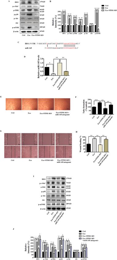
Pan et al. Cell Death and Disease (2021)12:38                                                                                          Page 6 of 15

  Fig. 3 Exo-SKF inhibits human umbilical vein endothelial cells (HUVEC) tube formation and migration through the miR-145 and IRS1
  pathways. A Representative time-course recording of intracellular Ca2+ fluorescence showing the inhibitory effect of SKF96365 on Ca2+ influx in
  MDA-MB-231 cells, which was determined with FluoForte Calcium Assay Kit. B Representative electron microscopic image of Exo-SKF. MDA-MB-231
  cells were treated with 10 μM of SKF96365. C The markers of exosomes in Exo and Exo-SKF, as determined by western blot. D SKF96365 did not affect
  the production of exosomes in MDA-MB-231 cells. E The uptake of Exo-SKF in HUVECs. F The Exo-SKF96365 inhibits tube formation in HUVECs.
  G Quantification of the results in (F). H Exo-SKF96365 reduces HUVEC migration. I Quantification of the results in (H). J Changes of miRNA level in
  MDA-MB-231 cells were detected by RT-qPCR after treatment with SKF96365. K The relative levels of miR-145 and miR-449 in Exo-SKF. L The relative
  levels of miR-145 and miR-449 in HUVECs after Exo or Exo-SKF treatment. M The expression of IRS1 and phosphorylation of c-Raf, ERK, p38, Akt, and
  mTOR in HUVECs after Exo or Exo-SKF treatment. N Quantification of the results in (M). Data represent mean ± SEM from three independent
  experiments (n = 3). *P < 0.05, **P < 0.01, and ***P < 0.001.

Official journal of the Cell Death Differentiation Association
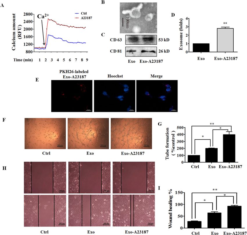
Pan et al. Cell Death and Disease (2021)12:38                                                                                                Page 7 of 15

  Fig. 4 Exo-STIM1-KO suppresses human umbilical vein endothelial cells (HUVEC) tube formation and increases miR-145 level. A Expression
  of STIM1 and phosphorylation of ERK in SKF96365-treated MDA-MB-231 cells. B Expression of STIM1 in STIM1-KO MDA-MB-231 cells. C Representative
  time-course recording of intracellular Ca2+ fluorescence in STIM1-KO-MDA-MB-231 cells. D Representative electron microscopic image of Exo-STIM1-
  KO. E The markers of exosomes in Exo-STIM1-KO, as determined by western blot. F The amount of Exo-STIM1-KO showed no significant change
  compared with Exo. G The uptake of Exo-STIM1-KO in HUVECs. HUVECs were cultured with PKH26-labeled Exo-STIM1-KO. H Tube formation was
  inhibited by Exo- STIM1-KO. I Quantification of the results in (H). J Relative levels of miRNAs in STIM1-silencing MDA-MB-231 cells. K The levels of miR-
  145 and miR-449 in Exo-STIM1-KO. L The levels of miR-145 and miR-449 in HUVECs after treatment with Exo and Exo-STIM1-KO. Data represent mean
  ± SEM from three independent experiments (n = 3). *P < 0.05, **P < 0.01.

Official journal of the Cell Death Differentiation Association
Pan et al. Cell Death and Disease (2021)12:38                                                                                              Page 8 of 15

  Fig. 5 Exo-STIM1-KO miR-145 inhibits angiogenesis through targeting IRS1 signal pathway in human umbilical vein endothelial cells
  (HUVECs). A Western blot analysis of IRS1 and its downstream signal proteins in HUVECs. B Quantification of the results in (A). C Schematic diagrams
  of miR-145 binding sites in the 3’-UTR of IRS1. D The levels of miR-145 in HUVECs after various treatments. E Images of tube formation of HUVECs after
  various treatments. F Quantification of the results in (E). G Representative migration of HUVECs after various treatments. H Quantification of the
  results in (G). I Western blot analysis of IRS1 and the downstream signal proteins in HUVECs treated with Exo-STIM1-KO plus/or miR-145 antagomir.
  J Quantification of the results in (I). Data represent mean ± SEM from three independent experiments (n = 3). *P < 0.05, **P < 0.01, and ***P < 0.001.

Official journal of the Cell Death Differentiation Association
Pan et al. Cell Death and Disease (2021)12:38                   Page 9 of 15

reduced by Exo-STIM1-KO. Furthermore, phosphoryla-
tion of IRS1 pathway proteins such as Raf, ERK, p38, Akt,
and mTOR was also elevated by Exo, but repressed by
Exo-STIM1-KO. These results are consistent with Exo-
SKF-treated HUVECs. Since several reports have shown
that miR-145 directly targets IRS129–32, and base-pairing
complement suggests that miR-145 binds on IRS1 3’-
untranslated regions (3’-UTR) (Fig. 5C), we hypothesized
that miR-145 might target IRS1 to play a pathological role
in the angiogenesis process. To confirm this hypothesis,
we used miR-145 antagomir to examine the angiogenic
effect of Exo-STIM1-KO in HUVECs. HUVECs were
treated with Exo, Exo-STIM1-KO, Exo-STIM1-KO plus
miR-145 antagomir, or miR-145 antagomir for 24 h. The
results showed that the Exo-STIM1-KO elevated level of
miR-145 compared to the Exo group, while Exo-STIM1-
KO plus miR-145 antagomir decreased the level of miR-
145 compared to the Exo-STIM1-KO group (Fig. 5D).
  Then HUVECs were treated with different exosomes for
24 h, and the tube formation and cell migration abilities
were investigated. Upregulation of tube formation by Exo
was suppressed by Exo-STIM1-KO, and Exo-STIM1-KO
plus miR-145 antagomir reversed this inhibition (Fig. 5E,
F). As shown in Fig. 5G, H, cell migration was significantly
suppressed in the Exo-STIM1-KO group in comparison
with the Exo group. Conversely, Exo-STIM1-KO plus
miR-145 antagomir group enhanced the migration ability
of HUVECs. These results suggested that miR-145 plays
an important role in endothelial cell migration mediated
by Exo-STIM1-KO.
  Next, we examined the expression of IRS1 pathway
proteins in HUVECs after incubation with different exo-
somes for 24 h. miR-145 antagomir upregulated the
expression of IRS1, and activated the IRS1-dependent
Akt/mTOR, Raf/ERK, and p38 MAPK. In addition, Exo-
STIM1-KO plus miR-145 antagomir obviously elevated
the expression of IRS1 and activated its downstream
molecules which had been suppressed by Exo-STIM1-KO
(Fig. 5I, J).

Exo-STIM1-KO prevents breast cancer angiogenesis in vivo
  To further determine the therapeutic potential of Exo-
STIM1-KO in vivo, we performed an in vivo Matrigel plug
assay to detect the newly formed blood vessels in the
transplanted gel plugs in BALB/c nude mice (Fig. 6A).
The hemoglobin content, which represents new vessel
formation, was significantly increased in Exo-treated mice
compared with the control group but was reduced in the
Exo-STIM1-KO group than the Exo-treated group. In
contrast, the hemoglobin concentration in the plugs
containing Exo-STIM1-KO plus miR-145 antagomir was
higher than the Exo-STIM1-KO group (Fig. 6B, C). H&E
staining assay revealed that Exo dramatically increased
plug vascularization compared to control, and Exo-STIM-

Official journal of the Cell Death Differentiation Association
Pan et al. Cell Death and Disease (2021)12:38 Page 10 of 15
Fig. 6 Exo-STIM1-KO inhibits angiogenesis in nude mice Matrigel HUVECs. Our results showed that treatment of MDA-
plug angiogenesis models. A Schematic description of the in vivo MB-231 cells with A23187 or SKF96365 in the presence
angiogenesis experiment. B Exo-STIM1-KO significantly reduced of extracellular Ca2+ led to an increase or decrease of
angiogenesis. Representative photographs of angiogenesis in the intracellular Ca2+, respectively. We also found that Exo-
nude mice are shown. C Plug hemoglobin amount representing A23187 promoted HUVECs migration and tube forma-
vascularity. D H&E staining of Matrigel plug sections. E Quantification
of the results in (D). F Representative immunohistochemical staining tion, and Exo-SKF inhibited them. These results sug-
of CD31. G Quantification of the results in (F). H Representative gested that change of Ca2+ level influenced exosome
immunohistochemical staining of IRS1. I Quantification of the results release from triple-negative breast cancer MDA-MB-231
in (H). Data represent mean ± SEM from three independent cells, and therefore affected angiogenesis of HUVECs.
experiments (n = 3). *P < 0.05, **P < 0.01, and ***P < 0.001.
STIM1 is a critical regulator of Ca2+ mobilization. Once
ER Ca2+ is depleted, the ER Ca2+ sensor STIM1 accu-
mulates in a junctional ER in close apposition to the
plasma membrane to activate the plasma membrane pore-
KO suppressed plug neovessel formation. However, Exo- forming unit Orai1, which induces SOCE36. Therefore, we
STIM-KO plus miR-145 antagomir reversed the Exo- deleted the STIM1 gene by CSISPR/Cas9, and found the
STIM-KO induced angiogenesis attenuation (Fig. 6D). intracellular Ca2+ was decreased in the STIM1-silencing
The blood vessel density which represents the relative MDA-MB-231 cells, the characteristic morphology and
tube length of the neovessels in Matrigel plugs was markers of exosomes were also observed. The exosome
quantified (Fig. 6E). Moreover, immunohistochemistry amounts did not change obviously.
analysis confirmed that the CD31 (a marker of the for- miRNAs are highly conserved small noncoding RNAs
mation of new vessels) positive cells in Exo-STIM1-KO- that induce post-transcriptional gene silencing canoni-
treated mice were significantly fewer than the Exo-treated cally by binding mRNA. miRNA/mRNA interaction hin-
mice, but increased when miR-145 antagomir was added ders protein synthesis and initiates mRNA degradation37.
(Fig. 6F, G). In addition, the expression of IRS1 was also Some miRNAs are known to be involved in tumorigenesis
lower in the Exo-STIM1-KO-treated mice compared with and tumor angiogenesis. For example, miR-34a is a tumor
the Exo group but was upregulated in Exo-STIM1-KO suppressor that is frequently downregulated in a number
plus miR-145 antagomir-treated mice (Fig. 6H, I). of tumor types, and ectopic expression of miR-34a in head
and neck squamous cell carcinoma (HNSCC) cell lines
Discussion inhibited tumor growth and tumor angiogenesis in vivo
Exosomes are known as potent cell–cell messenger. when introduced into SCID mouse xenograft models38;
Various cell types have the ability to release exosomes, miR-519c is a pivotal regulator of tumor angiogenesis39;
including immune cells, epithelial cells, and tumor cells. modulation of miR-126 expression can disrupt angio-
The mechanisms involved in the entry of exosomes in genesis and vascular integrity40. Some other miRNAs such
recipient cells include three main routes, such as recep- as miR-21, miR-221, and miR-222 are highly expressed in
tor/ligand signaling, fusion, phagocytosis/endocytosis33. the endothelial cells41. In the present study, we demon-
In this study, we found that PKH26-labeled Exo, Exo- strated that level of miR-145 increased in MDA-MB-231
A23187, Exo-SKF, and Exo-STIM1-KO from MDA-MB- cells treated with SKF, STIM1-KO, Exo-SKF, and Exo-
231 cells (donor cells) transferred to the HUVECs (reci- STIM1-KO, as well as in HUVECs treated with Exo-SKF
pient cells). Exosomes contribute to carcinogenesis by the and Exo-STIM1-KO. It was reported that miR-145 is
transport of oncogenic lipid, proteins, and nucleic acids, downregulated in breast cancer cancer42. These results
and the other bioactive molecules34. In this study, we support that Ca2+ sensor STIM1 might influence tumor
found that MDA-MB-231-derived exosomes promoted angiogenesis through regulating exosomal miR-145.
tube formation in HUVECs. The pathway of insulin-like growth factor I (IGF-I)/
Angiogenesis is a fundamental process involved in IRS1/Ras mediates vascular endothelial growth factor
carcinogenesis. Steps toward angiogenesis include endo- (VEGF) production, which is crucial in angiogenesis. IR is
thelial cell migration and proliferation, vascular tube expressed in endothelial cells as well as in cancer cells43.
formation, anastomosis of newly formed tubes35. An Insulin stimulation increases the tyrosine phosphorylation
increase of intracellular Ca2+ stimulates exosome secre- of IRS1 and IRS244. miR-145 has been repeatedly reported
tion14. However, the effects of exosomes from the breast to be a tumor suppressor, and to target gene IRS1 and
cancer cells with elevated or decreased Ca2+ conditions therefore inhibit its protein expression29. Through base-
on angiogenesis have not been reported yet. Therefore, we pairing complement, we also confirmed that IRS1 is the
treated MDA-MB-231 cells with calcium ionophore or potential target of miR-145. As expected, Exo promoted
inhibitor of store-operated Ca2+ channel, and isolated the IRS1 expression in HUVECs, which was attenuated by
exosomes to investigate their effects on angiogenesis in treatment with Exo-SKF or Exo-STIM1-KO. Tyrosin-
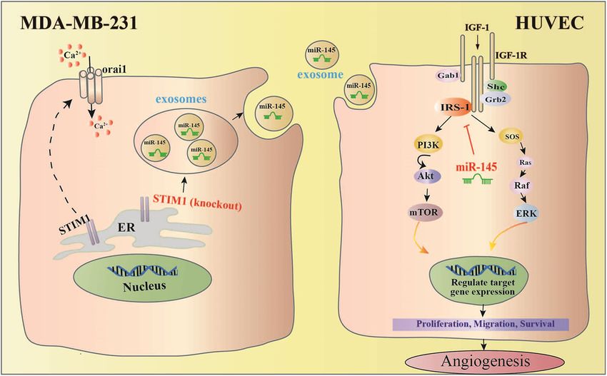
Official journal of the Cell Death Differentiation AssociationPan et al. Cell Death and Disease (2021)12:38 Page 11 of 15 Fig. 7 Exosomes from STIM1-KO-MDA-MB-231 breast cancer cells contain higher levels of miR-145, which targets IRS1 and its downstream signal to suppress tube formation of vascular endothelial cells, resulting in anti-angiogenesis effect. Predicted anti-angiogenic mechanism of exosomal miR-145 from STIM1-KO-MDA-MB-231 cells via targeting IRS1. phosphorylated IRS1 activates PI3K/Akt and Ras/Raf/ biomarker CD31, which was reversed by the addition of MAPK pathways45,46, which mediate endothelial cell miR-145 antagomir. These results confirmed that the proliferation, migration, survival, and vascular perme- exosomal miR-145 from STIM-KO-MDA-MB-231 cells ability47,48. In this study, we found that the phosphoryla- attenuated angiogenesis by targeting IRS1. tion of Raf, ERK, p38, Akt, and mTOR in HUVECs was Overall, our studies have demonstrated that exosomes increased by treatment with Exo, but was attenuated by from breast cancer cells with a lower level of Ca2+ contain concomitant treatment with SKF (Exo-SKF) and STIM1- more miR-145, which targets IRS1 to exhibit an anti- KO (Exo-STIM1-KO). These results suggest that miR-145 angiogenic effect (Fig. 7). Our results suggest that the might target IRS1 and therefore inhibit the angiogenesis reduction of Ca2+ level in cancer cells might contribute to of HUVECs via regulating IRS1/PI3K/Akt/mTOR and anti-angiogenic tumor therapy. IRS1/Raf/ERK pathways. We next used miR-145 antagomir to demonstrate Materials and methods whether Exo-STIM1-KO miR-145 could reduce angio- Cell culture and reagents genesis through directly repressing IRS1. The level of MDA-MB-231 cells were purchased from the cell bank miR-145 in Exo-STIM1-KO plus miR-145 antagomir of the Chinese academy of sciences (Shanghai, China). No group is lower than that in the Exo-STIM1-KO group. contamination of mycoplasma was confirmed by PCR test. Suppression of miR-145 in recipient HUVECs by miR-145 Cells were cultured in DMEM supplemented with 10% antagomir transfection abolished the anti-angiogenic fetal bovine serum (FBS), penicillin and streptomycin at effect of Exo-STIM1-KO in HUVECs, suggesting that 37 °C in a humidified atmosphere containing 5% CO2. miR-145 might play pivotal roles in the Exo-STIM1-KO DMEM, FBS, the enhanced chemiluminescence (ECL) attenuated tumor angiogenesis effect. reagent, and Total exosome isolation reagents were pur- The in vivo Matrigel plug assay is widely used to eval- chased from Thermo Fisher Scientific (4478359; Wal- uate the in vivo angiogenic potential49. To date, there has tham, MA, USA). Matrigel was purchased from BD been no report on the anti-angiogenic effects of exosomes Biosciences (San Josè, CA, USA). PKH26 Red fluorescent derived from STIM1-KO-tumor cells. By using in vivo cell linker kit was purchased from Sigma Chemicals (St. Matrigel plug assay, we found that Exo-STIM1-KO sig- Louis, MO, USA). TRIzol reagent was obtained from nificantly inhibited Exo-induced angiogenesis. Con- Invitrogen (Carlsbad, CA). The antibodies specific for sistently, immunohistochemical analysis showed a phospho-c-Raf (#9427), phospho-ERK (#4377), phospho- reduction of expression of IRS1 and angiogenesis p38 (#9211), phospho-Akt (#9271), Akt (#9272), Official journal of the Cell Death Differentiation Association
Pan et al. Cell Death and Disease (2021)12:38                                                                  Page 12 of 15

phospho-mTOR (#2971), STIM1 (#A5668) β-actin                    Tube-formation assay
(#4967), and the horseradish peroxidase-conjugated goat           HUVECs (1 × 10⁵ cells/well) were treated with exo-
anti-rabbit secondary antibody were purchased from Cell         somes derived from MDA-MB-231 cells after various
Signaling Technology, Inc. (Danvers, MA, USA). The              treatments for 24 h and then plated on MatrigelTM Matrix
anti-IRS1 (ab40777) and anti-CD31 (ab28364) antibodies          (50 μl/well) (BD Biosciences, San Josè, CA, USA) in 96-
were obtained from Abcam (Cambridge, MA, USA). The              well plates for 6 h at 37 °C in a 5% CO2 humidified
exosome-specific primary antibodies including CD9,               incubator. Tube formation was observed and imaged with
CD63, CD81, and HSP70 were obtained from System                 phase-contrast microscopy. For quantification, total tub-
Biosciences (EXOAB-KIT-1, Palo Alto, CA, USA).                  ular length and branch points per well were determined
                                                                using ImageJ software.
Cell proliferation assay
  Cell proliferation was assessed using an MTT assay as         Protein extraction and western blot
we previously reported50 with a small modification.                Western blot analysis was carried out as we previously
Briefly, cells were seeded onto 96-well plates and cultured      reported51 with a small modification. Cells were collected,
with exosomes from MDA-MB-231 cells for 24 h, and               and the protein concentration of each sample was deter-
then MTT was added to each well. After 4 h of incubation,       mined by the BCA protein assay kit. Equal amount of
the produced formazan was dissolved in DMSO, and OD             protein was separated by sodium dodecyl sulfate-
at 490 nm was monitored using microplate reader iMark           polyacrylamide gel electrophoresis (SDS-PAGE) and
(BIO-RAD, Hercules, CA, USA).                                   transferred to the PVDF membrane. After being blocked
                                                                with 5% skim milk, the membranes were incubated with
Isolation of exosomes                                           each primary antibody, and then the horseradish
  MDA-MB-231 cells were seeded at a density of 2 × 10⁶          peroxidase-conjugated secondary antibody. The signals
cells/100-mm dish and cultured for 6 h or 24 h without or       were detected with ChemiDocTM XRS + System (BIO-
with the treatment of A23187 (500 nM) or SKF96365               RAD, Hercules, CA, USA) after exposure to ECL reagent.
(10 μM) in serum-free medium. Exo, Exo-A23187, Exo-
SKF96365, and Exo-STIM1-KO in cell culture super-               Fluorescent imaging of exosome uptake
natants were isolated by use of total exosome isolation           Freshly isolated exosomes from MDA-MB-231 cells
reagents as described by the manufacturer. Exosome              were labeled with the PKH26 red fluorescent cell linker kit
protein amount was quantified using the PierceTM BCA             (Sigma-Aldrich, St. Louis, MO, USA) according to the
protein assay kit (23225; Thermo Fisher Scientific, Wal-         manufacturer’s instructions with a small modification,
tham, MA, USA).                                                 and then cultured with HUVEC for 3 h. After being
                                                                washed, HUVECs were stained with Hoechst for 15 min.
Wound-healing assay                                             The pictures were taken and the uptake of exosomes was
  HUVECs were cultured on 24-well plates (2 × 10⁵ cells/        observed using a fluorescence confocal microscope.
well) in DMEM medium. Wounds were made using a
sterile 10-µl pipette tip. After the cellular debris was        Generation of genetically modified cells using CRISPR/Cas9
removed by gently washing with PBS, the cells were cul-         genome editing
tured in high glucose serum-free DMEM with various                The knockout of STIM1 in MDA-MB-231 cell lines was
exosomes at 37 °C with 5% CO2. At least four images of          achieved by the CRISPR/Cas9 system. Oligonucleotide tar-
the scraped area were captured using phase-contrast             geting exon 4 (5’-ATACAATTGGACCGTGGATG-3’) was
microscopy after 24 h treatment, and cell migration dis-        designed as the CRISPR target site. MDA-MB-231 cells were
tances were determined using ImageJ software.                   transfected with the plasmid of the STIM1-targeted gRNA
                                                                encoding SpCas9 by Lipofectamine 3000. Following 72 h of
Intracellular Ca2+ measurement                                  puromycin selection, the cell culture was extended for 96 h
  Intracellular Ca2+ level was determined with the Fluo-        without puromycin, and the viable clonal cells were sub-
Forte Calcium Assay Kit (Enzo Life Sciences, Ann Arbor,         cultured in a 96-well plate with a density of one cell/well.
MI, United States). Briefly, MDA-MB-231 or STIM1-                Afterward, cells were cultured for 7–10 days. Individual
knockout MDA-MB-231 cells were cultured on 96 poly-             clones were expanded and screened for STIM1 depletion by
D-lysin-coated glass bottom plate, stained with Fluo-           genomic DNA sequencing and immunoblotting.
ForteTM dye-loading solution for 1 h, and then treated
with A23187 or SKF96365 for 1 h. After the addition of          miRNA isolation and real-time quantitative reverse
CaCl2 (8 mM), the fluorescence was determined with a             transcription-PCR (RT-qPCR) assay
multilabel plate reader VICTOR (Perkin Elmer, Waltham,            The total RNA from the cells and exosomes was isolated
MA, USA) at Ex = 485 nm/Em 535 nm at 20-s interval.             using the TRIzol reagent (Life Technologies, Carlsbad,

Official journal of the Cell Death Differentiation Association
Pan et al. Cell Death and Disease (2021)12:38 Page 13 of 15
Table 1 The sequences of several miRNA primers.
miRNA RT-Primer Forward Reverse
hsa-miR-21 gtcgtatccagtgcagggtccgaggtattcgcactggatacgactcaaca gcgcgtagcttatcagactga agtgcagggtccgaggtatt
hsa-miR-23a gtcgtatccagtgcagggtccgaggtattcgcactggatacgacggaaat gcgatcacattgccaggg agtgcagggtccgaggtatt
hsa-miR-155 gtcgtatccagtgcagggtccgaggtattcgcactggatacgacaacccc cgcgttaatgctaatcgtgata agtgcagggtccgaggtatt
hsa-miR-221 gtcgtatccagtgcagggtccgaggtattcgcactggatacgacgaaacc cgcgagctacattgtctgctg agtgcagggtccgaggtatt
hsa-miR-222 gtcgtatccagtgcagggtccgaggtattcgcactggatacgacacccag gcgcgagctacatctggcta agtgcagggtccgaggtatt
hsa-miR-449 gtcgtatccagtgcagggtccgaggtattcgcactggatacgacaccagc cgcgtggcagtgtattgtta agtgcagggtccgaggtatt
hsa-miR-494 gtcgtatccagtgcagggtccgaggtattcgcactggatacgacgaggtt cgcgtgaaacatacacggga agtgcagggtccgaggtatt
hsa-miR-9 gtcgtatccagtgcagggtccgaggtattcgcactggatacgactcatac gcgcgtctttggttatctagct agtgcagggtccgaggtatt
hsa-miR-34a gtcgtatccagtgcagggtccgaggtattcgcactggatacgacacaacc cgcgtggcagtgtcttagct agtgcagggtccgaggtatt
hsa-miR-125a-3p gtcgtatccagtgcagggtccgaggtattcgcactggatacgacagctcc gcgacgggttaggctcttg agtgcagggtccgaggtatt
hsa-miR-125a-5p gtcgtatccagtgcagggtccgaggtattcgcactggatacgactcacaa cgcgtccctgagaccctaac agtgcagggtccgaggtatt
hsa-miR-126 gtcgtatccagtgcagggtccgaggtattcgcactggatacgaccgcatt cgcgtcgtaccgtgagtaat agtgcagggtccgaggtatt
hsa-miR-145 gtcgtatccagtgcagggtccgaggtattcgcactggatacgacagggat cggtccagttttcccagga agtgcagggtccgaggtatt
hsa-miR-146a gtcgtatccagtgcagggtccgaggtattcgcactggatacgacaaccca cgcgtgagaactgaattcca agtgcagggtccgaggtatt
hsa-miR-148a gtcgtatccagtgcagggtccgaggtattcgcactggatacgacacaaag gcgcgtcagtgcactacagaa agtgcagggtccgaggtatt
hsa-miR-497 gtcgtatccagtgcagggtccgaggtattcgcactggatacgacacaaac gcgcagcagcacactgtg agtgcagggtccgaggtatt
hsa-miR-519c gtcgtatccagtgcagggtccgaggtattcgcactggatacgacatcctc gcgcgaaagtgcatcttttta agtgcagggtccgaggtatt
U6 ttcacgaatttgcgtgtcatc cgcttcggcagcacatatac ttcacgaatttgcgtgtcatc
CA, USA) or E.Z.N.A.TM miRNA Kit (Omega Bio-Tek, the Institutional Animal Care and Use Committee
Norcross, GA, USA), respectively. The cDNA synthesis guidelines. Mice were randomly assigned to four groups
was performed using M-MLV reverse transcriptase. Real- with three mice in each group. Briefly, 2 × 10⁶ HUVECs
time PCR was performed using the miScript SYBR Green were mixed with Exo, Exo-STIM1-KO, miR-145
PCR Kit on a CFX96TM Real-Time PCR Detection System antagomir, or antagomir NC, respectively. Then Lipo-
(BIO-RAD, Hercules, CA, USA). The sequences of several fectamine 6000 was added. The cell suspensions were
miRNA primers are described in Table 1. The expression mixed with 400 μl of High Concentration MatrigelTM
levels of U6 were used as an endogenous control for each Matrix at a ratio of 1:4, and then the mixture was
sample. The relative gene expression levels were calcu- subcutaneously injected into the dorsal region of nude
lated using the comparative Ct (ΔΔCt) method. mice (female, 6-week-old BALB/c). After 10 days, the
Matrigel plugs were harvested and processed for ana-
miRNA antagomir transfection lysis. The degree of vascularization was evaluated by
HUVECs were cultured on 6-well plates (1 × 10⁵ cells/ determining hemoglobin content. On the other hand,
well), and transfected with miR-145 antagomir or antag- plugs were fixed with 4% formaldehyde and embedded
omir negative control (NC) using Lipofectamine 6000 in paraffin, and stained with hematoxylin and eosin.
according to the manufacturer’s instructions. Simulta- The vessel area was quantified as mean relative tube
neously, Exo-STIM1-KO was added. After 24 h, cells were length by image analysis of six random microscopic
washed twice by PBS to remove remaining exosomes or fields using ImageJ software.
antagomir and collected to extract proteins and RNA.
Chemically modified miR-145 antagomir was purchased Statistical analysis
from Shanghai GenePharma Co., Ltd. All values are expressed as means ± SEM of triplicate
values. One-way ANOVA followed by Tukey’s Multiple
Matrigel plug assay for angiogenesis in nude mice Comparison Test was utilized to determine the statistical
All animal experiments were conducted at Laboratory significance with GraphPad Prism 5 (GraphPad, San
Animal Center, Institute of Radiation Medicine, Chi- Diego, CA, USA). Differences were considered statistically
nese Academy of Medical Sciences in accordance with significant when P < 0.05.
Official journal of the Cell Death Differentiation AssociationPan et al. Cell Death and Disease (2021)12:38 Page 14 of 15
Acknowledgements 17. Zou, C. et al. MiR-145 inhibits tumor angiogenesis and growth by N-RAS and
This study was supported by grants from the National Natural Science VEGF. Cell Cycle 11, 2137–2145 (2012).
Foundation of China (81672809, 81673464, 81373441), a grant for Major Project 18. Tiwari, A., Mukherjee, B. & Dixit, M. MicroRNA key to angiogenesis
of Tianjin for New Drug Development (17ZXXYSY00050). regulation: miRNA biology and therapy. Curr. Cancer Drug Targets 18,
266–277 (2018).
Author contributions 19. Welten, S. M., Goossens, E. A., Quax, P. H. & Nossent, A. Y. The multi-
M.J. and D.K. designed the experiments and acquired funding for the study; S. factorial nature of microRNAs in vascular remodelling. Cardiovasc. Res.
P., X.Z., C.S., and B.F. performed the experiments; Y.H., N.Z., and X.D. analyzed 110, 6–22 (2016).
the data; Z.Z., Y.Q., and R.W. provided technical assistances; M.J. wrote the 20. Wu, Z. et al. miR-497 suppresses angiogenesis in breast carcinoma by tar-
paper; D.K. edited the paper. geting HIF-1alpha. Oncol. Rep. 35, 1696–1702 (2016).
21. Yu, J. et al. MiR-148a inhibits angiogenesis by targeting ERBB3. J. Biomed. Res.
Ethics approval and consent to participate 25, 170–177 (2011).
This study does not involve human participants, human data, or human tissue. 22. Cosentino, G., Plantamura, I., Cataldo, A. & Iorio, M. V. MicroRNA and oxidative
Therefore, ethical approval is not necessary. stress interplay in the context of breast cancer pathogenesis. Int. J. Mol. Sci. 20,
5143 (2019).
23. Zhang, Z. et al. Downregulation of microRNA-449 promotes migration and
Conflict of interest
invasion of breast cancer cells by targeting tumor protein D52 (TPD52). Oncol.
The authors declare that they have no conflict of interest.
Res. 25, 753–761 (2017).
24. Etikala, D. M., Liu, R. & Wang, L. FOXP3-microRNA-146-NF-kappaB as onco-
target. Oncoscience 2, 839–840 (2015).
Publisher’s note 25. Jiang, Q. et al. MicroRNA-148a inhibits breast cancer migration and invasion by
Springer Nature remains neutral with regard to jurisdictional claims in directly targeting WNT-1. Oncol. Rep. 35, 1425–1432 (2016).
published maps and institutional affiliations. 26. Law, P. T. et al. MiR-145 modulates multiple components of the insulin-like
growth factor pathway in hepatocellular carcinoma. Carcinogenesis 33,
Supplementary Information accompanies this paper at (https://doi.org/ 1134–1141 (2012).
10.1038/s41419-020-03304-0). 27. Deshpande, N., Pysz, M. A. & Willmann, J. K. Molecular ultrasound assessment
of tumor angiogenesis. Angiogenesis 13, 175–188 (2010).
Received: 29 June 2020 Revised: 25 November 2020 Accepted: 27 28. Roos, J. et al. STIM1, an essential and conserved component of store-operated
November 2020 Ca2+ channel function. J. Cell Biol. 169, 435–445 (2005).
29. Shi, B. et al. Micro RNA 145 targets the insulin receptor substrate-1 and inhibits
the growth of colon cancer cells. J. Biol. Chem. 282, 32582–32590 (2007).
30. Xing, A. Y. et al. Deregulated expression of miR-145 in manifold human cancer
cells. Exp. Mol. Pathol. 95, 91–97 (2013).
References 31. Wang, Y. et al. MicroRNA-145 suppresses hepatocellular carcinoma by tar-
1. Torre, L. A. et al. Global cancer statistics, 2012. CA Cancer J. Clin. 65, 87–108 geting IRS1 and its downstream Akt signaling. Biochem. Biophys. Res. Commun.
(2015). 446, 1255–1260 (2014).
2. Carey, L. et al. Triple-negative breast cancer: disease entity or title of con- 32. Guo, Y. et al. Up-regulated miR-145 expression inhibits porcine preadipocytes
venience? Nat. Rev. Clin. Oncol. 7, 683–692 (2010). differentiation by targeting IRS1. Int. J. Biol. Sci. 8, 1408–1417 (2012).
3. Ramjiawan, R. R., Griffioen, A. W. & Duda, D. G. Anti-angiogenesis for cancer 33. Guay, C. & Regazzi, R. Exosomes as new players in metabolic organ cross-talk.
revisited: Is there a role for combinations with immunotherapy? Angiogenesis Diabetes Obes. Metab. 19, 137–146 (2017).
20, 185–204 (2017). 34. Marshall, H. T. & Djamgoz, M. B. A. Immuno-oncology: emerging targets and
4. Thery, C., Zitvogel, L. & Amigorena, S. Exosomes: composition, biogenesis and combination therapies. Front. Oncol. 8, 315 (2018).
function. Nat. Rev. Immunol. 2, 569–579 (2002). 35. Rajabi, M. & Mousa, S. A. The role of angiogenesis in cancer treatment. Bio-
5. Bae, S., Brumbaugh, J. & Bonavida, B. Exosomes derived from cancerous and medicines 5, 34 (2017).
non-cancerous cells regulate the anti-tumor response in the tumor micro- 36. Lunz, V., Romanin, C. & Frischauf, I. STIM1 activation of Orai1. Cell Calcium 77,
environment. Genes Cancer 9, 87–100 (2018). 29–38 (2019).
6. Guo, W. et al. Exosomes: new players in cancer (review). Oncol. Rep. 38, 37. Romano, G., Veneziano, D., Acunzo, M. & Croce, C. M. Small non-coding RNA
665–675 (2017). and cancer. Carcinogenesis 38, 485–491 (2017).
7. Adams, B. D., Kasinski, A. L. & Slack, F. J. Aberrant regulation and function of 38. Kumar, B. et al. Dysregulation of microRNA-34a expression in head and neck
microRNAs in cancer. Curr. Biol. 24, R762–R776 (2014). squamous cell carcinoma promotes tumor growth and tumor angiogenesis.
8. Berridge, M. J., Lipp, P. & Bootman, M. D. The versatility and universality of PLoS ONE 7, e37601 (2012).
calcium signalling. Nat. Rev. Mol. Cell Biol. 1, 11–21 (2000). 39. Cha, S. T. et al. MicroRNA-519c suppresses hypoxia-inducible factor-1alpha
9. Liou, J. et al. STIM is a Ca2+ sensor essential for Ca2+-store-depletion-triggered expression and tumor angiogenesis. Cancer Res. 70, 2675–2685 (2010).
Ca2+ influx. Curr. Biol. 15, 1235–1241 (2005). 40. Fish, J. E. et al. miR-126 regulates angiogenic signaling and vascular integrity.
10. Humeau, J. et al. Calcium signaling and cell cycle: progression or death. Cell Dev. Cell 15, 272–284 (2008).
Calcium 70, 3–15 (2018). 41. Kuehbacher, A., Urbich, C., Zeiher, A. M. & Dimmeler, S. Role of Dicer and
11. Wan, Z. et al. Exosome-mediated cell-cell communication in tumor progres- Drosha for endothelial microRNA expression and angiogenesis. Circ. Res. 101,
sion. Am. J. Cancer Res. 8, 1661–1673 (2018). 59–68 (2007).
12. Jia, Y. et al. Exosome: emerging biomarker in breast cancer. Oncotarget 8, 42. Iorio, M. V. et al. MicroRNA gene expression deregulation in human breast
41717–41733 (2017). cancer. Cancer Res. 65, 7065–7070 (2005).
13. Monteith, G. R., Davis, F. M. & Roberts-Thomson, S. J. Calcium channels and 43. King, G. L. & Johnson, S. M. Receptor-mediated transport of insulin across
pumps in cancer: changes and consequences. J. Biol. Chem. 287, 31666–31673 endothelial cells. Science 227, 1583–1586 (1985).
(2012). 44. Jiang, Z. Y. et al. Characterization of selective resistance to insulin signaling in
14. Savina, A., Furlan, M., Vidal, M. & Colombo, M. I. Exosome release is regulated the vasculature of obese Zucker (fa/fa) rats. J. Clin. Investig. 104, 447–457
by a calcium-dependent mechanism in K562 cells. J. Biol. Chem. 278, (1999).
20083–20090 (2003). 45. Obata, T. et al. Insulin signaling and its regulation of system A amino acid
15. Cui, C., Merritt, R., Fu, L. & Pan, Z. Targeting calcium signaling in cancer therapy. uptake in cultured rat vascular smooth muscle cells. Circ. Res. 79, 1167–1176
Acta Pharm. Sin. B 7, 3–17 (2017). (1996).
16. Harquail, J., Benzina, S. & Robichaud, G. A. MicroRNAs and breast cancer 46. Liang, L., Jiang, J. & Frank, S. J. Insulin receptor substrate-1-mediated
malignancy: an overview of miRNA-regulated cancer processes leading to enhancement of growth hormone-induced mitogen-activated protein kinase
metastasis. Cancer Biomark. 11, 269–280 (2012). activation. Endocrinology 141, 3328–3336 (2000).
Official journal of the Cell Death Differentiation AssociationPan et al. Cell Death and Disease (2021)12:38 Page 15 of 15
47. Iozzo, R. V. & Sanderson, R. D. Proteoglycans in cancer biology, tumour 50. Shao, C. et al. Alisol B 23-acetate inhibits IgE/Ag-mediated mast cell activation
microenvironment and angiogenesis. J. Cell. Mol. Med. 15, 1013–1031 (2011). and allergic reaction. Int. J. Mol. Sci. 19, 4092 (2018).
48. Patel-Hett, S. & D’Amore, P. A. Signal transduction in vasculogenesis and 51. Pan, S. et al. Inhibitory effect of taxifolin on mast cell activation and mast cell-
developmental angiogenesis. Int. J. Dev. Biol. 55, 353–363 (2011). mediated allergic inflammatory response. Int. Immunopharmacol. 71, 205–214
49. Kastana, P. et al. Matrigel plug assay for in vivo evaluation of angiogenesis. (2019).
Methods Mol. Biol. 1952, 219–232 (2019).
Official journal of the Cell Death Differentiation AssociationYou can also read