Melatonin alleviates intervertebral disc degeneration by disrupting the IL-1 β/NF-κB-NLRP3 inflammasome positive feedback loop - Nature
←
→
Page content transcription
If your browser does not render page correctly, please read the page content below
Bone Research                                                                                                                                   www.nature.com/boneres

                  ARTICLE                  OPEN

                  Melatonin alleviates intervertebral disc degeneration by
                  disrupting the IL-1β/NF-κB-NLRP3 inflammasome positive
                  feedback loop
                  Fan Chen1, Guowei Jiang1, Hui Liu1, Zemin Li1, Yuxin Pei2, Hua Wang1, Hehai Pan1, Haowen Cui1, Jun Long1, Jianru Wang1 and
                  Zhaomin Zheng1,3

                      The inflammatory response is induced by the overexpression of inflammatory cytokines, mainly interleukin (IL)-1β, and is one of the
                      main causes of intervertebral disc degeneration (IVDD). NLR pyrin domain containing 3 (NLRP3) inflammasome activation is an
                      important source of IL-1β. As an anti-inflammatory neuroendocrine hormone, melatonin plays various roles in different
                      pathophysiological conditions. However, its roles in IVDD are still not well understood and require more examination. First, we
                      demonstrated that melatonin delayed the progression of IVDD and relieved IVDD-related low back pain in a rat needle puncture
                      IVDD model; moreover, NLRP3 inflammasome activation (NLRP3, p20, and IL-1β levels) was significantly upregulated in severely
                      degenerated human discs and a rat IVDD model. Subsequently, an IL-1β/NF-κB-NLRP3 inflammasome activation positive feedback
                      loop was found in nucleus pulposus (NP) cells that were treated with IL-1β. In these cells, expression of NLRP3 and p20 was
1234567890();,:

                      significantly increased, NF-κB signaling was involved in this regulation, and mitochondrial reactive oxygen species (mtROS)
                      production increased. Furthermore, we found that melatonin disrupted the IL-1β/NF-κB-NLRP3 inflammasome activation positive
                      feedback loop in vitro and in vivo. Melatonin treatment decreased NLRP3, p20, and IL-1β levels by inhibiting NF-κB signaling and
                      downregulating mtROS production. Finally, we showed that melatonin mediated the disruption of the positive feedback loop of IL-
                      1β in vivo. In this study, we showed for the first time that IL-1β promotes its own expression by upregulating NLRP3 inflammasome
                      activation. Furthermore, melatonin disrupts the IL-1β positive feedback loop and may be a potential therapeutic agent for IVDD.

                      Bone Research (2020)8:10                               ; https://doi.org/10.1038/s41413-020-0087-2

                  INTRODUCTION                                                                               IL-1β and IL-18, is a canonical multimeric inflammasome complex
                  Low back pain (LBP), one of the most common health problems, is                            that is composed of the adaptor apoptosis-associated speck-like
                  a leading cause of disability worldwide and results in an enormous                         protein containing a CARD (ASC) and the effector pro-caspase-1.17,18
                  global burden to public health and the social economy, and ~84%                            When exposed to exogenous or endogenous stimuli, the NLRP3
                  of people experience LBP some point in their lifetime.1–6 LBP is a                         inflammasome becomes activated and drives caspase-1 activation,
                  multifactorial disease,7 and the disorder is strongly associated with                      which results in the cleavage and maturation of IL-1β and IL-18.19
                  intervertebral disc degeneration (IVDD), which is characterized by                         Dysregulated NLRP3 inflammasome activation is involved in diverse
                  a homeostatic imbalance between anabolism and catabolism,                                  diseases, including neurodegenerative diseases,20,21 osteoarthritis,22
                  including extracellular matrix (ECM) degradation8,9 or nucleus                             cancer,23,24 and inflammatory diseases.25,26 Few studies have
                  pulposus (NP) cell survival.10–12 The intervertebral disc (IVD) is a                       investigated the relationship between NLRP3 inflammasome activa-
                  special organ that consists of an outer fibrocartilaginous annulus                          tion and IVDD. However, numerous studies have confirmed that IL-
                  fibrosus (AF) and an inner gel-like NP.13,14 Inflammatory responses,                         1β is an important cause of IVDD,16,27 and so we hypothesized that
                  which are induced by inflammatory cytokine overexpression, are a                            the NLRP3 inflammasome activation/IL-1β inflammatory response
                  primary and important cause of IVDD. Recent studies have                                   axis may play an important role in IVDD progression and that
                  demonstrated that inflammatory cytokines, including tumor                                   eliminating stimuli that activate the NLRP3 inflammasome may
                  necrosis factor (TNF)-α and interleukin (IL)-1β, are strongly                              alleviate this progression.
                  correlated with ECM degradation or NP cell survival.15,16 Therefore,                          Melatonin (N-acetyl-5-methoxytryptamine), which is synthe-
                  a more profound understanding of the molecular mechanisms                                  sized by the pineal gland and many other organs, is a
                  underlying inflammatory cytokine secretion might provide new                                neuroendocrine hormone28,29 that is involved in a wide range
                  therapeutic targets for IVDD.                                                              of physiological functions, including anti-inflammatory,30,31 anti-
                     The NLR pyrin domain containing 3 (NLRP3) inflammasome, a                                degenerative,32,33 antioxidant,34,35 immunomodulatory,36,37 circa-
                  primary and crucial source of the highly inflammatory cytokines                             dian rhythm regulation,38 and cancer prevention activities.39–42

                  1
                   Department of Spine Surgery, The First Affiliated Hospital of Sun Yat-sen University, Guangzhou 510080, China; 2Department of Pediatric Intensive Care Unit, The First Affiliated
                  Hospital of Sun Yat-sen University, Guangzhou 510080, China and 3Pain Research Center, Sun Yat-Sen University, Guangzhou 510080, China
                  Correspondence: Jianru Wang (wangjru@mail.sysu.edu.cn) or Zhaomin Zheng (zhengzm1@163.com)
                  These author contributed equally: Fan Chen, Guowei Jiang

                  Received: 14 May 2019 Revised: 30 November 2019 Accepted: 16 December 2019

                  © The Author(s) 2020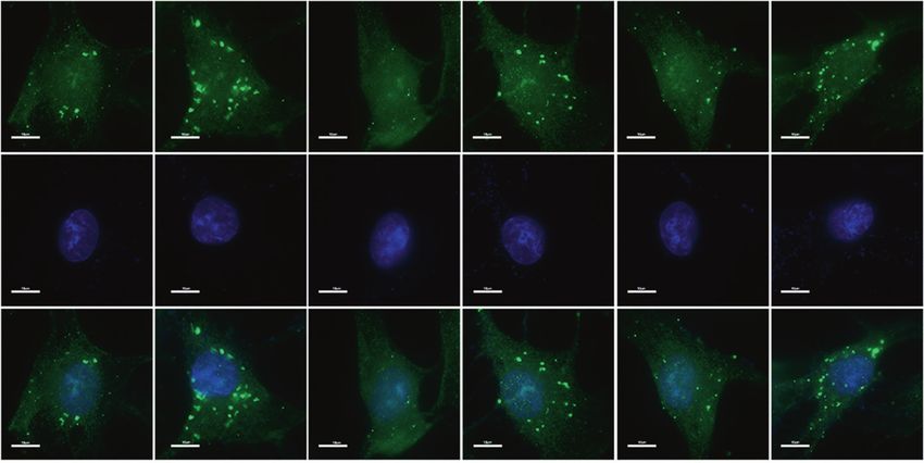
Melatonin suppresses the NLRP3 inflammasome in discs
                                                          F Chen et al.
2
    Notably, recent studies have demonstrated that melatonin              Melatonin suppresses NLRP3 inflammasome priming and
    attenuates the inflammatory response by inhibiting NLRP3               activation in vitro
    inflammasome activation during the progression of atherosclero-        First, we elucidated the effect by which melatonin affects NP cell
    sis43 and brain,44 liver45 and lung diseases.46 Moreover, melatonin   viability. Compared with the cytotoxicity in the control group,
    plays crucial roles in the IVDD process, including regulating NP      the groups treated with melatonin at concentrations below
    cell proliferation, remodeling the ECM,47 protecting vertebral        4 mM for either 24 or 48 h did not show any obvious cytotoxic
    endplate chondrocytes against apoptosis and calcification,48 and       effects (Fig. 3a, b). Subsequently, while investigating the effect
    preventing oxidative stress-induced NP cell apoptosis,49 indicat-     of melatonin on NLRP3 inflammasome priming and activation in
    ing a strong correlation between melatonin and IVDD. Although         NP cells, we found that NLRP3 and p20 expression were
    these published studies have indicated that melatonin partici-        decreased in NP cells treated with different melatonin doses,
    pates in the IVDD process by regulating NLRP3 inflammasome             with the lowest measured levels occurring at a dose of 1 mM
    activation in NP cells, this hypothesis has not been experimentally   (Fig. 3c, d). Furthermore, the expression levels of NLRP3 and
    examined.                                                             p20 started to decrease in NP cells treated with melatonin for
       Based on these observations, the objective of this study was to    different lengths of time and were significantly reduced at 24 h
    explore the molecular mechanisms underlying a novel activation        (Fig. 3e, f). The RT-qPCR results for NLRP3 were in agreement
    model of the NLRP3 inflammasome in IVDD. We also aimed to              with the western blot analysis results (Fig. 3g, h). Furthermore, IF
    investigate whether exogenous melatonin administration pre-           analysis also showed that melatonin suppressed NLRP3 inflam-
    vents IVDD by regulating NLRP3 inflammasome activation in vitro        masome activation in NP cells (Fig. 3i). These results showed that
    and in vivo.                                                          melatonin suppresses NLRP3 inflammasome activation in vitro.

                                                                          IL-1β induces NLRP3 inflammasome priming and activation
    RESULTS                                                               in vitro
    Melatonin ameliorates the progression of IVDD and LBP in vivo         Then, we investigated whether NLRP3 inflammasome priming and
    First, we established a rat IVDD model to determine whether           activation were induced in an IVDD cell model. As described in
    melatonin exerts a protective effect during the progression of        previous studies, IL-1β and TNF-α are classical cytokines that are
    IVDD in vivo. Melatonin was intraperitoneally injected into rats      used to establish an IVDD cell model. IL-1β is also produced by
    with AF puncture-induced IVDD. MRI images were obtained 4 or          NLRP3 inflammasome activation. LPS is a classical stimulator of the
    8 weeks post operation, and the IVDs from the rats treated with       NLRP3 inflammasome. Therefore, we selected IL-1β, TNF-α, and LPS
    melatonin displayed a significantly higher signal intensity than       to stimulate NP cells. First, NLRP3 and p20 expression was
    those without melatonin treatment (Fig. 1b, c). Histologically,       significantly increased in NP cells treated with IL-1β or LPS but only
    hematoxylin and eosin (H&E) and Safranin-O staining showed            slightly increased in NP cells treated with TNF-α (Fig. 4a, b).
    that the amounts of gelatinous NP tissue and the disc height in       Furthermore, the levels of NLRP3 and p20 gradually increased in NP
    the melatonin-treated rats were larger than those in the AF           cells treated with different doses of IL-1β and peaked at a dose of
    puncture rats, while the histologic score of the AF puncture rats     50 ng·mL−1 (Fig. 4c, d). After IL-1β treatment for different lengths of
    was higher than that of the melatonin-treated rats (Fig. 1d, e).      time, the expression of NLRP3 and p20 started to increase at 12 h
    IHC results showed that the expression of Aggrecan and                and exhibited an obvious increase at 24 h (Fig. 4e, f). The RT-qPCR
    Collagen II was decreased significantly in the AF puncture             results for NLRP3 expression were in agreement with the western
    group, and the levels of Aggrecan and Collagen II were                blot analysis results (Fig. 4g–i). In addition, IF staining also showed
    increased in rats that were treated with melatonin (Fig. 1f, g).      that IL-1β treatment increased NLRP3 expression in NP cells (Fig. 4j).
    Subsequently, the behavioral study results showed that mela-          These results suggest that IL-1β enhances NLRP3 inflammasome
    tonin significantly decreased mechanical hyperalgesia and              activation in vitro.
    thermal hyperalgesia compared to those of the AF puncture
    group (Fig. 1h). Therefore, these results indicated that melatonin    IL-1β upregulates NLRP3 inflammasome priming and activation by
    alleviates the progression of IVDD and LBP.                           increasing NF-κB signaling and mtROS production in vitro
                                                                          To examine the molecular mechanism by which IL-1β upregu-
    Melatonin alleviates IVDD by inhibiting NLRP3 inflammasome             lates NLRP3 inflammasome priming and activation, we detected
    priming and activation in vivo                                        NF-κB signaling and mitochondrial reactive oxygen species
    Next, we further investigated the correlation between NLRP3           (mtROS) production in NP cells. Previous studies have demon-
    inflammasome priming and activation and IVDD in vivo. First,           strated that IL-1β is strongly associated with the NF-κB signaling
    human discs with different grades of degeneration are shown as        pathway.50,51 In this study, we found that IL-1β activated the NF-
    representative MRI images in Fig. 2a (grades I–V). H&E staining       κB signaling pathway in NP cells (Fig. S1). Subsequently, to test
    showed that the number of NP cells was decreased in severely          the role of NF-κB signaling in NLRP3 inflammasome priming in
    degenerated human discs (Fig. 2b). Moreover, the expression of        IL-1β-treated NP cells, a silencing experiment was performed.
    NLRP3, p20, and IL-1β was increased in severely degenerated           Pretreatment with SM7368 (a specific inhibitor of the NF-κB
    human discs compared with that in mildly degenerative human           signaling pathway) or SM7368 plus IL-1β significantly decreased
    discs, as shown by IHC staining (Fig. 2c, d). Furthermore, the        NLRP3 levels in NP cells compared with those of untreated NP
    expression of NLRP3, p20, and IL-1β was increased in the IVDD rat     cells (Fig. 5a–c).
    model group compared with that of the control group, and                 As previously described, mtROS production plays an important
    melatonin administration reduced NLRP3, p20, and IL-1β expression     role in NLRP3 inflammasome activation.52,53 Therefore, we examined
    in the IVDD rat model (Fig. 2e, f). Then, to examine the mechanism    whether mtROS production was involved in NLRP3 inflammasome
    by which melatonin alleviates the progression of IVDD, we examined    activation in IL-1β-treated NP cells. We first detected that SOD2
    Aggrecan and Collagen II levels in the rat model. The expression of   expression was significantly decreased in the IL-1β treatment group
    Aggrecan and Collagen II was significantly decreased in the AF         compared with that of the control group, as measured by western
    puncture with melatonin and LPS groups (Fig. 2g, h). These results    blot and RT-qPCR analyses (Fig. 5d–e). MitoSOX Red staining
    demonstrated that NLRP3 inflammasome priming and activation            indicated that mtROS production was significantly upregulated in IL-
    were involved in the process of IVDD and that melatonin alleviated    1β-treated NP cells (Fig. 5g, h). Taken together, these results
    IVDD by inhibiting NLRP3 inflammasome priming and activation           confirmed that an IL-1β/NF-κB-NLRP3 inflammasome positive feed-
    in vivo.                                                              back loop is involved in the process of IVDD.

                                                                                                       Bone Research (2020)8:10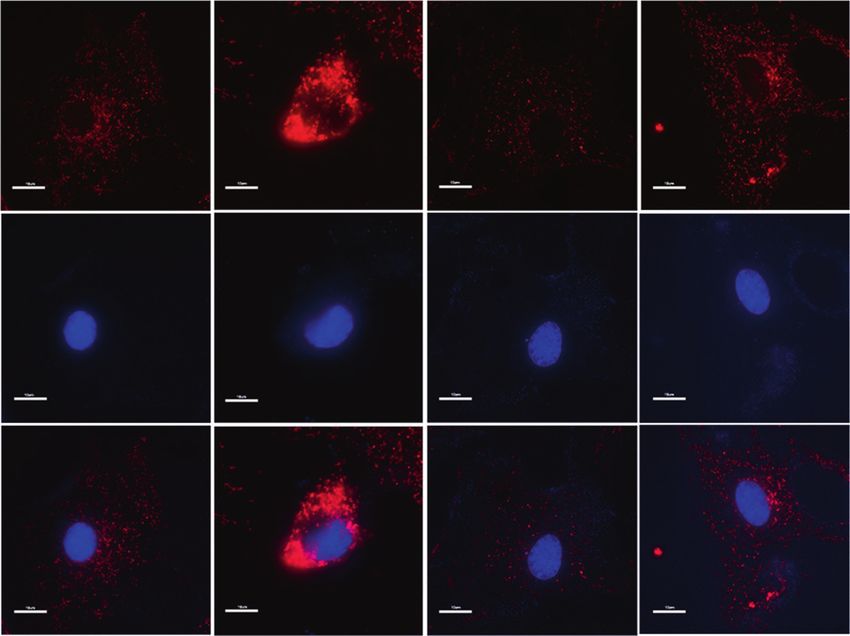
Melatonin suppresses the NLRP3 inflammasome in discs
F Chen et al.
3
b Sham surgery AF puncture AF puncture+Met c
CTR
AF puncture
AF puncture+Met
6
4 weeks
# #
** **
Pfrrimann grades
a
4
H
H3CO N CH3
O 2
N
H
8 weeks
0
4 weeks 8 weeks
d HE stain Safranin-O stain e
Sham surgery
10X
10X 10X
10X **
50X 50X 15
Histologic score
10
AF puncture+Met AF puncture
5
0
TR
re
et
+M
tu
C
nc
re
pu
tu
nc
AF
pu
AF
f Sham surgery AF puncture AF puncture+Met g
80 80
Collagen ll cell in rat
Aggrecan
Aggrecan+cell in rat
60 ** 60 **
NP tissue/%
NP tissue/%
+
40 40
20 20
Collagen II
0 0
y
t
et
TR
t
et
re
re
er
M
M
tu
tu
C
rg
t+
t+
nc
nc
su
re
re
pu
pu
tu
tu
am
nc
nc
AF
AF
Sh
pu
pu
AF
AF
h Thermal threshold Mechanical threshold
Paw withdrawal thermal latency/s
Sham surgery Sham surgery
AF puncturet
50% withdrawal threshold/g
25 25 AF puncturet
AF puncturet+Met AF puncturet+Met
20 20
15 15
* *
10
* * 10
5 5
0 0
0 5 10 15 0 5 10 15
Time after surgery/weeks Time after surgery/weeks
Fig. 1 Melatonin delays the progression of IVDD in vivo. a, b MRI images and Pfirrmann grade analysis of the rat models at 4 or 8 weeks after
the operation. c, d H&E staining, Safranin-O staining and histologic score analysis of the rat models at 8 weeks after the operation (original
magnification ×10, ×50; scale bar = 500 µm, 200 µm). e, f IHC staining and quantitative analysis of Aggrecan and Collagen II in IVDD rats
treated with melatonin (original magnification ×400; scale bar = 50 µm). g Behavioral study of the rat models at different time points after the
operation. CTR, Control. n = 5, *P < 0.05; **P < 0.01; #P < 0.05. The data are shown as the means ± SD
Melatonin disrupts the IL-1β-NLRP3 inflammasome positive inflammatory response by disrupting the IL-1β-NLRP3 inflam-
feedback loop in vitro masome positive feedback loop in NP cells. First, western blot
The preceding results indicated that melatonin suppresses analysis showed that melatonin obviously attenuated the IL-
NLRP3 inflammasome priming and activation in NP cells. 1β-induced increase in NLRP3, pro-IL-1β, and p20, while
Therefore, we determined whether melatonin reduces the MCC950 (a specific inhibitor of NLRP3 inflammasome
Bone Research (2020)8:10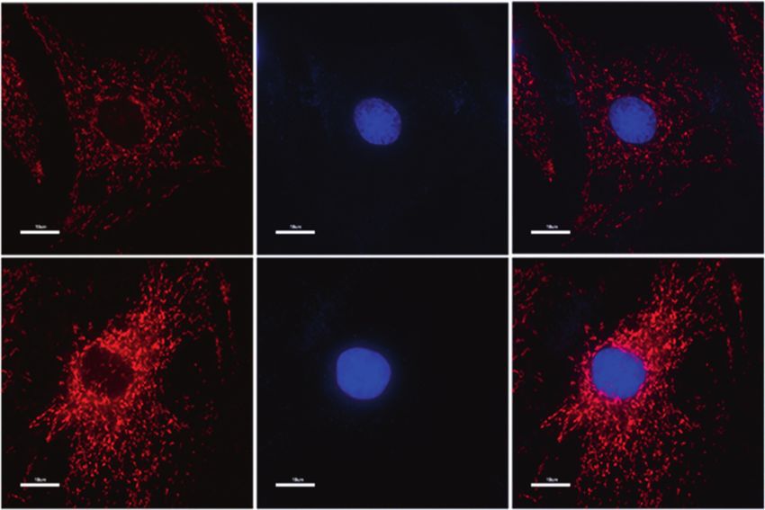
Melatonin suppresses the NLRP3 inflammasome in discs
F Chen et al.
4
a b H&E staining
Mild Severe 10X
50X
Grade I II III IV V
Mild
Severe
c NLRP3 p20 IL-1β
d
Mild
NLRP3* cells in human
IL-1β+ cells in human
p20+ cells in human
100 ** 100 100 **
***
NP tissue/%
NP tissue/%
NP tissue/%
80 80 80
60 60 60
40 40 40
Severe
20 20 20
0 0 0
Mild Severe Mild Severe Mild Severe
e g Aggrecan Collagen II
NLRP3 p20 IL-1β
AF puncture
Sham surgery
AF puncture+Met+LPS AF puncture+Met
AF puncture
AF puncture+Met
f h
Aggrecan+ cell in rat NP
100 100 80 100
NLRP3+ cell in rat NP
100
Collagen II+ cell in rat
* * *
IL-1β cell in rat NP
P20+ cell in rat NP
80 80 ** 80 80 *
60 NP tissue/%
tissue/%
tissue/%
tissue/%
tissue/%
60 60 60 60
40
40 40 40 40
+
20 20 20 20
20
0 0 0 0 0
y
e
et
ry
re
et
y
re
et
e
et
PS
re
et
PS
er
er
r
r
+M
M
M
M
+M
e
tu
tu
tu
tu
tu
+L
+L
rg
rg
rg
e+
e+
e+
nc
nc
nc
nc
nc
re
et
su
su
su
et
et
pu
pu
pu
pu
pu
ur
ur
ur
ur
u
M
+M
am
am
am
ct
ct
ct
ct
ct
e+
AF
AF
AF
AF
AF
n
n
n
n
t
n
re
Sh
pu
Sh
pu
Sh
pu
pu
ur
pu
tu
t
AF
AF
AF
AF
nc
AF
nc
pu
pu
AF
AF
Fig. 2 Melatonin alleviates IVDD by inhibiting NLRP3 inflammasome priming and activation in vivo. a MRI images of human discs. b H&E
staining of human discs (original magnification ×10, ×50; scale bar = 500 µm, 100 µm). c, d IHC staining and quantitative analysis of NLRP3,
P20, and IL-1β in human discs (original magnification ×400; scale bar = 50 µm). e, f IHC staining and quantitative analysis of NLRP3, P20, and IL-
1β in IVDD rats treated with melatonin (original magnification ×400, scale bar = 50 µm). g, h IHC staining and quantitative analysis of
Aggrecan and Collagen II in IVDD rats treated with melatonin or melatonin plus LPS (original magnification ×400; scale bar = 50 µm). n = 5,
*P < 0.05; **P < 0.01; ***P < 0.001. The data are shown as the means ± SD
activation) did not change the IL-1β-induced upregulation of Melatonin disrupts the IL-1β-NLRP3 inflammasome positive
NLRP3 and pro-IL-1β. In addition, a considerable decrease in p20 feedback loop by downregulating NF-κB signaling and mtROS
levels was observed in NP cells treated with melatonin plus IL-1β production
(Fig. 6a, b). The RT-qPCR and western blot analysis results of NLRP3 We next assessed the underlying mechanism by which melatonin
and pro-IL-1β are shown in Fig. 6c, d. In addition, IF staining also disrupts the IL-1β-NLRP3 inflammasome positive feedback loop.
confirmed that melatonin significantly attenuated IL-1β-induced First, we demonstrated that melatonin significantly suppressed
NLRP3 inflammasome activation (Fig. 6e). These data confirmed NF-κB signaling activation (Fig. S2). Subsequently, an siRNA
that melatonin effectively disrupted the IL-1β-NLRP3 inflamma- targeting P65 (si-P65) was established and verified in NP cells
some positive feedback loop in vitro. (Fig. S3). Consistently, the NLRP3 level significantly decreased
Bone Research (2020)8:10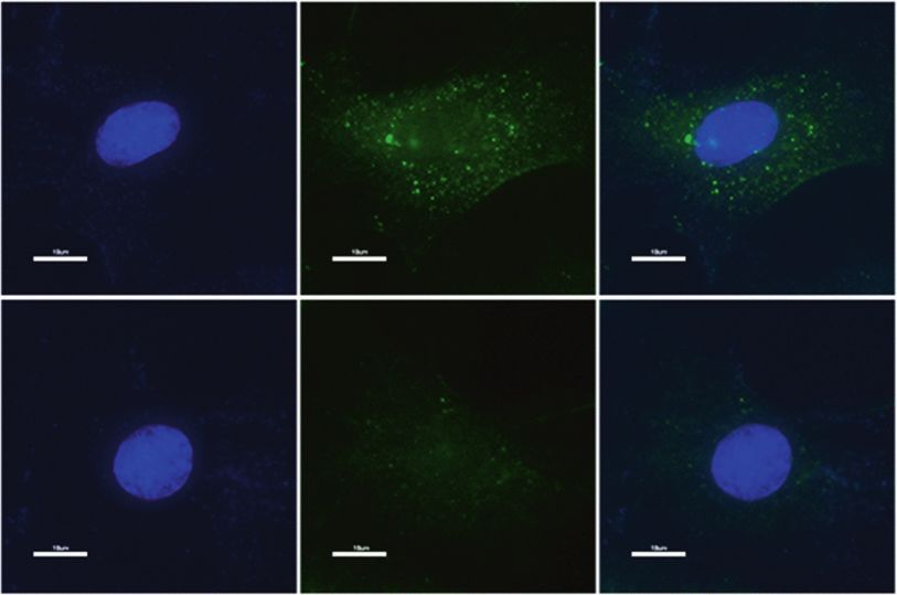
Melatonin suppresses the NLRP3 inflammasome in discs
F Chen et al.
5
a 150 b 150 c Melatonin/(mmol·L-1)
24 h 48 h
CTR 0.5 1.0 2.0
Cell viability/%
Cell viability/%
100 100 NLRP3 -110 kDa
* *
** **
50 50 p20 -20 kDa
β-actin -42 kDa
0 0
-1 -1
0 0.5 1.0 2.0 4.0 8.0 /(mmol·L ) 0 0.5 1.0 2.0 4.0 8.0 /(mmol·L )
d e f
(normalized to the β-actin level)
0.8 NLRP3
NLRP3 Melatonin/h Melatonin 1 mmol·L-1
(normalized to the β-actin level)
0.6 p20 p20
Melatonin 24 h
Relative protein level
CTR 12 24 48 0.6
Relative protein level
0.4 NLRP3 -110 kDa
** 0.4 *
* * *
* ** ** ** p20 -20 kDa ** *
*
0.2 0.2
β-actin -42 kDa
0.0
0.0
TR
h
h
h
TR
h
h
h
12
24
48
12
24
48
CTR 0.5 1.0 2.0 /(mmol·L-1)
C
C
CTR 0.5 1.0 2.0
g h
Relative mRNA level of NLRP3
1.5 Relative mRNA level of NLRP3 1.5
Melatonin 1 mmol·L-1 Melatonin 24 h
(fold change)
(fold change)
1.0 1.0
** ** **
0.5 *** *** 0.5 ***
0.0 0.0
CTR 12 h 24 h 48 h CTR 0.5 1.0 2.0 /(mmol·L-1)
i DAPI NLRP3 Merge
CTR
Melatonin
Fig. 3 Melatonin inhibits NLRP3 inflammasome priming and activation in NP cells. a The chemical structure of melatonin. b, c NP cell viability
after exposure to 0–8 mmol·L−1 NP for 24 h or 48 h. d–g Western blot and quantitative analysis of NLRP3 and P20 in NP cells treated with
different doses of melatonin or for different lengths of time. h, i RT-qPCR analysis of NLRP3 and P20 in NP cells treated with different doses of
melatonin or at different time points. j IF analysis of NLRP3 in NP cells treated with melatonin (original magnification ×1 000; scale bar =
10 µm). CTR, Control. *P < 0.05; **P < 0.01. The data are shown as the means ± SD
when these cells were transfected with si-P65, and melatonin or Melatonin disrupts the IL-1β/NF-κB-NLRP3 inflammasome positive
si-P65 obviously inhibited the IL-1β-induced increase in NLRP3 feedback loop in vivo
expression (Fig. 7a–c). Furthermore, IF staining also revealed IHC staining showed that, in human samples, p-P65 expression
that si-P65 attenuated NLRP3 expression in NP cells treated was higher in severely degenerated discs than in mildly
with si-P65 plus IL-1β (Fig. 7d, e). In addition, melatonin degenerated discs, while SOD2 expression was reduced (Fig. 8a,
prevented the IL-1β-induced decrease in SOD2 expression in NP b). Furthermore, the percentage of p-P65-positive cells decreased
cells (Fig. 7f–h). MitoSOX Red staining showed that mtROS significantly, while the percentage of SOD2-positive cells
production was significantly reduced in NP cells of the increased in melatonin plus AF puncture rats compared with
melatonin plus IL-1β treatment groups (Fig. 7i, j). Therefore, those of AF puncture rats (Fig. 8c, d). These results confirmed that
these results confirmed that melatonin disrupted the IL-1β melatonin suppresses the inflammatory response by disrupting
positive feedback loop by suppressing NF-κB signaling and the IL-1β/NF-κB-NLRP3 inflammasome positive feedback loop
mtROS production in vitro. in vitro and in vivo. In conclusion, these results indicated that
Bone Research (2020)8:10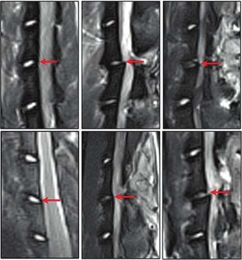
Melatonin suppresses the NLRP3 inflammasome in discs
F Chen et al.
6
a c IL-1β/(ng·mL-1) e IL-1β/h
α
F-
β
TR
S
-1
TN
LP
CTR 25 50 100 CTR 12 24 48
IL
C
NLRP3 -110 kDa -110 kDa NLRP3 -110 kDa
NLRP3
p20 -20 kDa p20 -20 kDa p20 -20 kDa
β-actin -42 kDa β-actin -42 kDa β-actin -42 kDa
b NLRP3
d f
(normalized to the β-actin level)
(normalized to the β-actin level)
(normalized to the β-actin level)
p20 1.5 1.0 NLRP3
2.0 NLRP3 p20
p20
**
Relative protein level
Relative protein level
**
Relative protein level
* * 0.8 * *
1.5
1.0 ** ** ** **
** * 0.6
1.0
0.4
0.5
0.5 * *
0.2
0.0 0.0 0.0
CTR 25 50 100 CTR 25 50 100 /(ng·mL-1)
TR
S
TR
S
TN β
α
TN β
α
TR
h
h
h
TR
h
h
h
F-
F-
LP
LP
-1
-1
12
24
48
12
24
48
C
C
C
C
IL
IL
g ** h i
Relative mRNA level of NLRP3
6
**
Relative mRNA level of NLRP3
Relative mRNA level of NLRP3
8 IL-1β 24 h 5 -1
IL-1β 50 ng·mL
4 * *
(fold change)
4 6 *
(fold change)
(fold change)
** 3
4
2 2
2
1
0 0 0
CTR IL-1 β TNF-α LPS 25 50 100 /(ng·mL-1)
TR
TR
h
h
h
12
24
48
C
C
j DAPI NLRP3 Merge
CTR
IL-1 β
Fig. 4 IL-1β enhances NLRP3 inflammasome priming and activation in vitro. a, b Western blot and quantitative analysis of NLRP3 and P20 in
NP cells treated with IL-1β, TNF-α or LPS. c–f Western blot and quantitative analysis of NLRP3 and P20 in NP cells treated with different doses
of IL-1β or at different time points. g–i RT-qPCR analysis of NLRP3 in NP cells treated with IL-1β, TNF-α or LPS, different doses of IL-1β or at
different time points. j IF analysis of NLRP3 in NP cells treated with IL-1β (original magnification ×1 000; scale bar = 10 µm). CTR, Control. *P <
0.05; **P < 0.01. The data are shown as the means ± SD
melatonin disrupts the IL-1β positive feedback loop by suppres- results were consistent with previous data and showed that the
sing NF-κB signaling and mtROS production and attenuating the application of melatonin in an IVDD model alleviated the
progression of IVDD, as illustrated in the proposed schematic progression of IVDD and LBP. However, whether NLRP3 inflamma-
representation of melatonin-mediated IVDD in vivo and in vitro some priming and activation are involved in this process is still
(Fig. 8e). unknown. Thus, we evaluated the markers of NLRP3 inflammasome
priming and activation and found that in the IVDD group, the
expression of NLRP3, P20, and IL-1β was elevated, indicating
DISCUSSION the priming and activation of the NLRP3 inflammasome. Further-
Melatonin is known to have multiple effects, including antioxidant, more, LPS, which is a proven NLRP3 inflammasome activator,
anti-inflammatory, and antiapoptotic impacts, in different sys- abrogated the effect of melatonin on IVDD, indicating that
tems.28,29 A recent study showed that melatonin downregulates melatonin acts by inhibiting NLRP3 inflammasome priming and
the gene expression of cyclin D1, PCNA, matrix metallopeptidase activation. Data from human disc samples also showed that in the
(MMP)-3, and MMP-9 and upregulates the gene expression of degenerative disc, NLRP3 inflammasome priming and activation
collagen type II alpha 1 chain and aggrecan in NP cells.47 Our marker expression were significantly increased, which gives us
Bone Research (2020)8:10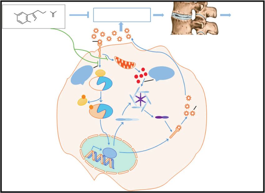
Melatonin suppresses the NLRP3 inflammasome in discs
F Chen et al.
7
a b c
2.0 5 *
Relative mRNA level of NLRP3
β1
**
L-
(normalized to β-actin)
Relative protein level
+I
4
1.5
68
68
(fold change)
73
73
β
TR
-1
SM
SM
3
IL
C
1.0
NLRP3 -110 kDa 2
0.5
1
β-actin -42 kDa
0.0 0
TR
68
TR
68
β
1β
β
1β
-1
-1
73
73
L-
L-
C
C
IL
IL
+I
+I
SM
SM
68
68
73
73
SM
SM
d e f
Relative mRNA level of SOD2
2.0 1.5
Relative protein level
(normalized to β-actin)
***
CTR IL-1β ***
1.5
(fold change)
1.0
SOD2 -22 kDa
1.0
0.5
β-actin -42 kDa 0.5
0.0 0.0
CTR IL-1 β CTR IL-1 β
g Mito SOX DAPI Merge
h
CTR
The mito SOX fluorescence
600
*
400
(% CTR)
200
IL-1β
0
CTR IL-1 β
Fig. 5 The IL-1β/NF-κB-NLRP3 inflammasome positive feedback loop is established in vitro. a, b Western blot and quantitative analysis of
NLRP3 in NP cells treated with IL-1β, SM7368, or IL-1β plus SM7368. c RT-qPCR analysis of NLRP3 in NP cells treated with IL-1β, SM7368, or IL-1β
plus SM7368. d–f Western blot and RT-qPCR analysis of SOD2 in NP cells treated with IL-1β. g, h MitoSOX red staining and quantitative analysis
of NP cells treated with IL-1β (original magnification ×1 000; scale bar = 10 µm). CTR, Control. *P < 0.05; **P < 0.01; ***P < 0.001. The data are
shown as the means ± SD
more confidence that the NLRP3 inflammasome plays an inhibiting IL-1β to prevent and reverse IVDD. Thus, it is essential to
important role in IVDD. study the expression and regulatory mechanism of IL-1β in IVDD.
One of the main causes of IVDD is an abnormal inflammatory We first found that in degenerated human NP tissue, the
response, which is induced by the overexpression of inflammatory expression of NLRP3, p20, and IL-1β was elevated. The NLRP3
cytokines, mainly IL-1β, causing NP cell apoptosis, downregulating inflammasome has been reported to be activated by LPS or
matrix gene expression or upregulating the expression of hyperosmotic stress in different systems. Dolunay et al. showed
collagen- and aggrecan-cleaving enzymes. For the first time, our that the inhibition of NLRP3/ASC/pro-caspase-1 inflammasome
study showed that IL-1β promotes its own expression by formation and activity prevents LPS-induced inflammatory hyper-
upregulating NLRP3 inflammasome activation. Furthermore, mel- algesia in mice.54,55 However, whether IL-1β activates the NLRP3
atonin, an anti-inflammatory molecule, disrupts the IL-1β positive inflammasome has seldom been studied. In this study, we showed
feedback loop by suppressing the NF-κB signaling pathway and for the first time that in NP cells, IL-1β treatment activated the
decreasing mtROS production. NLRP3 inflammasome in time- and dose-dependent manners. In
First, we found that IL-1β induced NLRP3 inflammasome addition, IL-1β treatment had an effect similar to that of LPS
priming and activation and thus promoted its own overexpres- treatment in upregulating NLRP3 and p20 expression. Interestingly,
sion. IL-1β has been widely studied in IVDD due to its catabolic according to previous studies, the NLRP3 inflammasome is a
effect. Le Maitre et al. reported that IL-1β is synthesized by native component of the innate immune system that processes pro-IL-1β
disc cells and that treating human disc cells with IL-1β induces an into a mature cytokine.17–19 Recently, Tang et al. reported that
imbalance between catabolism and anabolism.27 These responses honokiol suppresses activation of the TXNIP-NLRP3 inflammasome
represent the changes observed during IVDD. Hence, these in H2O2-stimulated NP cells, thereby inhibiting the activation of
findings provide a potential strategy for biological therapy by downstream inflammatory mediators such as IL-1β.56 Therefore, it
Bone Research (2020)8:10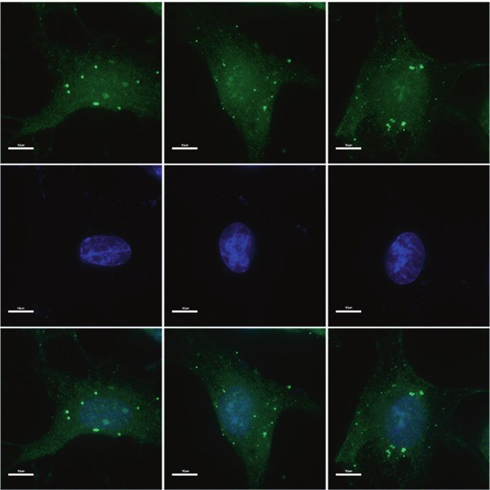
Melatonin suppresses the NLRP3 inflammasome in discs
F Chen et al.
8
0
95
C
a b
C
et
0
M
M
95
NLRP3
β+
β+
C
β
TR
Caspase-1
-1
-1
-1
et
C
Pro-IL-1 β
IL
IL
M
IL
M
C
1.5 ASC
NLRP3 -110 kDa p20
(normalized to β-actin)
NS
Relative protein level
NS #
Cas-1 -48 kDa 1.0 * * **
Pro-IL-1β -31 kDa
0.5
ASC -22 kDa
p20 -20 kDa 0.0
IL R
-1 M β
-1 M β
-1 M β
IL TR
-1 M β
IL TR
IL TR
IL TR
-1 M β
M C9 t
0
0
0
0
0
C 50
C 50
C 50
C 50
C 50
M 9 t
M C9 t
M C9 t
M C9 t
-1 M β+Met
-1 M β+Met
-1 M β+ et
-1 M β+ et
-1 M β+Met
β+ C Me
β+ CC e
β+ C e
β+ C Me
β+ C e
95
95
95
95
95
T
-1
-1
-1
1
-1
C
C
C
C
C
-
C
C
C
C
C
β-actin -42 kDa
IL
IL
IL
IL
IL
IL
IL
IL
IL
IL
c NS d #
Relative mRNA level of NLRP3
*
Relative mRNA level of IL-1β
6 6 **
(fold change)
(fold change)
4 4
2 2
0 0
β
et
0
0
β
et
TR
0
et
0
TR
et
95
95
95
95
-1
M
-1
M
M
M
C
C
C
IL
C
C
β+
IL
C
β+
C
C
C
C
M
M
-1
M
M
-1
β+
IL
β+
IL
-1
-1
IL
IL
e CTR IL-1β Met IL-1β+Met MCC950 IL-1β+MCC950
NLRP3
DAPI
Merge
Fig. 6 Melatonin disrupts the IL-1β-NLRP3 inflammasome positive feedback loop in vitro. a, b Western blot and quantitative analysis of NLRP3,
Caspase-1, pro-IL-1β, ASC, and P20 in NP cells treated with IL-1β, melatonin, IL-1β plus melatonin, MCC950, and MCC950 plus IL-1β. c, d RT-
qPCR analysis of NLRP3 and pro-IL-1β in NP cells treated with IL-1β, melatonin, IL-1β plus melatonin, MCC950, and MCC950 plus IL-1β. e IF
analysis of NLRP3 in NP cells with different treatments (original magnification ×1 000; scale bar = 10 µm). CTR, Control; Met, melatonin; Met +
IL-1β, melatonin plus IL-1β; NS, no statistical significance. *P < 0.05; **P < 0.01; #P < 0.05. The data are shown as the means ± SD
is reasonable that IL-1β promotes its own expression through inhibition of ROS production.59 Budai et al. also reported that LPS
NLRP3 inflammasome activation, forming a positive feedback loop. induces NLRP3 inflammasome regulation through the NF-κB, p38,
Furthermore, we studied the mechanism by which IL-1β JNK, and ERK signaling pathways.60 In this study, we found that IL-
induces NLRP3 inflammasome priming and activation. Our 1β induced NLRP3 expression through NF-κB activation. This
previous studies showed that in NP cells, IL-1β activates NF-κB conclusion was determined by the application of SM7368, a
signaling through p65 phosphorylation.51 There are some studies specific NF-κB pathway inhibitor. mtROS are the main activators of
concerning NLRP3 inflammasome regulation, and various path- the NLRP3 inflammasome;52,53 thus, it is important to know
ways, including the Smad, NFAT, NF-κB, and MAP kinase pathways, whether IL-1β regulates mtROS production. According to our
regulate NLRP3 expression.57,58 Yu et al. showed that hepatitis B e MitoSOX Red staining and western blotting results, IL-1β
antigen suppresses LPS-induced NLRP3 inflammasome activation treatment reduced SOD2 expression and induced mtROS produc-
and IL-1β production by repressing NLRP3 and pro-IL-1β expres- tion in NP cells. In conclusion, our results indicated that in NP cells,
sion through inhibition of NF-κB phosphorylation and by IL-1β activates the NLRP3 inflammasome through NF-κB signaling
repressing caspase-1 activation and IL-1β maturation through activation and mtROS production.
Bone Research (2020)8:10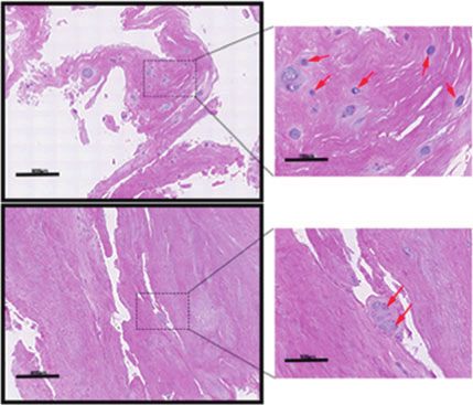
Melatonin suppresses the NLRP3 inflammasome in discs
F Chen et al.
9
a b c
##
Relative mRNA level of NLRP3
1β
1.5 8
L-
##
1β
**
+I
L-
65
65
(normalized to β-actin)
+I
β
Relative protein level
**
TR
-P
et
-1
-P
6
M
IL
si
si
C
(fold change)
NLRP3 -110 kDa 1.0
4
β-actin -42 kDa
0.5 2
0
d CTR si-p65 si-P65+IL-1β
0.0
65
65
β
TR
β
β
β
β
65
TR
1
-1
-P
-P
-1
C
L-
1
1
IL
-P
C
L-
L-
si
si
IL
+I
si
β+
+I
+I
et
65
et
M
-1
M
-P
NLRP3
IL
si
e f
et
M
150
β+
β
The NLRP3 fluorescence
-1
TR
et
-1
IL
M
IL
C
SOD2 -22 kDa
100
*
(% CTR)
DAPI
β-actin -42 kDa
50
g 2.0
0 **
#
(normalized to β-actin)
Relative protein level
1β
TR
65
Merge
L-
-P
C 1.5
+I
si
65
-P
si
1.0
0.5
i CTR IL-1β Met Met+IL-1β
0.0
β
TR
et
β
-1
1
M
C
L-
IL
Mito SOX
+I
et
M
j h
**
Relative mRNA level of SOD2
500 4
The mito SOX fluorescence
#
**
400
3
(fold CTR)
(% CTR)
DAPI
300
# 2
200
100 1
0 0
et
β
TR
β
et
β
Merge
1β
TR
M
1
-1
M
-1
L-
C
L-
C
IL
IL
+I
+I
et
et
M
M
Fig. 7 Melatonin disrupts the IL-1β-NLRP3 inflammasome positive feedback loop by downregulating NF-κB signaling and mtROS production.
a–c Western blot and RT-qPCR analysis of NLRP3 in NP cells cultured with IL-1β, si-P65, si-P65, and melatonin plus IL-1β. d IF analysis of NLRP3
in NP cells exposed to si-P65 and si-P65 plus IL-1β (original magnification ×1 000; scale bar = 10 µm). e–g Western blot and RT-qPCR analysis of
SOD2 in NP cells treated with IL-1β, melatonin, and melatonin plus IL-1β. h, i MitoSOX red staining and quantitative analysis of NP cells
cultured with melatonin and melatonin plus IL-1β (original magnification ×1 000; scale bar = 10 µm). CTR, Control; Met, melatonin; Met + IL-1β,
melatonin plus IL-1β. *P < 0.05; **P < 0.01; #P < 0.05; ##P < 0.01. The data are shown as the means ± SD
Furthermore, we found that melatonin disrupted the IL-1β melatonin treatment, indicating that melatonin exerted anti-
positive feedback loop and studied the mechanism of this inflammatory effects via the NF-κB-NLRP3 inflammasome-IL-1β axis.
disruption. Melatonin has also been reported to suppress NLRP3 There were several limitations to this study. First, the number of
inflammasome activation and IL-1β expression. Dong et al. showed human IVD tissue samples was relatively small due to the difficulty
that melatonin attenuates NLRP3, ASC, cleaved caspase-1, IL-1β, associated with acquiring grade I/II discs in clinical practice.
and IL-6 expression.44 Consistent with previous reports, our data Second, the detailed mechanisms by which melatonin suppresses
showed that melatonin suppressed NF-κB signaling activation and NF-κB signaling and mtROS production were not addressed and
reduced mtROS production to inhibit NLRP3 inflammasome might be elucidated in future studies.
activation and IL-1β expression. We confirmed these results by In conclusion, we found for the first time that IL-1β forms a
applying p65 siRNA and MCC950 (a specific inhibitor of NLRP3 positive feedback loop through NLRP3 inflammasome activation in
inflammasome activation). Furthermore, we confirmed our dis- IVDD and that melatonin disrupts this vicious cycle by suppressing
covery in vivo. Our data showed that in this IVDD model, p-p65 NF-κB signaling activation and mtROS production. Our research
expression was decreased and SOD2 expression was increased after showed a new mechanism by which IL-1β promotes the
Bone Research (2020)8:10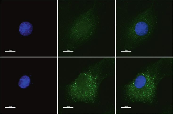
Melatonin suppresses the NLRP3 inflammasome in discs
F Chen et al.
10
a c CTR AF puncture AF puncture+Met
p-P65 Mild Severe
p-P65
SOD2
SOD2
b d **
p-P65 cells in rat NP tissue/%
80 100 ##
*
100 ** 100 # 80
** 60
human NP cells/%
human NP cells/%
rat NP cells/%
SOD2+cells in
80 80
SOD2 cells in
p-P65+cells in
60
60 60 40
+
40 40 40
20
20 20 20
+
0 0
0 0
Mild Severe Mild Severe
re
re
et
et
TR
TR
tu
tu
+M
+M
C
C
nc
nc
re
re
pu
pu
tu
tu
AF
AF
nc
nc
pu
pu
AF
AF
e
H
H3CO N CH3
O
Inflammation LBP
N
H
Melatonin
IL-1β IVDD
Dysfunctional
Mitochondrion
ep
st IκB Activation step
g ROS
in
im P50
Pr
P65 NLRP3 inflammasome
p
IκB IL-1β
p P50
P65 Active caspase-1
NLRP3
Pro-IL-1β
NF
-κB
Nucleus pulposus cell
Fig. 8 Melatonin disrupts the IL-1β/NF-κB-NLRP3 inflammasome positive feedback loop in vivo. a–d IHC staining and quantitative analysis of
p-P65 and SOD2 in human discs and rats at 8 weeks after the operation (original magnification ×400; scale bar = 50 µm). e Proposed
schematic representation of melatonin disruption of the IL-1β/NF-κB-NLRP3 inflammasome positive feedback loop to alleviate IVDD in vivo
and in vitro. CTR, Control; AF puncture + Met, AF puncture plus melatonin. n = 5. *P < 0.05; **P < 0.01; #P < 0.05; ##P < 0.01. The data are shown
as the means ± SD
inflammatory response in IVDD, and melatonin may be used as a Helsinki).61 Between March 2016 and April 2018, we collected 25
therapeutic agent for the treatment of inflammatory cytokine- disc samples (detailed information about the specimens is in Table 1)
related IVDD. from patients (male: female, 13: 12). The degree of disc
degeneration was evaluated by Pfirrmann classification. Normal
discs were obtained from patients with trauma and deformity, and
MATERIALS AND METHODS degenerated discs were obtained from patients with degenerative
Collection of human IVDs spinal diseases (disc herniation, spinal canal stenosis, and
Before tissue collection, each patient signed an informed consent degenerative scoliosis).
form, and the Medical Ethics Committee of The First Affiliated
Hospital of Sun Yat-sen University (no: [2017] 203) approved this Cell culture and cell viability assay
study. All studies in this paper were performed according to The As previously described,62 NP cells were isolated and cultured in
Code of Ethics of the World Medical Association (Declaration of DMEM (Invitrogen, CA, USA) containing 10% fetal bovine serum
Bone Research (2020)8:10Melatonin suppresses the NLRP3 inflammasome in discs
F Chen et al.
11
Table 1. Information of human disc samples from 25 patients Table 2. Specific primers
Human disc samples Sex Age Diagnosis Level Grade Primers sequences
1 M 17 y AIS T9/10 I NLRP3 R 5′- ATCAACAGGCGAGACCTCTG-3′
2 F 14 y AIS T7/8 I NLRP3 F 5′- ATCAACAGGCGAGACCTCTG-3′
3 M 16 y AIS T11/12 I SOD2 R 5′- GCGTTGATGTGAGGTTCCAG-3′
4 F 15 y AIS T11/12 II SOD2 F 5′- GCTCCGGTTTTGGGGTATCTG-3′
5 M 18 y Trauma L1/2 II P65 R 5′- GTTCACGGATGACCTCTTTGTTT-3′
6 F 19 y Trauma L3/4 II P65 F 5′- GGGCTTGGAAATAGAGACATTGA-3′
7 F 23 y Disc herniation L3/4 II IL-1β R 5′-CACACACTAGCAGGTCGTCA -3′
8 F 31 y Disc herniation L4/5 III IL-1β F 5′-CCTATGTCTTGCCCGTGGAG -3′
9 M 29 y Disc herniation L2/3 III β-Actin R 5′-GGATGGCTACGTACATGGCTG -3′
10 M 34 y Disc herniation L3/4 III β-Actin F 5′-CATTGTCACCAACTGGGACGATA -3′
11 F 42 y Disc herniation L4/5 III
12 F 36 y Disc herniation L4/5 III
13 M 27 y Disc herniation L4/5 III
described.63 Subsequently, the membranes were blocked with
3% bovine serum albumin and incubated with primary antibodies.
14 M 30 y Disc herniation L2/3 IV The primary antibodies included anti-pro-IL-1β (1:1 000, 12703,
15 M 39 y Disc herniation L3/4 IV Cell Signaling Technology), anti-IL-1β (1:1 000, ab8320, Abcam),
16 M 47 y Disc herniation L2/3 IV anti-phospho-P65 (1:1 000, 3033, Cell Signaling Technology), anti-
17 F 58 y Disc herniation L3/4 IV P65 (1:1 000, 8242, Cell Signaling Technology), anti-phospho-Erk1/
18 F 67 y Disc herniation L3/4 IV 2 (1:1 000, 4370, Cell Signaling Technology), anti-Erk1/2 (1:1 000,
4695, Cell Signaling Technology), anti-phospho-P38 (1:1 000, 4511,
19 M 59 y Disc herniation L4/5 IV Cell Signaling Technology), anti-P38 (1:1 000, 8690, Cell Signaling
20 F 69 y Disc herniation L3/4 V Technology), anti-NLRP3 (1:1 000, AG-20B-0014, AdipoGen), anti-
21 M 63 y Disc herniation L4/5 V cleaved Caspase-1 (p20) (1:1 000, AG-20B-0042, AdipoGen), anti-
22 M 83 y Disc herniation L3/4 V ASC (1:1 000, AG-25B-0006-C100, AdipoGen), anti-Caspase-1 (1:
23 F 68 y Disc herniation L3/4 V 1 000, ab1872, Abcam), anti-superoxide dismutase 2 (SOD2) (1:
1 000, 13141, Cell Signaling Technology), and anti-β-actin (1:3 000,
24 M 74 y Disc herniation L2/3 V 4970, Cell Signaling Technology). After washing with PBS, the
25 F 78 y Disc herniation L3/4 V membranes were incubated with the following secondary
AIS adolescent idiopathic scoliosis, M male, F female, y years antibodies: anti-rabbit IgG (1:5 000, 7074, Cell Signaling Technol-
ogy) or anti-mouse IgG (1:5 000, 7076, Cell Signaling Technology).
Finally, the Western blot bands were detected using enhanced
(Invitrogen, CA) and penicillin/streptomycin (Invitrogen, CA) at chemiluminescence detection reagents (Invitrogen, CA, USA) and
37 °C in a humidified incubator with 5% CO2. We harvested NP quantified using ImageJ software (National Institutes of Health,
cells using solutions containing trypsin (0.25%) and EDTA (1 mM) Bethesda, MD, USA).
(Invitrogen, CA) and subcultured the cells in 10 cm dishes. NP cells
were seeded in six-well plates, grown to ~80% confluence and Real-time quantitative polymerase chain reaction (RT-qPCR)
treated with melatonin (1 mmol·L−1, M5250, Sigma-Aldrich, USA), The isolation of total RNA from NP cells was performed using
MCC950 (10 nmol·L−1, Selleck, a specific inhibitor of NLRP3 RNAiso Plus (Takara, Japan). Complementary DNA synthesis was
inflammasome activation), lipopolysaccharide (LPS, 200 ng·mL−1, performed using a Prime Script RT Master Mix kit (Takara)
L2880, Sigma-Aldrich, USA), TNF-α (100 ng·mL−1, 210-TA-020, R&D according to the manufacturer’s instructions. SYBR green Premix
Systems, USA), or IL-1β (50 ng·mL−1, 201-LB-005, R&D Systems, Ex Taq II (Takara) was used to detect the relative mRNA levels of
USA) for 24 h for subsequent experiments. The cytotoxic effect of NLRP3, pro-IL-1β, SOD2, P65, and β-Actin, and the sequences of
melatonin on NP cells was detected using a cell counting kit the primer pairs are listed in Table 2 (Sangon Biotech, Shanghai,
(CCK)-8 assay (Dojindo Laboratories, Kumamoto, Japan) according China). RT-qPCR was performed on an ABI 7900HT Fast Real-Time
to the manufacturer’s instructions. PCR System (Applied Biosystems) for 40 cycles and quantified
using the 2−ΔΔCt method.
Small interfering RNA (siRNA) transfection
Rat NP cells were seeded at 5 × 10⁶ per well in a six-well plate and MitoSOX Red and immunofluorescence (IF) staining
transfected with negative control or siRNA targeting P65 (RiboBio, NP cells were treated as described, incubated with 2.0 mL of
Guangzhou, Guangdong, China) when the cells reached 60%–70% 5 μmol·L−1 MitoSOX Red reagent or fixed with 4% paraformalde-
confluence. The sequences for the P65-specific siRNAs were as hyde and then blocked with 5% normal goat serum. Subsequently,
follows: 1: GCTGCAGTTTGATGATGAA, 2: GCCCTATCCCTTTACGTCA, the cells were incubated with anti-NLRP3 (1:200, ab4207, Abcam)
and 3: GGACATATGAGACCTTCAA (10 nmol·L−1). Then, we used and anti-phospho-P65 (1:200, 3033, Cell Signaling Technology)
250 μL of serum-free optical-MEM (Invitrogen, CA, USA) to antibodies overnight, followed by incubation with Alexa Fluor-
individually dissolve 5 µL of siRNA or 10 μL of Lipofectamine 488-conjugated anti-rabbit and anti-goat secondary antibodies
3000 (Invitrogen, CA, USA). After mixing them together, the (Invitrogen, 1:2 000). Nuclear staining was performed using DAPI.
mixture was added to the cells. After treatment, the cells were Finally, the cells were photographed under an Olympus BX63
harvested for protein/RNA extraction. microscope (Tokyo, Japan) at ×400 and ×1 000 magnifications.
Western blot analysis Animal model and magnetic resonance imaging (MRI) evaluation
The proteins of treated NP cells were extracted and electrophor- As previously described,64 the rats (weighing 200–250 g, n = 5 per
etically separated via 10% or 15% SDS-PAGE, as previously group) were divided into three groups: the blank group received
Bone Research (2020)8:10Melatonin suppresses the NLRP3 inflammasome in discs
F Chen et al.
12
no puncture and was intraperitoneally injected with 0.9% normal REFERENCES
saline; the other two groups underwent AF puncture surgery with 1. Vlaeyen, J. W. S. et al. Low back pain. Nat. Rev. Dis. Primers 4, 52 (2018).
a 21-gauge needle inserted 3.0 mm into the L3/4 IVDs for 30 s. At 4 2. Hoy, D., Brooks, P., Blyth, F. & Buchbinder, R. The epidemiology of low back pain.
or 8 weeks post operation, MRI (T2-weighted images) signal Best. Pract. Res. Clin. Rheumatol. 24, 769–781 (2010).
change examinations were performed on the rats (n = 5 per 3. Maher, C., Underwood, M. & Buchbinder, R. Non-specific low back pain. Lancet
group). In addition, the degree of disc degeneration was evaluated 389, 736–747 (2017).
4. Hoy, D. et al. The global burden of low back pain: estimates from the Global
by Pfirrmann classification.
Burden of Disease 2010 study. Ann. Rheum. Dis. 73, 968–974 (2014).
Melatonin was diluted to 20 mg·mL−1 with normal saline and 5. Yang, G. et al. Rapid health transition in China, 1990–2010: findings from the
then intraperitoneally injected into the rats (30 mg·kg−1 per week), Global Burden of Disease Study 2010. Lancet 381, 1987–2015 (2013).
in addition to LPS (2 mg·kg−1 per week), until the day of euthanasia. 6. Foster, N. E. et al. Prevention and treatment of low back pain: evidence, chal-
lenges, and promising directions. Lancet 391, 2368–2383 (2018).
Immunohistochemistry (IHC) and histopathological analysis 7. Hartvigsen, J. et al. What low back pain is and why we need to pay attention.
The specimens were embedded in paraffin and cut into 5 µm Lancet 391, 2356–2367 (2018).
sections. Subsequently, the sections were deparaffinized and 8. Silagi, E. S., Shapiro, I. M. & Risbud, M. V. Glycosaminoglycan synthesis in the
rehydrated, followed by H&E and Safranin-O staining or antigen nucleus pulposus: Dysregulation and the pathogenesis of disc degeneration.
retrieval with 0.01 mol·L−1 sodium citrate. The sections were blocked Matrix Biol. 71–72, 368–379 (2018).
9. Roughley, P. J. Biology of intervertebral disc aging and degeneration: involve-
with 3% hydrogen peroxide and 5% normal goat serum. Then, the
ment of the extracellular matrix. Spine 29, 2691–2699 (2004).
slides were incubated with the following primary antibodies: anti- 10. Ding, F., Shao, Z. W. & Xiong, L. M. Cell death in intervertebral disc degeneration.
NLRP3 (1:200, AG-20B-0014, AdipoGen), anti-cleaved caspase-1 (p20) Apoptosis 18, 777–785 (2013).
(1:200, AG-20B-0042, AdipoGen), anti-IL-1β (1:200, ab8320, Abcam), 11. Zhao, C. Q., Jiang, L. S. & Dai, L. Y. Programmed cell death in intervertebral disc
anti-SOD2 (1:1 000, 13141, Cell Signaling Technology), and anti- degeneration. Apoptosis 11, 2079–2088 (2006).
phospho-P65 (1:200, 3033, Cell Signaling Technology). The sections 12. Ji, M. L. et al. Preclinical development of a microRNA-based therapy for inter-
were incubated with a secondary antibody and then developed with vertebral disc degeneration. Nat. Commun. 9, 5051 (2018).
DAB solution. Hematoxylin was used for nuclear staining. Finally, the 13. Adams, M. A., Dolan, P. & McNally, D. S. The internal mechanical functioning of
sections were observed and imaged under an Olympus BX63 intervertebral discs and articular cartilage, and its relevance to matrix biology.
microscope at ×10, ×50, and ×400 magnifications, and the Matrix Biol. 28, 384–389 (2009).
percentages of NLRP3+, IL-1β+, p20+, SOD2+, and phospho-P65+ 14. Roberts, S., Evans, H., Trivedi, J. & Menage, J. Histology and pathology of the
human intervertebral disc. J. Bone Jt. Surg. Am. Vol. 88 (Suppl 2), 10–14 (2006).
cells in the IVD samples were quantified using ImageJ software 15. Risbud, M. V. & Shapiro, I. M. Role of cytokines in intervertebral disc degeneration:
(National Institutes of Health, Bethesda, MD, USA).65 The histologic pain and disc content. Nat. Rev. Rheumatol. 10, 44–56 (2014).
scores were assessed as previously described: normal disc, five; 16. Yang, W. et al. Interleukin-1beta in intervertebral disk degeneration. Clin. Chim.
moderately degenerated disc, 6–11; and severely degenerated disc, Acta 450, 262–272 (2015).
12–14.66,67 17. de Zoete, M. R., Palm, N. W., Zhu, S. & Flavell, R. A. Inflammasomes. Cold Spring
Harb. Perspect. Biol. 6, a016287 (2014).
Behavioral study 18. He, Y., Hara, H. & Nunez, G. Mechanism and regulation of NLRP3 inflammasome
The rats were subjected to behavioral tests as described in our activation. Trends Biochem Sci. 41, 1012–1021 (2016).
previous study.63 Reflex reactions to both mechanical and harmful 19. Prochnicki, T., Mangan, M. S. & Latz, E. Recent insights into the molecular
thermal stimuli applied to both hind paws were measured in five mechanisms of the NLRP3 inflammasome activation. F1000Res. https://doi.org/
10.12688/f1000research.8614.1 (2016).
rats from each group within a week of surgery and every 2 weeks 20. Freeman, L. C. & Ting, J. P. The pathogenic role of the inflammasome in neuro-
during the following 10 weeks. degenerative diseases. J. Neurochem. 136 (Suppl 1), 29–38 (2016).
21. Heneka, M. T., McManus, R. M. & Latz, E. Inflammasome signalling in brain
Statistical analysis function and neurodegenerative disease. Nat. Rev. Neurosci. 19, 610–621
The differences among the groups were assessed by one-way (2018).
analysis of variance (ANOVA), which was performed using SPSS 22. McAllister, M. J., Chemaly, M., Eakin, A. J., Gibson, D. S. & McGilligan, V. E. NLRP3 as
software version 19.0 for Windows (IL, USA). If the ANOVA results a potentially novel biomarker for the management of osteoarthritis. Osteoarthr.
were statistically significant, the differences between the two Cartil. 26, 612–619 (2018).
groups were examined by using Bonferroni’s post hoc test. The 23. Moossavi, M., Parsamanesh, N., Bahrami, A., Atkin, S. L. & Sahebkar, A. Role of the
NLRP3 inflammasome in cancer. Mol. Cancer 17, 158 (2018).
data are presented as the mean ± SD. P values < 0.05 were
24. Karki, R., Man, S. M. & Kanneganti, T. D. Inflammasomes and cancer. Cancer
considered statistically significant. Immunol. Res. 5, 94–99 (2017).
25. Mangan, M. S. J. et al. Targeting the NLRP3 inflammasome in inflammatory dis-
eases. Nat. Rev. Drug Discov. 17, 588–606 (2018).
ACKNOWLEDGEMENTS 26. Mao, L., Kitani, A., Strober, W. & Fuss, I. J. The role of NLRP3 and IL-1beta in the
We thank Qifeng Ou, Zheng Yu, and Haitao Cui (Sun Yat-sen University) for assisting
pathogenesis of inflammatory bowel disease. Front. Immunol. 9, 2566 (2018).
with the established IVDD rat model and immunohistochemical staining. This work
27. Le Maitre, C. L., Freemont, A. J. & Hoyland, J. A. The role of interleukin-1 in the
was supported by grants from the Natural Science Foundation of Guangdong
pathogenesis of human intervertebral disc degeneration. Arthritis Res. Ther. 7,
Province (Grant no: 2017A030313670) awarded to J.W. and the National Natural
R732–R745 (2005).
Science Foundation of China (81572175 and 81772386), awarded to Z.Z.
28. Acuna-Castroviejo, D. et al. Extrapineal melatonin: sources, regulation, and
potential functions. Cell. Mol. life Sci. 71, 2997–3025 (2014).
29. Venegas, C. et al. Extrapineal melatonin: analysis of its subcellular distribution and
AUTHOR CONTRIBUTIONS daily fluctuations. J. Pineal Res. 52, 217–227 (2012).
Study design: J.W. and Z.Z. Study conduction: F.C., G.J., H.L., and Z.L. Data collection: F.C., 30. Favero, G., Franceschetti, L., Bonomini, F., Rodella, L. F. & Rezzani, R. Melatonin as
Y.P., and Z.L., H.P., J.L., and H.C. Data analysis: F.C., Z.Z., and J.W. Data interpretation: F. an anti-inflammatory agent modulating inflammasome activation. Int. J. Endo-
C., H.L., Z.Z., and J.W. Drafting the paper: F.C. and J.W. crinol. 2017, 1835195 (2017).
31. Hardeland, R Melatonin and inflammation-story of a double-edged blade. J.
Pineal Res. 65, e12525 (2018).
ADDITIONAL INFORMATION 32. Sarlak, G., Jenwitheesuk, A., Chetsawang, B. & Govitrapong, P. Effects of melatonin
The online version of this article (https://doi.org/10.1038/s41413-020-0087-2) on nervous system aging: neurogenesis and neurodegeneration. J. Pharmacol.
contains supplementary material, which is available to authorized users. Sci. 123, 9–24 (2013).
33. Wang, J. Z. & Wang, Z. F. Role of melatonin in Alzheimer-like neurodegeneration.
Competing interests: The authors declare no competing interests. Acta Pharmacol. Sin. 27, 41–49 (2006).
Bone Research (2020)8:10Melatonin suppresses the NLRP3 inflammasome in discs
F Chen et al.
13
34. Karaaslan, C. & Suzen, S. Antioxidant properties of melatonin and its potential 55. Zheng, Q. et al. Hyperosmotic stress-induced TRPM2 channel activation stimu-
action in diseases. Curr. Top. Med. Chem. 15, 894–903 (2015). lates NLRP3 inflammasome activity in primary human corneal epithelial cells.
35. Ganie, S. A. et al. Melatonin: a potential anti-oxidant therapeutic agent for mito- Investig. Ophthalmol. Vis. Sci. 59, 3259–3268 (2018).
chondrial dysfunctions and related disorders. Rejuvenation Res. 19, 21–40 (2016). 56. Tang, P. et al. Honokiol alleviates the degeneration of intervertebral disc via
36. Giannoulia-Karantana, A., Vlachou, A., Polychronopoulou, S., Papassotiriou, I. & suppressing the activation of TXNIP-NLRP3 inflammasome signal pathway. Free
Chrousos, G. P. Melatonin and immunomodulation: connections and potential Radic. Biol. Med. 120, 368–379 (2018).
clinical applications. Neuroimmunomodulation 13, 133–144 (2006). 57. Tschopp, J. & Schroder, K. NLRP3 inflammasome activation: the convergence of
37. Calvo, J. R., Rafii-el-Idrissi, M., Pozo, D. & Guerrero, J. M. Immunomodulatory role multiple signalling pathways on ROS production? Nat. Rev. Immunology 10,
of melatonin: specific binding sites in human and rodent lymphoid cells. J. Pineal 210–215 (2010).
Res. 18, 119–126 (1995). 58. Mezzasoma, L., Antognelli, C. & Talesa, V. N. A novel role for brain natriuretic
38. Poza, J. J., Pujol, M., Ortega-Albas, J. J. & Romero, O. Melatonin in sleep disorders. peptide: inhibition of IL-1beta secretion via downregulation of NF-kB/Erk 1/2 and
Neurologia. https://doi.org/10.1016/j.nrl.2018.08.002 (2018). NALP3/ASC/Caspase-1 activation in human THP-1 monocyte. Mediators Inflamm.
39. Talib, W. H. Melatonin and cancer hallmarks. Molecules. https://doi.org/10.3390/ 2017, 5858315 (2017).
molecules23030518 (2018). 59. Yu, X. et al. HBV inhibits LPS-induced NLRP3 inflammasome activation and IL-
40. Su, S. C. et al. Cancer metastasis: mechanisms of inhibition by melatonin. J. Pineal 1beta production via suppressing the NF-kappaB pathway and ROS production. J.
Res. https://doi.org/10.1111/jpi.12370 (2017). Hepatol. 66, 693–702 (2017).
41. Reiter, R. J. et al. Melatonin, a full service anti-cancer agent: inhibition of initiation, 60. Budai, M. M., Varga, A., Milesz, S., Tozser, J. & Benko, S. Aloe vera downregulates
progression and metastasis. Int. J. Mol. Sci. https://doi.org/10.3390/ijms18040843 LPS-induced inflammatory cytokine production and expression of NLRP3
(2017). inflammasome in human macrophages. Mol. Immunol. 56, 471–479 (2013).
42. Farhood, B. et al. Melatonin and cancer: from the promotion of genomic stability to 61. Human Experimentation. Code of ethics of the world medical association
use in cancer treatment. J. Cell. Physiol. https://doi.org/10.1002/jcp.27391 (2018). (Declaration of Helsinki). Can. Med. Assoc. J. 91, 619 (1964).
43. Ma, S. et al. Melatonin ameliorates the progression of atherosclerosis via mito- 62. Zhang, J. et al. TNF-alpha enhances apoptosis by promoting CHOP expression in
phagy activation and NLRP3 inflammasome inhibition. Oxid. Med. Cell. Longev. nucleus pulposus cells: role of the MAPK and NF-kappaB pathways. J. Orthop. Res.
2018, 9286458 (2018). https://doi.org/10.1002/jor.24204 (2018).
44. Dong, Y. et al. Melatonin attenuated early brain injury induced by subarachnoid 63. Zhang, J. et al. TGF-beta1 suppresses CCL3/4 expression through the ERK sig-
hemorrhage via regulating NLRP3 inflammasome and apoptosis signaling. J. naling pathway and inhibits intervertebral disc degeneration and inflammation-
Pineal Res. 60, 253–262 (2016). related pain in a rat model. Exp. Mol. Med. 49, e379 (2017).
45. Cao, Z. et al. Melatonin alleviates cadmium-induced liver injury by inhibiting the 64. Li, Z. et al. Both expression of cytokines and posterior annulus fibrosus rupture
TXNIP-NLRP3 inflammasome. J. Pineal Res. https://doi.org/10.1111/jpi.12389 are essential for pain behavior changes induced by degenerative intervertebral
(2017). disc: an experimental study in rats. J. Orthop. Res. 32, 262–272 (2014).
46. Zhang, Y. et al. Melatonin alleviates acute lung injury through inhibiting the 65. Li, Z. et al. Wnt5a suppresses inflammation-driven intervertebral disc degenera-
NLRP3 inflammasome. J. Pineal Res. 60, 405–414 (2016). tion via a TNF-alpha/NF-kappaB-Wnt5a negative-feedback loop. Osteoarthr. Cartil.
47. Li, Z. et al. Melatonin inhibits nucleus pulposus (NP) cell proliferation and extra- 26, 966–977 (2018).
cellular matrix (ECM) remodeling via the melatonin membrane receptors mediated 66. Singh, K., Masuda, K. & An, H. S. Animal models for human disc degeneration.
PI3K-Akt pathway. J. Pineal Res. https://doi.org/10.1111/jpi.12435 (2017). Spine J. 5, 267s–279s (2005).
48. Zhang, Z. et al. Melatonin protects vertebral endplate chondrocytes against 67. Chen, D. et al. Metformin protects against apoptosis and senescence in nucleus
apoptosis and calcification via the Sirt1-autophagy pathway. J. Cell. Mol. Med. pulposus cells and ameliorates disc degeneration in vivo. Cell Death Dis. 7, e2441
https://doi.org/10.1111/jcmm.13903 (2018). (2016).
49. He, R. et al. Melatonin resists oxidative stress-induced apoptosis in nucleus pul-
posus cells. Life Sci. 199, 122–130 (2018).
50. Tian, Y. et al. Inflammatory cytokines associated with degenerative disc disease Open Access This article is licensed under a Creative Commons
control aggrecanase-1 (ADAMTS-4) expression in nucleus pulposus cells through Attribution 4.0 International License, which permits use, sharing,
MAPK and NF-kappaB. Am. J. Pathol. 182, 2310–2321 (2013). adaptation, distribution and reproduction in any medium or format, as long as you give
51. Wang, J. et al. Tumor necrosis factor alpha- and interleukin-1beta-dependent appropriate credit to the original author(s) and the source, provide a link to the Creative
induction of CCL3 expression by nucleus pulposus cells promotes macrophage Commons license, and indicate if changes were made. The images or other third party
migration through CCR1. Arthritis Rheum. 65, 832–842 (2013). material in this article are included in the article’s Creative Commons license, unless
52. Shi, Q. et al. Mitochondrial ROS activate interleukin-1beta expression in allergic indicated otherwise in a credit line to the material. If material is not included in the
rhinitis. Oncol. Lett. 16, 3193–3200 (2018). article’s Creative Commons license and your intended use is not permitted by statutory
53. Oh, J. Y. et al. Mesenchymal stem/stromal cells inhibit the NLRP3 inflammasome regulation or exceeds the permitted use, you will need to obtain permission directly
by decreasing mitochondrial reactive oxygen species. Stem Cells 32, 1553–1563 from the copyright holder. To view a copy of this license, visit http://creativecommons.
(2014). org/licenses/by/4.0/.
54. Dolunay, A. et al. Inhibition of NLRP3 inflammasome prevents LPS-induced
inflammatory hyperalgesia in mice: contribution of NF-kappaB, Caspase-1/11,
ASC, NOX, and NOS isoforms. Inflammation 40, 366–386 (2017). © The Author(s) 2020
Bone Research (2020)8:10You can also read