Silencing microRNA 29b 3p expression protects human trabecular meshwork cells against oxidative injury via upregulation of RNF138 to activate the ...
←
→
Page content transcription
If your browser does not render page correctly, please read the page content below
INTERNATIONAL JOURNAL OF MOlecular medicine 47: 101, 2021

          Silencing microRNA‑29b‑3p expression protects human
           trabecular meshwork cells against oxidative injury via
           upregulation of RNF138 to activate the ERK pathway
        HETING LIU1, YANGHUI XIU2, QING ZHANG1, YUXIN XU1, QIANQIAN WAN1 and LIMING TAO1

          1
           Department of Ophthalmology, The Second Hospital of Anhui Medical University, Hefei, Anhui 230601;
           2
           Eye Institute and Xiamen Eye Center, Affiliated Xiamen University, Xiamen, Fujian 361000, P.R. China

                                  Received November 23, 2020; Accepted February 26, 2021

                                                  DOI: 10.3892/ijmm.2021.4934

Abstract. In recent years, the potential involvement of           Introduction
numerous microRNAs (miRNAs) in glaucoma has been
widely reported. However, the role of microRNA‑29b‑3p             Glaucoma is a progressive optic neuropathy characterized
(miR‑29b‑3p) in the pathogenesis of glaucoma remains              by the damage of optic nerve and visual function, which can
unknown. This study aimed to explore the biological role and      lead to irreversible vision loss (1,2). Over 70 million indi‑
regulatory mechanism of miR‑29b‑3p in the oxidative injury        viduals worldwide are affected by glaucoma (3). Elevated
of human trabecular meshwork (HTM) cells induced by               intraocular pressure (IOP) is the primary risk factor for
H2O2 stimulation. By establishing a glaucoma rat model, the       glaucoma, and reducing IOP is an effective therapy for
effects of miR‑29‑3p in glaucoma were detected in vivo. Our       glaucoma (4,5). The high IOP results from the imbalance
findings demonstrated that miR‑29b‑3p was upregulated in a        of the aqueous humor inflow and outflow. Trabecular
glaucoma model and antagomiR‑29b‑3p alleviated the symp‑          meshwork (TM), responsible for the extracellular matrix
toms of glaucoma. In vitro assays revealed that miR‑29b‑3p        (ECM) production, plays an important role in aqueous
expression was significantly upregulated in HTM cells with        humor outflow (6,7). It has been suggested that the exces‑
H 2 O 2 stimulation. Knockdown of miR‑29b‑3p alleviated           sive deposition of ECM in TM cells causes the resistance
H2O2 ‑induced oxidative injury in HTM cells by promoting          of outflow (8,9). Thus, it is of great significance to clarify
cell viability, and inhibiting cell apoptosis, reactive oxygen    the potential mechanism underlying ECM deposition in TM
species generation and extracellular matrix production.           cells for glaucoma treatment.
Subsequently, it was found that E3 ubiquitin‑protein ligase           MicroR NAs (miR NAs/miRs), a family of small
RNF138 (RNF138) was a downstream target of miR‑29b‑3p.            non‑coding RNAs with ~22 nucleotides in length, play
RNF138 expression was downregulated in HTM cells with             crucial roles in the regulation of posttranscriptional gene
H2O2 stimulation. RNF138 knockdown significantly rescued          silencing (10,11). miRNAs serve as key regulators in various
the protective effect of miR‑29b‑3p inhibitor on HTM cells        cellular processes, including cell growth, metabolism, migra‑
under oxidative injury. Additionally, miR‑29b‑3p silencing        tion and apoptosis in a number of human diseases (12,13).
activated the ERK pathway via upregulating RNF138.                Previously, increasing number of miRNAs are reported to
Collectively, silencing of miR‑29b‑3p protected HTM cells         be involved in the progression of glaucoma. miR‑21a‑5p
against oxidative injury by upregulation of RNF138 to acti‑       exerts neuroprotective effects on mesenchymal stem cells
vate the ERK pathway.                                             by targeting programmed cell death protein 4 in acute glau‑
                                                                  coma (14). miR‑1298 protects human trabecular meshwork
                                                                  (HTM) cells against chronic oxidative injury by targeting
                                                                  eukaryotic translation initiation factor 4E type 3 (15).
                                                                  miR‑483‑3p targeting Smad4 has a suppressive effect on
                                                                  ECM production in HTM cells (16). Moreover, the functions
                                                                  of miR‑29b‑3p in a variety of diseases have been eluci‑
                                                                  dated. For example, miR‑29b‑3p expression is upregulated
Correspondence      to: Dr Liming Tao, Department of
Ophthalmology, The Second Hospital of Anhui Medical University,   in patients with congenital heart disease and miR‑29b‑3p
678 Furong Road, Hefei, Anhui 230601, P.R. China                  silencing promotes cardiomyocyte proliferation via targeting
E‑mail: taoliming@ahmu.edu.cn                                     NOTCH2 (17). miR‑29b‑3p contributes to the inflammatory
                                                                  response of human bronchial epithelial cells stimulated by
Key words: microRNA‑29b‑3p, E3 ubiquitin‑protein ligase           particulate matter by suppressing the AMPK pathway (18).
RNF138, ERK pathway, glaucoma, oxidative stress                   miR‑29b‑3p promotes the apoptosis of retinal microvascular
                                                                  endothelial cells via downregulating sirtuin‑1 in an in vitro
                                                                  model of diabetic retinopathy (19).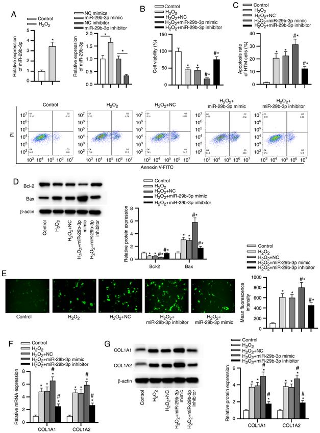
2 LIU et al: miR-29b-3p KNOCKDOWN PROTECTS HTM CELLS AGAINST OXIDATIVE INJURY
The aim of the present study was to investigate the role HTM cell culture and treatment. HTM cells (ScienCell
and regulatory mechanism of miR‑29b‑3p in HTM cells under Research Laboratories, Inc.) were cultured in Dulbecco's
oxidative stress in order to find potential novel therapeutic modified Eagle's medium (DMEM) containing 15% fetal
targets for glaucoma treatment. bovine serum, 2 mM L‑glutamine, 0.05% gentamicin, 1 ng/ml
FGF‑2 and 0.25 µg/ml amphotericin B (all from ScienCell
Materials and methods Research Laboratories, Inc.) at 37˚C with 5% CO 2. The
medium was replaced every day when the culture reached 70%
Laboratory animals. A total of 18 Wistar rats (8 weeks confluence. To induce oxidative stress, cells were incubated
old, male, weighing 220~260 g) were provided by Beijing in DMEM supplemented with H 2O2 (Beyotime Institute of
Vital River Laboratory Animal Technology Co., Ltd. Biotechnology) at concentrations of 100, 200, 300, 400 µM for
Before the experiments, all rats received 1 week of adap‑ 24 h after reaching 85% confluence.
tive feeding, with four to six rats in each cage. Rats were
raised with ad libitum access to water and food in a temper‑ Cell transfection. The mature miR‑29b‑3p mimic (for
ature‑controlled room (22±2˚C) with a 12 h light/dark cycle miR‑29b‑3p overexpression) and its NC mimic, miR‑29b‑3p
and a relative humidity of 40‑60%. All animal studies inhibitor (for miR‑29b‑3p silencing) and its NC inhibitor,
were performed following the animal guidelines of the small interfering (si)RNA against E3 ubiquitin‑protein ligase
International Association for the Study of Pain (20), and RNF138 (RNF138; siRNF138, for RNF138 knockdown) and
approved by the Ethics Committee of the Second Hospital its NC (si‑NC) were constructed by Shanghai GenePharma
of Anhui Medical University (approval no. 2019‑051; Hefei, Co., Ltd. The following sequences were included in the present
China). study: NC mimic, 5'‑CAGUACUUUUGUGUAGUACAAA‑3';
miR‑29b‑3p mimic, 5'‑UAGCACCAUU UGA AAUCAGUG
Establishment of glaucoma models and IOP measurement. U‑3'; NC inhibitor, 5'‑UUUGUACUACACAAAAGUACUG‑3';
For establishment of glaucoma models, rats were anesthe‑ miR‑29b‑3p inhibitor, 5'‑UAGCACCAUUUGAAAUCAGUG
tized with intraperitoneal injection of 3% pentobarbital U‑3'; si‑NC, 5'‑UAGA AGU UAACUUCACAGCAU‑3'; and
sodium (30 mg/kg; Sigma‑Aldrich; Merck KGaA). The right siRNF138, 5'‑ACAU UUUCUACAGAAA ACGUG‑3'. HTM
eyes of rats underwent operation. 0.1 ml aqueous fluid was cells were seeded into the 6‑well plates at a density of 1x10⁶
extracted from the anterior temporal horn of iris through a and subsequently transfected with miR‑29b‑3p mimic/inhibitor
cannula, and 14 rats were slowly injected with 3% compound (50 nM), NC mimic/inhibitor (50 nM), siRNF138 and si‑NC
carbomer solution through the cannula, while 4 rats were (100 ng) using Lipofectamine® 3000 (Invitrogen; Thermo
treated with the same amount of normal saline. After admin‑ Fisher Scientific, Inc.) for 48 h. The HTM cells were assigned
istration, all rats were treated with 0.5% moxifloxacin three into the following groups: i) Control group, cells without H2O2
times a day for 3 days. IOP measurement was operated using a stimulation; ii) H2O2 group, cells with 300 µM H2O2 stimu‑
tonometer (Mentor O&O, Inc.). IOP was measured at 10 AM lation; iii) H 2 O 2 + NC group, cells with 300 µM H 2 O 2
and 10 PM every day. A mean IOP was calculated from five stimulation + NC; iv) H2O2 + miR‑29b‑3p mimic group, cells
automatically averaged measurements. A total of 12 rats with 300 µM H2O2 stimulation + miR‑29b‑3p mimic; v) H2O2
with high IOP >30 mmHg were chosen. When the steadily + miR‑29b‑3p inhibitor group, cells with 300 µM H2O2 stimu‑
increased IOP was achieved, four glaucoma model rats were lation + miR‑29b‑3p inhibitor; vi) H2O2 + siRNF138 group,
injected with 5 µl antagomiR‑29b‑3p (Shanghai GenePharma cells with 300 µM H2O2 stimulation + siRNF138; and v) H2O2
Co., Ltd.), whereas the control rats were injected with 5 µl + miR‑29b‑3p inhibitor + siRNF138 group, cells with 300 µM
antagomiR‑negative control (NC) (Shanghai GenePharma H2O2 stimulation + miR‑29b‑3p inhibitor + siRNF138. The
Co., Ltd.). Rats were anesthetized with an intraperitoneal time interval between transfection and subsequent experimen‑
injection of 30 mg/kg pentobarbital sodium and were sacri‑ tation was 48 h.
ficed by decapitation.
Reverse transcription quantitative polymerase chain
Hematoxylin and eosin (H&E) staining. The right eyeballs reaction (RT‑qPCR). TRIzol reagent (Invitrogen; Thermo
of rats were isolated after the rats were sacrificed, and then Fisher Scientific, Inc.) was employed for the extraction of
fixed in 4% paraformaldehyde for 24 h at room temperature. total RNA from HTM cells. First‑strand cDNA synthesis was
The cornea was incised along the sclera and the iris and performed using M‑MLV reverse transcriptase (Invitrogen;
lens were removed 30 min later. The retinal tissues were Thermo Fisher Scientific, Inc.), according to the manufac‑
paraffin‑embedded and cut into 5‑µm sections for H&E turer's protocol. qPCR was conducted using the TaqMan™
staining assay. The sections were stained with hematoxylin for Universal Master Mix II (Applied Biosystems; Thermo Fisher
2 min and stained with eosin for 10 min at room temperature Scientific, Inc.) in 7500 Fast Real‑Time PCR System (Applied
(Beijing Zhongshan Golden Bridge Biotechnology Co., Ltd.). Biosystems; Thermo Fisher Scientific, Inc.). For quantification
After dehydration with gradient alcohol, the sections were of miRNAs, cDNA was obtained from 3 µg RNA using a
sealed using neutral resin. The morphology of retinal tissues specific stem‑loop primer. The 2‑ΔΔCq method (21) was used
was observed under a light microscope at x20 magnification for quantification and miRNA expression was normalized to
(Olympus Corporation), and the results were quantified using U6, while GAPDH acted as the endogenous reference gene
ImageJ software (version 1.46; National Institutes of Health). for mRNA. The following primers were used for RT‑qPCR:
The mean value was obtained from five randomly selected miR‑29b‑3p forward, 5'‑UAGCACCAUU UGA AAUC‑3' and
fields. reverse, 5'‑GTGCAGGTCCGAGGT‑3'; U6 forward, 5'‑CGC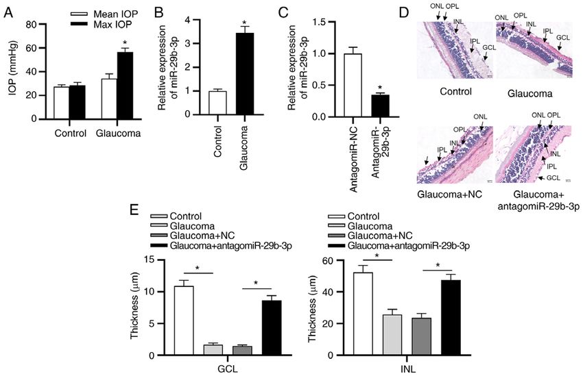
INTERNATIONAL JOURNAL OF MOlecular medicine 47: 101, 2021 3
TTCGGCAGCACATATAC‑3' and reverse, 5'‑TTCACGA AT dark. Cell apoptosis was determined using the Attune™ NxT
TTGCGTGTCAT‑3'; RNF138 forward, 5'‑TATAGTTCTGGC Flow Cytometer (Invitrogen; Thermo Fisher Scientific, Inc.)
TCTCTGAA‑3' and reverse, 5'‑CTGACTTGGGTAAATGCT and the apoptosis rate was analyzed with FlowJo software
CAAT‑3'; collagen α‑1(I) chain (COL1A1) forward, 5'‑CCT (version 10.0; FlowJo LLC). Apoptosis rate = apoptosis rate in
GTCTGCTTCCTGTAAAC‑3' and reverse, 5'‑ATGTTCGGT quadrant (Q)2 + apoptosis rate in Q3.
TGGTCAAAGATAAAT‑3'; collagen α‑2(I) chain (COL1A2)
forward, 5'‑CAGTCGTATG CGCGTATAG C‑3' and reverse, Detection of reactive oxygen species (ROS). ROS levels
5'‑CGTAGTC GTAGCTAGC TAGAGA‑3'; and GAPDH in HTM cells were examined using a ROS assay kit
forward, 5'‑GGAGCGAGATCCCTCCAAAAT‑3' and reverse, (cat. no. S0033M; Beyotime Institute of Biotechnology). After
5'‑GGCTGT TGTCATACT TCTCATG G‑3'. The thermocy‑ transfection and H2O2 stimulation, HTM cells were washed in
cling conditions used were as follows: 94˚C for 5 min; followed PBS and subsequently cultured in DMEM containing 10 µM
by 40 cycles of 94˚C for 1 min, 56˚C for 1 min and 72˚C for 2,7‑dichlorodihydrofluorescein diacetate (DCFHDA) for
1 min. 20 min at 37˚C. After washing with PBS twice, the cells were
treated with 0.25% trypsin‑EDTA, followed by centrifugation
Western blot analysis. The treated HTM cells were collected at 800 x g for 6 min at 37˚C. Then, cells were resuspended
and washed with pre‑cooling PBS. After centrifugation at in 500 µl PBS. The fluorescence intensity, regarded as ROS
600 x g for 5 min at 4˚C, the cells were lysed with radio immu‑ generation, was measured by an Infinite M200 microplate
noprecipitation assay buffer (Sigma‑Aldrich; Merck KGaA). reader (485 and 525 nm emission).
Then, cell lysates were centrifuged at 12,000 x g for 12 min
at 4˚C, followed by measurement of proteins in the supernatant Bioinformatics analysis. Targets of miR‑29b‑3p were
using the BCA protein assay (Pierce; Thermo Fisher Scientific, predicted using miRanda version 3.3a (http://cbio.mskcc.
Inc.). After proteins (20 µg per lane) were separated via org/microrna_data/miRanda‑aug2010.tar.gz), microT‑CDS
sodium dodecyl sulfate‑polyacrylamide gel electrophoresis on DIANA Tools version 5.0 (http://diana.imis.athena‑innovation.
10% gel, proteins were transferred to polyvinylidene difluoride gr/DianaTools/index.php?r=microT_CDS/index) and RNA22
membranes. Afterwards, the membranes were blocked with version 2.0 (https://cm.jefferson.edu/rna22/Interactive/).
5% non‑fat dry milk at 4˚C overnight, and incubated with the
following primary antibodies at 4˚C overnight: Anti‑RNF138 Luciferase reporter assay. The wild‑type (Wt) and mutated
(cat. no. K107111P; 1:5,000; Beijing Solarbio Science (Mut) sequences of the 3'UTR of RNF138 complementary
& Technology Co., Ltd.), anti‑Bcl‑2 (cat. no. ab185002; to miR‑29b‑3p were subcloned into the pmirGLO‑luciferase
1:1,000; Abcam), anti‑Bax (cat. no. ab32503; 1:1,000; plasmids (Promega Corporation) to generate Wt RNF138
Abcam), anti‑COL1A1 (cat. no. ab210966; 1:1,000; Abcam), (RNF138‑Wt) and Mut type RNF138 (RNF138‑Mut).
anti‑COL1A2 (cat. no. ab96723; 1:1,000; Abcam), anti‑ERK RNF138‑Wt and RNF138‑Mut reporters were co‑transfected
(cat. no. ab115799; 1:1,000; Abcam), anti‑phosphorylated with NC or miR‑29b‑3p mimic into 293T cells (American Type
(p)‑ERK (cat. no. ab214036; 1:1,000; Abcam) and anti‑β‑actin Culture Collection) using Lipofectamine 3000. The interaction
(cat. no. ab179467; 1:1,000; Abcam). The membranes were between miR‑29b‑3p and 3'‑UTR of RNF138 was detected
subsequently cultured with the corresponding secondary using a Luciferase Assay system (Promega Corporation) 48 h
antibodies (cat. no. ab205718; 1:5,000; Abcam) for 1.5 h at post‑transfection. Relative luciferase activity was defined
room temperature. The proteins were detected using the as the ratio of firefly luciferase activity to Renilla luciferase
chemiluminescence detection (ECL Plus; Pierce; Thermo activity.
Fisher Scientific, Inc.). Semi‑quantification was performed
using ImageJ software (version 1.46; National Institutes of Statistical analysis. Data are presented as the mean ± standard
Health). deviation of three independent experiments and analyzed using
the SPSS 21.0 software (IBM Corp.). Comparisons between
3‑(4,5‑Dimethylthiazol‑2‑yl)‑2,5‑diphenyltetrazolium bromide 2 groups were analyzed using unpaired Student's t‑test, while
(MTT) assay. HTM cell viability was assessed using an MTT comparisons among ≥3 groups were analyzed using one‑way
assay. After transfection and H2O2 stimulation, cells (1,000 cells analysis of variance. Dunnett's or Tukey's post hoc tests were
per well) were seeded in the 96‑well plates, followed by incuba‑ used following one‑way analysis of variance. P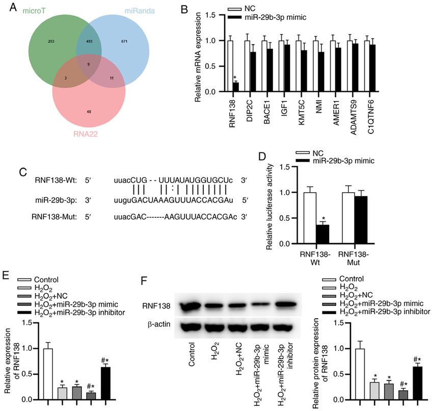
4                          LIU et al: miR-29b-3p KNOCKDOWN PROTECTS HTM CELLS AGAINST OXIDATIVE INJURY

Figure 1. miR‑29b‑3p is upregulated in a rat glaucoma model and antagomiR‑29b‑3p alleviates the symptoms of glaucoma. (A) Mean and max IOP in the
control and glaucoma groups. *P
INTERNATIONAL JOURNAL OF MOlecular medicine 47: 101, 2021                                                         5

Figure 3. miR‑29b‑3p facilitates H2O2‑induced injury and promotes ECM deposition, whereas silencing of miR‑29b‑3p exerts the opposite effects. (A) The
expression of miR‑29b‑3p in HTM cells following different treatments was determined by RT‑qPCR. (B) HTM cell viability in each group was assessed by a
3‑(4,5‑dimethylthiazol‑2‑yl)‑2,5‑diphenyltetrazolium bromide assay. (C) The apoptotic rate of HTM cells in each group was evaluated using flow cytometry.
(D) The levels of apoptosis‑related proteins in HTM cells in each group were measured by western blotting. (E) Reactive oxygen species generation in HTM
cells in each group was detected by 2,7‑dichlorodihydrofluorescein diacetate staining. (F) The mRNA expression of COL1A1 and COL1A2 in HTM cells
was detected by RT‑qPCR. (G) The protein levels of ECM‑related genes in HTM cells were evaluated by western blotting. *P
6                        LIU et al: miR-29b-3p KNOCKDOWN PROTECTS HTM CELLS AGAINST OXIDATIVE INJURY

Figure 4. RNF138 is a target gene of miR‑29b‑3p. (A) Venn diagram showing nine mRNAs as possible targets of miR‑29b‑3p as predicted by microT, miRanda
and RNA22 analyses. (B) RNF138 expression in miR‑29b‑3p mimic‑transfected HTM cells was detected using RT‑qPCR. (C) The predicted binding site
between miR‑29b‑3p and RNF138. (D) Luciferase reporter assay was performed to confirm the binding ability between miR‑29b‑3p and RNF138. (E and F) The
expression of RNF138 at the mRNA and protein levels in HTM cells of each group was determined by RT‑qPCR and western blotting, respectively. *P
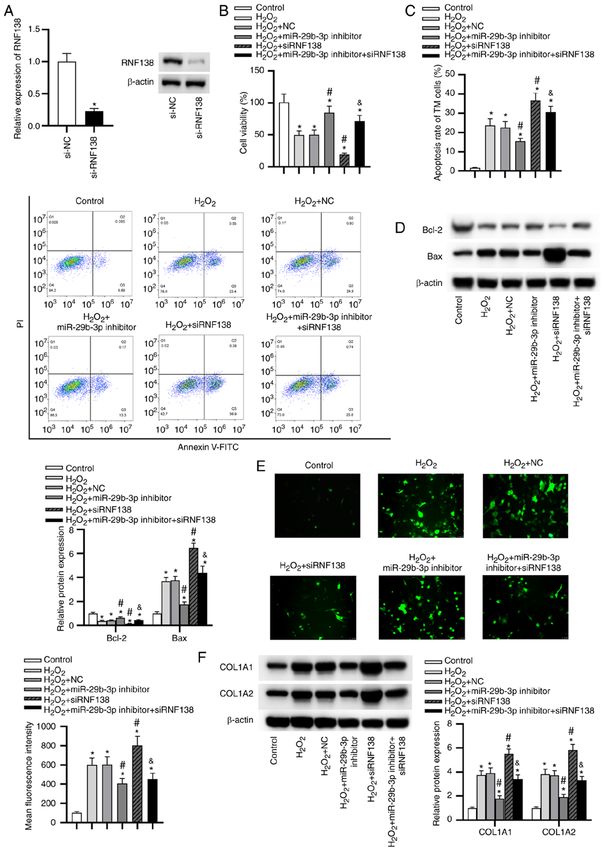
INTERNATIONAL JOURNAL OF MOlecular medicine 47: 101, 2021                                                            7

Figure 5. Silencing RNF138 expression promotes H2O2‑induced injury and silencing of miR‑29b‑3p ameliorates it by upregulating RNF138. (A) The silencing
efficiency of RNF138 in HTM cells was verified by reverse transcription quantitative polymerase chain reaction and western blotting. *P
8                        LIU et al: miR-29b-3p KNOCKDOWN PROTECTS HTM CELLS AGAINST OXIDATIVE INJURY

Figure 6. Downregulation of miR‑29b‑3p activates the ERK pathway by upregulating E3 ubiquitin‑protein ligase RNF138. The expression of t‑ERK and p‑ERK
proteins in human trabecular meshwork cells of each group was detected by western blotting and the ratio of p‑ERK/t‑ERK was semi‑quantified. *P
INTERNATIONAL JOURNAL OF MOlecular medicine 47: 101, 2021                                             9

whereas the H 2O2 + siRNF138 group showed the opposite               Therefore, miR‑29b‑3p regulated the ERK pathway
results. Moreover, the effect of miR‑29b‑3p inhibitor on the         by targeting RNF138 in HTM cells under oxidative injury.
ERK pathway was significantly rescued by siRNF138.                       In conclusion, the present study revealed that silencing of
                                                                     miR‑29b‑3p alleviated glaucoma and suppressed apoptosis,
Discussion                                                           ROS generation, ECM deposition, as well as promoting the
                                                                     viability of HTM cells under oxidative injury. Mechanistically,
The loss of vision caused by glaucoma is irreversible.               miR‑29b‑3p was demonstrated to target RNF138 3'UTR and
Multiple miRNAs are related to retinal damage, retinal               downregulates its expression to inactive the ERK pathway.
homeostasis and retinogenesis (22,23). Previous reports iden‑        The protective role of silencing of miR‑29b‑3p in HTM cells
tified a series of miRNAs that may be potential biomarkers           may offer a novel therapeutic strategy for glaucoma.
in glaucoma (24,25). Additionally, oxidative stress is a key
pathophysiological mechanism in glaucoma (26). The high              Acknowledgements
IOP results from the imbalance of the aqueous humor inflow
and outflow, and the oxidative injury of TM is responsible           Not applicable.
for aqueous humor outflow (6,7). Therefore, the present study
explored the role of miR‑29b‑3p in the oxidative injury of           Funding
TM cells. miRNAs act as key regulators of TM cells in the
progression of glaucoma (15,27). The present study found that        This study was supported by the Research Fund of Anhui
miR‑29b‑3p expression was increased in glaucoma model rats           Medical University (grant no. 2020xkj202).
and antagomiR‑29b‑3p alleviated the symptoms of glaucoma.
Moreover, miR‑29b‑3p expression was significantly upregu‑            Availability of data and materials
lated in H 2O2 ‑stimulated HTM cells. Downregulation of
miR‑29b‑3p alleviated H2O2 ‑induced oxidative injury in HTM          The datasets used and/or analyzed during the current study are
cells by promoting cell viability, and inhibiting cell apoptosis     available from the corresponding author on reasonable request.
and ROS generation as well as ECM production. Previous
studies have reported that oxidative damage aggravates glau‑         Authors' contributions
coma progression by increasing TM cell apoptosis and ECM
production (28,29). These findings suggested that inhibition of      HL, YX, QZ and LT performed the experiments. HL, QZ,
miR‑29b‑3p may play a protective role in glaucoma.                   QW, YX and LT contributed to data analysis and wrote
     Based on bioinformatics analysis, RNF138 was predicted          the paper. HL and LT made substantial contributions to
as a downstream target of miR‑29b‑3p. RNF138 serves as               the design of the present study and acquired experimental
an anti‑apoptotic gene in cancers. Upregulation of RNF138            materials. All authors read and approved the final
promotes cell proliferation and inhibits apoptosis in                manuscript. HL and LT confirm the authenticity of all the raw
cisplatin‑sensitive gastric cancer cells (30). Additionally, down‑   data.
regulation of RNF138 induces apoptosis of spermatogenic
cells in mice (31). In the present study, RNF138 was confirmed       Ethics approval and consent to participate
to be the functional downstream gene of miR‑29b‑3p. RNF138
expression was downregulated in HTM cells with H2O2 stimu‑           All animal studies were performed following the animal
lation. Silencing of RNF138 inhibited viability, promoted            guidelines of the International Association for the Study of
apoptosis, ROS generation and ECM production in HTM                  Pain, and approved by the Ethics Committee of the Second
cells under oxidative injury. In addition, RNF138 knockdown          Hospital of Anhui Medical University (approval no. 2019‑051;
significantly reversed the protective effects of miR‑29b‑3p          Hefei, China).
inhibitor on the oxidative injury in HTM cells, which was
consistent with the anti‑apoptotic role of RNF138 identified in      Patient consent for publication
previous literatures.
     The ERK signaling pathway is an important intra‑                Not applicable.
cellular pathway, which has the ability to promote
proliferation in various cell types (32,33). The activation          Competing interests
of the ERK pathway induced by Vitamin D attenuates the
H2O2 ‑stimulated oxidative injury in human endothelial cells (34).   The authors declare that they have no competing interests.
The ERK pathway activated by myeloid cell leukemia1 protects
rat pheochromocytoma cells from H2O2 oxidant injury (35).            References
RNF138 silencing suppresses the development of glioma by
repressing the ERK pathway (36). Therefore, the present study         1. Yanagi M, Kawasaki R, Wang JJ, Wong TY, Crowston J and
detected the changes of key proteins in the ERK pathway in               Kiuchi Y: Vascular risk factors in glaucoma: A review. Clin Exp
                                                                         Ophthalmol 39: 252‑258, 2011.
HTM cells stimulated by H2O2. The ERK pathway was signifi‑            2. Liao Q, Wang DH and Sun HJ: Association of genetic polymor‑
cantly suppressed in HTM cells under H 2O2 stimulation,                  phisms of eNOS with glaucoma. Mol Vis 17: 153‑158, 2011.
and this suppression was rescued by miR‑29b‑3p suppres‑               3. Williams PA, Harder JM, Foxworth NE, Cochran KE, Philip VM,
                                                                         Porciatti V, Smithies O and John SW: Vitamin B3 modulates
sion. Additionally, RNF138 knockdown inhibited the                       mitochondrial vulnerability and prevents glaucoma in aged mice.
activation of ERK pathway induced by miR‑29b‑3p knockdown.               Science 355: 756‑760, 2017.
10 LIU et al: miR-29b-3p KNOCKDOWN PROTECTS HTM CELLS AGAINST OXIDATIVE INJURY
4. Heijl A, Leske MC, Bengtsson B, Hyman L, Bengtsson B and 22. Izzotti A, Ceccaroli C, Longobardi MG, Micale RT, Pulliero A,
Hussein M; Early Manifest Glaucoma Trial Group: Reduction of La Maestra S and Saccà SC: Molecular damage in glaucoma:
intraocular pressure and glaucoma progression: Results from the From anterior to posterior eye segment. Microrna 4: 3‑17, 2015.
early manifest glaucoma trial. Arch Ophthalmol 120: 1268‑1279, 23. Genini S, Guziewicz KE, Beltran WA and Aguirre GD: Altered
2002. miRNA expression in canine retinas during normal development
5. Hysi PG, Cheng CY, Springelkamp H, Macgregor S, Bailey JNC, and in models of retinal degeneration. BMC Genomics 15: 172,
Wojciechowski R, Vitart V, Nag A, Hewitt AW, Höhn R, et al: 2014.
Genome‑wide analysis of multi‑ancestry cohorts identifies 24. Hindle A, Thoonen R, Jasien JV, Grange RMH, Amin K, Wise J,
new loci influencing intraocular pressure and susceptibility to Ozaki M, Ritch R, Malhotra R and Buys ES: Identification of
glaucoma. Nat Genet 46: 1126‑1130, 2014. candidate miRNA biomarkers for glaucoma. Invest Ophthalmol
6. Villarreal G Jr, Oh DJ, Kang MH and Rhee DJ: Coordinated Vis Sci 60: 134‑146, 2019.
regulation of extracellular matrix synthesis by the microRNA‑29 25. Romano G, Platania CB, Forte S, Salomone S, Drago F and
family in the trabecular meshwork. Invest Ophthalmol Vis Bucolo C: MicroRNA target prediction in glaucoma. Prog Brain
Sci 52: 3391‑3397, 2011. Res 220: 217‑240, 2015.
7. Vidal‑Sanz M, Salinas‑Navarro M, Nadal‑Nicolás FM, 26. Kim KY, Perkins GA, Shim MS, Bushong E, Alcasid N, Ju S,
Alarcón‑Martínez L, Valiente‑Soriano FJ, de Imperial JM, Ellisman MH, Weinreb RN and Ju WK: DRP1 inhibition rescues
Avilés‑Trigueros M, Agudo‑Barriuso M and Villegas‑Pérez MP: retinal ganglion cells and their axons by preserving mitochon‑
Understanding glaucomatous damage: Anatomical and func‑ drial integrity in a mouse model of glaucoma. Cell Death Dis 6:
tional data from ocular hypertensive rodent retinas. Prog Retin e1839, 2015.
Eye Res 31: 1‑27, 2012. 27. Wang Y, Li F and Wang S: MicroRNA‑93 is overexpressed and
8. Vranka JA, Kelley MJ, Acott TS and Keller KE: Extracellular induces apoptosis in glaucoma trabecular meshwork cells. Mol
matrix in the trabecular meshwork: Intraocular pressure regula‑ Med Rep 14: 5746‑5750, 2016.
tion and dysregulation in glaucoma. Exp Eye Res 133: 112‑125, 28. Ueda J, Wentz‑Hunter K and Yue BY: Distribution of myocilin
2015. and extracellular matrix components in the juxtacanalicular
9. Medina‑Ortiz WE, Belmares R, Neubauer S, Wordinger RJ and tissue of human eyes. Invest Ophthalmol Vis Sci 43: 1068‑1076,
Clark AF: Cellular fibronectin expression in human trabecular 2002.
meshwork and induction by transforming growth factor‑ β2. 29. Fatma N, Kubo E, Toris CB, Stamer WD, Camras CB
Invest Ophthalmol Vis Sci 54: 6779‑6788, 2013. and Singh DP: PRDX6 attenuates oxidative stress‑ and
10. Schanen BC and Li X: Transcriptional regulation of mammalian TGFbeta‑induced abnormalities of human trabecular meshwork
miRNA genes. Genomics 97: 1‑6, 2011. cells. Free Radic Res 43: 783‑795, 2009.
11. Wahid F, Khan T and Kim YY: MicroRNA and diseases: 30. Lu Y, Han D, Liu W, Huang R, Ou J, Chen X, Zhang X, Wang X,
Therapeutic potential as new generation of drugs. Biochimie 104: Li S, Wang L, et al: RNF138 confers cisplatin resistance in gastric
12‑26, 2014. cancer cells via activating Chk1 signaling pathway. Cancer Biol
12. Ha M and Kim VN: Regulation of microRNA biogenesis. Nat Ther 19: 1128‑1138, 2018.
Rev Mol Cell Biol 15: 509‑524, 2014. 31. Xu L, Lu Y, Han D, Yao R, Wang H, Zhong S, Luo Y,
13. Chen K and Rajewsky N: The evolution of gene regulation by Han R, Li K, Fu J, et al: Rnf138 deficiency promotes apoptosis
transcription factors and microRNAs. Nat Rev Genet 8: 93‑103, of spermatogonia in juvenile male mice. Cell Death Dis 8: e2795,
2007. 2017.
14. Su W, Li Z, Jia Y, Zhu Y, Cai W, Wan P, Zhang Y, Zheng SG and 32. Kim EK and Choi EJ: Compromised MAPK signaling in human
Zhuo Y: MicroRNA‑21a‑5p/PDCD4 axis regulates mesenchymal diseases: An update. Arch Toxicol 89: 867‑882, 2015.
stem cell‑induced neuroprotection in acute glaucoma. J Mol Cell 33. Sun Y, Liu WZ, Liu T, Feng X, Yang N and Zhou HF: Signaling
Biol 9: 289‑301, 2017. pathway of MAPK/ERK in cell proliferation, differentiation,
15. Ruibin W, Zheng X, Chen J, Zhang X, Yang X and Lin Y: migration, senescence and apoptosis. J Recept Signal Transduct
Micro RNA‑1298 opposes the effects of chronic oxidative stress Res 35: 600‑604, 2015.
on human trabecular meshwork cells via targeting on EIF4E3. 34. Polidoro L, Properzi G, Marampon F, Gravina GL, Festuccia C,
Biomed Pharmacother 100: 349‑357, 2018. Di Cesare E, Scarsella L, Ciccarelli C, Zani BM and Ferri C:
16. Shen W, Han Y, Huang B, Qi Y, Xu L, Guo R, Wang X and Vitamin D protects human endothelial cells from H2O2 oxidant
Wang J: MicroRNA‑483‑3p inhibits extracellular matrix produc‑ injury through the Mek/Erk‑Sirt1 axis activation. J Cardiovasc
tion by targeting Smad4 in human trabecular meshwork cells. Transl Res 6: 221‑231, 2013.
Invest Ophthalmol Vis Sci 56: 8419‑8427, 2015. 35. Li R, Yin F, Guo YY, Zhao KC, Ruan Q and Qi YM: Knockdown
17. Yang Q, Wu F, Mi Y, Wang F, Cai K, Yang X, Zhang R, Liu L, of ANRIL aggravates H 2O2‑induced injury in PC‑12 cells by
Zhang Y, Wang Y, et al: Aberrant expression of miR‑29b‑3p targeting microRNA‑125a. Biomed Pharmacother 92: 952‑961,
influences heart development and cardiomyocyte proliferation 2017.
by targeting NOTCH2. Cell Prolif 53: e12764, 2020. 36. Wu H, Li X, Feng M, Yao L, Deng Z, Zao G, Zhou Y,
18. Wang J, Zhu M, Ye L, Chen C, She J and Song Y: MiR‑29b‑3p Chen S and Du Z: Downregulation of RNF138 inhibits cellular
promotes particulate matter‑induced inflammatory responses proliferation, migration, invasion and EMT in glioma cells
by regulating the C1QTNF6/AMPK pathway. Aging (Albany via suppression of the Erk signaling pathway. Oncol Rep 40:
NY) 12: 1141‑1158, 2020. 3285‑3296, 2018.
19. Zeng Y, Cui Z, Liu J, Chen J and Tang S: MicroRNA‑29b‑3p
promotes human retinal microvascular endothelial cell apoptosis
via blocking SIRT1 in diabetic retinopathy. Front Physiol 10: This work is licensed under a Creative Commons
1621, 2019. Attribution-NonCommercial-NoDerivatives 4.0
20. Orlans FB: Ethical decision making about animal experiments. International (CC BY-NC-ND 4.0) License.
Ethics Behav 7: 163‑171, 1997.
21. Livak KJ and Schmittgen TD: Analysis of relative gene expres‑
sion data using real‑time quantitative PCR and the 2(‑Delta Delta
C(T)) method. Methods 25: 402‑408, 2001.You can also read