Proapoptotic effect of endocannabinoids in prostate cancer cells
←
→
Page content transcription
If your browser does not render page correctly, please read the page content below
ONCOLOGY REPORTS 33: 1599-1608, 2015

   Proapoptotic effect of endocannabinoids in prostate cancer cells
                    O. Orellana-Serradell1, C.E. Poblete1, C. Sanchez1, E.A. Castellón1,
                        I. Gallegos2, C. Huidobro3, M.N. Llanos4 and H.R. Contreras1

       1
           Physiology and Biophysics Program, Institute of Biomedical Sciences, Faculty of Medicine, University of Chile;
                   2
                     Pathological Anatomy Service, 3Urology Service, Clinic Hospital of the University of Chile;
              4
               Laboratory of Nutrition and Metabolic Regulation, INTA, University of Chile, Santiago 8389100, Chile

                                       Received October 13, 2014; Accepted December 29, 2014

                                                        DOI: 10.3892/or.2015.3746

Abstract. In the early stages, prostate cancer is androgen‑              of the apoptotic pathway. Finally, we observed that endocan-
dependent; therefore, medical castration has shown significant           nabinoid treatment activated the Erk pathway and at the same
results during the initial stages of this pathology. Despite this        time, produced a decrease in the activation levels of the Akt
early effect, advanced prostate cancer is resilient to such treat-       pathway. Based on these results, we suggest that endocannabi-
ment. Recent evidence shows that derivatives of Cannabis                 noids may be a beneficial option for the treatment of prostate
sativa and its analogs may exert a protective effect against             cancer that has become nonresponsive to common therapies.
different types of oncologic pathologies. The purpose of the
present study was to detect the presence of cannabinoid recep-           Introduction
tors (CB1 and CB2) on cancer cells with a prostatic origin and
to evaluate the effect of the in vitro use of synthetic analogs. In      Prostate cancer (PrC) is a highly prevalent oncologic pathology
order to do this, we used a commercial cell line and primary             in most countries throughout the world (1). During its early
cultures derived from prostate cancer and benign prostatic               stages this disease is usually asymptomatic and exhibits slow
hyperplasia. The presence of the CB1 and CB2 receptors                   progression, which carries the risk of having it diagnosed at
was determined by immunohistochemistry where we showed                   an advanced stage. Research indicates a direct correlation
a higher expression of these receptors in later stages of the            between the appearance of symptoms and the spreading of
disease (samples with a high Gleason score). Later, treat-               the cancer or metastasis. This late diagnosis may decrease the
ments were conducted using anandamide, 2-arachidonoyl                    treatment options available for patients and also the chances
glycerol and a synthetic analog of anandamide, methanan-                 of recovery (2).
damide. Using the MTT assay, we proved that the treatments                    Due to the above factors and in light of the aggressive
produced a cell growth inhibitory effect on all the different            nature of this disease in its late stages, it has become critical to
prostate cancer cultures. This effect was demonstrated to be             search for more effective tools with which to detect PrC while
dose-dependent. The use of a specific CB1 receptor blocker               it is still in its early stages, and for better treatment options.
(SR141716) confirmed that this effect was produced primarily             These two aspects would greatly improve the quality of life
from the activation of the CB1 receptor. In order to understand          and the overall survival expectancy of these patients (3,4).
the MTT assay results, we determined cell cycle distribution by               Regarding the molecular pathogenesis of PrC, it has been
flow cytometry, which showed no variation at the different cell          observed that certain genetic alterations may provoke the
cycle stages in all the cultures after treatment. Treatment with         transformation of normal prostatic cells into cancerous cells.
endocannabinoids resulted in an increase in the percentage of            In this context, it has been reported that certain mutations
apoptotic cells as determined by Annexin V assays and caused             in key specific genes such as PTEN, TP53, E-cadherin and
an increase in the levels of activated caspase-3 and a reduc-            β-catenin (5) are important in this transformation process. In
tion in the levels of Bcl-2 confirming that the reduction in cell        addition, it has been demonstrated that several growth factors,
viability noted in the MTT assay was caused by the activation            such as insulin growth factor I (IGF-I), transforming growth
                                                                         factor α and β (TGFα or β) and members of the fibroblast
                                                                         growth factor (FGF) family, may be involved in the prolifera-
                                                                         tion and metastasis of PrC (6).
                                                                              The molecular factors involved in the development of
Correspondence to: Dr Héctor R. Contreras, Physiology and                PrC are various and as the transformation process in the cell
Biophysics Program, Institute of Biomedical Sciences, Faculty of
                                                                         evolves, it acquires a malignant phenotype with the ability
Medicine, University of Chile, Clasificador 7, Santiago 8389100, Chile
                                                                         to invade and generate metastasis in different parts of the
E-mail: hcontrer@med.uchile.cl
                                                                         body, but with a preference to form metastatic lesions in the
Key words: prostate cancer, endocannabinoid receptors                    bones (5,6). For this reason, many compounds have been used
                                                                         to try and control the progression of prostate cancer. It has
                                                                         been suggested that marijuana (Cannabis sativa) through some
1600 orellana-serradell et al: Endocannabinoids and prostate cancer
of its active compounds could be involved in halting tumoral its demonstrated effects on cancer cells. Another member of
growth and thus delaying its progression to more advanced this family is 2-arachidonyl glyceryl ether, the endogenous
and aggressive stages (7-9). relevance of which is currently being studied (9).
Marijuana acts in the organism as a psychoactive agent Due to the previous effects noted in other types of cancer
through the production of its active component, the cannabi- and increased knowledge of anandamide (and its synthetic
noid (-)-∆ 9-tetrahidrocanabinol (or THC), which is an aromatic analog methanandamide) and 2-AG, further investigation of
terpenoid compound with a very low solubility in water. Two the effects of these endocannabinoids for the treatment of PrC
different cannabinoid receptors have been described, charac- is warranted. Recently, it was reported that an increment in
terized and cloned: cannabinoid receptor 1 (or CB1), originally intracellular levels of 2-AG inhibited the invasive ability of the
found in the brain, and cannabinoid receptor 2 (or CB2), which PrC cell lines PC3 and DU-145 by a mechanism involving the
was first described in the spleen. Both receptors are part of the activation of the CB1 receptor and through the inactivation of
superfamily of G protein-coupled receptors (10,11). protein kinase A (22). In the same manner, the direct participa-
Other than its exogenous ligands such as THC, the organism tion of anandamide (Ana) in decreasing the proliferative action
can produce similar compounds that have been termed as of EGF in cell lines has been proven. Following treatment with
endocannabinoids, which are capable of modulating several anandamide in DU-145 and LNCaP cells, a downregulating
physiological mechanisms through the CB1 and CB2 membrane effect on the expression levels for the receptor of the EGF
receptors (12), although it has also been reported recently that growth factor accompanied by a proliferative arrest in the G1
they may also act in an independent receptor manner (13). phase of the cell cycle and a rise in the levels of apoptosis and
Endocannabinoids are molecules that are derived from necrosis in the cells were found (26-28).
unsaturated fatty acids acting as endogenous ligands for Notwithstanding these results, information concerning the
cannabinoid receptors CB1 and CB2 and originate in the signaling pathways mediating these effects in PrC is still scarce
plasma membrane from phospholipids in response to a rise in and somewhat contradictory in nature. Some information has
the intracellular concentrations of calcium. These endogenous been reported in other types of cancer, for example, the activa-
ligands bind to the CB1 and CB2 receptors with great affinity tion of the AKT pathway in astrocytoma cells after treatment
and participate in many biological processes in the immune, with THC or Ana (29), activation of the ERK pathway when
respiratory, circulatory and reproductive systems (9,14-17). using THC in glioma cell lines (30) or the activation of the
Endocannabinoids are then eliminated in a two step process: JNK pathway when using endocannabinoids in different types
first, an intracellular accumulation, followed by an enzymatic of nerve cells that express the CB1 receptor (31).
metabolization by an enzyme that belongs to the serine To date, all of the studies using endocannabinoids have
hydrolase family, the fatty acid amide hydrolase (or FAAH) been made in cell lines. The objective of the present study was
or by the monoacyl glycerol lipase (a soluble serine hydrolase to analyze the effect of endocannabinoids, not only on cell
enzyme). These two enzymes are the main proteins in charge lines, but also on primary cultures of PrC and the signaling
of degrading endocannabinoids (18). pathways involved in order to obtain a better understanding of
On a cellular level, it has been found that endocannabinoids the possible effects following treatment with these molecules
may modulate cell proliferation, viability and differentiation. against prostate cancer.
This evidence suggests that endocannabinoids may also be
involved in controlling the growth and transformation of Materials and methods
tumor cells (8,19,20). Regarding this, evidence shows that
endocannabinoids may inhibit the growth of several types of Materials. Endocanabinoids anandamide (Ana) and 2-AG were
tumors through the inhibition of proliferative pathways such purchased from Calbiochem (San Diego, CA, USA) (cat. nos.
as adenylate cyclase (21) and protein kinase A (22), arrest of 172100 and 181251, respectively) and methanandamide (Me)
the cell cycle by induction of p27 (23), downregulation of the was purchased from Biomol (Plymouth Meeting, PA, USA)
EGF receptor (EGF-R) and other molecules related to growth (cat. no. FA-021). CB1 receptor antagonist, SR141716, was
pathways such as the nerve growth factor receptor (NGF-R), purchased from Sanofi (Montpellier, France).
the vascular endothelial growth factor (VEGF) and prolac- Primary antibodies for the CB1 and CB2 receptors
tine (24). Likewise, when tumor cells are treated with specific were obtained from Cayman Chemical (Ann Arbor, MI,
antagonists for endocannabinoids, the invasive ability of USA) (cat. nos. 10006590 and 101550, respectively), and
tumors increases. The great advantage that the use of endocan- caspase-3 and Bcl-2 from Cell Signaling Technology, Inc.
nabinoids may bring to the battle against prostate cancer is that (Danvers, MA, USA) (cat. nos. 9661 and 2876, respectively).
it has been demonstrated that the receptors for these molecules Phospho-p44/42 MAPK (Erk1/2) was purchased from Cell
are substantially overexpressed in cancerous prostatic cells Signaling Technology, Inc. (Danvers) (cat. no. 9101) and
when compared to normal, healthy prostate tissues (9,25). p-Akt 1/2/3 (Thr308)-R from Santa Cruz Biotechnology,
There are two classes of endocannabinoids. Among those Inc. (Dallas, TX, USA) (cat. no. 16646-R). Anti-mouse and
that are derived from fatty acids, anandamide is the most studied anti-rabbit secondary peroxidase‑conjugated antibodies were
and was the first to be described. Other members of this family obtained from Jackson Immuno Research (West Grove,
include N-oleoylethanolamide and N-palmitoylethanolamide, PA, USA) (cat. nos. 115-035-003 and 111-035‑003, respec-
which have shown strong dietetic effects independent of CB1 tively). Anti‑rabbit FITC‑conjugated secondary antibodies
or CB2 activation. The second class of endocannabinoids were obtained from Jackson Immuno Research (cat. no.
are those bound to glycerol, of which 2-arachidonoyl glyc- 305-095-003). For immunohistochemistry the subsequent
erol (2-AG) is the most commonly used in research due to kits were used: Histostain®-Plus Bulk kit (cat. no. 85-8943)ONCOLOGY REPORTS 33: 1599-1608, 2015 1601
and the DAB-Plus Substrate kit (cat. no. 00-2020) (both from antagonist SR141716 (diluted in DMSO) for 30 min at 37˚C in
Invitrogen, Carlsbad, CA, USA). Annexin V-FITC Apoptosis the absence of light. Afterwards, the antagonist was removed
Detection kit was obtained from BD Pharmingen (Franklin and the cells were washed with PBS. Finally, the cells were
Lakes, NJ, USA) (cat. no. 556547). treated with the different endocannabinoids under the same
conditions as explained above.
Methods
Immunohistochemistry. Formalin-fixed and paraffin‑embedded Cell viability. The effect of endocannabinoids on cell viability
prostate specimens were obtained from the archives of the was determined by 3-(4,5-dimethylthiazol‑2-yl)‑2,5-diphenyl
Pathological Anatomy Service, Clinic Hospital of the University tetrazoliumbromide (MTT) assays. The cells were plated at
of Chile, with the corresponding authorization. All samples 5x10³/well in 200 µl of complete culture medium containing
were evaluated by an expert pathologist (I.G.) and grouped 2.5, 5.0 and 10.0 µM concentrations of Ana, 2-AG or Me in
as following: benign prostate hyperplasia (BPH) as a non- 96-well microtiter plates for 48 h at 37˚C in a humidified
malignant control and PrC samples of high and low Gleason chamber. Each condition was repeated 5 times. After incuba-
score. Tumor and control samples were cut into 5-µm sections, tion, MTT reagent (100 µl 5 mg/ml in PBS) was added to each
mounted on silane-treated slides, deparaffinized in xylene and well, and the microplates were incubated for 3 h at 37˚C in
dehydrated in a series of ethanol solutions with increasing the dark. The MTT solution was removed from the wells by
ethanol content up to 100%. The sections were washed with aspiration and the crystals were dissolved in DMSO (150 µl).
phosphate-buffered solution (PBS) (5 min, 3 times). The Absorbance was recorded on a microplate reader (Mod.
sections were incubated in a steam bath for 10 min at 95-100˚C DNM-9602; Perlong, Beijing, China) at a 550 nm wavelength.
in retrieval buffer (10 mM citrate buffer, pH 6.0). After cooling The effect of the three endocannabinoids on growth inhibition
down, the samples were incubated with 3% H2O2 for 10 min, in was assessed as the percentage of inhibition in regards to the
order to inhibit the activity of endogenous peroxidase. Then, the untreated controls (100%).
sections were washed with PBS 2-3 times, incubated in blocking
solution (PBS 2% BSA) at room temperature for 1 h and washed Western blot analysis. Following the treatment of cells with
again with PBS 2-3 times. Primary antibodies were added and endocannabinoids at a concentration of 5 µM for 48 h, the
the sections were incubated for 1 h at 37˚C or overnight at 4˚C. medium was aspirated and the cells were washed with PBS
After incubation with the primary antibodies, a secondary and then trypsinized and centrifuged at 2,500 rpm for 5 min.
antibody was added to the sections for 30 min at 37˚C. Then, The resulting pellet was resuspended in a lysis buffer with
the samples were washed 3 times with PBS for 5 min. Next, a protease inhibitor cocktail. Later, the cells were scraped
the sections were stained with the strepavidin‑biotin system and the lysate was collected in a microfuge tube and passed
followed by counterstaining with hematoxylin. Finally, all of through a syringe to break up the cell aggregates. The lysate
the specimens were sealed with neutral glue. was cleared by centrifugation at 13,500 x g for 15 min at 4˚C,
and the supernatant was discarded and the protein pellet was
Cell cultures. PrC primary cell cultures were obtained from collected for protein quantification using the Bradford method
fresh samples of patients with prostate adenocarcinoma. The at 570 nM in a Rayleigh spectrophotometer (UV-1600 model).
protocol for obtaining the sample and its use was approved For western blot analysis, 40 µg of protein was resolved over
by the Universidad de Chile Bioethics Committee including 10% polyacrylamide gels, with a molecular weight standard
the required informed consent (DI MULT 05/36-2 project (cat. no. 161-0374; Pierce, Rockford, IL, USA), and electro-
Universidad de Chile). The human prostate carcinoma cell transferred onto a nitrocellulose membrane (cat. no. 162-0115;
line (PC3) was obtained from the American Type Culture Bio-Rad, Berkeley, CA, USA). The nonspecific sites on the
Collection (Rockville, MD, USA). PC3 cells were cultured in membranes were blocked by being incubated with a blocking
Dulbecco's modified Eagle's medium (DMEM) supplemented buffer for 1 h at room temperature. Then, the membranes
with 10% fetal bovine serum and 1% antibiotic penicillin and were incubated with the corresponding primary antibody in
streptomycin. PrC primary cell cultures were grown in DMEM blocking buffer, overnight at 4˚C, followed by incubation with
supplemented with 7% fetal bovine serum and 1% antibiotic anti-mouse or anti-rabbit peroxidase-conjugated secondary
penicillin and streptomycin. All the cells were maintained antibodies and detected by chemiluminescence (EZ-ECL
under standard cell culture conditions at 37˚C in 5% CO2 in a kit, cat. no. 20-500‑120; Biological Industries, Beit‑Haemek,
humid environment (32). Israel). The bands were scanned and then analyzed using
the scientific software program Un-Scan-It (Silk Scientific
Cell treatments. Anandamide (dissolved in DMSO), Corporation, Orem, UT, USA).
2-arachidonoyl glycerol (2-AG; dissolved in DMSO) and
methanandamide (Me; dissolved in methanol) were used for Quantification of cell cycle distribution by flow cytometry. The
the treatment of cells. The final concentrations of DMSO cells were grown at a 106 density in culture dishes and were
and the methanol used were proven harmless for the treated treated with endocannabinoids at a 5.0 µM concentration for
cells. For the dose‑dependent studies, the cells were treated 48 h. Then, the cells were trypsinized, washed with PBS and
with Ana, 2-AG and Me at final concentrations of 2.5, 5.0 fixed in cold ethanol (70% v/v) and stored in cytometry tubes at
and 10.0 µM for 48 h. For the rest of the experiments 5.0 µM -20˚C. At the time of the analysis, the cells were centrifuged and
was used since it showed the best concentration/effect ratio. resuspended in a labeling solution (propidium iodide 0.5 mg/ml
To establish the role of CB1 and CB2 receptors in the endo- and RNase 100 µg/ml). The labeled cells were incubated for
cannabinoid effects, cells were treated with 20 µM of CB1 30 min at 37˚C in the dark and then analyzed in a FACScan1602 orellana-serradell et al: Endocannabinoids and prostate cancer
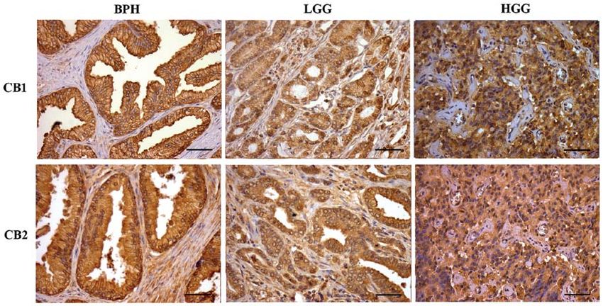
Figure 1. Immunohistochemical staining for CB1-R and CB2-R in BPH, LGG and HGG (x40 magnification). Scale bar, 50 µm. BPH, benign prostatic
hyperplasia tissue; LGG, low Gleason grade prostatic cancer tissue; HGG, high Gleason grade prostatic cancer tissue; CB1-R, cannabinoid receptor 1; CB2-R,
cannabinoid receptor 2.
cytometer (Becton-Dickinson, Franklin Lakes, NJ, USA). Data types of cells showed the presence of the receptors. In addi-
analysis was assessed using WinMD1 version 2.8 software. tion, the receptors appeared to be homogeneously distributed
throughout the cell surface of PC3, BPH and PrC cells in a
Annexin V assay. Cells were grown at a 106 density and treated similar way (data not shown).
with endocannabinoids at a 5.0 µM concentration for 6 h.
After the treatment, the cells were trypsinized, washed with Effect of endocannabinoids on the cell viability of PC3
cold PBS and then centrifuged at 2,500 rpm for 5 min. The cells and primary cultures of BPH and PrC. To assess the
supernatant was discarded and the pellet was resuspended in cell viability response of the PC3 cell line and the primary
1 ml of cold PBS. The cells were counted with a Neubauer cultures of PrC and BPH and the primary cultures to the
chamber and 105 cells were transferred to a cytometry tube. different endocannabinoids, MTT assay was employed. Fig. 2
Then, the cells were processed and labeled according to the shows that treatment of PC3 cells and primary cultures of
BD Pharmingen Annexin V-FITC Apoptosis Detection kit PrC and BPH for 48 h with the different endocannabinoids
that was used for this assay. The labeled cells were analyzed in (at 2.5, 5.0 and 10 µM concentrations) significantly decreased
a FACScan cytometer (Becton-Dickinson). Data analysis was the viability of the cells (PONCOLOGY REPORTS 33: 1599-1608, 2015 1603 Figure 2. Effect of endocannabinoids on the viability of PC3 cells and primary cultures of BPH and PrC. PC3 cells and cultures of BPH and PrC were treated with different doses of endocannabinoids (2.5, 5.0 and 10.0 µM) for 48 h at 37˚C. After the treatments, cell viability was evaluated through MTT assay (n=4). The results are expressed comparing the results to the viability of the control cells (cells that were left untreated) (P
1604                             orellana-serradell et al: Endocannabinoids and prostate cancer

Figure 4. Effect of endocannabinoids on the cell cycle distribution of PC3 cells and BPH and PrC cultures. Cell cycle analysis was assessed to determine
whether the effect on cell viability noted in the MTT assays could be cell cycle-dependent. PC3 cells and primary cultures of BPH and PrC were treated and
then labeled. The labeled cells were analyzed using a FACScan cytometer, and the percentage of cells in the G1, S and G2 phases were calculated using the
WinMDI v2.8 software (n=3, P>0.05). BPH, benign prostatic hyperplasia tissue; PrC, prostate cancer; C, control; Me, methanandamide; 2-AG, 2-arachidonoyl
glycerol; Ana, anandamide.

Figure 5. Flow cytometric analysis of FITC Annexin V staining. Primary cell cultures and cell lines (PC3) of prostate cancer were left untreated (Control; first
panels at the left) or treated for 6 h with endocannabinoids at a concentration of 5 µM (second, third and fourth panels from the left). Cells were incubated with
FITC Annexin V in a buffer containing propidium iodide (PI) and then analyzed by flow cytometry. Untreated cells were mostly FITC- and PI-negative which
indicates that they were not undergoing any type of cell death process. After the 6-h treatment there were two different groups of cells: one composed of viable
cells (FITC- and PI-negative, lower left quadrant) and one composed of cells undergoing apoptosis (FITC-positive and PI-negative, lower right quadrant).
Another very small population of cells was observed to be both FITC- and PI-positive (upper right quadrant) which indicates that they were in the final stage
of apoptosis or undergoing necrosis (n=3). Me, methanandamide; 2-AG, 2-arachidonoyl glycerol; Ana, anandamide.

research from our group showed a decrease in the expression                        Annexin V assay. As shown in Fig. 5, the number of cells in
of PCNA (a cell proliferation marker) in response to endocan-                      the apoptotic state was higher in all of the treated cell cultures
nabinoid treatment (data not shown). Due to these results, we                      when compared with the untreated ones, while the number of
hypothesized that the effect on cell viability demonstrated                        necrotic cells remained statistically without variation (P>0.05).
by the endocannabinoids on the different cell cultures could                       This effect was noted in both types of cells, but it was greater
be produced through induction of apoptosis by cell cycle                           in the PC3 cells than that noted in the primary cultures of PrC.
arrest. PC3 cells and primary cultures of BPH and PrC were                         In addition, none of the different endocannabinoids showed a
treated with the different endocannabinoids for 48 h at a 5 µM                     higher effect when compared with the others in the cell lines,
concentration. As shown in Fig. 4 there were no significant                        yet the results varied among the different endocannabinoids
variations in the distribution of cells throughout the cell cycle                  in the primary cultures, with Me showing a higher effect than
when comparing control untreated cells with PC3 cells and                          the other two agents. These results suggest that the effect of
PrC and BPH cultures that were treated with the different                          endocannabinoids on PC3 cells and primary cultures of PrC
endocannabinoids (P>0.05).                                                         may be caused by the activation of the apoptotic pathway.

Flow cytometric analysis of FITC Annexin V staining in the                         Effect of endocannabinoids on the expression of active
PC3 cells and primary cultures of PrC. To analyze the action                       caspase-3 and Bcl-2 in primary cultures of PrC and BPH.
of endocannabinoids on the different cultures of PrC cells                         The results of the Annexin V assay indicated that endocan-
and the possibility of a proapoptotic effect we used a FITC                        nabinoids may exert their action through the activation of the
ONCOLOGY REPORTS 33: 1599-1608, 2015                                                             1605

Figure 6. Expression of active caspase-3 and Bcl-2 in primary cultures of PrC and BPH after treatment with endocannabinoids. Primary cultures of PrC and
BPH were treated with endocannabinoids at a 5 µM concentration for 48 h. After the treatments, the cells were collected and proteins were extracted and
analyzed through western blot analysis. The figures show the variation in active caspase-3 and Bcl-2 expression in the treated cells compared to the expression
in the control untreated cells. The dotted lines represent the control untreated cells (100%). Each image shows a representative experiment repeated three
times with similar results (n=3) (P
1606                        orellana-serradell et al: Endocannabinoids and prostate cancer

nabinoids for 48 h at a 5 µM concentration. After the treatments,        Olea-Herrero et al (36) recently postulated that the effect of
we observed that Erk protein expression was augmented in the         methanandamide on the survival of PC3 cells may be carried
treated cells when compared to the control-untreated cells           out specifically by activation of the CB2 receptor. This finding
(Fig. 7). In contrast, the levels of Akt decreased following         not only differs from our results but also of the published
all the treatments in contrast to the increase noted in the Erk      data by Agudelo et al (37); who despite not using methanan-
expression. These results indicate that Erk activation/Akt           damide used its endogenous analog anandamide. This group
downregulation may be involved in the suppresive effect that         reported that the effect of anandamide was produced by the
endocannabinoids have on the cell viability of PrC cells.            activation of CB1 receptors. Furthermore, the affinity of
                                                                     methanandamide for the CB2 receptor was very low (with a
Discussion                                                           Ki of 815 nM for CB2 and a Ki of 20 nM for CB1). Some
                                                                     researchers even consider this molecule to be a specific agonist
Prostate cancer has become the most commonly diagnosed               for CB1. In previous experiments from our laboratory using
cancer in men and is one of the most threatening diseases in         radioligand binding studies, we observed that in both cell lines
Western countries. Although there are therapies that have been       and primary cultures, endocannabinoids bound more readily
proven effective, such as androgen deprivation, these treatment      to CB1 receptors than to CB2 receptors (data not shown). This
strategies are not able to eliminate all tumor cells. In addition,   may account for the effect observed when using the specific
current chemotherapy treatments cause many undesirable side          inhibitor for CB1.
effects for patients.                                                    Although previous results depicted a decrease in PCNA in
    It has been reported that endocannabinoids have a wide           PrC cell lines following endocannabinoid treatment, the cell
range of regulatory effects in a variety of physiological            cycle analysis assessed by flow cytometry showed no variations
processes. One of their most promising actions has to do with        in the distribution of the different stages of the cell cycle. Other
their effect on various types of cancers and their potential use     studies have reported cell cycle arrest following treatment with
in the treatment of these diseases (18).                             different THC analogs but using them at higher concentra-
    In the present study, it was demonstrated by immunohisto-        tions (39,40), which may be one reason why even though we
chemistry that CB1 and CB2 receptors are highly expressed in         previously observed that levels of PCNA were lower after the
prostate cancer samples with different degrees of malignancy,        treatments, that was not enough to modify the cell cycle distri-
as well as in BPH tissue. The presence of receptors CB1/CB2 in       bution of the different cell cultures. Another reason for this may
commercial cell lines (PC3), as well as in primary cultures of       be that the decrease in the PCNA levels was not high enough to
PrC, was additionally demonstrated by immunocytochemistry            have a significant effect on the cell cycle (38).
and western blot analysis (data not shown) as it was coinci-             Once we concluded that the effect of endocannabinoids on
dentally reported by other studies (25,30,33). Furthermore, we       cell viability was not exerted through a modification of the cell
observed that the expression of these receptors was associated       cycle, we analyzed whether this effect may be due to activa-
with the degree of malignancy in PrC, with higher expression         tion of the apoptotic pathway. Through Annexin V assays we
in the most aggressive samples of PrC. To analyze the effect         observed no change in the percentage of necrotic cells after the
that the analogs of endocannabinoids have on the various PrC         treatment with endocannabinoids, but we observed an increase
cell cultures, the sensitivity to three different concentrations     in the number of cells in early apoptosis after the treatments.
of endocannabinoids was evaluated, which established that            These results were confirmed by evaluating the expression
primary cultures were more resistant to endocannabinoids             of caspase-3 and Bcl-2 post-treatment with the endocan-
than PC3 cells. This differential effect may be explained by a       nabinoids. The results revealed an increase in the levels of
different expression of metabolizing enzymes, such as FAAH,          active caspase-3 and a decrease in the expression levels of
which metabolize and degrade endocannabinoids in situ.               Bcl-2. This simultaneous effect observed in both proteins
Several studies have illustrated differences in the expres-          may be explained by a change in the activity of the nuclear
sion of these enzymes in prostate cancer cell lines and the          factor-κ B (NF-κ B) induced by the endocannabinoids. NF-κ B
effect this has on treatment with endocannabinoids in cancer         is involved in cell proliferation through multiple mechanisms,
cells (34,35). The action of these enzymes, their expression         some of which control the expression of Bcl-2 and also the
and related metabolism of endocannabinoids may account               activation of proteins involved in the apoptotic pathway such
for the weaker effect that the treatments had on the primary         as caspases. It has been reported that in prostate cancer NF-κ B
cultures. Moreover, it was also observed that the effect of the      is overactivated, thus endocannabinoids may act through this
endocannabinoids in regards to the viability of the different        pathway, simultaneously activating caspase-3, downregulating
cell cultures occurred in a dose-dependent manner and that           Bcl-2 expression; and other proteins that may also be involved
there may be a saturation of the receptors associated with the       in the apoptotic process (7,41).
increasing concentration of each treatment.                              All of our results suggest that endocannabinoids may
    Furthermore, the effect of the different treatments on both      exert their effect by activating the apoptotic pathway without
cell lines and primary cultures was almost totally reversed          modifying the cell cycle stage or inducing necrosis. Moreover,
when they were previously incubated with the CB1R antagonist         a decrease in the levels of AKT in the primary cultures was
SR141716. As this is a selective antagonist for the CB1 receptor,    detected after treatment with endocannabinoids. AKT func-
the results obtained lead us to conclude that the observed effect    tions as a critical positive regulator of metabolism and cell
of the different treatments was produced mainly by the activa-       proliferation (39) which is positively correlated with the effect
tion of CB1 and not CB2; these results also discard a possible       caused by the different treatments on prostate cancer cultures.
receptor-independent effect of the endocannabinoids.                 These results are supported by other reports that have shown
ONCOLOGY REPORTS 33: 1599-1608, 2015 1607
that AKT is decreased in other types of cancer following treat- 14. Kirkham TC and Tucci SA: Endocannabinoids in appetite control
and the treatment of obesity. CNS Neurol Disord Drug Targets 5:
ment with THC (41). Furthermore, increases in the activation 272-292, 2006.
of Erk and treatment with cannabinoids have been correlated 15. Bovolin P, Cottone E, Pomatto V, et al: Endocannabinoids are
with antiproliferative processes in other types of cancer (42). involved in male vertebrate reproduction: regulatory mechanisms
at central and gonadal level. Front Endocrinol (Lausanne) 5: 54,
The results observed in our study depict a simultaneous 2014.
activation of the ERK pathway and a decrease in AKT levels. 16. Jiang W, Zhang Y, Xiao L, Van Cleemput J, Ji SP, Bai G
The combination of these two events may contribute to the and Zhang X: Cannabinoids promote embryonic and adult
hippocampus neurogenesis and produce anxiolytic- and antide-
activation of antiproliferative pathways and a decrease in the pressant-like effects. J Clin Invest 115: 3104-3116, 2005.
proliferative pathways in primary cultures of PrC. A more 17. Cridge BJ and Rosengren RJ: Critical appraisal of the potential
in-depth analysis of the proteins involved in these pathways use of cannabinoids in cancer management. Cancer Manag
Res 30: 301-313, 2013.
may be important to better describe the intracellular mecha- 18. Batista LA, Gobira PH, Viana TG, et al: Inhibition of endo-
nisms that may be activated by endocannabinoids in prostate cannabinoid neuronal uptake and hydrolysis as strategies for
cancer cells and thus search for other possible targets of inter- developing anxiolytic drugs. Behav Pharmacol 25: 425-433,
2014.
vention to potentiate the effects depicted by the treatment of 19. Bifulco M, Laezza C, Pisanti S and Gazzerro P: Cannabinoids
cancer cells with endocannabinoids. and cancer: pros and cons of an antitumor strategy.
Finally and in accordance with our findings, we conclude Br J Pharmacol 148: 123-135, 2006.
20. Ramos JA and Bianco FJ: The role of cannabinoids in prostate
that endocannabinoids are capable of halting the growth of cancer: Basic science perspective and potential clinical appli-
prostate cancer cells through activation of apoptotic mecha- cations. Indian J Urol 28: 9-14, 2012
nisms. Furthermore, we suggest that this effect may be through 21. Melck D, Rueda D, Galve-Roperh I, et al: Involvement of the
cAMP/protein kinase A pathway and of mitogen-activated
the modulation of the Erk and Akt signaling pathways by protein kinase in the anti-proliferative effects of anandamide in
endocannabinoids. Therefore, endocannabinoids appear to be human breast cancer cells. FEBS Lett 463: 235-240, 1999.
a powerful tool for investigation in the development of drugs 22. Nithipatikom K, Endsley M, Isbell M, et al: 2-Arachidonoyl-
glycerol: a novel inhibitor of androgen-independent prostate
and treatments against advanced PrC. cancer cell invasion. Cancer Res 15: 8826-8830, 2004.
23. Portella G, Laezza C, Laccetti P, et al: Inhibitory effects of
Acknowledgements cannabinoid CB1 receptor stimulation on tumor growth and
metastatic spreading: actions on signals involved in angiogenesis
and metastasis. FASEB J 17: 1771-1773, 2003
We acknowledge Ms. Graciela Caroca for her excellent technical 24. Melck D, De Petrocellis L, Orlando P, et al: Suppression of nerve
assistance. The present study was supported by Vicerrectoria growth factor Trk receptor and prolactin receptors by endocan-
nabinoids leads to inhibition of human breast and prostate cancer
de Investigación y Desarrrollo of Universidad de Chile (VID) cell proliferation. Endocrinology 141: 118-126, 2000.
Grant DI MULT 05/36-2. Grants FONDECYT, 1060500 (H.C.), 25. Czifra G, Varga A, Nyeste K, et al: Increased expressions
1110269 (H.C.) and 1140417 (E.C.). of cannabinoid receptor-1 and transient receptor potential
vanilloid-1 in human prostate carcinoma. J. Cancer Res Clin
Oncol 135: 507-514, 2009.
References 26. Mimeault M, Pommery N, Wattez N, et al: Anti-proliferative and
apoptotic effects of anandamide in human prostatic cancer cell
1. Jemal A, Bray F, Center MM, et al: Global cancer statistics. CA lines: implication of epidermal growth factor receptor down‑regu-
Cancer J Clin 61: 69-90, 2011. lation and ceramide production. Postate 15: 1-12, 2003.
2. Sark W: Epidemiology of prostate cancer and its precursors. 27. Ruiz L, Miguel A and Díaz-Laviada I: Delta9‑tetrahydro
Mod Pathol 10: 1038, 2004. (Epub ahead of print). doi: cannabinol induces apoptosis in human prostate PC-3 cells via a
10.1038/modpathol.3800074. receptor‑independent mechanism. FEBS Lett 458: 400-404, 1999.
28. Sarfaraz S, Afaq F, Adhami VM and Mukhtar H: Cannabinoid
3. Tefekli A and Tunc M: Future prospects in the diagnosis and receptor as a novel target for the treatment of prostate cancer.
management of localized prostate cancer. Sci World J 2013: Cancer Res 65: 1635-1641, 2005.
347263, 2013. 29. Gómez del Pulgar T, Velasco G and Guzmán M: The CB1 canna-
4. Hsing A and Chokkalingam A: Prostate cancer epidemiology. binoid receptor is coupled to the activation of protein kinase
Front Biosci 11: 1388-1413, 2006. B/Akt. Biochem J 347: 369-373, 2000.
5. Porkka K and Visakorpi T: Molecular mechanisms of prostate 30. Gómez del Pulgar T, Velasco G, Sánchez C, et al: De
cancer. Eur Urol 45: 683-691, 2004. novo‑synthesized ceramide is involved in cannabinoid-induced
6. Lee C, Jia Z, Rahmatpanah F, Zhang Q, Zi X, McClelland M and apoptosis. Biochem J 363: 183-188, 2002.
Mercola D: Role of the adjacent stroma cells in prostate cancer 31. Rueda D, Galve-Roperh I, Haro A and Guzmán M: The CB1
development and progression: synergy between TGF-β and IGF cannabinoid receptor is coupled to the activation of c-Jun
signaling. Biomed Res Int 2014: 502093, 2014. N-terminal kinase. Mol Pharmacol 58: 814-820, 2000.
7. Bifulco M, Malfitano A, Pisanti S and Laezza C: Endo 32. Mendoza P, Sánchez C, Contreras HR, et al: Evaluation of MENT
cannabinoids in endocrine and related tumours. Endocr Relat on primary cell cultures from benign prostatic hyperplasia and
Cancer 15: 391-408, 2008. prostate carcinoma. Int J Androl 32: 607-615, 2009.
8. Hermanson DJ and Marnett LJ: Cannabinoids, endocan- 33. Chunga S, Hammarstenb P, Josefssonb A, et al: A high canna-
nabinoids, and cancer. Cancer Metastasis Rev 30: 599-612, 2011. binoid CB(1) receptor immunoreactivity is associated with
9. Díaz-Laviada I: The endocannabinoid system in prostate cancer. disease severity and outcome in prostate cancer. Eur J Cancer 45:
Nat Rev Urol 8: 553-561, 2011. 174-182, 2009.
10. Devane WA, Dysarz FA, Johnson MR, et al: Determination 34. Brown L, Cascio M, Wahle K, et al: Cannabinoid receptor‑
and characterization of a cannabinoid receptor in rat brain. Mol dependent and -independent anti-proliferative effects of omega-3
Pharmacol 34: 605-613, 1998. ethanolamides in androgen receptor-positive and‑negative
11. Van Dross R, Soliman E, Jha S, et al: Receptor-dependent and prostate cancer cell lines. Carcinogenesis 31: 1584‑1591, 2010.
receptor-independent endocannabinoid signaling: A therapeutic 35. Nithipatikom K, Isbell M, Endsleya M, et al: Anti-proliferative
target for regulation of cancer growth. Life Sci 92: 463-466, effect of a putative endocannabinoid, 2-arachidonylglyceryl ether
2013. in prostate carcinoma cells. Prostaglandins Other Lipid Mediat
12. Guindon J and Hohmann AG: The endocannabinoid system and 94: 34-43, 2011.
cancer: therapeutic implication. Br J Pharmacol 16: 1447-1463, 36. Olea-Her rero N, Vara D, Malagarie- Cazenave S and
2011. Díaz‑Laviada I: Inhibition of human tumour prostate PC-3 cell
13. Pisanti S and Bifulco M: Endocannabinoid system modulation growth by cannabinoids R(+)-Methanandamide and JWH-015:
in cancer biology and therapy. Pharmacol Res 60: 107-116, 2009. involvement of CB2. Br J Cancer 101: 940-950, 2009.1608 orellana-serradell et al: Endocannabinoids and prostate cancer
37. Agudelo M, Newton C, Widen R, et al: Cannabinoid receptor 2 41. De Petrocellis L, Ligresti A, Moriello A, et al: Non-THC
(CB2) mediates immunoglobulin class switching from IgM to IgE cannabinoids inhibit prostate carcinoma growth in vitro and
in cultures of murine-purified B lymphocytes. J. Neuroimmune in vivo: pro-apoptotic effects and underlying mechanisms. Br J
Pharmacol 3: 35-42, 2008. Pharmacol 168: 79-102, 2013.
38. Kelman Z: PCNA: structure, function and interactions. Oncogene 42. Hsu SS, Huang CJ, Cheng HH, et al: Anandamide-induced
6: 629-640, 1997. Ca 2+ elevation leading to p38 MAPK phosphorylation and
39. Song G, Ouyang G and Bao S: The activation of Akt/PKB signaling subsequent cell death via apoptosis in human osteosarcoma cells.
pathway and cell survival. J Cell Mol Med 1: 59-71, 2005. Toxicology 28: 21-29, 2007.
40. Greenhough A, Patsos H, Ann C, et al: The cannabinoid
D9-tetrahydrocannabinol inhibits RAS-MAPK and PI3K-AKT
survival signalling and induces BAD-mediated apoptosis in
colorectal cancer cells. Int J Cancer 121: 2172-2180, 2007.You can also read