G Protein-Coupled Estrogen Receptor Correlates With Dkk2 Expression and Has Prognostic Impact in Ovarian Cancer Patients - Frontiers
←
→
Page content transcription
If your browser does not render page correctly, please read the page content below
ORIGINAL RESEARCH
                                                                                                                                        published: 19 February 2021
                                                                                                                                   doi: 10.3389/fendo.2021.564002

                                           G Protein-Coupled Estrogen
                                           Receptor Correlates With Dkk2
                                           Expression and Has Prognostic
                                           Impact in Ovarian Cancer Patients
                                           Patricia Fraungruber 1, Till Kaltofen 1, Sabine Heublein 1,2, Christina Kuhn 1, Doris Mayr 3,
                                           Alexander Burges 1, Sven Mahner 1, Philipp Rathert 4, Udo Jeschke 1,5* and Fabian Trillsch 1
                                           1 Department of Obstetrics and Gynecology, University Hospital, Ludwig-Maximilians-University (LMU) Munich, Munich,
                                           Germany, 2 Department of Gynecology and Obstetrics, University of Heidelberg, Heidelberg, Germany, 3 Department of
                                           Pathology, LMU Munich, Munich, Germany, 4 Department of Biochemistry, University Stuttgart, Stuttgart, Germany,
                                           5 Department of Obstetrics and Gynecology, University Hospital Augsburg, Augsburg, Germany

                                           Purpose: Wnt pathway modulator Dickkopf 2 (Dkk2) and signaling of the G protein-
                           Edited by:
                   Sarah H. Lindsey,
                                           coupled estrogen receptor (GPER) seem to have essential functions in numerous cancer
      Tulane University, United States     types. For epithelial ovarian cancer (EOC), it has not been proven if either Dkk2 or the
                       Reviewed by:        GPER on its own have an independent impact on overall survival (OS). So far, the
                         Ian Campbell,
                                           correlation of both factors and their clinical significance has not systematically been
    Peter MacCallum Cancer Centre,
                              Australia    investigated before.
              Edward Joseph Filardo,
 The University of Iowa, United States
                                           Methods: Expression levels of Dkk2 were immunohistochemically analyzed in 156 patient
                *Correspondence:
                                           samples from different histologic subtypes of EOC applying the immune-reactivity score
                      Udo Jeschke          (IRS). Expression analyses were correlated with clinical and pathological parameters to
       Udo.Jeschke@uk-augsburg.de          assess for prognostic relevance. Data analysis was performed using Spearman’s
                                           correlations, Kruskal-Wallis-test and Kaplan-Meier estimates.
                   Specialty section:
         This article was submitted to     Results: Highest Dkk2 expression of all subtypes was observed in clear cell carcinoma. In
             Molecular and Structural
                        Endocrinology,
                                           addition, Dkk2 expression differed significantly (p4) and GPER (IRS>8),
        Accepted: 05 January 2021
       Published: 19 February 2021         had a significantly better overall survival compared to patients with low expression (61
                             Citation:     months vs. 33 months; p=0.024).
Fraungruber P, Kaltofen T, Heublein S,
Kuhn C, Mayr D, Burges A, Mahner S,
                                           Conclusion: Dkk2 and GPER expression correlates in EOC and combined expression of
   Rathert P, Jeschke U and Trillsch F     both is associated with improved OS. These findings underline the clinical significance of
  (2021) G Protein-Coupled Estrogen        both pathways and indicate a possible prognostic impact as well as a potential for
       Receptor Correlates With Dkk2
      Expression and Has Prognostic        treatment strategies addressing interactions between estrogen and Wnt signaling in
   Impact in Ovarian Cancer Patients.      ovarian cancer.
        Front. Endocrinol. 12:564002.
    doi: 10.3389/fendo.2021.564002         Keywords: Dickkopf 2, G protein-coupled estrogen receptor, Wnt signaling, estrogen, epithelial ovarian cancer

Frontiers in Endocrinology | www.frontiersin.org                                 1                                     February 2021 | Volume 12 | Article 564002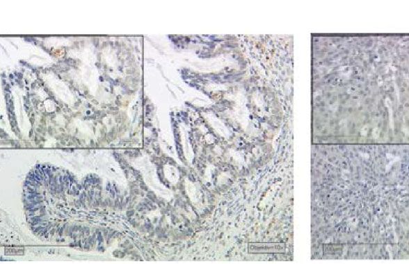
Fraungruber et al.                                                                                                             Dkk2 and GPER in Ovarian Cancer

INTRODUCTION                                                                                 GPER is a transmembrane receptor with intracellular
                                                                                         domains binding E2 (26), which mediates rapid non-genomic
Epithelial ovarian cancer (EOC) causes most deaths of                                    estrogen signaling (27). Its activation via agonists like G1 or E2
gynecological malignancies (1) with a relative 5-year survival of                        (28) leads to cAMP production (29), activation of extracellular
almost 45% (2). The need to identify suitable screening methods,                         signal-related kinase 1 and 2 (Erk1/2) (28), mobilization of
prognostic markers and efficient therapies is crucial. So far,                            intracellular Ca 2+ , phosphatidylinositol 3-kinase (PI3K)
standard treatment for primary disease consists of debulking                             activation (26) and the induction of metalloproteinases which
surgery and a platinum-based chemotherapy with                                           then transactivates the epidermal growth factor receptor (30).
antiangiogenics and/or Poly-ADP-Ribose-Polymerase (PARP)                                 GPER can also indirectly impact gene transcription (31). Since its
inhibitors (3). Apart from clinicopathological aspects such as                           role in ovarian cancer has been conflicting so far (32–34) this
the stage in the system of the International Federation of                               analysis focused on the correlation of Dkk2 with GPER to
Gynecology and Obstetrics (FIGO), volume of residual disease                             identify a possible link between Wnt and estrogen and
after debulking surgery, patients’ age, and histological subtype                         investigating their potential prognostic significance.
(4–7), there are no reliable prognostic factors to predict the
clinical course. With regards to the molecular background and
specific gene mutations, EOC is histologically separated into
clear cell, endometrioid, mucinous, and serous carcinoma of low                          METHODS
or high grade (LGSC/HGSC) (8).
    Revealing molecular events that cause ovarian cancer and are                         Patients
responsible for its progression represent a major challenge for                          In this study 156 formalin-fixated and paraffin-embedded tissue
translational research. One approach is to understand the                                specimens of epithelial ovarian cancer from patients who had
importance and complexity of the Wnt signaling pathway and                               been treated in the Department of Obstetrics and Gynecology at
its regulation (9–11). Secreted Wnt glycoproteins translate their                        Ludwig-Maximilians-University of Munich between 1990 and
function via binding to Frizzled receptors and co-receptors such                         2002 were analyzed. Numerous markers were already examined
as low-density-lipoprotein-related protein 5/6 (LRP5/6) (11).                            in this collective in preceding studies (35–37). Clinical data was
Subsequently, Wnt proteins exhibit their effects on several                              collected from the patient’s charts and information about the
cellular processes by activating either the canonical Wnt/b-                             follow up was acquired from the Munich Cancer Registry.
catenin or at least two non-canonical b-catenin-independent                                  Only patients with malignant, non-borderline tumors were
pathways (12). Alterations in Wnt signaling components, such as                          included in the study. Seventy-three patients (46.8%) were older
APC (adenomatous polyposis coli) protein, AXIN and b-catenin                             or age 60 years at the initial diagnosis and 83 patients (53.2%)
and downregulation of modulatory Wnt antagonists have been                               were younger than 60 years. There were no data available about
described to be involved in the onset of several cancer types (10,                       estrogen replacement therapy in postmenopausal women.
13, 14). As a consequence, modulators of the Wnt pathway like                            Pathologists categorized the histological subtypes of the
members of the Dickkopf family (Dkk1-4) may play an essential                            samples: LGSC (n=24), HGSC (n=80), endometrioid (n=21),
role during development (15, 16) and tumorigenesis (17, 18).                             clear cell (n=12), mucinous (n=13). According to the updated
Dkks bind to LRP5/6 with higher affinity than Wnt (19). Dkk2                              FIGO classification from 2014, specimens of serous ovarian
seems able to act as agonist as well as antagonist for Wnt/LRP6                          cancer were re-evaluated and attributed to low-grade (G1) and
signaling depending on the cellular context and therefore co-                            high-grade (G3) histology. Endometrioid and mucinous ovarian
factors such as krm2 (18–20). In EOC Zhu et al. suggest that                             cancer samples were related to G1, G2, and G3. Clear cell cancer
Dkk2 may functions as a Wnt pathway inhibitor (13).                                      was always categorized as G3 (38). Staging was done following
    Estrogen (E2, 17b-estradiol) has numerous cellular functions                         the FIGO classification: I (n=35), II (n=10), III (n=103), IV (n=3)
in the human body including gynecologic cancer biology (21)                              (Table 1).
and interactions between estrogen and Wnt signaling have been
described (22–25). In this context an interplay of Dkk2 and                              Sampling and Microarray Construction
estrogen receptors (ER) could link these two mechanisms and                              Three core biopsies for each EOC patient were taken from
classical nuclear ERa or ERb as well as the G protein-coupled                            paraffin-embedded and formalin-fixed tumor blocks in our
estrogen receptor (GPER) could be involved in this process.                              archive. The biopsies were assembled in tissue microarrays
                                                                                         (TMA) paraffin blocks. Those TMA paraffin blocks were cut
                                                                                         into serial sections at 2 mm and fixed on slides. A pathologist
                                                                                         verified that representative areas of the tumor were aligned on
Abbreviations: Dkk2, Dickkopf2; EOC, epithelial ovarian cancer; E2, estrogen,
                                                                                         the slides.
17b-estradiol; ERa, nuclear estrogen receptor alpha; ERb, nuclear estrogen
receptor alpha beta; Erk1, extracellular signal-related kinase 1; Erk2,
extracellular signal-related kinase 2; GPER, G protein-coupled estrogen                  Immunohistochemistry
receptor; HGSC, high-grade serous carcinoma; IRS, immune-reactivity score;               Immunohistochemical staining of paraffin-embedded and
LGSC, low-grade serous carcinoma; LRP5, low-density-lipoprotein-related
protein 5; LRP6, low-density-lipoprotein-related protein 6; OS, overall survival;
                                                                                         formalin-fixed tissue micro arrays of ovarian cancer specimens
PI3K, phosphatidylinositol 3-kinase; ROC curve, receiver operating characteristics       for Dkk2 was performed as previously described (39). The TMA
curve; TCF, T-cell factor; TMA, tissue microarrays.                                      slides were dewaxed in Roticlear (Carl Roth Karlsruhe,

Frontiers in Endocrinology | www.frontiersin.org                                     2                                February 2021 | Volume 12 | Article 564002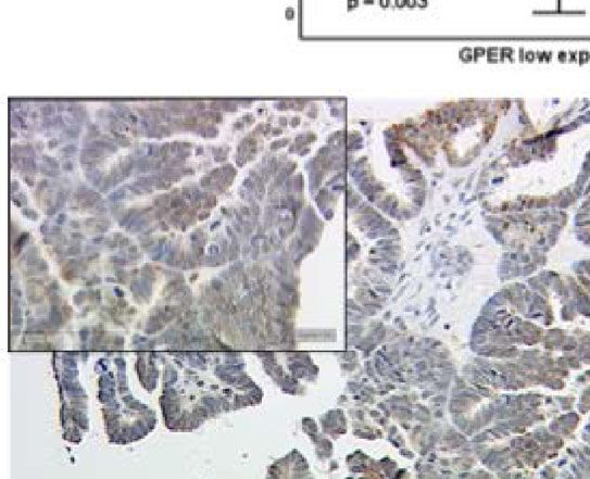
Fraungruber et al.                                                                                                              Dkk2 and GPER in Ovarian Cancer

TABLE 1 | Correlation between Dkk2 expression and clinicopathologic                       2=moderate, 3=strong) multiplied with the percentage of
characteristics of ovarian cancer patients.
                                                                                          stained cells (0=no staining, 1%≤10% positive cells, 2 = 11%–
Characteristics                Total        Dkk2 low        Dkk2 high         P           50% positive cells, 3 = 51%–80% positive cells, 4%≥81% positive
                                           expression       expression      value         cells). The immunoreactivity score ranges from 0 to 2: negative, 3
                            Number of      Number of        Number of                     to 4: weak, 6 to 8: moderate, and 9 to 12: strong (40). Formerly
                            cases(%)         cases            cases
                                                                                          published staining results of GPER in this panel recorded in the
Age(y)                                                                                    archive of the laboratory were recaptured (36).
            ≥60y             73 (46.8)          28               21        8)      70 (44.9)          11               31
                                                                                          Statistical analysis was operated with SPSS 25 (IBM, Chicago, IL,
                                                                                          USA). With the Kruskal-Wallis analysis the null hypothesis was
                                                                                          tested against its opposite. Further Spearman’s correlation
Dkk2, Dickkopf2; GPER, G protein-coupled estrogen receptor; HGSC, high-grade serous
carcinoma; LGSC, low-grade serous carcinoma; FIGO, International Federation of            analysis and Kruskal-Wallis analysis was applied for testing
Gynecology and Obstetrics.                                                                correlation of Dkk2 and GPER scores. The Kaplan-Meier
Bold numbers represent p-values < 0.05.                                                   estimate was used for analyzing times to event variables.
                                                                                          Correlations between mean Dkk2 expression and
                                                                                          clinicopathologic characteristics were assessed with Chi-Square
Germany) for 20 min. The endogenous peroxidase was
                                                                                          tests (Table 1, crosstab). For all tests p-values ≤ 0.05 were
suppressed with 3% hydrogen peroxide (Merck, Darmstadt,
                                                                                          considered as statistically significant. Figures were designed
Germany) in methanol (20 min). The specimens were
                                                                                          with SPSS 25 and Microsoft Power Point 2016 (Microsoft,
rehydrated in a descending alcohol series (100%, 70%,
                                                                                          Redmond, WA, USA).
50% ethanol). The epitopes were retrieved by putting the
slides in a pressure cooker with sodium citrate buffer (pH
6.0) for 5 min. After cooling to room temperature, the                                    RESULTS
slides were washed in in distilled water and phosphate-buffered
saline (PBS). To evade unspecific staining reagent 1 of the                                Correlations between Dkk2 expression and clinicopathologic
polymer kit (ZytoChem Plus HRP Polymer System, Berlin,                                    characteristics of EOC patients are displayed in Table 1. A
Germany) was administered for 5 min. Next the slides                                      median IRS of 6 for anti-Dkk2 staining was observed in the
incubated at +4°C for 16 h with the primary anti-body Anti-                               131 of 152 cases (86%) with adequate staining. Applying ROC
Dkk2 polyclonal rabbit IgG (ProteinTech, Manchester, UK). As                              curve analysis, an IRS>4 was selected as cut-off.
negative controls the primary antibody was replaced by normal                                 Dkk2 expression differed significantly between the
rabbit immunoglobulin G([IgG] supersensitive rabbit negative                              histological cancer subtypes (Figures 1A–F) with clear cell
control; BioGenex, Fremont, California). Washing in PBS and                               carcinomas showing the highest median IRS of 12 compared to
the application of reagents 2 (20 min) and 3 (30 min) of the                              the other subtypes (range: 9–12; p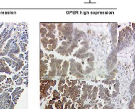
Fraungruber et al.                                                                                                                  Dkk2 and GPER in Ovarian Cancer

                         A                                             B                                   C

                                  D                                    E                                   F

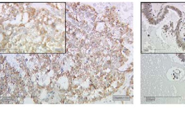
 FIGURE 1 | Dkk2 expression patterns in different histological subtypes of EOC after immunohistochemical staining was performed as shown in a Kruskal-Wallis analysis
 for histological subtypes (A). Clear cell carcinomas (B) presented the strongest staining patterns. Low-grade serous carcinomas (LGSC; C) had shown moderate Dkk2
 expression. For endometrioid (D), mucinous (E) and high-grade serous carcinomas (HGSC; F) the median IRS was lower. Scale bares equal 200 mm.

markers with a possible impact on the prognosis of EOC.                               Dkk2 did not correlate with either ERa or ERb expression
Cytoplasmatic Dkk2 was observed to correlate significantly                             (Table 2).
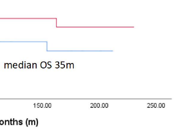
with cytoplasmic GPER expression (cc=0.304, p=0.001).                                    Patients with high Dkk2 expression (IRS>4) exhibited longer
Further analysis revealed that high Dkk2 expression is                                OS with a median of 65 months compared to 35 months in
correlated to high GPER expression (Figure 2). In contrast,                           Kaplan-Meier analysis, although this difference was not

                                            A

                              B                                                       C

 FIGURE 2 | Kruskal-Wallis analysis for correlation of Dkk2 and GPER expression (A). High expression of Dkk2 (B) correlates with high GPER expression (C) in
 tissue samples of the same patient. Scale bares equal 200 mm.

Frontiers in Endocrinology | www.frontiersin.org                                  4                                       February 2021 | Volume 12 | Article 564002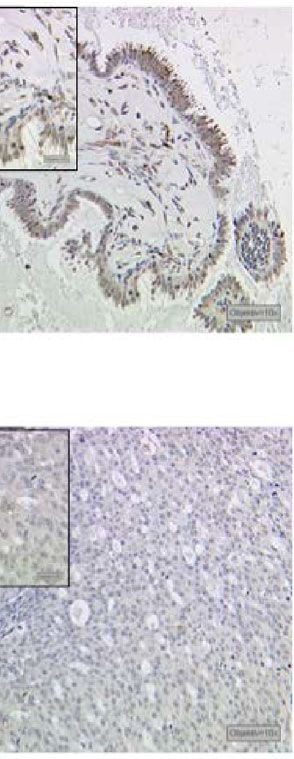
Fraungruber et al.                                                                                                                    Dkk2 and GPER in Ovarian Cancer

TABLE 2 | Results of Spearman’s correlation analysis of Dkk2 with the different                significance (36). When the expression analyses of the two
estrogen receptors (GPER, ERa, ERb).
                                                                                               markers were combined, patients with high Dkk2 (IRS>4) as
Staining              DKK2                  GPER              ERa                 ERb          well as high GPER (IRS>8) expression had a significantly longer
                                                                                               OS with 61 months compared to 33 months in patients with low
DKK2
                                                                                               expression of both influenced OS (p=0.024; Figure 3B).
cc                    1.000                 0.304            0.092              -0.080
p                        .                  0.001            0.298              0.366
n                      125                   124              131                128

Dkk2, Dickkopf 2; GPER, G protein-coupled estrogen receptor; cc, correlation coefficient;
                                                                                               DISCUSSION
p, two-tailed significance; n, number of patients.
Bold numbers represent p-values < 0.05.                                                        Dkk2 as a Wnt/b-catenin antagonist may play an important role
                                                                                               in ovarian cancer (13, 18, 43). In this analysis, we investigated the
statistically significant (p=0.207; Figure 3A). The same trend was                              expression of Dkk2 in the different histological subtypes of
observed for GPER expression as published before with longer                                   epithelial ovarian cancer, its relation to clinicopathological
OS for patients with high expression but without statistical                                   aspects and its impact on OS. Clear cell carcinoma exhibited

                                        A

                                        B

 FIGURE 3 | Kaplan-Meier estimates of Dkk2 (A) and Dkk2 combined with GPER expression (B) were analyzed. Though not statistically significant, high cytoplasmic
 Dkk2 (A) and GPER (36) expression was connoted with a longer OS. Patients with carcinomas highly expressing both Dkk2 and GPER in the cytoplasm compared
 to patients with carcinomas lowly expressing Dkk2 and GPER showed significantly (61 months vs. 33 months, p=0.024) increased OS (B).

Frontiers in Endocrinology | www.frontiersin.org                                           5                                 February 2021 | Volume 12 | Article 564002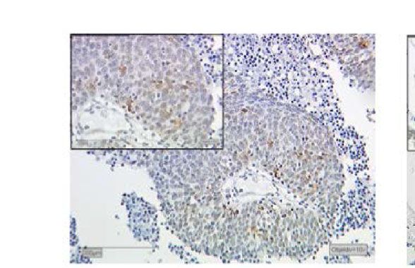
Fraungruber et al.                                                                                               Dkk2 and GPER in Ovarian Cancer

the highest Dkk2 expression at all and LGSC showed                         combined with agents modulating GPER. Although promising in
significantly higher expression compared to the other                       early stage development, previous strategies targeting Wnt
histologies, which could reflect the different pathogenesis and             proteins like tumor associated MUC1 (TA-MUC1) inhibitor
origins of the histological subtypes (44).                                 gatipotuzumab and others have not led to durable responses
    In a previous study from Zhu et al. it has been shown that             and not reached clinical significance so far (48). Very recently, a
Dkk2 is frequently methylated and therefore epigenetically                 Wnt modulator of Dkk1 (DKN-01) has shown interesting
silenced in ovarian cancer. Lower Dkk2 expression levels                   activity and is currently in a phase 2 basket trial which still
correlated with tumor progression and advanced tumor stages                supports the rationale for this approach (49).
(FIGO III-IV). By treating mice with the DNA methyltransferase                 In renal cancer cells, the selective estrogen receptor
inhibitor 5- aza-2′-deoxycitidine (decitabine) in order to re-             modulator genistein reportedly abolished miR-1260b, which is
establish Dkk2 expression in mice tumor growth was impaired                able to suppress Wnt signaling modulators like Dkk2, and
(13). This is in accordance with our findings, suggesting an                therefore preserved levels of these proteins (24). Genistein is
impact of Dkk2 on OS although this was not significant.                     not exclusively binding to GPER though, it also inflects ERa and/
Seemingly aberrant DNA methylation patterns also play a                    or ERb (50). In hepatocytes administering the GPER antagonist
major role in platinum resistance, therefore the potential of              G15 attenuated b-cat Ser675 phosphorylation and T-cell factor
epigenetic modulator decitabine to restore sensitivity towards             (TCF) expression suggesting an involvement of GPER in b-cat/
platinum has been successfully tested in a phase II clinical trial         TCF activities (51). Beside cell culture experiments, analyzing
(45). So far, agents for epigenetic therapy may cause severe               methylation patterns with methylation-specific polymerase chain
adverse effects, in particular when they are administered in               reaction could help to further investigate the suggested
combination with chemotherapy. This underscores the                        interactions of GPER and Dkk2. Implementing TCF/LEF
necessity of more selective epigenetic modulators (46).                    (lymphoid enhancing factor) reporter assays, could be assessed
    The impact of GPER on the OS of ovarian cancer patients has            to evaluate possible effects of GPER agonists or antagonists on
been controversially discussed so far (32–34). The conflicting              the Wnt signaling pathway.
results in these studies may arise from application of different               There are some factors limiting our study. First of all, it is
concentrations for the agonists E2 and G1 and the investigation            retrospective based on a single dataset with a relatively low
in different cancer cell lines. Accounting for these and the current       sample size which may not be sufficient to elucidate all
results, GPER may not be sufficient to predict OS on its own.               subtype-specific differences in an heterogenous tumor like
However, in combination with other factors like Luteinizing                ovarian cancer (44). Additional specific information of patient
Hormone/Choriogonadotropin Receptor and Follicle                           characteristics like an history of hormonal replacement therapy
Stimulating Hormone Receptor (36) or Dkk2, it could serve as               could enrich the investigation how estrogen levels interact with
a positive prognostic factor for patients suffering from epithelial        Dkk2 and better account for possible environmental toxicants. In
ovarian cancer.                                                            Kaplan-Meier analysis, subtype-specific evaluation did not reveal
    As previous studies elucidated a possible connection between           significant differences regarding OS between patients with high
estrogen and Wnt signaling (22–25), we investigated the                    and low Dkk2 expression so that results can be considered as a
relationship of Dkk2 with estrogen receptors. Subcellular                  base for further research in ovarian cancer. Further methods will
localization of the DKK2 staining pattern was noted which has              be necessary capture the extensive complexity of GPER and Wnt
been previously attributed to the Golgi apparatus (www.                    signaling pathways with their possible interaction as indicated.
proteinatlas.org). Unlike other studies in breast cancer which                 However, aside from these limitations our data is in
have shown an association between plasma membrane                          accordance with previous findings in EOC literature (13, 33,
expression and outcome, plasma membrane expression of                      36, 45, 52) and elucidate that targeting the GPER receptor as well
GPER was not detected in the ovarian cancer samples                        as the Wnt pathway could represent promising therapeutic
evaluated here (47). We could demonstrate a strong correlation             strategy in ovarian cancer. The study might provide an
of high cytoplasmic Dkk2 and high cytoplasmic GPER                         impetus to further investigate the crosstalk between estrogen
expression levels in EOC samples. In contrast, no correlation              and Wnt signaling in regard to the therapeutic potential in EOC.
of Dkk2 with the traditional estrogen receptors ERa or ERb was
noted. To the best of our knowledge, a possible connection of
GPER and Dkk2 has not yet been investigated. The described                 DATA AVAILABILITY STATEMENT
association of higher Dkk2 expression in younger patients may
be reflected by more patients in premenopausal status and                   The raw data supporting the conclusions of this article will be
therefore relate to the estrogen levels in these patients.                 made available by the authors, without undue reservation.
    In our study, a high Dkk2 expression in combination with a
high cytoplasmic level of GPER had a significant prognostic
impact on OS which might help to find new approaches for                    ETHICS STATEMENT
possible treatment strategies accounting for the correlation of
estrogen and Wnt signaling pathways. As Dkk2 is a modulator of             The studies involving human participants were reviewed and
the Wnt pathway, therapeutics addressing this cascade could be             approved by Ludwig-Maximilians-University, Munich,

Frontiers in Endocrinology | www.frontiersin.org                       6                                February 2021 | Volume 12 | Article 564002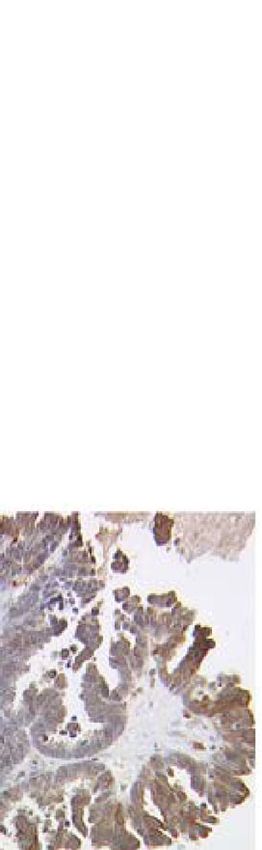
Fraungruber et al.                                                                                                                          Dkk2 and GPER in Ovarian Cancer

Germany. Written informed consent for participation was not                                 and UJ wrote the first draft of the manuscript. All authors
required for this study in accordance with the national legislation                         contributed to the article and approved the submitted version.
and the institutional requirements.
                                                                                            FUNDING
                                                                                            This study was funded by the Medical Faculty of the
AUTHOR CONTRIBUTIONS                                                                        LMU Munich.

PF, UJ, SH, AB, SM, and FT contributed conception and design                                ACKNOWLEDGMENTS
of the study. DM performed histological examinations on the
patient tumour tissue. PF, CK, UJ, and PR did the laboratory                                We wish to thank Martina Rahmeh, Sabine Finck, and Cornelia
work. PF, TK, UJ, and PR performed the statistical analysis. PF                             Herbst for excellent technical assistance.

REFERENCES                                                                                  16. Hassler C, Cruciat CM, Huang YL, Kuriyama S, Mayor R, Niehrs C. Kremen is
                                                                                                required for neural crest induction in Xenopus and promotes LRP6-mediated
 1. Siegel RL, Miller KD, Jemal A. Cancer statistics, 2019. CA Cancer J Clin (2019)             Wnt signaling. Development (2007) 134(23):4255–63. doi: 10.1242/dev.005942
    69: (1):7–34. doi: 10.3322/caac.21551                                                   17. Park H, Jung HY, Choi HJ, Kim DY, Yoo JY, Yun CO, et al. Distinct roles of
 2. Baldwin LA, Huang B, Miller RW, Tucker T, Goodrich ST, Podzielinski I,                      DKK1 and DKK2 in tumor angiogenesis. Angiogenesis (2014) 17(1):221–34.
    et al. Ten-year relative survival for epithelial ovarian cancer. Obstet Gynecol             doi: 10.1007/s10456-013-9390-5
    (2012) 120(3):612–8. doi: 10.1097/AOG.0b013e318264f794                                  18. Shao YC, Wei Y, Liu JF, Xu XY. The role of Dickkopf family in cancers: from
 3. Kim JY, Cho CH, Song HS. Targeted therapy of ovarian cancer including                       Bench to Bedside. Am J Cancer Res (2017) 7(9):1754–68.
    immune check point inhibitor. Korean J Intern Med (2017) 32(5):798–804.                 19. Niehrs C. Function and biological roles of the Dickkopf family of Wnt
    doi: 10.3904/kjim.2017.008                                                                  modulators. Oncogene (2006) 25(57):7469–81. doi: 10.1038/sj.onc.1210054
 4. du Bois A, Reuss A, Pujade-Lauraine E, Harter P, Ray-Coquard I, Pfisterer J.             20. Mao B, Niehrs C. Kremen2 modulates Dickkopf2 activity during Wnt/LRP6
    Role of surgical outcome as prognostic factor in advanced epithelial ovarian                signaling. Gene (2003) 302(1-2):179–83. doi: 10.1016/s0378-1119(02)01106-x
    cancer: a combined exploratory analysis of 3 prospectively randomized phase             21. Hamilton KJ, Hewitt SC, Arao Y, Korach KS. Estrogen Hormone Biology.
    3 multicenter trials: by the Arbeitsgemeinschaft Gynaekologische Onkologie                  Curr Top Dev Biol (2017) 125:109–46. doi: 10.1016/bs.ctdb.2016.12.005
    Studiengruppe Ovarialkarzinom (AGO-OVAR) and the Groupe                                 22. Shen HH, Yang CY, Kung CW, Chen SY, Wu HM, Cheng PY, et al. Raloxifene
    d’Investigateurs Nationaux Pour les Etudes des Cancers de l’Ovaire                          inhibits adipose tissue inflammation and adipogenesis through Wnt
    (GINECO). Cancer (2009) 115(6):1234–44. doi: 10.1002/cncr.24149                             regulation in ovariectomized rats and 3 T3-L1 cells. J BioMed Sci (2019) 26
 5. Aletti GD, Gostout BS, Podratz KC, Cliby WA. Ovarian cancer surgical                        (1):62. doi: 10.1186/s12929-019-0556-3
    resectability: relative impact of disease, patient status, and surgeon. Gynecol         23. Bhukhai K, Suksen K, Bhummaphan N, Janjorn K, Thongon N,
    Oncol (2006) 100(1):33–7. doi: 10.1016/j.ygyno.2005.07.123                                   Tantikanlayaporn D, et al. A phytoestrogen diarylheptanoid mediates
 6. Vergote I, De Brabanter J, Fyles A, Bertelsen K, Einhorn N, Sevelda P, et al.                estrogen receptor/Akt/glycogen synthase kinase 3beta protein-dependent
    Prognostic importance of degree of differentiation and cyst rupture in stage I               activation of the Wnt/beta-catenin signaling pathway. J Biol Chem (2012)
    invasive epithelial ovarian carcinoma. Lancet (Lond Engl) (2001) 357                         287(43):36168–78. doi: 10.1074/jbc.M112.344747
    (9251):176–82. doi: 10.1016/s0140-6736(00)03590-x                                       24. Hirata H, Ueno K, Nakajima K, Tabatabai ZL, Hinoda Y, Ishii N, et al. Genistein
 7. Dembo AJ, Davy M, Stenwig AE, Berle EJ, Bush RS, Kjorstad K. Prognostic                     downregulates onco-miR-1260b and inhibits Wnt-signalling in renal cancer
    factors in patients with stage I epithelial ovarian cancer. Obstet Gynecol (1990)           cells. Br J Cancer (2013) 108(10):2070–8. doi: 10.1038/bjc.2013.173
    75(2):263–73.                                                                           25. Scott EL, Brann DW. Estrogen regulation of Dkk1 and Wnt/beta-Catenin
 8. Duska LR, Kohn EC. The new classifications of ovarian, fallopian tube, and                   signaling in neurodegenerative disease. Brain Res (2013) 1514:63–74.
    primary peritoneal cancer and their clinical implications. Ann Oncol (2017)                 doi: 10.1016/j.brainres.2012.12.015
    28(suppl_8):viii8–viii12. doi: 10.1093/annonc/mdx445                                    26. Revankar CM, Cimino DF, Sklar LA, Arterburn JB, Prossnitz ER. A
 9. Ricken A, Lochhead P, Kontogiannea M, Farookhi R. Wnt signaling in the                      transmembrane intracellular estrogen receptor mediates rapid cell signaling.
    ovary: identification and compartmentalized expression of wnt-2, wnt-2b,                     Science (2005) 307(5715):1625–30. doi: 10.1126/science.1106943
    and frizzled-4 mRNAs. Endocrinology (2002) 143(7):2741–9. doi: 10.1210/                 27. Albanito L, Lappano R, Madeo A, Chimento A, Prossnitz ER, Cappello AR,
    endo.143.7.8908                                                                             et al. G-protein-coupled receptor 30 and estrogen receptor-alpha are involved
10. Ying Y, Tao Q. Epigenetic disruption of the WNT/beta-catenin signaling                      in the proliferative effects induced by atrazine in ovarian cancer cells. Environ
    pathway in human cancers. Epigenetics (2009) 4(5):307–12.                                   Health Perspect (2008) 116(12):1648–55. doi: 10.1289/ehp.11297
11. Clevers H, Nusse R. Wnt/beta-catenin signaling and disease. Cell (2012) 149             28. Barton M. Not lost in translation: Emerging clinical importance of the G
    (6):1192–205. doi: 10.1016/j.cell.2012.05.012                                               protein-coupled estrogen receptor GPER. Steroids (2016) 111:37–45.
12. Komiya Y, Habas R. Wnt signal transduction pathways. Organogenesis (2008)                   doi: 10.1016/j.steroids.2016.02.016
    4(2):68–75. doi: 10.4161/org.4.2.5851                                                   29. Aronica SM, Kraus WL, Katzenellenbogen BS. Estrogen action via the cAMP
13. Zhu J, Zhang S, Gu L, Di W. Epigenetic silencing of DKK2 and Wnt signal                     signaling pathway: stimulation of adenylate cyclase and cAMP-regulated gene
    pathway components in human ovarian carcinoma. Carcinogenesis (2012) 33                     transcription. Proc Natl Acad Sci USA (1994) 91(18):8517–21. doi: 10.1073/
    (12):2334–43. doi: 10.1093/carcin/bgs278                                                    pnas.91.18.8517
14. Martin-Orozco E, Sanchez-Fernandez A, Ortiz-Parra I, Ayala-San Nicolas M.               30. Prenzel N, Zwick E, Daub H, Leserer M, Abraham R, Wallasch C, et al. EGF
    WNT Signaling in Tumors: The Way to Evade Drugs and Immunity. Front                         receptor transactivation by G-protein-coupled receptors requires
    Immunol (2019) 10:2854(2854). doi: 10.3389/fimmu.2019.02854                                  metalloproteinase cleavage of proHB-EGF. Nature (1999) 402(6764):884–8.
15. Devotta A, Hong CS, Saint-Jeannet JP. Dkk2 promotes neural crest                            doi: 10.1038/47260
    specification by activating Wnt/beta-catenin signaling in a GSK3beta                     31. Filardo EJ, Thomas P. Minireview: G protein-coupled estrogen receptor-1,
    independent manner. Elife (2018) 7:e3440. doi: 10.7554/eLife.34404                          GPER-1: its mechanism of action and role in female reproductive cancer, renal

Frontiers in Endocrinology | www.frontiersin.org                                        7                                        February 2021 | Volume 12 | Article 564002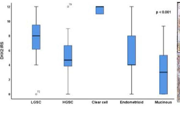
Fraungruber et al.                                                                                                                               Dkk2 and GPER in Ovarian Cancer

      and vascular physiology. Endocrinology (2012) 153(7):2953–62. doi: 10.1210/              47. Sjostrom M, Hartman L, Grabau D, Fornander T, Malmstrom P,
      en.2012-1061                                                                                 Nordenskjold B, et al. Lack of G protein-coupled estrogen receptor (GPER)
32.   Fujiwara S, Terai Y, Kawaguchi H, Takai M, Yoo S, Tanaka Y, et al. GPR30                     in the plasma membrane is associated with excellent long-term prognosis in
      regulates the EGFR-Akt cascade and predicts lower survival in patients with                  breast cancer. Breast Cancer Res Treat (2014) 145(1):61–71. doi: 10.1007/
      ovarian cancer. J Ovarian Res (2012) 5(1):35. doi: 10.1186/1757-2215-5-35                    s10549-014-2936-4
33.   Ignatov T, Modl S, Thulig M, Weissenborn C, Treeck O, Ortmann O, et al.                  48. Jung YS, Park JI. Wnt signaling in cancer: therapeutic targeting of Wnt
      GPER-1 acts as a tumor suppressor in ovarian cancer. J Ovarian Res (2013) 6                  signaling beyond beta-catenin and the destruction complex. Exp Mol Med
      (1):51. doi: 10.1186/1757-2215-6-51                                                          (2020) 52(2):183–91. doi: 10.1038/s12276-020-0380-6
34.   Kolkova Z, Casslen V, Henic E, Ahmadi S, Ehinger A, Jirstrom K, et al. The G             49. ClinicalTrials. A Study of DKN-01 as a Monotherapy or in Combination With
      protein-coupled estrogen receptor 1 (GPER/GPR30) does not predict survival in                Paclitaxel in Patients With Recurrent Epithelial Endometrial or Epithelial
      patients with ovarian cancer. J Ovarian Res (2012) 5:9. doi: 10.1186/1757-2215-5-9           Ovarian Cancer or Carcinosarcoma (P204). (2020). https://clinicaltrials.gov/
35.   Czogalla B, Kuhn C, Heublein S, Schmockel E, Mayr D, Kolben T, et al. EP3                    ct2/show/NCT03395080.
      receptor is a prognostic factor in TA-MUC1-negative ovarian cancer. J Cancer             50. Prossnitz ER, Barton M. The G-protein-coupled estrogen receptor GPER in
      Res Clin Oncol (2019) 145(10):2519–27. doi: 10.1007/s00432-019-03017-8                       health and disease. Nat Rev Endocrinol (2011) 7(12):715–26. doi: 10.1038/
36.   Heublein S, Mayr D, Vrekoussis T, Friese K, Hofmann SS, Jeschke U, et al. The                nrendo.2011.122
      G-protein coupled estrogen receptor (GPER/GPR30) is a gonadotropin                       51. Tian L, Shao W, Ip W, Song Z, Badakhshi Y, Jin T. The developmental Wnt
      receptor dependent positive prognosticator in ovarian carcinoma patients.                    signaling pathway effector beta-catenin/TCF mediates hepatic functions of the
      PLoS One (2013) 8(8):e71791. doi: 10.1371/journal.pone.0071791                               sex hormone estradiol in regulating lipid metabolism. PLoS Biol (2019) 17(10):
37.   Deuster E, Mayr D, Hester A, Kolben T, Zeder-Goss C, Burges A, et al.                        e3000444. doi: 10.1371/journal.pbio.3000444
      Correlation of the Aryl Hydrocarbon Receptor with FSHR in Ovarian Cancer                 52. Wang C, Lv X, He C, Hua G, Tsai MY, Davis JS. The G-protein-coupled
      Patients. Int J Mol Sci (2019) 20(12):2862. doi: 10.3390/ijms20122862                        estrogen receptor agonist G-1 suppresses proliferation of ovarian cancer cells
38.   Meinhold-Heerlein I, Fotopoulou C, Harter P, Kurzeder C, Mustea A,                           by blocking tubulin polymerization. Cell Death Dis (2013) 4:e869.
      Wimberger P, et al. The new WHO classification of ovarian, fallopian tube,                    doi: 10.1038/cddis.2013.397
      and primary peritoneal cancer and its clinical implications. Arch Gynecol
      Obstet (2016) 293(4):695–700. doi: 10.1007/s00404-016-4035-8
                                                                                               Conflict of Interest: SM received research support, advisory board, honoraria and
39.   Heidegger H, Dietlmeier S, Ye Y, Kuhn C, Vattai A, Aberl C, et al. The
                                                                                               travel expenses from AstraZeneca, Clovis, Medac, MSD, PharmaMar, Roche,
      Prostaglandin EP3 Receptor Is an Independent Negative Prognostic Factor for
                                                                                               Sensor Kinesis, Tesaro, and Teva. FT declares research support, advisory board,
      Cervical Cancer Patients. Int J Mol Sci (2017) 18(7):1571. doi: 10.3390/
                                                                                               honoraria and travel expenses from AstraZeneca, Medac, PharmaMar, Roche, and
      ijms18071571
                                                                                               Tesaro. SH reports grants from Baden-Württemberg Ministry of Science, Research
40.   Remmele W, Hildebrand U, Hienz HA, Klein PJ, Vierbuchen M, Behnken LJ,
                                                                                               and the Arts, from StuRa Ruprecht-Karls-University of Heidelberg, FöFoLe LMU
      et al. Comparative histological, histochemical, immunohistochemical and
                                                                                               Munich Medical Faculty, grants from FERRING, personal fees from Roche, other
      biochemical studies on oestrogen receptors, lectin receptors, and Barr
                                                                                               from Astra Zeneca, grants from Novartis Oncology, grants and non-financial
      bodies in human breast cancer. Virchows Arch A Pathol Anat Histopathol
                                                                                               support from Apceth GmbH, non-financial support from Addex and grants from
      (1986) 409(2):127–47. doi: 10.1007/bf00708323
                                                                                               Heuer Stiftung. She further reports grants from Deutsche Forschungsgemeinschaft
41.   Hoo ZH, Candlish J, Teare D. What is an ROC curve? Emerg Med J (2017) 34
                                                                                               within the funding program Open Access Publishing, by the Baden-Württemberg
      (6):357–9. doi: 10.1136/emermed-2017-206735
                                                                                               Ministry of Science, Research and the Arts and by Ruprecht-Karls-University
42.   Fluss R, Faraggi D, Reiser B. Estimation of the Youden Index and its associated
                                                                                               Heidelberg, outside the submitted work. TK receives a grant from the Friedrich-
      cutoff point. Biom J (2005) 47(4):458–72. doi: 10.1002/bimj.200410135
                                                                                               Baur-Stiftung.
43.   Wu W, Glinka A, Delius H, Niehrs C. Mutual antagonism between dickkopf1
      and dickkopf2 regulates Wnt/beta-catenin signalling. Curr Biol (2000) 10                 The remaining authors declare that the research was conducted in the absence of
      (24):1611–4. doi: 10.1016/s0960-9822(00)00868-x                                          any commercial or financial relationships that could be construed as a potential
44.   Kurman RJ, Shih Ie M. Molecular pathogenesis and extraovarian origin of                  conflict of interest.
      epithelial ovarian cancer–shifting the paradigm. Hum Pathol (2011) 42
      (7):918–31. doi: 10.1016/j.humpath.2011.03.003                                           Copyright © 2021 Fraungruber, Kaltofen, Heublein, Kuhn, Mayr, Burges, Mahner,
45.   Matei D, Fang F, Shen C, Schilder J, Arnold A, Zeng Y, et al. Epigenetic                 Rathert, Jeschke and Trillsch. This is an open-access article distributed under the terms
      resensitization to platinum in ovarian cancer. Cancer Res (2012) 72(9):2197–             of the Creative Commons Attribution License (CC BY). The use, distribution or
      205. doi: 10.1158/0008-5472.Can-11-3909                                                  reproduction in other forums is permitted, provided the original author(s) and the
46.   Moufarrij S, Dandapani M, Arthofer E, Gomez S, Srivastava A, Lopez-                      copyright owner(s) are credited and that the original publication in this journal is
      Acevedo M, et al. Epigenetic therapy for ovarian cancer: promise and                     cited, in accordance with accepted academic practice. No use, distribution or
      progress. Clin Epigenet (2019) 11(1):7. doi: 10.1186/s13148-018-0602-0                   reproduction is permitted which does not comply with these terms.

Frontiers in Endocrinology | www.frontiersin.org                                           8                                          February 2021 | Volume 12 | Article 564002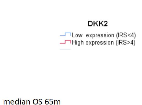
You can also read


