The Actin/Spectrin Membrane-Associated Periodic Skeleton in Neurons - UBA
←
→
Page content transcription
If your browser does not render page correctly, please read the page content below
MINI REVIEW
                                                                                                                                              published: 23 May 2018
                                                                                                                                      doi: 10.3389/fnsyn.2018.00010

                                           The Actin/Spectrin
                                           Membrane-Associated Periodic
                                           Skeleton in Neurons
                                           Nicolas Unsain 1,2,3 , Fernando D. Stefani 4,5 and Alfredo Cáceres 1,2,3 *
                                           1
                                            Instituto de Investigación Médica Mercedes y Martín Ferreyra (INIMEC), Consejo Nacional de Investigaciones Científicas y
                                           Técnicas (CONICET), Córdoba, Argentina, 2 Universidad Nacional de Córdoba, Córdoba, Argentina, 3 Instituto Universitario
                                           Ciencias Biomédicas de Córdoba (IUCBC), Córdoba, Argentina, 4 Centro de Investigaciones en Bionanociencias (CIBION),
                                           Consejo Nacional de Investigaciones Científicas y Técnicas (CONICET), Buenos Aires, Argentina, 5 Departamento de Física,
                                           Facultad de Ciencias Exactas y Naturales, Universidad de Buenos Aires, Buenos Aires, Argentina

                                            Neurons are the most asymmetric cell types, with their axons commonly extending over
                                            lengths that are thousand times longer than the diameter of the cell soma. Fluorescence
                                            nanoscopy has recently unveiled that actin, spectrin and accompanying proteins form
                                            a membrane-associated periodic skeleton (MPS) that is ubiquitously present in mature
                                            axons from all neuronal types evaluated so far. The MPS is a regular supramolecular
                                            protein structure consisting of actin “rings” separated by spectrin tetramer “spacers”.
                                            Although the MPS is best organized in axons, it is also present in dendrites, dendritic
                             Edited by:
                                            spine necks and thin cellular extensions of non-neuronal cells such as oligodendrocytes
                       Shelley Halpain,     and microglia. The unique organization of the actin/spectrin skeleton has raised the
   University of California, San Diego,
                                            hypothesis that it might serve to support the extreme physical and structural conditions
                          United States
                                            that axons must resist during the lifespan of an organism. Another plausible function of
                      Reviewed by:
               Christophe Leterrier,        the MPS consists of membrane compartmentalization and subsequent organization of
            NeuroCyto Lab, France           protein domains. This review focuses on what we know so far about the structure of the
                    Andreas Prokop,
           University of Manchester,        MPS in different neuronal subdomains, its dynamics and the emerging evidence of its
                    United Kingdom          impact in axonal biology.
              Pirta Elina Hotulainen,
     Minerva Foundation Institute for       Keywords: actin, spectrin, axon, dendrites, cytoskeleton, super resolution microscopy, fluorescence nanoscopy
         Medical Research, Finland

                *Correspondence:
                    Alfredo Cáceres        INTRODUCTION
           acaceres@immf.uncor.edu
                                           Plasma membrane domain specialization is determinant for key cellular activities such as adhesion,
         Received: 02 January 2018         signaling, membrane excitability, endo/exocytosis and stress resistance, among others. A set of
           Accepted: 04 May 2018           filamentous proteins have emerged early in evolution to form and maintain these domains; they
           Published: 23 May 2018          are organized in a scaffold known as the actin/spectrin-based membrane skeleton, which is located
                            Citation:      at the inner surface of plasma membranes (Bennett and Baines, 2001; Baines, 2010).
 Unsain N, Stefani FD and Cáceres A            Most of what we know about this skeleton comes from research characterizing the organization
           (2018) The Actin/Spectrin
                                           of the erythrocyte membrane-cortical cytoskeleton (EMCC). The EMCC is responsible for the
     Membrane-Associated Periodic
                Skeleton in Neurons.
                                           remarkable mechanical properties of the erythrocyte, including its resistance to high shear
    Front. Synaptic Neurosci. 10:10.       stress and rapid changes in shape. The vital importance of the EMCC is evidenced by the fact
     doi: 10.3389/fnsyn.2018.00010         that mutations affecting its constituents cause hereditary hemolytic anemia (Delaunay, 2007).

Frontiers in Synaptic Neuroscience | www.frontiersin.org                          1                                                 May 2018 | Volume 10 | Article 10
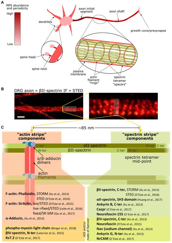
Unsain et al.                                                                                              The Membrane-Associated Periodic Skeleton in Neurons

   The detailed knowledge of the EMCC can be used as a                              interaction with Band 3, whereas a second link to the membrane
template for studying and interpreting the distinctive cortical                     is formed through an ankyrin complex close to the middle
cytoskeleton in neurons. The two-dimensional (2D) organization                      point of the spectrin tetramer (Figure 1B). The principal
of the EMCC was definitively demonstrated in the mid-1980s                          components of the EMCC are also found in other cell types
using negative staining electron microscopy (Byers and Branton,                     suggesting it provides essential biological functions (Baines,
1985; Shen et al., 1986), which together with additional                            2009, 2010).
biochemical work established a model with a repetitive 2D                              In the nervous system, spectrins have key roles in membrane
structure consisting of short, ∼37 nm long actin filaments                          domain organization. Experimental deletion of αII-, βII-, βIII-
(composed of 12 to 14 monomers) that coordinate 5 or 6 αI/βI-                       and βIV-spectrin forms, as well as mutations found in human
spectrin tetramers (Figure 1A). Accessory proteins, namely                          patients, induce drastic phenotypic defects in the nervous system,
protein 4.1 and dimers of α/β-adducin, further stabilize the                        mostly related to mislocalization of neurotransmitter receptors
actin-spectrin interaction. The whole structure is attached to                      or components of Ranvier nodes or affecting the formation of
the plasma membrane through two protein complexes that                              the axon initial segment (AIS; Parkinson et al., 2001; Komada and
interact with the anion transport protein Band 3. At the                            Soriano, 2002; Ikeda et al., 2006; Zhang et al., 2013; Huang et al.,
actin-spectrin junction, the protein 4.1 complex mediates the                       2017).

  FIGURE 1 | Overview of the actin/spectrin skeleton of erythrocytes. (A) Basic arrangement of the actin/spectrin skeleton underlying the erythrocyte membrane.
  (B) Schematic representation of the principal components of the erythrocyte membrane-cortical cytoskeleton (EMCC) and its attachments to the plasma membrane
  by means of the protein 4.1 and ankyrin complexes. For further details on this structure please refer to other reviews (Baines, 2010; Lux, 2016).

Frontiers in Synaptic Neuroscience | www.frontiersin.org                        2                                                May 2018 | Volume 10 | Article 10
Unsain et al.                                                                                 The Membrane-Associated Periodic Skeleton in Neurons

ORGANIZATION OF THE                                                       only after the establishment of the lattice along the axon (Zhong
MEMBRANE-ASSOCIATED PERIODIC                                              et al., 2014).
SKELETON                                                                      Current evidence suggests that the minimal components
                                                                          required for organizing the periodical lattice of the
In 2013, a seminal study using stochastic optical reconstruction          MPS are F-actin and spectrins, because pharmacological
microscopy (STORM) revealed the nanoscale organization of the             depolymerization of F-actin breaks the periodicity of spectrins,
actin-spectrin skeleton in axons as a periodic arrangement of             and βII-spectrin knock down affects F-actin periodic distribution
F-actin rings separated by ∼190 nm spectrin tetramer spacers              (Xu et al., 2013; Zhong et al., 2014). Adducin, although found
(Xu et al., 2013), now referred to as the membrane-associated             in the mature MPS arrangement, is not likely necessary
periodical skeleton (MPS, Figure 2). The resemblance to the               to form the MPS, since adducin null neurons can build
EMCC components, together with the distance between F-actin               an MPS (Leite et al., 2016; Qu et al., 2017) and the MPS
rings equivalent to the size of a stretched spectrin tetramer,            of immature axons lack adducin (Zhong et al., 2014).
supported the conception of a structural working model of the             Instead, adducin may help in keeping F-actin filaments at
MPS, which has been corroborated and improved by others since             a fixed length and to increase the stability of the actin-
then: the MPS is composed of numerous short actin filaments               spectrin interaction, as shown for erythrocytes (Gardner
organized in ring-like structures transverse to the axon, and             and Bennett, 1987). The idea that adducin is important
separated by various αII/βII-spectrin tetramers extended along            for the structural stability of the MPS is also supported
the axon (Figure 2).                                                      by the fact that adducin null neurons present a decreased
    A number of complementary fluorescence nanoscopy studies              fraction of axonal segments with regular MPS (Qu et al.,
have revealed that the MPS is ubiquitously present in mature              2017).
axons and, to a less extent, in dendrites from all neuronal types             The MPS is not fully regular along a single axon, but
evaluated so far (Xu et al., 2013; D’Este et al., 2016; He et al.,        displays different degrees of organization and may even be
2016). Notably, the MPS has been also observed in brain slices            absent in short segments (Barabas et al., 2017; Qu et al.,
(Xu et al., 2013; D’Este et al., 2015) and in living cells using          2017). Whether this irregularity reflects a meaningful biological
either permeable F-actin probes (D’Este et al., 2015) or tagged           design remains to be established, and highlights the need
βII-spectrin expression (Zhong et al., 2014).                             to determine MPS abundance (fraction of an axon/dendrite
    The MPS is also present at the AIS and Ranvier nodes, and             with MPS) and regularity in an unbiased and quantitative
‘‘markers’’ of these domains, such as ankyrin G, βIV-spectrin,            manner. Different approaches have been used to tackle these
neurofascin and Nav channels organize periodically (Leterrier             challenges, such as quantifying autocorrelation amplitudes
et al., 2015; D’Este et al., 2017). Interestingly, glial membranes        (Zhong et al., 2014), sinusoid fit to selected regions (Leterrier
at the juxtaparanode also contain proteins organized periodically         et al., 2015) and computing the two-dimensional Pearson
(D’Este et al., 2017), probably arranged by their interaction to          correlation against a predefined periodic pattern (Barabas et al.,
periodical protein lattices in the axon.                                  2017; Unsain et al., 2018). Also, for an unbiased and high
    In cultured neurons, the MPS has been detected soon after             volume sampling of neurites, an open-source software was
axon outgrowth, but almost exclusively in regions proximal                developed that integrates batch analysis, using an automated and
to the cell body. Over time, the periodical organization                  unbiased interrogation of neurite segments by two-dimensional
spreads distally, and by 12 days in vitro (DIV) the MPS                   Pearson correlation against a modeled MPS (Barabas et al.,
can be found along the entire axonal length (Zhong et al.,                2017).
2014). This proximal to distal developmental pattern awaits                   The lower abundance and organization of the MPS in
confirmation using unbiased sampling approaches and                       dendrites compared to axons (D’Este et al., 2015, 2016) cannot be
quantitative analysis of the MPS (Barabas et al., 2017; Unsain            explained by their different diameters (Han et al., 2017). Instead,
et al., 2018). Interestingly, the first proteins to show such             the reason may be the lower levels of βII-spectrin in dendrites.
a periodic arrangement during axonal growth are F-actin                   Experiments where the level of βII-spectrin was increased in
and βII-spectrin. αII-spectrin distributes periodically in                dendrites resulted in a significant improvement of their MPS
the mature axon and seems to be the partner of βII- and                   organization (Zhong et al., 2014). It is worth noting that under
βIV-spectrins in their respective tetramers (Huang et al.,                these conditions, dendrites maintain their molecular identity. On
2017).                                                                    the other hand, the most common actin/spectrin organization
    Adducin is a barbed-end capping protein known to stabilize            of the somato-dendritic compartment seems to be a 2D lattice
actin filaments and to prevent further incorporation of                   similar to the EMCC (Han et al., 2017).
monomers; interestingly, α-adducin deletion results in actin                  Dendritic spines are specialized structures harboring most
‘‘rings’’ with increased diameter (Leite et al., 2016). Adducin is        excitatory synapses. Changes in synaptic strength correlate with
found with multiple copies per ring, suggesting that each actin           modifications of spine shape, which tightly depends on the
‘‘ring’’ might be composed of several short filaments. In favor of        organization of the underlying actin cytoskeleton. Interestingly,
this, platinum replica electron microscopy (PREM) revealed the            dendritic spines necks, but not the head, show a periodical
existence of short actin filaments at the AIS with lengths similar        organization of actin and spectrin (Bär et al., 2016; He et al.,
to those found at the EMCC (Jones et al., 2014). Interestingly, the       2016; Sidenstein et al., 2016); the periodical lattice can be
incorporation of adducin into the MPS is a late event occurring           found even if the dendritic shaft from which it sprouts

Frontiers in Synaptic Neuroscience | www.frontiersin.org              3                                           May 2018 | Volume 10 | Article 10
Unsain et al. The Membrane-Associated Periodic Skeleton in Neurons FIGURE 2 | Overview of the membrane-associated periodic skeleton (MPS) of neurons and its associated proteins. (A) The MPS abundance and organization in different domains of a neuron, from being robust and well organized in the axon initial segment to being completely absent in the cell soma. (B) Axon shafts from sensory neurons in culture, stained against βII-spectrin and imaged by Stimulated Emission Depletion microscopy (STED) (Unsain et al., 2018), reveals the MPS. Scale bar 1 µm. (C) List of proteins arranged with a ∼190 nm periodicity in axons, indicating their location with respect to the alternating actin and spectrin “stripes.” Note that the same protein can have one end in one stripe, and the other in the other stripe, as in the case of spectrins. Also, some proteins can have one of its ends interacting with the MPS and thus being periodically arranged, but have the other end freely extending out of the MPS, and thus showing no periodicity. Such is the case of ankyrin G, which has its N-terminus periodically distributed, but not its C-terminus. The components on the bottom of the list under yellow shading are exclusively found at the AIS and/or at Ranvier nodes. Frontiers in Synaptic Neuroscience | www.frontiersin.org 4 May 2018 | Volume 10 | Article 10
Unsain et al.                                                                                 The Membrane-Associated Periodic Skeleton in Neurons

lacks a clear MPS. By contrast, current evidence suggests                     Fluorescence recovery after photobleaching (FRAP) and
that the MPS cannot form at either side of the synapse,                   live-cell STORM experiments with tagged βII-spectrin have
probably due to the high levels of synaptic proteins. For                 revealed that spectrin is relatively stable in the MPS (30 min
example, the AIS receives axo-axonic GABAergic synapses and               timeframe) and that the MPS structure itself may not move
at these postsynaptic sites the MPS is disrupted (D’Este et al.,          along the axon (6 min timeframe; Zhong et al., 2014). In
2015). Also, the MPS is interrupted at presynaptic elements               support of the observed low turnover of spectrin at the MPS,
of en-passant synapses (He et al., 2016; Sidenstein et al.,               mathematical modeling has suggested that tetramer exchange
2016).                                                                    would be difficult in an already formed MPS because the
                                                                          ‘‘resident’’ tetramers are held under tension in a fully-stretched
DYNAMICS OF THE MPS                                                       conformation, energetically unfavorable for a free tetramer (Lai
                                                                          and Cao, 2014; Zhang et al., 2017).
The actin filament (F-actin) is a dynamic polymer, constantly
incorporating and releasing monomers (G-actin). Incorporation             THE MPS AND AXONAL STABILITY
of monomers occurs preferentially at the plus (barbed) end,
while monomer release is more frequent at the opposite                    It was initially suggested that the MPS could be necessary for
(minus) end. A variety of actin binding proteins, in turn, can            the structural stability of the axon as it may provide distinctive
organize F-actin into higher order arrangements and regulate              mechanical properties. However, the structural consequences
their interaction to different cellular structures (Winder and            of MPS disassembly are not that easy to grasp. For instance,
Ayscough, 2005). Drugs affecting the actin cytoskeleton are               actin-destabilizing regimes that acutely deplete axons from
widely used tools to estimate the impact of disturbing F-actin            MPS are not accompanied by axon destruction (Zhong et al.,
dynamics in a given cellular structure. Interestingly, the response       2014; Valakh et al., 2015; Qu et al., 2017). Indeed, reports
of the MPS to actin-depolymerizing drugs such as cytochalasin             on the systematic deletion of structural components of the
D and latrunculin varies depending on the neuronal region.                MPS or enzymes related to actin polymerization in Drosophila
In the distal axon, doses of latrunculin that are sufficient to           melanogaster reported no evidence of axon destruction (Qu
affect filopodial or intra-axonal longitudinal actin filaments do         et al., 2017). This evidence suggests that the MPS is not
not disrupt the MPS; higher doses do. On the other hand,                  essential for the structural stability of the axon in the basal
cytochalasin D affects the MPS at lower doses and faster than             state.
latrunculin, while still affecting other F-actin arrangements                 However, axons innervating moving parts of the body of
in the axon (Xu et al., 2013; Qu et al., 2017). In contrast,              Caenorhabditis elegans or zebra fish depleted from βII-spectrin
the MPS of the AIS is particularly resistant to high doses of             are damaged by physical stress associated with animal movement
both cytochalasin D and latrunculin (Leterrier et al., 2015).             (Hammarlund et al., 2007; Krieg et al., 2017). A recent study has
To correctly interpret these results it is important to consider          evaluated the relationship between the MPS and axon stability in
the different modes of action of latrunculins and cytochalasins.          a more physiological setting. Developmental axon pruning can
Latrunculins bind exclusively to actin monomers, preventing               be modeled in cell culture by withdrawing dorsal root ganglion
their incorporation into filaments (Peterson and Mitchison,               explants from Nerve Growth Factor (Glebova and Ginty, 2005).
2002), while cytochalasin D also caps the barbed end of actin             Under this condition, the axonal MPS shows a significant
filaments, decreasing both addition and release of monomers at            decrease in abundance during most of the degeneration period,
this end (Peterson and Mitchison, 2002; Scherlach et al., 2010).          and is fully dismantled in fragmenting axons. Interestingly, acute
On the other hand, both actin depolymerizing drugs have a                 pharmacological stabilization of F-actin prevents both MPS loss
greater effect on the MPS of immature axons than of mature                and axonal fragmentation (Unsain et al., 2018). Taken together,
ones, which correlates with the later incorporation of the actin-         these studies suggest that the MPS is not required to keep
capping protein α-adducin (Zhong et al., 2014; Qu et al., 2017)           axon integrity under basal conditions, but instead it might
suggesting that it may protect the barbed-end from cytochalasin           be necessary for coping with physical stress or degenerative
D activity.                                                               triggers.
    These actin-depolymerizing drugs may not target actin                     From a structural perspective, it is yet not clear how
filaments that are part of the MPS. Instead, they may                     the MPS and microtubules (MT) work together to provide
affect a pool of newly synthetized short polymers that are                the extraordinary mechanical properties of the axon.
subsequently exchanged within the MPS. This latter possibility            Small molecule-induced depolymerization approaches have
is supported by experiments assessing MPS abundance in                    highlighted their inter-dependance. In Drosophila neurons,
fly axons, which have shown that deletion of the actin                    disruption of the MPS with low doses of cytochalasin
elongating factor profilin does not affect MPS abundance but              D affects MT polymerization, ultimately leading to MT
deletion of actin nucleators (like formin and Arp2/3) decreases           ‘‘gaps’’ (Qu et al., 2017). In mouse neurons, deletion of
MPS abundance (Qu et al., 2017). These results support the                α-adducin affects MT-based transport (Leite et al., 2016).
hypothesis that the actin rings of the MPS are composed                   Also, the concomitant disruption of the actin cytoskeleton
of short actin filaments, and that nucleation of new actin                and the deletion of the MT stabilizing protein Shot induce
filaments would be more important than their growth for MPS               axonal loss. Conversely, MT depolymerization induced by
stability.                                                                nocodazole disrupts the MPS (Zhong et al., 2014; Qu et al.,

Frontiers in Synaptic Neuroscience | www.frontiersin.org              5                                           May 2018 | Volume 10 | Article 10
Unsain et al. The Membrane-Associated Periodic Skeleton in Neurons
2017). Additional evidence for an interaction between MT CONCLUDING REMARKS
and the MPS comes from the observation that MPS-bound
ankyrin G regulates MT dynamics, protein transport and Current challenges concerning the MPS can be divided
AIS stability/maintenance through physical interactions with in two main aspects. First, its detailed structure must
EB1 and EB3 at their deep C-terminus tail (Leterrier et al., 2011, be determined, including the molecular interactions that
2015) or by functional interaction with CRMPs (Maniar et al., hold it together and linked to the plasma membrane. For
2011). instance, Band 3 is a transmembrane protein that is key
The MPS would allow the axon membrane to sustain shear to tether the EMCC to the plasma membrane. In neurons,
and torsional efforts, since membrane stiffness measurements however, Band 3 is not expressed and it is still unknown
with atomic force microscopy (AFM) have established which proteins, if any, play an equivalent role. Second, the
that the axon has a superior resistance than the somato- biological functions of the MPS remain uncertain, both in
dendritic compartment (Zhang et al., 2017). Also, it has been terms of structure and mechanical properties, as well as
proposed that myosin-dependent contraction of actin rings, signaling, including action potential propagation, and cell-cell
counteracted by the underlying MT bundle, would induce interactions.
a torsional tension necessary to maintain axon volume in To assess these questions, it will be critical to develop
response to external stretch or compression forces (Fan et al., approaches to specifically disrupt the MPS structure without
2017). Direct evidence that actin rings may function as a affecting the behavior of its components outside of the MPS. For
contractile actomyosin complex comes from the finding of instance, the use of F-actin depolymerizing drugs to disassemble
a periodical co-localization of phosphorylated myosin light the MPS also affects actin filaments elsewhere in the cell,
chain (pMLC) with actin at the AIS-MPS (Berger et al., and hence conclusions regarding the MPS are limited. The
2018). same drawback can be said when evaluating the deletion of
spectrins.
Another challenge is to understand what are (if any) the
PLASMA MEMBRANE BIOLOGY functional consequences of a periodicity in the longitudinal
aspect of an axon, both structurally and functionally. For
The EMCC effectively confines the movement of peripheral instance, the membrane stripes organized by the underlying MPS
or integral membrane proteins in erythrocyte (Tomishige would likely confine periodically ion channels in the longitudinal
et al., 1998). The importance of this function is highlighted axis of the axon, which may impact, or actually be crucial for
by hereditary pathologies showing lack of confinement of proper action potential propagation.
erythrocyte membrane proteins (Kodippili et al., 2009). Confined In summary, the recent discovery of the MPS has created
phospholipid diffusion by the underlying actin/spectrin cortical new, exciting avenues of research that are just beginning to
skeleton was also recently observed in other cell types using be explored and will likely provide solid ground to tackle
super-resolution microscopy (Andrade et al., 2015). Also, it has long-lasting questions of neurobiology.
been found that membrane-bound probes in the AIS follow
transversally oriented trajectories confined to stripes spaced by
190 nm. These trajectories co-localize with the spectrin of the AUTHOR CONTRIBUTIONS
MPS, with the underlying actin stripes serving as the confinement
NU, FS and AC wrote and revised the manuscript.
‘‘fence’’ (Albrecht et al., 2016). Interestingly, the authors failed
to observe such motion confinement outside the AIS, suggesting
that AIS-exclusive protein/s organized by the MPS but not the FUNDING
MPS itself is/are responsible for the effective confinement of
lateral diffusion (see Huang and Rasband, 2016, for a review). This work received financial support from Agencia Nacional de
A non-excluding possibility is that the probes were actively Promoción Científica y Tecnológica grant nos. PICT-V-2014-
anchored by spectrin related components specific of the AIS. 3729 (FS, AC and NU), PICT-2013-0792 (FS), PICT-2016-0838
Extensive evidence supports a critical role of the actin/spectrin (NU) and by the International Society for Neurochemistry, grant
skeleton in ligand-receptor signaling, membrane budding and ISN-CAEN Category 1B 2017 (NU).
endocytosis. However, it is still unknown how these functions
are affected when this cortical network is organized in periodical ACKNOWLEDGMENTS
stripes, which likely produce parallel equivalent subdomains of
membrane proteins and/or spatio-temporal structures key for We thank Gaby F. Martinez and Luciano A. Masullo for the
signaling (Grecco et al., 2011). STED image displayed in Figure 2.
REFERENCES Andrade, D. M., Clausen, M. P., Keller, J., Mueller, V., Wu, C., Bear, J. E.,
et al. (2015). Cortical actin networks induce spatio-temporal confinement
Albrecht, D., Winterflood, C. M., Sadeghi, M., Tschager, T., Noé, F., and Ewers, H. of phospholipids in the plasma membrane—a minimally invasive
(2016). Nanoscopic compartmentalization of membrane protein motion at the investigation by STED-FCS. Sci. Rep. 5:11454. doi: 10.1038/srep
axon initial segment. J. Cell Biol. 215, 37–46. doi: 10.1083/jcb.201603108 11454
Frontiers in Synaptic Neuroscience | www.frontiersin.org 6 May 2018 | Volume 10 | Article 10Unsain et al. The Membrane-Associated Periodic Skeleton in Neurons Baines, A. J. (2009). Evolution of spectrin function in cytoskeletal and membrane Jones, S. L., Korobova, F., and Svitkina, T. (2014). Axon initial segment networks. Biochem. Soc. Trans. 37, 796–803. doi: 10.1042/BST0370796 cytoskeleton comprises a multiprotein submembranous coat containing sparse Baines, A. J. (2010). The spectrin-ankyrin-4.1-adducin membrane skeleton: actin filaments. J. Cell Biol. 205, 67–81. doi: 10.1083/jcb.201401045 adapting eukaryotic cells to the demands of animal life. Protoplasma 244, Kodippili, G. C., Spector, J., Sullivan, C., Kuypers, F. A., Labotka, R., 99–131. doi: 10.1007/s00709-010-0181-1 Gallagher, P. G., et al. (2009). Imaging of the diffusion of single band Bär, J., Kobler, O., van Bommel, B., and Mikhaylova, M. (2016). Periodic 3 molecules on normal and mutant erythrocytes. Blood 113, 6237–6245. F-actin structures shape the neck of dendritic spines. Sci. Rep. 6:37136. doi: 10.1182/blood-2009-02-205450 doi: 10.1038/srep37136 Komada, M., and Soriano, P. (2002). βIV-spectrin regulates sodium channel Barabas, F. M., Masullo, L. A., Bordenave, M. D., Giusti, A. S., Unsain, N., clustering through ankyrin-G at axon initial segments and nodes of Ranvier. Refojo, D., et al. (2017). Automated quantification of protein periodic J. Cell Biol. 156, 337–348. doi: 10.1083/jcb.200110003 nanostructures in fluorescence nanoscopy images: abundance and regularity Krieg, M., Stühmer, J., Cueva, J. G., Fetter, R., Spilker, K., Cremers, D., et al. (2017). of neuronal spectrin membrane-associated skeleton. Sci. Rep. 7:16029. Genetic defects in β-spectrin and tau sensitize C. elegans axons to movement- doi: 10.1038/s41598-017-16280-x induced damage via torque-tension coupling. Elife 6:e20172. doi: 10.7554/eLife. Bennett, V., and Baines, A. J. (2001). Spectrin and ankyrin-based pathways: 20172 metazoan inventions for integrating cells into tissues. Physiol. Rev. 81, Lai, L., and Cao, J. (2014). Spectrins in axonal cytoskeletons: dynamics revealed 1353–1392. doi: 10.1152/physrev.2001.81.3.1353 by extensions and fluctuations. J. Chem. Phys. 141:15101. doi: 10.1063/1.48 Berger, S. L., Leo-Macias, A., Yuen, S., Khatri, L., Pfennig, S., Zhang, Y., et al. 85720 (2018). Localized myosin II activity regulates assembly and plasticity of Leite, S. C. C., Sampaio, P., Sousa, V. F. F., Nogueira-Rodrigues, J., Pinto-Costa, R., the axon initial segment. Neuron 97, 555.e6–570.e6. doi: 10.1016/j.neuron. Peters, L. L. L., et al. (2016). The actin-binding protein α-adducin is required for 2017.12.039 maintaining axon diameter. Cell Rep. 15, 490–498. doi: 10.1016/j.celrep.2016. Byers, T. J., and Branton, D. (1985). Visualization of the protein associations in 03.047 the erythrocyte membrane skeleton. Proc. Natl. Acad. Sci. U S A 82, 6153–6157. Leterrier, C., Potier, J., Caillol, G., Debarnot, C., Rueda Boroni, F., and Dargent, B. doi: 10.1073/pnas.82.18.6153 (2015). Nanoscale architecture of the axon initial segment reveals an organized D’Este, E., Kamin, D., Balzarotti, F., and Hell, S. W. (2017). Ultrastructural and robust scaffold. Cell Rep. 13, 2781–2793. doi: 10.1016/j.celrep.2015.11.051 anatomy of nodes of Ranvier in the peripheral nervous system as revealed Leterrier, C., Vacher, H., Fache, M.-P., D’Ortoli, S. A., Castets, F., Autillo- by STED microscopy. Proc. Natl. Acad. Sci. U S A 114, E191–E199. Touati, A., et al. (2011). End-binding proteins EB3 and EB1 link microtubules doi: 10.1073/pnas.1619553114 to ankyrin G in the axon initial segment. Proc. Natl. Acad. Sci. U S A 108, D’Este, E., Kamin, D., Göttfert, F., El-Hady, A., and Hell, S. W. (2015). STED 8826–8831. doi: 10.1073/pnas.1018671108 nanoscopy reveals the ubiquity of subcortical cytoskeleton periodicity in living Lux, S. E. IV. (2016). Anatomy of the red cell membrane skeleton: neurons. Cell Rep. 10, 1246–1251. doi: 10.1016/j.celrep.2015.02.007 unanswered questions. Blood 127, 187–199. doi: 10.1182/blood-2014-12- D’Este, E., Kamin, D., Velte, C., Göttfert, F., Simons, M., and Hell, S. W. (2016). 512772 Subcortical cytoskeleton periodicity throughout the nervous system. Sci. Rep. Maniar, T. A., Kaplan, M., Wang, G. J., Shen, K., Wei, L., Shaw, J. E., et al. (2011). 6:22741. doi: 10.1038/srep22741 UNC-33 (CRMP) and ankyrin organize microtubules and localize kinesin to Delaunay, J. (2007). The molecular basis of hereditary red cell membrane polarize axon-dendrite sorting. Nat. Neurosci. 15, 48–56. doi: 10.1038/nn.2970 disorders. Blood Rev. 21, 1–20. doi: 10.1016/j.blre.2006.03.005 Parkinson, N. J., Olsson, C. L., Hallows, J. L., McKee-Johnson, J., Keogh, B. P., Fan, A., Tofangchi, A., Kandel, M., Popescu, G., and Saif, T. (2017). Coupled Noben-Trauth, K., et al. (2001). Mutant α-spectrin 4 causes auditory and motor circumferential and axial tension driven by actin and myosin influences in vivo neuropathies in quivering mice. Nat. Genet. 29, 61–65. doi: 10.1038/ng710 axon diameter. Sci. Rep. 7:14188. doi: 10.1038/s41598-017-13830-1 Peterson, J. R., and Mitchison, T. J. (2002). Review small molecules, big impact: a Galiano, M. R., Jha, S., Ho, T. S.-Y., Zhang, C., Ogawa, Y., Chang, K.-J., et al. history of chemical inhibitors and the cytoskeleton. Chem. Biol. 9, 1275–1285. (2012). A distal axonal cytoskeleton forms an intra-axonal boundary that doi: 10.1016/S1074-5521(02)00284-3 controls axon initial segment assembly. Cell 149, 1125–1139. doi: 10.1016/j.cell. Qu, Y., Hahn, I., Webb, S. E. D., Pearce, S. P., and Prokop, A. (2017). Periodic actin 2012.03.039 structures in neuronal axons are required to maintain microtubules. Mol. Biol. Gardner, K., and Bennett, V. (1987). Modulation of spectrin-actin assembly by Cell 28, 296–308. doi: 10.1091/mbc.E16-10-0727 erythrocyte adducin. Nature 328, 359–362. doi: 10.1038/328359a0 Scherlach, K., Boettger, D., Remme, N., and Hertweck, C. (2010). The Glebova, N. O., and Ginty, D. D. (2005). Growth and survival signals ontrolling chemistry and biology of cytochalasans. Nat. Prod. Rep. 27, 869–886. sympathetic nervous system development. Annu. Rev. Neurosci. 28, 191–222. doi: 10.1039/b903913a doi: 10.1146/annurev.neuro.28.061604.135659 Shen, B. W., Josephs, R., and Steck, T. L. (1986). Ultrastructure of the intact Grecco, H. E., Schmick, M., and Bastiaens, P. I. H. (2011). Signaling from the living skeleton of the human erythrocyte membrane. J. Cell Biol. 102, 997–1006. plasma membrane. Cell 144, 897–909. doi: 10.1016/j.cell.2011.01.029 doi: 10.1083/jcb.102.3.997 Hammarlund, M., Jorgensen, E. M., and Bastiani, M. J. (2007). Axons break Sidenstein, S. C., D’Este, E., Böhm, M. J., Danzl, J. G., Belov, V. N., and Hell, S. W. in animals lacking β-spectrin. J. Cell Biol. 176, 269–275. doi: 10.1083/jcb. (2016). Multicolour multilevel STED nanoscopy of actin/spectrin organization 200611117 at synapses. Sci. Rep. 6:26725. doi: 10.1038/srep26725 Han, B., Zhou, R., Xia, C., and Zhuang, X. (2017). Structural organization of the Tomishige, M., Sako, Y., and Kusumi, A. (1998). Regulation mechanism of actin-spectrin-based membrane skeleton in dendrites and soma of neurons. the lateral diffusion of band 3 in erythrocyte membranes by the membrane Proc. Natl. Acad. Sci. U S A 114, E6678–E6685. doi: 10.1073/pnas.1705043114 skeleton. J. Cell Biol. 142, 989–1000. doi: 10.1083/jcb.142.4.989 He, J., Zhou, R., Wu, Z., Carrasco, M. A., Kurshan, P. T., Farley, J. E., et al. (2016). Unsain, N., Bordenave, M. D., Martinez, G. F., Jalil, S., von Bilderling, C., Prevalent presence of periodic actin-spectrin-based membrane skeleton in a Barabas, F. M., et al. (2018). Remodeling of the actin/spectrin membrane- broad range of neuronal cell types and animal species. Proc. Natl. Acad. Sci. associated periodic skeleton, growth cone collapse and F-actin decrease during U S A 113, 6029–6034. doi: 10.1073/pnas.1605707113 axonal degeneration. Sci. Rep. 8:6002. doi: 10.1038/s41598-018-23781-w Huang, Y. M., and Rasband, M. N. (2016). Organization of the axon initial Valakh, V., Frey, E., Babetto, E., Walker, L. J., and DiAntonio, A. (2015). segment: actin like a fence. J. Cell Biol. 215, 9–11. doi: 10.1083/jcb.201609084 Cytoskeletal disruption activates the DLK/JNK pathway, which promotes Huang, C. Y.-M., Zhang, C., Ho, T. S.-Y., Oses-Prieto, J., Burlingame, A. L., axonal regeneration and mimics a preconditioning injury. Neurobiol. Dis. 77, Lalonde, J., et al. (2017). αII spectrin forms a periodic cytoskeleton at the axon 13–25. doi: 10.1016/j.nbd.2015.02.014 initial segment and is required for nervous system function. J. Neurosci. 37, Winder, S. J., and Ayscough, K. R. (2005). Actin-binding proteins. J. Cell Sci. 118, 11311–11322. doi: 10.1523/JNEUROSCI.2112-17.2017 651–654. doi: 10.1242/jcs.01670 Ikeda, Y., Dick, K. A., Weatherspoon, M. R., Gincel, D., Armbrust, K. R., Xu, K., Zhong, G., and Zhuang, X. (2013). Actin, spectrin, and associated Dalton, J. C., et al. (2006). Spectrin mutations cause spinocerebellar ataxia type proteins form a periodic cytoskeletal structure in axons. Science 339, 452–456. 5. Nat. Genet. 38, 184–190. doi: 10.1038/ng1728 doi: 10.1126/science.1232251 Frontiers in Synaptic Neuroscience | www.frontiersin.org 7 May 2018 | Volume 10 | Article 10
Unsain et al.                                                                                                        The Membrane-Associated Periodic Skeleton in Neurons

Zhang, Y., Abiraman, K., Li, H., Pierce, D. M., Tzingounis, A. V., and                      Conflict of Interest Statement: The authors declare that the research was
  Lykotrafitis, G. (2017). Modeling of the axon membrane skeleton structure and             conducted in the absence of any commercial or financial relationships that could
  implications for its mechanical properties. PLoS Comput. Biol. 13:e1005407.               be construed as a potential conflict of interest.
  doi: 10.1371/journal.pcbi.1005407
Zhang, C., Susuki, K., Zollinger, D. R., Dupree, J. L., and Rasband, M. N.                  Copyright © 2018 Unsain, Stefani and Cáceres. This is an open-access article
  (2013). Membrane domain organization of myelinated axons requires                         distributed under the terms of the Creative Commons Attribution License (CC BY).
  α II spectrin. J. Cell Biol. 203, 437–443. doi: 10.1083/jcb.2013                          The use, distribution or reproduction in other forums is permitted, provided the
  08116                                                                                     original author(s) and the copyright owner are credited and that the original
Zhong, G., He, J., Zhou, R., Lorenzo, D., Babcock, H. P., Bennett, V., et al. (2014).       publication in this journal is cited, in accordance with accepted academic practice.
  Developmental mechanism of the periodic membrane skeleton in axons. Elife                 No use, distribution or reproduction is permitted which does not comply with these
  3:e04581. doi: 10.7554/elife.04581                                                        terms.

Frontiers in Synaptic Neuroscience | www.frontiersin.org                                8                                                    May 2018 | Volume 10 | Article 10
You can also read