Construction of a novel chimeric dextransucrase fused to the carbohydrate-binding module CBM2a
←
→
Page content transcription
If your browser does not render page correctly, please read the page content below
Preprints (www.preprints.org) | NOT PEER-REVIEWED | Posted: 1 September 2021 doi:10.20944/preprints202109.0013.v1
Article
Construction of a novel chimeric dextransucrase fused to the
carbohydrate-binding module CBM2a
Reinaldo Fraga Vidal 1,*, Roberto C. Arísticas Ribalta 1, Lisandra Martínez Valdés 1, Meinardo Lafargue Gámez 1,
Amanda Montes Alvarez 1, Arianne Rubio Sánchez 1, Eric Dubreucq 2, Benoît Moreau 3
1 Cuban Research Institute on Sugarcane By-Products (ICIDCA). P.O. Box 4026, 11000, Havana, Cuba. Tel.
053-7-696-7015; Fax: 053-7-988243; reynaldo.fraga@icidca.azcuba.cu
2 SupAgro Montpellier, 2 Place Viala, 34060 Montpellier Cedex 2, France; Eric.Dubreucq@supagro.fr
3 Haute Ecole Province de Henaut – Condorcet, Rue Paul Pastur 11, 7800 Ath, Belgium. E. mail: benoit.mo-
reau@condorcet.be
* Correspondence: reynaldo.fraga@icidca.azcuba.cu (R.F.); Tel. 053-7-696-7015
Abstract: The lactic acid bacteria (LAB) have great potential to produce homoexopolysaccharides
(HoPS), have been the subject of extensive research efforts, given their health benefits and physico-
chemical properties. The HoPS functional properties are determined by structural characteristics of
varied molecular weights, types of glycosidic linkages, degrees of branching and chemical compo-
sition. The dextransucrases (DSases) are responsible of the synthesis of a kind of HoPS (dextran
polymers), which are among the first biopolymers produced at industrial scale with applications in
medicine and biotechnology. The concept of glycodiversification opens additional applications for
DSases. In that sense the design and characterization of new DSases is of prime importance. Previ-
ously, we described the isolation and characterization of a novel extracellular dextransucrase (DSR-
F) encoding gene. In this study, from DSR-F, we design a novel chimeric dextransucrase DSR-F-
∆SP-∆GBD-CBM2a, where DSR-F-∆SP-∆GBD is fused to the carbohydrate-binding module (CBM2a)
of the β-1-4 exoglucanase/xylanase Cex (Xyn10A) of Cellulomonas fimi ATCC 484. This dextransu-
crase variant is active and without alteration in its specificity. The DSR-F-∆SP-∆GBD-CBM2a is pu-
rified by cellulose affinity chromatography for the very first time. Our results indicate that new
hybrids and chimeric DSases with novel binding capacity to cellulose can be designed to obtain
glyco-biocatalysts from renewable lignocellulosic materials.
Keywords: dextransucrases; GH70; lactic acid bacteria; sucrose-active enzymes; carbohydrate bind-
ing module; glucansucrase; cellulose binding domain; Leuconostoc
1. Introduction
Homoexopolysaccharides (HoPS) produced by lactic acid bacteria (LAB) have re-
ceived great research efforts into their physicochemical and bioactive properties [1]. The
individual functional properties of HoPS are determined by their chemical composition,
molecular weights, types of glycosidic linkages as well as degree and arrangement of
branches [2]. The structural diversity of HoPS is a result of the unmatched variety of pos-
sible osidic bonds between sugar monomers, offering an extensive range of functionalities
of interest for food, feed, pharmaceuticals, cosmetics and chemicals industries [3-6].
Dextrans are among the first microbial HoPS produced at industrial scale [7]. Micro-
organisms of the genera Lactobacillus, Streptococcus, Weissella, Leuconostoc, Pediococcus, Oe-
nococcus and Acetobacter [8] produce these polysaccharides. The Leuconostoc mesenteroides
NRRL B-512F is used for the synthesis of the most common and widespread commercial
dextran. The biopolymer’s main chain contains (1-6) linked glucosyl residues with only
5% of (1-3) linked branches [7]. The dextran fractions of controlled molecular weight and
their numerous derivatives are mainly used in medicine, pharmaceuticals and fine chem-
© 2021 by the author(s). Distributed under a Creative Commons CC BY license.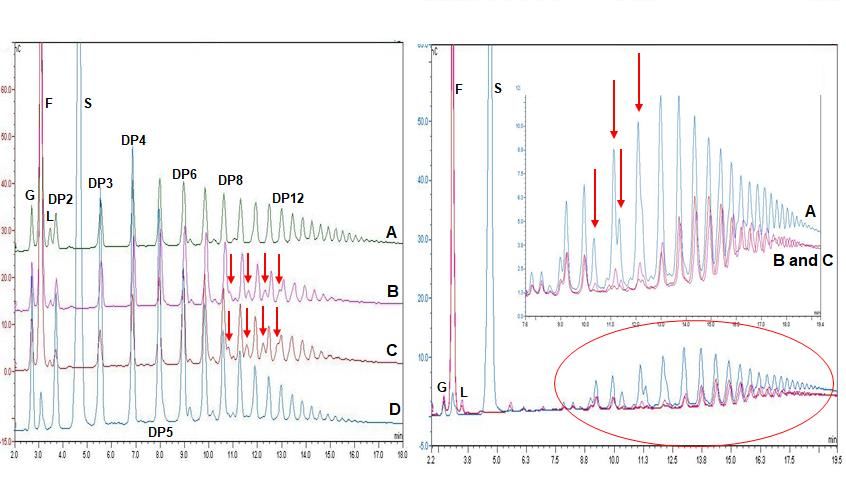
Preprints (www.preprints.org) | NOT PEER-REVIEWED | Posted: 1 September 2021 doi:10.20944/preprints202109.0013.v1
istry [9,10]. The extracellular glucansucrase (dextransucrase) DSR-S, a 6--D-glucosyl-
transferase (EC 2.4.1.5) is responsible for the polymer production [11]. This enzyme be-
longs to the glycoside hydrolase family 70 (GH70) according to the CAZy classification
(http://www.cazy.org/) [12]. The GH70 family consists of a large and diverse group of
polymerases and branching enzymes, some of them being mainly active on sucrose and
others on starch substrates [13,14].
The catalytic domain of GH70 presents the typical (/)8 barrel of glucansucrases, the
three amino acids (D551, E589, D662, DSR-S numbering) forming the catalytic triad are
highly conserved in the GH70 family [15-17].
The study of GH70 enzymes with different specificities from new LAB strains and
from mining genome data sets could provide new insights in structure-function relation-
ships of glucansucrases as well as enlarge the natural dextransucrase repertoire available
for industrial application [18]. The Cuban Research Institute on Sugarcane By-products
(ICIDCA) has a collection of LAB strains isolated from sugarcane and sugarcane deriva-
tives. Some of them have already been characterized to some extent [19-21]. In the present
study, a chimeric dextransucrase fused to the carbohydrate-binding module (CBM2a) of
the exoglucanase/xylanase Cex (Xyn10A) of Cellulomonas fimi ATCC 484 was obtained.
This variant, fully active, was purified by cellulose affinity chromatography and partially
characterized.
2. Results and Discussion
2.1. Design of a Chimeric Dextransucrase (DSR-F-∆SP-∆GBD-CBM2a) Fused to the Carbohy-
drate Binding Module CBM2a
Based on the 3D structural model of DSR-F-∆SP-∆GBD (Fig. 1B), a chimeric dextransu-
crase fused to the carbohydrate binding module CBM2a (DSR-F-∆SP-∆GBD-CBM2a) was
constructed (Fig. 1A and 1C). Deleting the two APY repeat units from the C-terminal end
of DSR-F and adding the CBM2a, allowed further investigation of this domain fusion on
the specificity of the chimeric fused variant.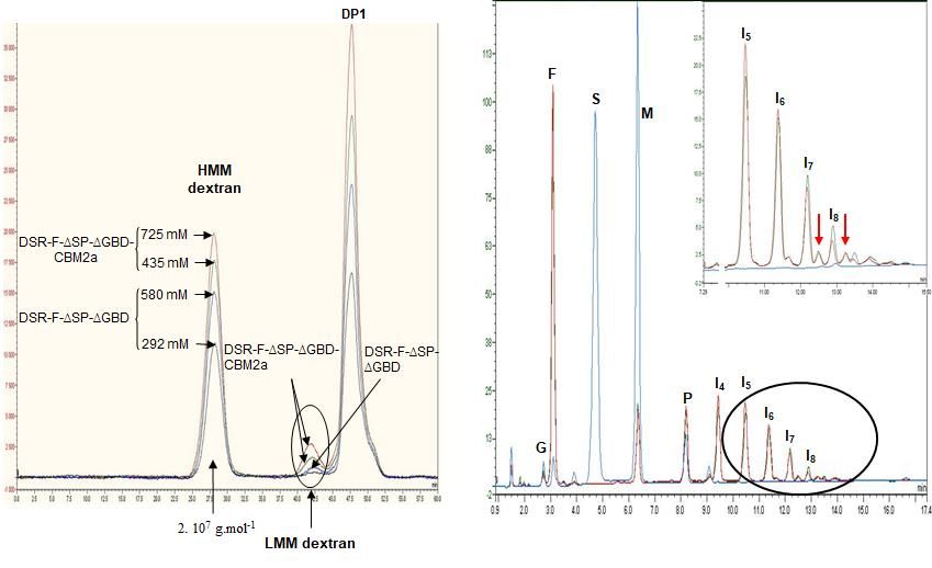
Preprints (www.preprints.org) | NOT PEER-REVIEWED | Posted: 1 September 2021 doi:10.20944/preprints202109.0013.v1
(A) (B) (C)
Figure 1. (A). Primary structure representation of the dextransucrase DSR-F of Leu-
conostoc citreum B/110-1-2 and its truncated variant DSRF-∆SP-∆GBD. (B). 3D structure
model of DSRF-∆SP-∆GBD, five different domains are highlighted, in red (domain V), yel-
low (domain IV), green (domain B), blue (domain A), and magenta (domain C) (C). Linear
schematic representation of DSRF-∆SP-∆GBD domain organization with CBM2a fused to
the C-terminal end.
The recombinant DSR-F enzyme was previously produced without part of its C-ter-
minal glucan binding domain (Fig. 1A, DSR-F-∆SP-∆GBD). This deletion of the C-terminal
domain V including a cell wall (CW) repeat and the two APY repeats did not affect the
DSR-F-∆SP-∆GBD specificity and efficiency [19]. Other examples of APY repeats that have
been deleted without adverse effect on other GH70 and related enzymes have been ob-
served in alternansucrase, inulosucrase, and branching sucrase BSR-B [22-24].
2.2. Subcloning of DSR-F-∆SP-∆GBD to Obtain DSR-F-∆SP-∆GBD-CBM2a in the Ex-
pression Vector pdsrF-CBM2a. Purification of the Fusion Protein DSR-F-∆SP-∆GBD-CBM2a
The construction of plasmid pdsrF-CBM2a allows the inducible expression of DsrF-
ΔSP-ΔGBD fused in its carboxyl end region with the carbohydrate binding module
CBM2a of Cex, a -1,4-exo-glucanase of Cellulomonas fimi ATCC 484. The fusion of the
CBM2a to a dextransucrase is reported for the very first time. The presence of the CBM2a
module seems to not affect the ability to form dextran polymer by the fusion enzyme DsrF-
ΔSP-ΔGBD-CBM2a (Fig. 2_A and B), and it will permit its purification for further charac-
terization and future immobilization on cellulosic or lignocellulosic materials. A success-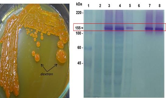
Preprints (www.preprints.org) | NOT PEER-REVIEWED | Posted: 1 September 2021                doi:10.20944/preprints202109.0013.v1

                                 ful dextransucrase fusion for immobilization is the case of DSR-S from Leuconostoc mesen-
                                 teroides B-512FMC to glutathione S-transferase (GST), resulting in a novel and completely
                                 active fused truncated variant [25].
                                       The purification of DsrF-ΔSP-ΔGBD-CBM2a from the soluble fraction of the E. coli
                                 DH10B (pdsrF-CBM2a) was performed using regenerated amorphous cellulose (RAC) as
                                 an affinity matrix. The purified protein migrated as a single band with an apparent mo-
                                 lecular mass of 155 kDa and a 90-95% purity estimated by densitometry (Fig. 2_B). This is
                                 the first report of the use of a cellulose-based matrix for the purification of a dextransu-
                                 crase by affinity chromatography. The levels of purified protein are greater than those
                                 achieved by the immobilized metal ion affinity chromatography (IMAC) (data not
                                 shown). This may be because RAC has a higher binding capacity (365 mg of protein per
                                 gram of RAC) than any other commercial resin used to purify proteins (10-40 mg of pro-
                                 teins per gram of resin) [26].

                                                      (A)                                         (B)

                                       Figure 2. Production of DsrF-ΔSP-ΔGBD-CBM2a in E. coli DH10B. (A) Detection of
                                 dextransucrase activity through the formation of dextran on LBT agar plate. (B) SDS-
                                 PAGE of DsrF-ΔSP-ΔGBD-CBM2a purification, stained combining two detection meth-
                                 ods. Zymography of the proteins with dextransucrase activity (colored bands), followed
                                 by detection of total proteins by negative staining with imidazole and zinc sulfate salts
                                 (transparent bands). The formation of the dextran polymer demonstrates the activity of
                                 the fusion. (1) Molecular Weight Marker (ColorBurstTM, Sigma-Aldrich Co.). (2) Soluble
                                 fraction of E. coli DH10B (pSE380) cell lysate as negative control. (3) Soluble fraction of E.
                                 coli DH10B (pdsrF-CBD) cell lysate. (4) Flowthrough fraction, not retained by the RAC. (5)
                                 Fraction of the 1st washed of column (8 Column Volumes, CV). (6) - Fraction of the 2nd
                                 washed of column (12 CV). (7 and 8) - Fractions eluted.

                                      2.3. Action of DSR-F-ΔSP-ΔGBD-CBM2a in Polymerization and Acceptor Reactions

                                      A polymerization reaction with sucrose was carried out to ensure the chimeric DSR-
                                 F-ΔSP-ΔGBD-CBM2a behave in the same way as the DSR-F-ΔSP-ΔGBD variant. As dex-
                                 transucrase GTF180 from Lb. reuteri 180 [27] and alternansucrase (ASR) from L. citreum
                                 NRRL B-1355 [28], DSR-F-∆SP-∆GBD-CBM2a and DSR-F-∆SP-∆GBD are also striking ex-
                                 amples of α-transglucosylases among GH70 glucansucrases. They catalyze a bi-modal
                                 population of glucan, comprising a high molar mass (HMM, 2x10 7 g.mol-1) dextran and a
                                 low molar mass gluco-oligosaccharides (LMM, DP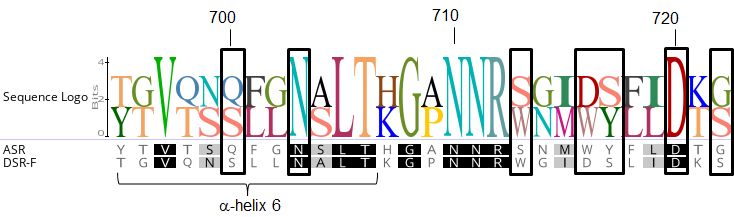
Preprints (www.preprints.org) | NOT PEER-REVIEWED | Posted: 1 September 2021               doi:10.20944/preprints202109.0013.v1

                                 gation mechanism, as for other GH70 enzymes, where smaller oligosaccharides are pro-
                                 duced in a non-processive mode at the beginning of a reaction. When a critical length is
                                 reached a processive mechanism starts helped by glucan-binding domains [29].

                                        (I)                                                                      (II)

                                      Figure 3. (I)-Analysis of HPSEC chromatograms synthesized using the chimeric dex-
                                 transucrase DSR-F-∆SP-∆GBD-CBM2a (from sucrose 580 mM and 725 mM) and DSR-F-
                                 ∆SP-∆GBD (from sucrose 292 mM and 435 mM) with 50 mM sodium acetate buffer, pH
                                 5.5 and 1 U.mL-1 of enzyme. Enzymatic reactions were stopped after 24 h. Peak identifica-
                                 tion: HMM dextran high molecular mass dextran (2.107 g.mol-1 as estimated by HPSEC-
                                 MALLS), LMM dextran low molecular mass dextran (oligosaccharides of DP
Preprints (www.preprints.org) | NOT PEER-REVIEWED | Posted: 1 September 2021 doi:10.20944/preprints202109.0013.v1
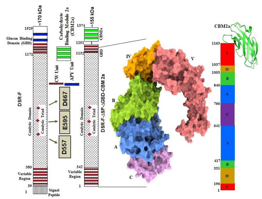
reaction products showed that a low amount of leucrose could be detected due to the
action of fructose as an acceptor (Fig. 4-I). This kind of transglycosylation has been re-
ported to be favored at the start of the dextransucrase reactions [29, 30]. It has been re-
cently suggested that leucrose serves as the substrate for further dextran formation or
elongation of IMOs [31]. The IMOs from DP8 to DP12 were glucosyl decorated in the same
way by the chimeric dextransucrase DSR-F-∆SP-∆GBD-CBM2a and DSR-F-∆SP-∆GBD,
leading to formation of products clearly observable on HPAEC-PAD chromatograms (Fig.
4-I, arrows). Such chromatographic patterns resulting from the acceptor reaction (oligo-
dextrans as acceptor molecules) is reported for the first time for a DSR-F variant enzyme.
According to the specificity of both analyzed enzymes, these modified IMOs could be
products being glucosylated to some extent and probably (1-4) branched glucose units.
As a specific population of IMOs seems to act as acceptor molecules to be branched, two
factors could be involved with this observation. Structural analyses of dextrans have iden-
tified a relation between the degree of branching and the rms radii of the polymers where
a comparatively higher proportion of more branched and more compact dextran mole-
cules is increasingly formed. On the other hand branching may also depend on the steric
properties of the glucosyl-donor in the active site of the enzyme [31]. However, the struc-
ture of these products (DP8-DP12) must be elucidated in more detail.
(I) (II)
Figure 4. (I)- Analysis of HPAEC-PAD chromatograms products synthesized using
the chimeric dextransucrase DSR-F-∆SP-∆GBD-CBM2a, DSR-F-∆SP-∆GBD and native
dextransucrase DSR-S from Leuconostoc mesenteroides NRRL B-512F from sucrose (292
mM) and dextran 1500 g.mol-1 (66.6 mM) with 50 mM sodium acetate buffer, pH 5.5 and
1 U.mL-1 of enzyme. Enzymatic reactions were stopped after 24 h, nC, nanocoulombs. I-
(A)- Enzymatic reaction using DSR-S from L. mesenteroides NRRL–B512; I-(B)- Enzymatic
reaction using DSR-F-∆SP-∆GBD recombinant; I-(C)- Enzymatic reaction using chimeric
dextransucrase DSR-F-∆SP-∆GBD-CBM2a; I-(D)- Enzymatic reaction using chimeric dex-
transucrase DSR-F-∆SP-∆GBD-CBM2a at the initial time (t = 0 min). Arrows indicate four
extra peaks immediately after the products with degrees of polymerization (DP) of 8-12,
respectively. (II)-Analysis of HPAEC-PAD chromatogram products synthesized using the
chimeric dextransucrase DSR-F-∆SP-∆GBD-CBM2a and DSR-F-∆SP-∆GBD sucrose (292
mM) and dextran 6000 g.mol-1 (66.6 mM) with 50 mM sodium acetate buffer, pH 5.5 and
1 U.mL-1 of enzyme. Enzymatic reactions were stopped after 24 h. II-(A)- Enzymatic reac-
tion using chimeric dextransucrase DSR-F-∆SP-∆GBD-CBM2a at the initial time (t = 0
min); II-(B)- and II-(C)- Enzymatic reaction using DSR-F-∆SP-∆GBD-CBM2a and DSR-F-
∆SP-∆GBD, respectively. Arrows indicate peaks corresponding to modified dextrans fromPreprints (www.preprints.org) | NOT PEER-REVIEWED | Posted: 1 September 2021 doi:10.20944/preprints202109.0013.v1
the initial reaction time. Peak identification: G Glucose, F Fructose, L Leucrose, S Sucrose,
DP2-DP12 Isomaltooligosaccharides with a degree of polymerization (DP) from 2 to 12.
A reaction using sucrose (292 mM) and 6000 g.mol-1 dextran (66.6 mM), a linear (1-
6) glucan or oligodextran with a mean degree of polymerization of 37 was performed. As
seen on HPAEC-PAD chromatograms, the product profile after 24 hours of reaction was
different from the initial reaction profile. As in the previous reaction, a low amount of
leucrose was also detected. Analyses of the final reaction products showed the intensity
of several peaks corresponding to isomaltooligosaccharides decreased, suggesting those
products were modified in a similar way by both enzymes DSR-F-∆SP-∆GBD-CBM2a and
DSR-F-∆SP-∆GBD (Fig. 4-II, arrows). So far, only a few studies reported disproportiona-
tion reactions of glucansucrases [32,33] which were already studied in detail in other pol-
ymerizing enzymes such as levansucrases and inulosucrases [34]. Previous modification
of linear (1-6) short dextrans have only been reported on branching sucrases [13,24].
Therefore, further studies are needed to corroborate a potential utilization of short IMOs
as glucosyl donors during dextran synthesis and dextran modifications by dextransu-
crase-α-transglucosylases enzymes.
From the products analysis of the reactions performed in this study, it seems that on
dextransucrase DSR-F variants some kind of interactions with IMOs are taking place. Re-
cently, the first functional surface binding site (SBS-A1) for a GH70 family enzyme was
described, it binds isomaltose, isomaltotriose, isomaltononaose, panose and oligoalternan
[35]. An alignment sequence of SBS-A1 and the equivalent residues in dextransucrase
DSR-F is shown in Fig. 5. Several residues are very conserved in both sequences which
could be associated with a similar functionality in DSR-F.
Figure 5. Surface binding site (SBS-A1) sequence comparison. Alignment of the resi-
dues corresponding to SBS-A1 of alternansucrase ASR (in black boxes) from L. citreum
NRRL B-1355 (numbered and -helix 6 according to ASR in complex with isomaltotriose,
PDB entry 6SYQ) with the equivalent residues in dextransucrase DSR-F.Preprints (www.preprints.org) | NOT PEER-REVIEWED | Posted: 1 September 2021 doi:10.20944/preprints202109.0013.v1
3. Materials and Methods
3.1. Bacterial Strains and Culture Media
Escherichia coli DH10B was used for the subcloning steps and for protein expression
with the pSE380 vector (Invitrogen, USA). The screening of E. coli colonies expressing
functional dextransucrase enzymes was done [20]. LBT medium was used instead of Lu-
ria-Bertani (LB) medium [11]. The method relies on the addition of the pH indicator bro-
mothymol blue to LBT media, supplemented with 5% sucrose and 1% glycerol as extra
carbon sources. E. coli was grown and maintained on LB medium supplemented when
needed with ampicillin (100 g mL-1). All strains were stored at -80 °C in 15% glycerol.
3.2. Subcloning to Express the dsrF-ΔSP-ΔGBD-CBM2a Gene in E. coli
The 3.7 kb fragment encoding the truncated variant of DsrF (DsrF-ΔSP-ΔGBD) was
digested with the restriction enzymes NcoI-EcoRI from the pSEdsrF plasmid and inserted
into the plasmid pET38b (+) (Novagen), also digested with the same restriction enzymes,
to create the 9.5 kb pETdsrF plasmid (Kmr) after the ligation of both fragments. The latter
contains the truncated variant dsrF-ΔSP-ΔGBD under the inducible promoter PT7lac,
fused at the 3' end to the signal peptide of the exo--1,4-glucanase Cex from Cellulomonas
fimi ATCC 484, at the 5' end of the cellulose binding module (CBM2a) of said enzyme, and
to the His8 tag. Likewise, the truncated variant is also fused to the 5'-terminus of the tran-
scription terminator of bacteriophage T7. The strain of E. coli DH10B was transformed
with the plasmid pETdsrF, which was subsequently purified and digested with the re-
striction enzymes NcoI-AvrII releasing the dsrF-ΔSP-ΔGBD fused with the CBM2a and the
His8 tag. This fragment was ligated to the equally digested pSE380 plasmid NcoI-AvrII,
giving rise to the 8.4 kb pdsrF-CBM2a plasmid (Apr) containing the dsrF-ΔSP-ΔGBD-
CBM2a fragment under the inducible Ptrc promoter, fused at the end 3' to the terminator
of the transcription of E. coli rrnBt1-t2. The strain of E. coli DH10B was transformed with
this plasmid.Preprints (www.preprints.org) | NOT PEER-REVIEWED | Posted: 1 September 2021 doi:10.20944/preprints202109.0013.v1
Table 1. Plasmids used in this work.
Plasmids Description Reference
Replicon colE1, vector used for protein production in E. coli,
fused to the signal secretion Cex downstream the PT7lac
pET38b(+) promoter. It harbours the encoding region for the Novagen
Carbohydrate-Binding Module. (CBM2a) and a His8 tag, Kmr,
size: 5,8 kb.
Replicon colE1, vector used for protein production in E. coli
pSE380 Invitrogen
downstream the PTrc promoter, Apr, size: 4,4 kb.
pSE380 NcoI-EcoRI, fused to the 3,7 kb amplicon NcoI-EcoRI
pSEdsrF obtained from pGEMdsrF. Harbours the truncated variant [19]
DsrF-∆SP-∆GBD, Apr, size: 8,1 kb
pET38b(+) NcoI-EcoRI, fused to the 3,7 kb (DsrF-∆SP-∆GBD)
pETdsrF This work
NcoI-EcoRI from pSEdsrF, Kmr, size: 9,5 kb.
pSE380 NcoI-AvrII, fused to the 4,2 kb DNA fragment (DsrF-
pdsrF-CBM2a ∆SP-∆GBD-CBM2a) NcoI-AvrII from pETdsrF, Apr, size: 8,6 This work
kb.
3.3. Inducible production of DSR-F-ΔSP-ΔGBD-CBM2a in E. coli
For the production of the recombinant dextransucrase DsrF-ΔSP-ΔGBD-CBM2a, a
colony of the recombinant strain of E. coli DH10B (pdsrF-CBM2a), was inoculated in 100
mL invaginated flasks with 10 mL of 2xYT medium supplemented with 100 mM Tris/HCl,
pH 6.4, 100 g.mL-1 ampicillin at 30 °C, and grown overnight at 175 r.min-1. The next day
the cultures were diluted 1:100 with the same medium, supplemented with ampicillin 200
g.mL-1 and grown at 30 °C to an OD(600nm) 0.5 with shaking at 175 r.min-1. Isopropyl-1-
thio--D-galactopyranoside (IPTG) (inducer of the Ptrc promoter) was added at a final
concentration of 2.4 mmol.L-1 and incubated 12-14 h at 20 °C with shaking at 175 r.min-1.
The cells were collected by centrifugation (Eppendorf 5804 R centrifuge) at 10,000 x g 15
min at 4 °C and resuspended in the rupture buffer [50 mmol.L-1 NaAc, pH 5.4, Triton X
100 0.1% (v/v), CaCl2 0.05 gL-1, cocktail of protease inhibitors (Roche)] up to an OD(600nm)
of 80. Cell disruption was performed by ultrasound in a disruptor (MSP, England). The
sample (always kept on ice) was exposed to seven cycles of 1 min, at a constant amplitude
of 28 microns, with one minute of rest between cycles. The lysate extract was centrifuged
(Eppendorf 5804 R centrifuge) at 21 390 x g for 40 min, and the soluble and insoluble frac-
tion of each cell extract was recovered. The samples were stored at -20 ºC until they were
used in other analyses.
3.4. Cellulose Affinity Chromatography (CAC)
The chimeric DSR-F-ΔSP-ΔGBD-CBM2a was purified by cellulose affinity chroma-
tography (CAC). For this, a matrix of regenerated amorphous cellulose (RAC) obtained
from Microgranular Cellulose CC31 (Whatman) was used [36]. The soluble fraction of the
sonicate extract (10 mL) was mixed by slow agitation with 5 mL of RAC for 2 hours at 4
°C. The enzyme bound to the matrix was recovered by centrifugation (Eppendorf 5804 R)
at 3 500 x g for 5 min and packed in a 10 mL column (10 cm X 2 cm , Bio-Rad). The
protein was eluted with 1 mL of glycerol 99 %.
3.5. Determination of Enzymatic Activity Dextransucrase
The enzymatic activity was performed at 30 °C and 40 °C, in 50 mmol.L -1 sodium
acetate buffer (pH 5.4), 0.05 gL-1 CaCl2, 1 gL-1 NaN3, and 100 gL- 1 sucrose. The saccharolyticPreprints (www.preprints.org) | NOT PEER-REVIEWED | Posted: 1 September 2021 doi:10.20944/preprints202109.0013.v1
activity was determined by detecting the levels of free reducing sugars with the dinitrosal-
icylic acid (DNSA) method [37]. One unit of activity was defined as the amount of enzyme
that catalyzes the release of 1 μmol.min-1 of fructose under the conditions tested. The con-
centration of proteins was detected according to [38] using BSA as a standard. All deter-
minations were made in triplicate.
3.6. SDS-PAGE and Zymograms
Protein electrophoresis under denaturing conditions (SDS-PAGE) was performed
with the XCell SureLockTM Mini-Cell system, with NOVEX Tris–Acetate gels of 1.5-mm
thickness, NuPAGE® anti-oxidant and NuPAGE®Tris-Acetate SDS Running Buffer (for
Tris-Acetate gels) from Invitrogen. The NuPAGE® Sample Reducing Agent (3 ml) and Nu-
PAGE®LDS Sample Buffer (X4) sample buffer (7.5 ml) were mixed with 20 l of sample
and heated at 70 °C for 10 min prior to being loaded onto gels. Samples containing sus-
pended cells were centrifuged at 10,000 x g in a micro-centrifuge before being applied to
the gels. Approximately 2 mU of enzyme was loaded onto the gels and electrophoresis
was carried out for 1 h at 150 V. The gels were then stained for dextransucrase activity in
situ according to the procedure of [39] in combination with a reversible negative protein
staining method [40]. Precision Plus Protein™ All Blue Standard was included in all elec-
trophoresis runs.
3.7. Production of High Molecular Weight Dextrans, Maltooligosaccharides, and Linear Dextran
Modification
High-molecular-weight dextran synthesis was performed at 40 °C for 24 h, in buffer
20 mmol.L-1 NaAc (pH 5.4), CaCl2 0.05 gL-1, NaN3 1 gL-1, sucrose 100 gL-1 and 1 U.ml-1 of
enzyme. Malto-oligosaccharides were produced under the same conditions, except malt-
ose 50 gL-1 was added to the reaction. Linear dextrans were modified under the same con-
ditions but 66.6 mM of -1,6 dextrans (1500 g.mol-1 and 6000 g.mol-1; Sigma) was added.
3.8. High Performance Anion-Exchange Chromatography with Pulsed Amperometric Detection
(HPAEC-PAD)
Synthesized oligosaccharides were analyzed by high HPAEC-PAD using a Dionex
Carbo-Pack PA100 4x250 mm column at room temperature. A gradient of sodium acetate
(from 6 to 300 mmol.L-1 in 28 min) was applied in 150 mmol.L-1 NaOH at a flow rate of 1
mL.min-1. Detection was performed with an ED40 Doinex module gold electrode and a
reference pH Ag/AgCl. Standards used were glucose, fructose, sucrose, panose, leucrose,
prepared as 10 mg.mL-1 in buffer 20 mmol.L-1 NaAc (pH 5.4). The samples were diluted
10 times in water and were filtered through membranes with pores of 0.20 m (Sartorius)
before injection.
3.9. High-Performance Size Exclusion Chromatography (HPSEC)
Glucan molecular weight distributions were determined by HPSEC. For dextran
analyses, two Shodex OH-Pack SB-805 and SB-802.5 columns were maintained in series,
using an eluent containing 0.45 M of NaN 3 and 1% of ethylene glycol at a flow rate of 0.3
ml min-1. Columns and guard columns were maintained at 70 °C, and samples were fil-
tered through a 0.45-m-pore-size filter (Sartorius) before injection [29]. The reaction was
stopped after 24 h by heating 5 min at 95 °C in a boiling water bath. Calibration standards
of commercial dextrans of 2.106 g.mol-1, 530.103 g.mol-1, 70x103 g.mol-1, and 10x103 g.mol-1
(Sigma-Aldrich) were used.
4. ConclusionsPreprints (www.preprints.org) | NOT PEER-REVIEWED | Posted: 1 September 2021 doi:10.20944/preprints202109.0013.v1
Both enzymes were able to produce HMM and LMM polymers from sucrose. They
were also able to modify at least two kinds of linear dextrans (1500 and 6000 g.mol-1),
making DSR-F-∆SP-∆GBD-CBM2a and DSR-F-∆SP-∆GBD biocatalysts to act as efficient -
transglucosidases in the presence of sucrose and linear oligodextrans. The additional cel-
lulose-binding domain does not affect the modification capacity on linear oligodextrans
of the fusion enzyme. These findings give new insights into the versatile reactions cata-
lyzed by dextransucrases. Investigation of the relationship between structure and function
of DSR-F variants will undoubtedly improve understanding of the polymerization mech-
anism of this enzyme and of GH70 glucansucrases in general. Considering its specificity,
this fusion variant hold a great potential for the production of novel functional foods.
Author Contributions: Conceptualization, R.F. and R.A.; methodology, L.M.; software, R.F., E.D.;
validation, M.L., A.M. and A.R.; formal analysis, R.F.; investigation, R.F., M.L., A.M. and A.R; re-
sources, E.D.; data curation, R.F.; writing—original draft preparation, R.F.; writing—review and ed-
iting, R.F.; supervision, E.D.; project administration, B.M.; funding acquisition, B.M. All authors
have read and agreed to the published version of the manuscript.
Funding: Please add: This research and the ACP were funded by Development Research Project
financed by the Development Cooperation Program of ARES (Académie de Recherche et d’En-
seigment Supérieur) from Belgium (2017-2022).
Acknowledgments: Reinaldo Fraga Vidal and Roberto C. Aríticas Ribalta were supported by a De-
velopment Research Project financed by the Development Cooperation Program of ARES (Acadé-
mie de Recherche et d’Enseigment Supérieur) from Belgium (2017-2022). Reinaldo Fraga Vidal
thanks William David Rau (MS Rau Antiques, New Orleans, USA) for his kind support. We thank
Magali Remaud-Simeon and Pierre Monsan for their technical support. We also thank Dr. Joan
Combie for the style correction of the manuscript.
Conflicts of Interest: The authors declare no conflict of interest. The funders had no role in the
design of the study; in the collection, analyses, or interpretation of data; in the writing of the manu-
script, or in the decision to publish the results.
References
1. Pu Y.; Zou Q.; Hou D., Zhang Y. and Chen S. Molecular weight kinetics and chain scission models for dextran polymers
during ultrasonic degradation. Carbohydr. Polym. 2017, 156, 71–76. https://doi.org/10.1016/j.carbpol.2016.09.017
2. Zannini E.; Waters D.M.; Coffey A.; Arendt E.K. Production, properties, and industrial food application of lactic acid bac-
teria-derived exopolysaccharides. Appl. Microbiol. Biotechnol. 2016, 100, 1121–1135. http://doi.org./10.1007/s00253-015-7172-
2
3. Vettori M.H.P.B.; Blanco K.C.; Cortezi M.; De Lima C.J.B. and Contiero J. Dextran: effect of process parameters on produc-
tion, purification and molecular weight and recent applications. Diálogos Cienc. 2012, 171–186. http://doi.org./
10.7447/dc2012.018
4. Ryan P.M.; Ross R.P.; Fitzgerald G.F.; Caplice N.M.; Stanton C. Sugar-coated: exopolysaccharide producing lactic acid bac-
teria for food and human health applications. Food Funct. 2015, 6 (3):679-93. http://doi.org./10.1039/c4fo00529e
5. Salazar N.; Gueimonde M.; De Los Reyes-Gavilán C.G.; Ruas-Madiedo P. Exopolysaccharides Produced by Lactic Acid
Bacteria and Bifidobacteria as Fermentable Substrates by the Intestinal Microbiota. Crit. Rev. Food Sci. Nutr. 2016, 56 (9):1440-
53. https://doi.org/10.1080/10408398.2013.770728
6. Xu L. and Zhang J. Bacterial glucans: production, properties, and applications. Appl. Microbiol. Biotechnol. 2016, 100
(21):9023-9036. https://doi.org/10.1007/s00253-016-7836-6
7. Naessens M.; Cerdobbek A.; Soetaert W.; Vandamme E.J. Leuconostoc dextransucrase and dextran: production, properties
and applications. J. Chem. Technol. Biotechnol. 2005, 80:845-860. https://doi.org/10.1002/jctb.1322
8. Semor N.; Azmi W.; Gautan M. Characterization and structural analysis of unique dextran synthesized by purified dex-
transucrase of newly isolated Acetobacter tropicalis. Curr. Biotechnol. 2018, 7:376-386.
https://doi.org/10.2174/2211550107666180905100531
9. Badel S.; Bernardi T.; Michaud P. New perspectives for Lactobacilli exopolysaccharides. Biotechnol. Adv. 2011, 29 (1):54–66.
https://doi.org/10.1016/j.biotechadv.2010.08.011
10. Zdolsek H.J.; Vegfors M.; Lindahl T.L.; Tornquist T.; Bortnik P. & Hahn R.G. Hydroxyethyl starches and dextran during
hip replacement surgery: effects on blood volume and coagulation. Acta Anaesthesiol. Scand. 2011, 55 (6):677-685.
https://doi.org/10.1111/j.1399-6576.2011.02434.xPreprints (www.preprints.org) | NOT PEER-REVIEWED | Posted: 1 September 2021 doi:10.20944/preprints202109.0013.v1
11. Monchois V.; Remaud-Simeon M.; Russel R.R.; Monsan P.; Willemot R.M. Characterization of Leuconostoc mesenteroides
NRRL B-512F dextransucrase (DSRS) and identification of amino-acid residues playing a key role in enzyme activity. Appl.
Microbiol. Biotechnol. 1997, 48 (4):465-72. https://doi.org/10.1007/s002530051081
12. Lombard V.; Golaconda Ramulu H.; Drula E.; Coutinho P.M.; Henrissat B. The Carbohydrate-active enzymes database
(CAZy) in 2013. Nucl. Acids Res. 2014, 42:D490-D495. https://doi.org/10.1093/nar/gkt1178
13. Passerini D.; Vuillemin M.l.; Ufarté L.; Morel S.; Loux V.; Fontagné-Faucher C.; Monsan P.; Remaud-Siméon M.; Moulis C.
Inventory of the GH70 enzymes encoded by Leuconostoc citreum NRRL B-1299–identification of three novel-transglucosyl-
ases. FEBS J. 2015, 282 (11):2115-2130. https://doi.org/10.1111/febs.13261
14. Gangoiti J.; van Leeuwen S.; Gerwig G.; Duboux S.; Vafiadi C.; Pijning T.; Dijkhuizen L. 4,3-α-Glucanotransferase, a novel
reaction specificity in glycoside hydrolase family 70 and clan GH-H. Sci. Rep. 2017, 7, 39761;
https://doi.org/10.1038/srep39761
15. Macgregor E.A.; Jespersen H.M.; Svensson B. A circularly permuted alpha-amylase-type alpha/beta-barrel structure in glu-
can-synthesizing glucosyltransferases. FEBS Lett. 1996, 378:263-266. https://doi.org/10.1016/0014-5793(95)01428-4
16. Vujicić-Zagar A. and Dijkstra B.W. Monoclinic crystal form of Aspergillus niger alpha-amylase in complex with maltose at
1.8 angstroms resolution. Acta Crystallogr. Sect. F Struct. Biol. Cryst, Commun. 2006, 62(Pt8):716-21.
https://doi.org/10.1107/s1744309106024729
17. Pijning T.; Vujicic-Zagar A.; Kralj S.; Eeuwema W.; Dijkhuizen L.; Dijkstra, B.W. Biochemical and crystallographic charac-
terization of a glucansucrase from Lactobacillus reuteri 180. Biocat. Biotrans. 2008, 26(1-2):12-17.
https://doi.org/10.1080/10242420701789163
18. Molina M.; Cioci G.; Moulis C.; Séverac E.; Remaud-Simeon M. Bacterial -Glucan and branching sucrases from GH70
Family: Discovery, structure-function relationship studies and engineering. Microorganisms 2021, 9:1607.
https://doi.org/10.3390/microorganisms9081607
19. Fraga R.; Moulis C.; Escalier P.; Remaud-Simeón M.; Monsan P. Isolation of a Gene from Leuconostoc citreum B/110-1-2
Encoding a Novel Dextransucrase Enzyme. Curr. Microbiol. 2011, 62:1260-1266. https://doi.org/10.1007/s00284-010-9851-7
20. Fraga R.; Martínez A.; Moulis C.; Escalier P.; Morel S.; Remaud-Siméon M.; Monsan P. A novel dextransucrase is produced
by Leuconostoc citreum strain B/110-1-2: an isolate used for the industrial production of dextran and dextran-derivatives. J.
Ind. Microbiol. Biotechnol. 2011, 38:1499-1503. https://doi.org/10.1007/s10295-010-0936-x
21. Fraga R.; Pacios S.; Arísticas R.C.; Martínez L.; Lafargue M.; Montes A.; Remaud-Simeon M.; Monsan P. Cloning and Partial
Characterization of an Extracellular Dextransucrase Coding Region (DSR-V) from Leuconostoc citreum M-3. In: Microbial
Exopolysaccharides: Current Research and Developments. Chapter 11, 1st ed.; Ed. Özlem Ateş Duru; Caister Academic Press,
Norfolk, UK, 2019; pp. 295-314. https://doi.org/10.21775/9781912530267 https://doi.org/10.21775/9781912530267.11
22. Joucla G.; Pizzut S.; Monsan P.; Remaud-Simeon M. Construction of a fully active truncated alternansucrase partially de-
leted of its carboxy-terminal domain. FEBS Lett. 2006, 580:763-768. https://doi.org/10.1016/j.febslet.2006.01.001
23. Olivares-Illana V.; López-Munguía A.; Olvera C. Molecular characterization of inulosucrase from Leuconostoc citreum: a
fructosyltransferase within a glucosyltransferase. J. Bacteriol. 2003, 185:3606-3612.
https://dx.doi.org/10.1128%2FJB.185.12.3606-3612.2003
24. Vuillemin M.; Claverie M.; Brison Y.; Séverac E.; Bondy P.; Morel S.; Monsan P.; Moulis, C.; and Remaud-Siméon M. Char-
acterization of the first -(1→3) branching sucrases of the GH70 family. J. Biol. Chem. 2016, 291:7687-7702.
https://doi.org/10.1074/jbc.m115.688044
25. Parlak M.; Ustek D.; Tanriseven A. Designing of a novel dextransucrase efficient in acceptor reactions. Carbohydr. Res. 2014,
386:41-47. https://doi.org/10.1016/j.carres.2014.01.003
26. Hong J.; Wang Y.; Ye X.; Percival Zhang Y.H. Simple protein purification through affinity adsorption on regenerated amor-
phous cellulose followed by intein self-cleavage. J. Chromatogr. A 2008, 1194:150-154.
https://doi.org/10.1016/j.chroma.2008.04.048
27. Meng X.; Dobruchowska J.M.; Pijning T.; Gerwig G.J.; Kamerling J.P.; Dijkhuizen L. Truncation of domain V of the mul-
tidomain glucansucrase GTF180 of Lactobacillus reuteri 180 heavily impairs its polysaccharide-synthesizing ability. Appl.
Microbiol. Biotechnol. 2015, 99:5885-5894. https://doi.org/10.1007/s00253-014-6361-8
28. Molina M.; Moulis C.; Monties N.; Pizzut-Serin S.; Guieysse D.; Morel S.; Cioci G.; Remaud-Simeon M. Deciphering an
undecided enzyme: Investigations of the structural determinants involved in the linkage specificity of alternansucrase. ACS
Catal. 2019, 9 (3):2222-2237. https://doi.org/10.1021/acscatal.8b04510
29. Moulis C.; Joucla G.; Harrison D.; Fabre E.; Potocki-Veronese G.; Monsan P.; Remaud-Simeon M. Understanding the
polymerization mechanism of glycoside-hydrolase family 70 glucansucrases. J. Biol. Chem. 2006, 281:31254-31267.
https://doi.org/10.1074/jbc.m604850200
30. Tsuchiya H.M.; Hellman N.N.; Koepsell H.J. Factors affecting the molecular weight of enzymatically synthesized dextran.
J. Am. Chem. Soc. 1953, 75 (3):757–758. https://doi.org/10.1021/ja01099a521
31. Bechtner J.; Hassler V.; Wefers D.; Vogel R.F.; Jakob F. Insights into extracellular dextran formation by Liquorilactobacillus
nagelii TMW 1.1827 using secretomes obtained in the presence or absence of sucrose. Enzyme Microb. Technol. 2021,
143:109724. https://doi.org/10.1016/j.enzmictec.2020.109724
32. Binder T.P.; Cote G.L.; Robyt G.F. Disproportionation reactions catalyzed by Leuconostoc and Streptococcus glucansucrases.
Carbohydr. Res. 1983, 124 (2): 275-286. https://doi.org/10.1016/0008-6215(83)88463-8Preprints (www.preprints.org) | NOT PEER-REVIEWED | Posted: 1 September 2021 doi:10.20944/preprints202109.0013.v1
33. López-Munguía A.; Pelenc V.; Remaud-Simeon M.; Biton J.; Michel J.M.; Lang C.; Paul F.; Monsan P. Production and puri-
fication of alternansucrase, a glucosyltransferase from Leuconostoc mesenteroides NRRL B-1355, for the synthesis of oli-
goalternans. Enzyme Microb. Technol. 1993, 15 (1):77-85.
34. Ozimek, L.K.; Krajl S.; van der Maarel M.J.E.C.; Dijkhuizen L. The levansucrase and inulosucrase enzymes of Lactobacillus
reuteri 121 catalyse processive and non-processive transglycosylation reactions. Microbiology 2006, 152 (4):1187-1196.
https://doi.org/10.1099/mic.0.28484-0
35. Molina M.; Moulis C.; Monties N.; David G.; Morel S.; Cioci G.; Remaud-Simeon M. A specific oligosaccharide-binding site
in the alternansucrase catalytic domain mediates alternan elongation. J. Biol. Chem. 2020, 295:9474-9489.
https://doi.org/10.1074/jbc.ra120.013028
36. Zhang Y-H. P.; Cui J.; Lynd L.R.; Kuang L.R. A transition from cellulose swelling to cellulose dissolution by o-phosphoric
acid: evidence from enzymatic hydrolysis and supramolecular structure. Biomacromolecules 2006, 7(2):644-648.
https://doi.org/10.1021/bm050799c
37. Sumner J.; Howell S. A method for determination of invertase activity. J. Biol. Chem. 1935, 108:51-54.
38. Bradford M.M. A rapid and sensitive method for the quantitation of microgram quantities of protein utilizing the principle
of protein-dye binding. Anal. Biochem. 1976, 72: 248-254. https://doi.org/10.1016/0003-2697(76)90527-3
39. Miller A.W.; Robyt J.F. Detection of dextransucrase and levansucrase on polyacrylamide gels by the periodic acid-Schiff
stain: staining artifacts and their prevention. Anal. Biochem. 1986, 156:357-363. https://doi.org/10.1016/0003-2697(86)90266-6
40. Fernandez-Patrón C.; Hardy E.; Seoane J.; Castellanos L. Double staining of coomassie blue-stained polyacrylamide gels
by imidazole-sodium dodecyl sulfate-zinc reverse staining: sensitive detection of coomassie blue-undetected proteins. Anal.
Biochem. 1995, 224 (1):263-269. https://doi.org/10.1006/abio.1995.1039You can also read