A Bile Duct-on-a-Chip with Organ-Level Functions - bioRxiv
←
→
Page content transcription
If your browser does not render page correctly, please read the page content below
bioRxiv preprint first posted online Mar. 30, 2019; doi: http://dx.doi.org/10.1101/594291. The copyright holder for this preprint
 (which was not peer-reviewed) is the author/funder, who has granted bioRxiv a license to display the preprint in perpetuity.
                                    All rights reserved. No reuse allowed without permission.

                                                                                                                                    1

                      A Bile Duct-on-a-Chip with Organ-Level Functions

              Yu Du1,8, Gauri Khandekar1,8, Jessica Llewellyn1,8, William Polacheck2,3,4, Christopher S.
                                    Chen3,5,8, Rebecca G. Wells1,6,7,8,*

   1
    Division of Gastroenterology, Department of Medicine, Perelman School of Medicine at
   the University of Pennsylvania, Philadelphia, PA
   2
    The Wyss Institute for Biologically Inspired Engineering, Harvard University, Boston,
   MA
   3
    The Biological Design Center and Department of Biomedical Engineering, Boston
   University, Boston, MA
   4
    Joint Department of Biomedical Engineering, University of North Carolina at Chapel Hill
   and North Carolina State University, Chapel Hill, NC
   5
    Tissue Microfabrication Laboratory, Department of Biomedical Engineering, Boston
   University, Boston, MA 02215.
   6
    Department of Bioengineering, School of Engineering and Applied Sciences, The
   University of Pennsylvania, Philadelphia, PA
   7
    Department of Pathology and Laboratory Medicine, Perelman School of Medicine at
   the University of Pennsylvania, Philadelphia, PA
   8
    Center for Engineering MechanoBiology, The University of Pennsylvania, Philadelphia,
   PA

   Author emails:
   yudu@pennmedicine.upenn.edu
   gaurik@pennmedicine.upenn.edu
   llj@pennmedicine.upenn.edu
   polachec@ad.unc.edu
   chencs@bu.edu
   rgwells@pennmedicine.upenn.edu

   Key words: Microfluidics, glycocalyx, biliatresone, permeability, monolayer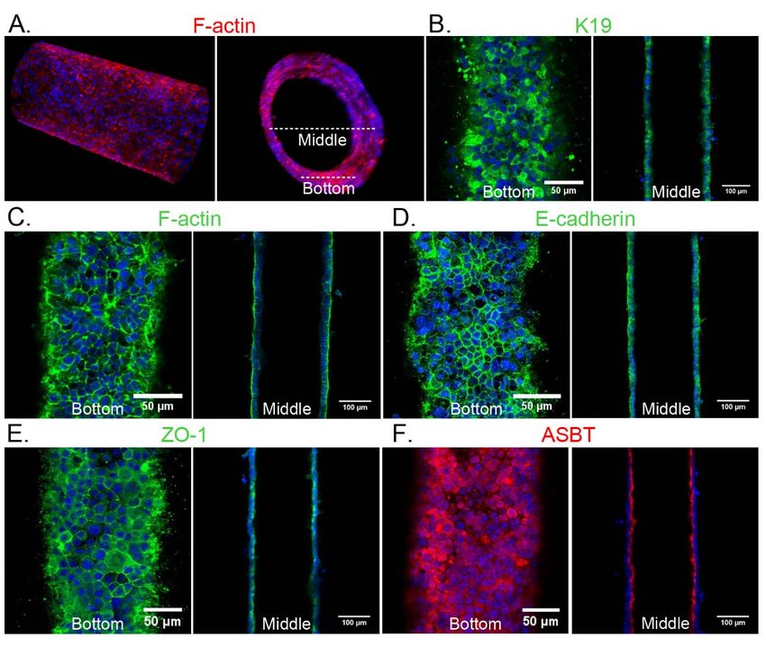
bioRxiv preprint first posted online Mar. 30, 2019; doi: http://dx.doi.org/10.1101/594291. The copyright holder for this preprint
 (which was not peer-reviewed) is the author/funder, who has granted bioRxiv a license to display the preprint in perpetuity.
                                    All rights reserved. No reuse allowed without permission.

                                                                                                                                    2

   ADDRESS CORRESPONDENCE AND REPRINT REQUESTS TO:

   Rebecca G. Wells
   Department of Medicine (GI)
   Perelman School of Medicine, University of Pennsylvania
   905 BRB II/III
   421 Curie Blvd.
   Philadelphia, PA 19104-6140
   Email: rgwells@pennmedicine.upenn.edu
   Tel: +1-215-573-1860

   Abbreviations: PBC, primary biliary cholangitis; PSC, primary sclerosing cholangitis;
   ECM, extracellular matrix; EHBD, extrahepatic bile duct; DMEM, Dulbecco’s Modified
   Eagle Medium; PDMS, polydimethylsiloxane; FITC, fluorescein isothiocyanate; PBS,
   phosphate buffered saline; Pd, permeability coefficient; PFA, paraformaldehyde; BSA,
   bovine serum albumin; DAPI, 4 ,6-diamidino-2-phenylindole; SNA, Sambucus nigra
                                                        ′

   lectin; SBA, soybean agglutinin; ASBT, apical sodium-dependent bile salt transporter ;
   GCDC, glycochenodeoxycholic acid.

   Grant support: This work was supported by grant R56 DK119290 from the National
   Institutes of Diabetes and Digestive and Kidney Diseases (to RGW), EB08396 from the
   National Institute of Biomedical Imaging and Bioengineering, the Fred and Suzanne
   Biesecker Foundation for Pediatric Liver Diseases at the Children’s Hospital of
   Philadelphia, and the Center for Engineering MechanoBiology (CEMB), an NSF Science
   and Technology Center, under grant agreement CMMI: 15-48571. WJP acknowledges
   support from a Ruth L. Kirchstein National Research Service Award (F32 HL129733)
   and from the NIH through the Organ Design and Engineering Training program (T32
   EB16652).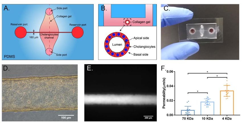
bioRxiv preprint first posted online Mar. 30, 2019; doi: http://dx.doi.org/10.1101/594291. The copyright holder for this preprint
 (which was not peer-reviewed) is the author/funder, who has granted bioRxiv a license to display the preprint in perpetuity.
                                    All rights reserved. No reuse allowed without permission.

                                                                                                                                    3

   Abstract
   Chronic cholestatic liver diseases such as primary biliary cholangitis (PBC) and primary

   sclerosing cholangitis (PSC) are frequently associated with damage to the barrier

   function of the biliary epithelium, but barrier function is difficult to study in vivo and has

   not been recapitulated in vitro. Here we report the development of a bile duct-on-a-chip

   that phenocopies not only the tubular architecture of the bile duct in three dimensions,

   but also its barrier functions. We demonstrated that mouse cholangiocytes in the

   channel of the device became polarized and formed mature tight junctions, and that the

   permeability of the cholangiocyte monolayer was comparable to that measured ex vivo

   for the rat bile duct. Permeability decreased significantly when cells formed a

   compacted monolayer with cell densities comparable to that seen in vivo. This device

   enabled independent access to the apical and basolateral surfaces of the cholangiocyte

   channel, allowing proof-of-concept toxicity studies with the biliary toxin biliatresone and

   the bile acid glycochenodeoxycholic acid. The cholangiocyte basolateral side was more

   vulnerable than the apical side to treatment with either agent, suggesting a protective

   adaptation of the apical surface that is normally exposed to bile. Further studies

   revealed a protective role of the cholangiocyte apical glycocalyx, wherein disruption of

   the glycocalyx with neuraminidase increased the permeability of the cholangiocyte

   monolayer after treatment with glycochenodeoxycholic acid. Conclusion: This bile duct-

   on-a-chip captured essential features of a simplified bile duct in structure and organ-

   level functions and represents a novel in vitro platform to study the pathophysiology of

   the bile duct using cholangiocytes from a variety of sources.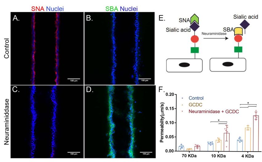
bioRxiv preprint first posted online Mar. 30, 2019; doi: http://dx.doi.org/10.1101/594291. The copyright holder for this preprint
 (which was not peer-reviewed) is the author/funder, who has granted bioRxiv a license to display the preprint in perpetuity.
                                    All rights reserved. No reuse allowed without permission.

                                                                                                                                    4

   Introduction
   Chronic cholestatic liver diseases such as primary biliary cholangitis (PBC) and primary

   sclerosing cholangitis (PSC) are often associated with alterations in the tight junctions of

   bile duct epithelial cells (cholangiocytes). Data from mouse models similarly suggest

   that impaired tight junction integrity plays an important role in the pathogenesis of

   cholangiopathies(1-3). Most in vitro research on biliary physiology and pathology,

   however, employs cells either cultured in 2D monolayers or as organoids in 3D

   extracellular matrix (ECM). These conventional methods fail to replicate many key

   aspects of bile duct structural organization or to recapitulate important tissue-level

   integrated physiological functions such as forming a protective barrier and

   compartmentalizing bile.

   Microfluidic organs-on-chips can overcome some of these limitations(4). Soft lithography,

   which is used to design organs-on-chips, allows control of surface features over a range

   of physiologically-relevant scales. Organs-on-chips can mimic aspects of the physiology

   of cell-cell and cell-ECM junctions in tissues including the alveolar-capillary interface(5),

   the blood-brain barrier(6-8), and liver sinusoids, in a controllable way, allowing both high-

   resolution imaging and biochemical and metabolic analyses in real time. The technology

   has great potential to advance the study of tissue development, physiology and

   pathophysiology.

   To capture the structure and environment of the bile ducts, we used organ-on-chip

   technology to develop a microengineered bile duct with controllable architecture and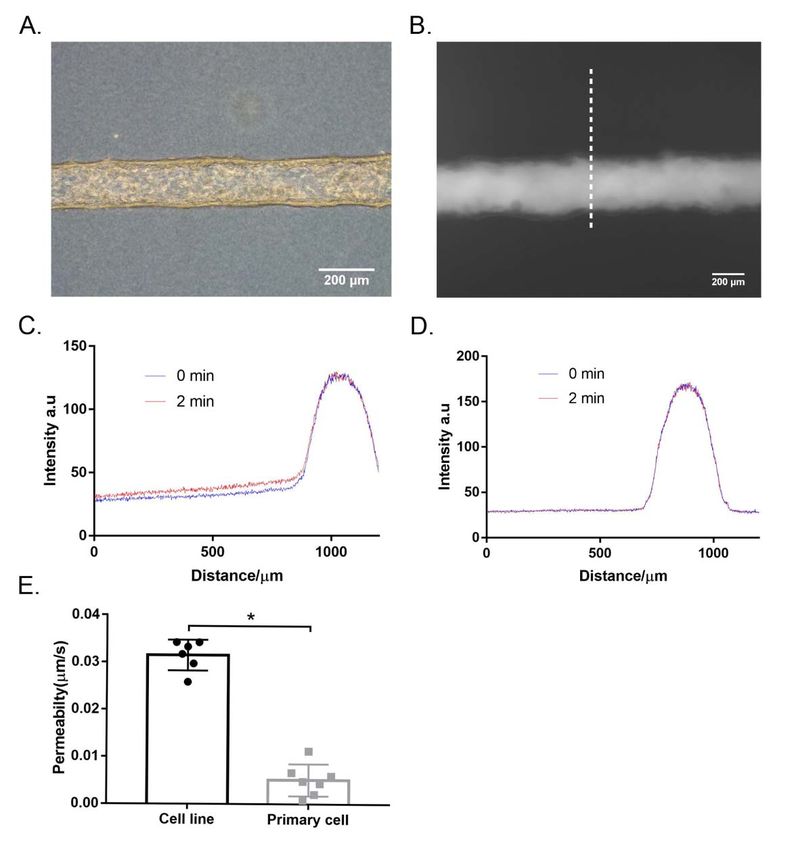
bioRxiv preprint first posted online Mar. 30, 2019; doi: http://dx.doi.org/10.1101/594291. The copyright holder for this preprint
 (which was not peer-reviewed) is the author/funder, who has granted bioRxiv a license to display the preprint in perpetuity.
                                    All rights reserved. No reuse allowed without permission.

                                                                                                                                     5

   surrounding matrix. We demonstrate that a self-organized cholangiocyte-lined channel

   faithfully recapitulates key functions of the bile duct and enables us to study the barrier

   function of cholangiocytes monolayer quantitatively and independently from either the

   apical or basolateral side. As a proof-of-concept, we use this model to demonstrate the

   protective role of the cholangiocyte apical glycocalyx.

   Materials and Methods

   CELL ISOLATION AND CULTURE

   The small cholangiocyte cell line was originally isolated from normal mice (BALB/c) and

   immortalized by transfection with the SV40 large-T antigen(9). Primary cholangiocytes

   were isolated from extrahepatic bile ducts (EHBD) of normal adult mice (BALB/c) as

   described previously(10). Cells were cultured in low glucose Dulbecco’s Modified Eagle

   Medium (DMEM; Thermo Fisher Scientific, Waltham, MA) supplemented as previously

   reported(9-11).

   FABRICATION OF THE BILE DUCT-ON-A-CHIP

   Microfluidic          devices         were        fabricated          using       soft      lithography           as      described

   previously(12,       13)
                              (Fig. 1A). Polydimethylsiloxane (PDMS, Sylgard 184, Dow-Corning,

   Midland, MI) devices were treated with 0.01% (v/v) poly-L-lysine for 1 h and 0.5% (v/v)

   glutaraldehyde for 20 min to promote collagen adhesion. After the devices were

   washed overnight in water and for 30 min in 70% ethanol, steel acupuncture needles

   (160     μ m diameter, Seirin, Kyoto, Japan) were inserted and the devices were then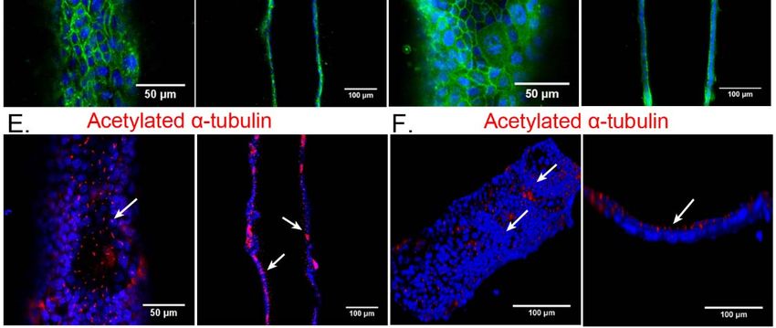
bioRxiv preprint first posted online Mar. 30, 2019; doi: http://dx.doi.org/10.1101/594291. The copyright holder for this preprint
 (which was not peer-reviewed) is the author/funder, who has granted bioRxiv a license to display the preprint in perpetuity.
                                    All rights reserved. No reuse allowed without permission.

                                                                                                                                          6

   sterilized under UV for 20 min. A solution of 2.5 mg/ml rat tail type 1 collagen (Thermo

   Fisher Scientific), 1X DMEM medium, 10 mM HEPES, 0.1 M NaOH and NaHCO3 (0.035%

   w/v) was infused via the side ports and allowed to polymerize for 20 min at 37°C.

   Needles were removed to form channels, which were then coated with 100                                                            g/ml
                                                                                                                                     μ

   laminin overnight (via the large reservoir ports) at 37°C. A                                           suspension            of       0.5

   million/ml cholangiocytes was introduced into the reservoir ports. Cells were allowed

   to adhere to the top surface of the channel for 2 min; devices were then flipped to allow

   cells to adhere to the bottom surface of the channel for 5 min. Non-adherent cells were

   removed by rinsing with cell culture medium and the devices were filled with fresh

   medium. Devices were maintained at 37°C (5% CO2) for 7 d on a rocker at 5 rpm with

   daily medium changes until the development of compact monolayers.

   PERMEABILITY MEASUREMENTS

   To measure the permeability of the cholangiocyte monolayers in the device, fluorescent

   dextran (70 kDa, 10 kDa and 4 kDa, labeled with fluorescein isothiocyanate (FITC)

   (Sigma, St. Louis, MO)) in phosphate buffered saline (PBS) was introduced into the

   devices at a concentration of 20 g/ml. Diffusion of the dextran was imaged in real time
                                                      μ

   with an          EVOS FL Auto 2 Imaging System (Thermo Fisher Scientific) at 10X

   magnification. The diffusive permeability coefficient (Pd) was calculated by measuring

   the flux of dextran into the collagen gel and fitting the resulting diffusion profiles to a

   dynamic mass conservation equation, as described previously(12, 14).

   IMMUNOSTAINING AND LECTIN STAINING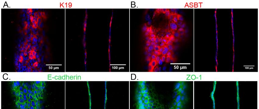
bioRxiv preprint first posted online Mar. 30, 2019; doi: http://dx.doi.org/10.1101/594291. The copyright holder for this preprint
 (which was not peer-reviewed) is the author/funder, who has granted bioRxiv a license to display the preprint in perpetuity.
                                    All rights reserved. No reuse allowed without permission.

                                                                                                                                    7

   Cholangiocyte monolayers in the device were fixed with 4% Paraformaldehyde (PFA) at

   37oC for 20 min with rocking. Cells were rinsed 3X with PBS and permeabilized with 0.1%

   Triton X-100 for 3 d, then blocked with 2% bovine serum albumin (BSA) (Sigma) in PBS

   at 4oC overnight with rocking. Primary antibodies together with 4 ,6-diamidino-2-                                ′

   phenylindole (DAPI; Thermo Fisher Scientific) diluted in 2% BSA in PBS were incubated

   overnight at 4oC, and rinsed 3X with PBS for 5 min each with rocking, followed by an

   overnight rinse. Secondary antibodies were diluted in 2% BSA in PBS and incubated

   overnight at 4oC, and rinsed 3X with PBS for 5 min each on a rocker, followed by an

   overnight rinse. Primary antibodies and the concentrations used are listed in

   Supplemental Table S1. Cy3- and Cy5-conjugated secondary antibodies were used at

   1:400 (Vector Laboratories, Burlington, CA).

   Extrahepatic bile ducts were frozen in O.C.T. (Tissue Tek, VWR, Bridgeport, NJ). 5 µm

   thick sections were brought to room temperature and fixed in 10% neutral buffered

   formalin for 4 min. Tissue was blocked with StartingBlock™ T20/PBS Blocking Buffer

   (Thermo Scientific) before being incubated overnight with K19 primary antibody (1:100,

   Troma III, DSHB, Iowa city, Iowa) in staining buffer (0.2% Triton X-100, 0.1% bovine

   serum albumin, in PBS) at 4°C. Cy™2 AffiniPure Donkey Anti-Rat IgG (H+L) (1:500,

   Jackson ImmunoResearch Lab, West Grove, PA) was then used as the secondary

   antibody and incubated for 1 hr at RT.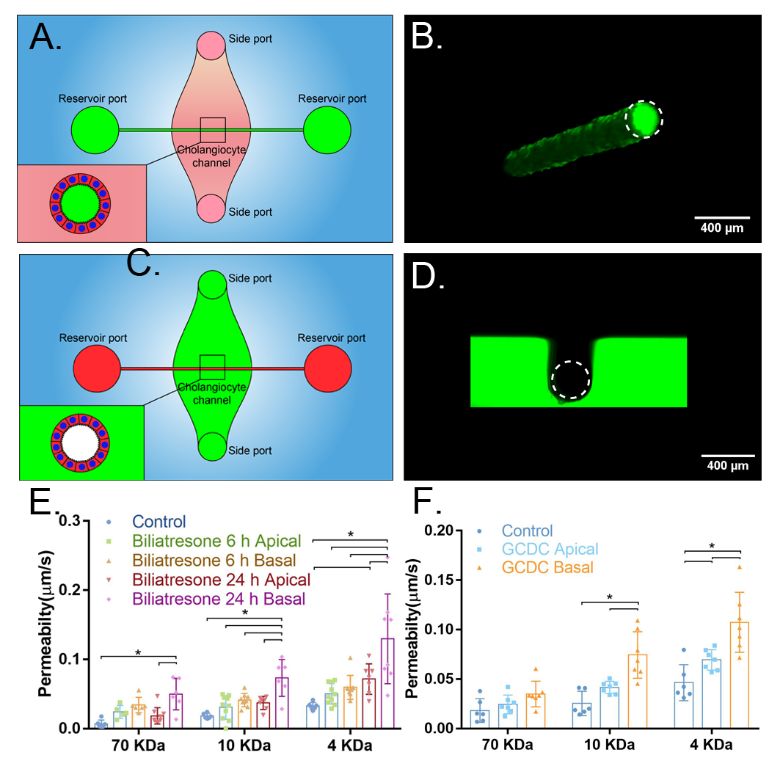
bioRxiv preprint first posted online Mar. 30, 2019; doi: http://dx.doi.org/10.1101/594291. The copyright holder for this preprint
 (which was not peer-reviewed) is the author/funder, who has granted bioRxiv a license to display the preprint in perpetuity.
                                    All rights reserved. No reuse allowed without permission.

                                                                                                                                      8

   For      Sambucus            nigra      (SNA)         and      soybean          agglutinin         (SBA)        lectin      staining,

   cholangiocytes were fixed with 4% PFA at 37oC for 20 min with rocking. Cells were

   rinsed with PBS for 1 h, then blocked with 1X Carbo-Free Blocking Solution (Vector

   Laboratories) for 1 h at RT on a rocker. After 3X 15 min PBS rinses, cells were

   incubated with SBA-FITC or SNA-Cy5 (Vector Laboratories) at 20 g/ml for 1 h at RT                         μ

   with rocking. Cells were rinsed with PBS for 1 h, then the channel was filled with

   mounting medium containing DAPI (Vector Laboratories). Images were acquired using a

   SCTR Leica confocal microscope and Leica application suite (LAS X) (Leica, Buffalo

   Grove, IL).

   STATISTICAL ANALYSIS

   Statistical significance was assessed using one-way ANOVA. p
bioRxiv preprint first posted online Mar. 30, 2019; doi: http://dx.doi.org/10.1101/594291. The copyright holder for this preprint
(which was not peer-reviewed) is the author/funder, who has granted bioRxiv a license to display the preprint in perpetuity.
All rights reserved. No reuse allowed without permission.
9
Results
FABRICATION OF THE BILE DUCT-ON-A-CHIP
In order to study the function of the apical and basal sides of cholangiocytes
independently, we developed a microfluidic device consisting of a PDMS support with a
160 μm cholangiocyte-lined channel through a matrix bulk; apical and basal sides of the
channel were accessible through separate ports (Figure 1A, B). The device was
designed as per previously-described models of endothelium-lined, vascular-mimetic
channels(12, 13)
. To generate a bile duct-on-a-chip, the channel was formed within a
collagen plug and the lumen was coated with laminin (100 μ g/ml) for 4 hours. The
channels were seeded with a line of mouse cholangiocytes. Cholangiocytes in the
channel formed a confluent and then compact epithelial monolayer (nearly cubical cells
with similar height and width) (Figure 1C, D).
To determine whether the bile duct-on-a-chip recapitulated the barrier function of the
bile duct, we perfused the lumen with FITC-dextran ranging in size from 4 to 70 kDa.
There was no obvious leakage of fluorescent dextran into the collagen matrix even after
10 minutes (Figure 1E). Quantification of permeability showed an increase as the
molecular weight of the dextran decreased (Figure 1F). The measured permeability of
cholangiocyte cell line was comparable with that measured in rat ex vivo systems using
insulin (5.8 kDa; 0.45 μ m/s)(15). This model of the bile duct thus mimics the barrier
function of ducts in vivo.bioRxiv preprint first posted online Mar. 30, 2019; doi: http://dx.doi.org/10.1101/594291. The copyright holder for this preprint
(which was not peer-reviewed) is the author/funder, who has granted bioRxiv a license to display the preprint in perpetuity.
All rights reserved. No reuse allowed without permission.
10
CHARACTERIZATION OF THE BILE DUCT-ON-A-CHIP
We used immunofluorescence staining to characterize the cholangiocytes in the
channel. F-actin staining showed that cholangiocytes grew into a compact monolayer,
forming a cylindrical tube with two open ends connected to the large reservoirs (Figure
2A, C). Cells maintained expression of the cholangiocyte marker K19 (Figure 2B), and
demonstrated staining for ZO-1 (TJP-1) and E-cadherin 1 (CDH1, also known as
cadherin 1) at cell-cell junctions (Figure 2D, E), confirming the formation of tight
junctions. Staining for the apical sodium-dependent bile acid transporter (ASBT)
confirmed that cells in the bile duct-on-a-chip were polarized (Figure 2F).
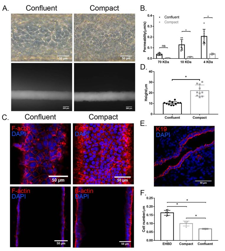
A SUPERCONFLUENT MONOLAYER IS REQUIRED FOR OPTIMAL BARRIER
FUNCTION
Full barrier function required that cholangiocytes were not just confluent but
“superconfluent” and compact (Figure 3A); as the cholangiocyte monolayer grew more
dense, permeability decreased significantly (Figure 3B). By F-actin staining, we
confirmed that the permeability difference between what we term a confluent versus a
compact monolayer was not the result of gaps in the monolayer (Figure 3C). Cell height
increased more than two-fold in the compact compared to the confluent monolayer
(Figure 3D). To determine the cell density of cholangiocyte monolayers in vivo, we
counted cell number as a function of monolayer length in adult mouse EHBD and in
confluent and compact devices (Figure 3E). We found that compact monolayers were
more dense than confluent ones, and that mice EHBD (Figure 3E, F) had an evenbioRxiv preprint first posted online Mar. 30, 2019; doi: http://dx.doi.org/10.1101/594291. The copyright holder for this preprint
(which was not peer-reviewed) is the author/funder, who has granted bioRxiv a license to display the preprint in perpetuity.
All rights reserved. No reuse allowed without permission.
11
higher cell density. These results suggested that the biliary epithelium forms a dense,
compact monolayer – beyond confluence – in order to form a mature barrier.
THE APICAL AND BASAL SURFACES OF THE MONOLAYER CAN BE ACCESSED
INDEPENDENTLY
In order to determine whether the apical and basal surfaces of cholangiocytes in the
device could be accessed independently, we applied FITC-dextran through either the
large reservoir ports, which are directly connected to the lumen, or the small side ports,
which connect to the basal surface of the monolayer through the collagen bulk. We
found that FITC-dextran added through the reservoir ports remained within the lumen
(Figure 4A, B), making contact only with the apical side of the cholangiocytes in the
device and unable to penetrate through the monolayer. Dextran applied via the side
ports remained within the collagen plug, in contact with only the basal side of the
monolayer (Figure 4C, D).
Biliatresone is a plant isoflavonoid that is toxic to cholangiocytes and causes a
syndrome mimicking the pediatric disease biliary atresia in neonatal livestock and larval
zebrafish(16). We previously demonstrated that biliatresone applied to the basal surface
of a cholangiocyte spheroid causes mislocalization of the apical markers ZO-1 and E-
cadherin and increases cholangiocyte monolayer permeability(11). A limitation of these
spheroid studies was that biliatresone could only be added to the basal surface. As a
demonstration of the potential use of the bile duct-on-a-chip device, we showed that
biliatresone treatment caused cholangiocyte damage, as evidenced by increasedbioRxiv preprint first posted online Mar. 30, 2019; doi: http://dx.doi.org/10.1101/594291. The copyright holder for this preprint
(which was not peer-reviewed) is the author/funder, who has granted bioRxiv a license to display the preprint in perpetuity.
All rights reserved. No reuse allowed without permission.
12
permeability of monolayers in the device, and that this damage was worse with basal as
opposed to apical administration (Figure 4E).
Cholangiocytes are adapted to tolerate exposure to toxic bile at their apical surfaces
though it has been difficult to study in cell culture. Impaired duct barrier function may
lead to bile leakage through the monolayer, and cause damage from the basolateral
(17-19)
side, which is less tolerant of bile . As a second illustration of the potential use of
the bile duct-on-a-chip device, we exposed cholangiocytes to 1 mM of the bile acid
glycochenodeoxycholic acid (GCDC) from either the apical or basal side. GCDC
treatment caused significantly more damage, as defined by increased permeability,
when applied to the basal side rather than the apical side (Figure 4F), suggesting that
once bile leaks through the epithelial monolayer, it can cause additional damage
through the basolateral side of the barrier, leading to a feedback loop of further damage
and increased leakage.
THE GLYCOCALYX PROTECTS CHOLANGIOCYTES FROM BILE ACID-INDUCED
DAMAGE
Cholangiocytes have an apical glycocalyx that protects them from bile acid toxicity(20, 21).
We stained the apical surfaces of the cholangiocyte monolayer in the bile duct-on-a-chip
using the lectin Sambucus nigra agglutinin (SNA), which binds to α 2-6-bound (terminal)
sialic acid residues, and demonstrated that the sialic acid-enriched glycocalyx was
maintained in cholangiocytes in the device and was appropriately localized to the apical
surface (Figure 5A). We also stained cells in the channel with the lectin soybeanbioRxiv preprint first posted online Mar. 30, 2019; doi: http://dx.doi.org/10.1101/594291. The copyright holder for this preprint
(which was not peer-reviewed) is the author/funder, who has granted bioRxiv a license to display the preprint in perpetuity.
All rights reserved. No reuse allowed without permission.
13
agglutinin (SBA), which only detects carbohydrates lacking terminal sialic acid residues
(Figure 5E), confirming a lack of cholangiocyte staining at baseline (Figure 5B). We then
treated cells with neuraminidase, applied apically, to remove the terminal sialic acid.
Post-treatment staining with SNA was minimal, while there was new staining with SBA,
confirming the presence of apical sialic acid residues in untreated cells and the efficacy
of neuraminidase in this system (Figure 5C, D). To test the role of the glycocalyx in
protecting cholangiocytes from bile acids, we perfused the device lumen with GCDC (1
mM) before or after treatment with neuraminidase, and then measured monolayer
permeability. We found no effect of GCDC, with or without neuraminidase treatment, on
permeability to 70 kDa FITC-dextran; however, GCDC alone increased the permeability
to 4 kDa FITC-dextran, and GCDC exposure of neuraminidase-treated cholangiocytes
increased permeability to both 4 kDa and 10 kDa FITC-dextran (Figure 5F). Taken
together, these findings demonstrate that the cholangiocyte-lined channel is resistant to
bile acid toxicity and that the glycocalyx plays a protective role.
CONSTRUCTION OF A BILE DUCT-ON-A-CHIP WITH PRIMARY MURINE
EXTRAHEPATIC CHOLANGIOCYTES
To demonstrate that the bile duct-on-a-chip can be used with cells from various sources,
we also used primary murine extrahepatic cholangiocytes (Figure S1A). Primary
cholangiocytes, like the cell line, formed a compact tubular monolayer. Cells maintained
expression of the cholangiocyte marker K19 (Figure 6A), and demonstrated staining for
E-cadherin and ZO-1 at cell-cell junctions (Figure 6C, D), confirming the formation of
tight junctions. Staining for ASBT (Figure 6B) and acetylated -tubulin (Figure 6E, F) αbioRxiv preprint first posted online Mar. 30, 2019; doi: http://dx.doi.org/10.1101/594291. The copyright holder for this preprint
(which was not peer-reviewed) is the author/funder, who has granted bioRxiv a license to display the preprint in perpetuity.
All rights reserved. No reuse allowed without permission.
14
confirmed that primary cholangiocytes in the bile duct-on-a-chip were ciliated and highly
polarized. In permeability assays, there was no visible leakage of 4kDa FITC-dextran
from the lumen even after 10 mins (Figure S1B, Movie S1). Analysis of the intensity
change along a line perpendicular to the channel showed almost no increase in intensity
across the gel after 2 mins (Figure S1D), in contrast to the cholangiocyte cell line
(Figure S1C) and the permeability to 4 kDa FITC-dextran of primary cholangiocyte was
about only 16% of that for the cholangiocyte cell line (Figure S1E). Thus, the primary
cell-lined device demonstrated even better barrier function than the device lined with the
cholangiocyte cell line (Figure S1E).
Discussion
We have developed an in vitro model of the bile duct by employing organ-on-chip
technology, recapitulating not only the three-dimensional architecture of the bile duct but
also its barrier function. We demonstrated that cholangiocytes are polarized in the
device and that permeability is comparable to in vivo values for the bile duct, requires a
dense monolayer, and can be quantified. We also showed that the device enables
apical vs. basolateral treatments and that cells respond differently depending on the
side of exposure. As a demonstration of the potential use of the device in studying
biliary physiology, we showed that the apical glycocalyx plays a protective role against
bile acid toxicity and that the biliary toxin biliatresone has greater toxicity when applied
to the basal surface. The bile duct-on-a-chip thus represents a novel in vitro model forbioRxiv preprint first posted online Mar. 30, 2019; doi: http://dx.doi.org/10.1101/594291. The copyright holder for this preprint
(which was not peer-reviewed) is the author/funder, who has granted bioRxiv a license to display the preprint in perpetuity.
All rights reserved. No reuse allowed without permission.
15
studying cholangiopathies. The in vivo-like permeability barrier and the potential to
quantify changes in barrier function particular strengths of this model.
Most in vitro studies of cholangiopathies have relied on 2D cell culture or on 3D
organoid culture. 2D culture, although convenient, fails to replicate bile duct physiology
with respect to duct structure, extracellular matrix composition, and stiffness, and does
not support fluid flow. Polarized cell organization and permeability measurements,
although possible, require specialized culture systems. Cholangiocytes in 3D spheroid
culture differentiate well, can be cultured in physiological matrices, and demonstrate
barrier functions and transport activities. However, spheroids are highly variable in
shape and size, and it is difficult to control their positions in a gel (for microscopy),
sample lumen content, or treat the cholangiocyte apical surface. The bile duct-on-a-chip
provides uniform ducts that are accessible for imaging, with two sets of ports that
enable sampling of luminal contents as well as selective (apical vs. basolateral)
exposures. Additionally, the chemical composition and mechanics of the surrounding
matrix as well as the fluid flow rate can be varied.
Several different bioengineered bile ducts, including acellular constructs using
biodegradable materials and constructs using human cholangiocytes, have been
reported. Sampaziotis et al. reconstructed the biliary epithelium with a bioengineered
bile duct consisting of extrahepatic cholangiocytes derived from the common bile duct or
the gallbladder. These results suggested that bioengineered bile ducts may have
promise for transplantation applications, but the difficulty in manipulating this ductbioRxiv preprint first posted online Mar. 30, 2019; doi: http://dx.doi.org/10.1101/594291. The copyright holder for this preprint
(which was not peer-reviewed) is the author/funder, who has granted bioRxiv a license to display the preprint in perpetuity.
All rights reserved. No reuse allowed without permission.
16
construct makes it unsuitable for in vitro study. Chen et al. constructed a bile duct using
organoid-derived cholangiocyte-like cells on a collagen-coated polyethersulfone hollow
fiber membrane, yielding a tubular structure with polarized bile acid transport activity.
However, the cholangiocytes cultured on the fiber have outward-facing apical surfaces,
in contrast to ducts in vivo and the bile duct-on-a-chip.
Previous studies have suggested that epithelial cell confluence is associated with
morphology, polarity and barrier function(22-25). Although changes in transepithelial
electronic resistance have been reported to be minimal after cells reach confluence(24),
we found that permeability decreased significantly when cells progressed from confluent
to compact, with associated changes in height, suggesting changes in tight junction
molecules(25). These results demonstrate that compactness is required for an optimal
barrier function, and suggest that confluence may be an intermediate rather than
ultimate goal in the repair of an injured epithelial layer.
We demonstrated that this device can be constructed with primary murine extrahepatic
cholangiocytes and that in the device these cells stably expressed biliary markers,
junctional molecules and bile salt transport proteins and also developed cilia on their
apical surfaces, facing the lumen. The bile duct-on-a-chip thus offers the opportunity to
carry out experiments using genetically-modified cholangiocytes and to compare intra-
vs. extra-hepatic cholangiocytes. Human cholangiocytes, including those derived from
induced pluripotent stem cells, could also be studied using this device.bioRxiv preprint first posted online Mar. 30, 2019; doi: http://dx.doi.org/10.1101/594291. The copyright holder for this preprint
(which was not peer-reviewed) is the author/funder, who has granted bioRxiv a license to display the preprint in perpetuity.
All rights reserved. No reuse allowed without permission.
17
In conclusion, our bile duct-on-a-chip mimics the basic features of the bile duct, making
it a novel platform for in vitro studies of biliary physiopathology.
ACKNOWLEDGEMENTS:
We are grateful to the UPenn Cell and Developmental Biology Microscopy Core, the
Singh Center for Nanotechnology, and the UPenn NIDDK Center for Molecular Studies
in Digestive and Liver Disease (NIH-P30-DK050306). The small cholangiocyte cell line
was generously provided by Gianfranco Alpini (Texas A&M Health Science Center
College of Medicine and Baylor Scott & White Digestive Disease Research Center).
REFERENCES:
1. Matsumoto K, Imasato M, Yamazaki Y, Tanaka H, Watanabe M, Eguchi H, Nagano H, et
al. Claudin 2 deficiency reduces bile flow and increases susceptibility to cholesterol gallstone
disease in mice. Gastroenterology 2014;147:1134-1145.e1110.
2. Herr KJ, Tsang YH, Ong JW, Li Q, Yap LL, Yu W, Yin H, et al. Loss of alpha-catenin
elicits a cholestatic response and impairs liver regeneration. Sci Rep 2014;4:6835.
3. Nakagawa H, Hikiba Y, Hirata Y, Font-Burgada J, Sakamoto K, Hayakawa Y, Taniguchi
K, et al. Loss of liver E-cadherin induces sclerosing cholangitis and promotes carcinogenesis.
Proc Natl Acad Sci U S A 2014;111:1090-1095.
4. Bhatia SN, Ingber DE. Microfluidic organs-on-chips. Nat Biotechnol 2014;32:760-772.
5. Huh D, Matthews BD, Mammoto A, Montoya-Zavala M, Hsin HY, Ingber DE.
Reconstituting organ-level lung functions on a chip. Science 2010;328:1662-1668.
6. Griep LM, Wolbers F, de Wagenaar B, ter Braak PM, Weksler BB, Romero IA, Couraud
PO, et al. BBB on chip: microfluidic platform to mechanically and biochemically modulate blood-
brain barrier function. Biomed Microdevices 2013;15:145-150.
7. Brown JA, Codreanu SG, Shi M, Sherrod SD, Markov DA, Neely MD, Britt CM, et al.
Metabolic consequences of inflammatory disruption of the blood-brain barrier in an organ-on-
chip model of the human neurovascular unit. J Neuroinflammation 2016;13:306.
8. Booth R, Kim H. Characterization of a microfluidic in vitro model of the blood-brain
barrier (muBBB). Lab Chip 2012;12:1784-1792.
9. Ueno Y, Alpini G, Yahagi K, Kanno N, Moritoki Y, Fukushima K, Glaser S, et al.
Evaluation of differential gene expression by microarray analysis in small and large
cholangiocytes isolated from normal mice. Liver Int 2003;23:449-459.
10. Karjoo S, Wells RG. Isolation of neonatal extrahepatic cholangiocytes. J Vis Exp 2014.
11. Waisbourd-Zinman O, Koh H, Tsai S, Lavrut PM, Dang C, Zhao X, Pack M, et al. The
toxin biliatresone causes mouse extrahepatic cholangiocyte damage and fibrosis through
decreased glutathione and SOX17. Hepatology 2016;64:880-893.bioRxiv preprint first posted online Mar. 30, 2019; doi: http://dx.doi.org/10.1101/594291. The copyright holder for this preprint
(which was not peer-reviewed) is the author/funder, who has granted bioRxiv a license to display the preprint in perpetuity.
All rights reserved. No reuse allowed without permission.
18
12. Polacheck WJ, Kutys ML, Yang J, Eyckmans J, Wu Y, Vasavada H, Hirschi KK, et al. A
non-canonical Notch complex regulates adherens junctions and vascular barrier function.
Nature 2017;552:258-262.
13. Nguyen DH, Stapleton SC, Yang MT, Cha SS, Choi CK, Galie PA, Chen CS. Biomimetic
model to reconstitute angiogenic sprouting morphogenesis in vitro. Proc Natl Acad Sci U S A
2013;110:6712-6717.
14. Adamson RH, Lenz JF, Curry FE. Quantitative laser scanning confocal microscopy on
single capillaries: permeability measurement. Microcirculation 1994;1:251-265.
15. Smith ND, Boyer JL. Permeability characteristics of bile duct in the rat. Am J Physiol
1982;242:G52-57.
16. Lorent K, Gong W, Koo KA, Waisbourd-Zinman O, Karjoo S, Zhao X, Sealy I, et al.
Identification of a plant isoflavonoid that causes biliary atresia. Sci Transl Med 2015;7:286ra267.
17. Benedetti A, Alvaro D, Bassotti C, Gigliozzi A, Ferretti G, La Rosa T, Di Sario A, et al.
Cytotoxicity of bile salts against biliary epithelium: a study in isolated bile ductule fragments and
isolated perfused rat liver. Hepatology 1997;26:9-21.
18. Xia X, Francis H, Glaser S, Alpini G, LeSage G. Bile acid interactions with
cholangiocytes. World J Gastroenterol 2006;12:3553-3563.
19. Hopkins AM, Walsh SV, Verkade P, Boquet P, Nusrat A. Constitutive activation of Rho
proteins by CNF-1 influences tight junction structure and epithelial barrier function. J Cell Sci
2003;116:725-742.
20. Hohenester S, Wenniger LM, Paulusma CC, van Vliet SJ, Jefferson DM, Elferink RP,
Beuers U. A biliary HCO3- umbrella constitutes a protective mechanism against bile acid-
induced injury in human cholangiocytes. Hepatology 2012;55:173-183.
21. Maillette de Buy Wenniger LJ, Hohenester S, Maroni L, van Vliet SJ, Oude Elferink RP,
Beuers U. The Cholangiocyte Glycocalyx Stabilizes the 'Biliary HCO3 Umbrella': An Integrated
Line of Defense against Toxic Bile Acids. Dig Dis 2015;33:397-407.
22. Pepperkok R, Bre MH, Davoust J, Kreis TE. Microtubules are stabilized in confluent
epithelial cells but not in fibroblasts. J Cell Biol 1990;111:3003-3012.
23. Ishibe S, Haydu JE, Togawa A, Marlier A, Cantley LG. Cell confluence regulates
hepatocyte growth factor-stimulated cell morphogenesis in a beta-catenin-dependent manner.
Mol Cell Biol 2006;26:9232-9243.
24. Hidalgo IJ, Raub TJ, Borchardt RT. Characterization of the human colon carcinoma cell
line (Caco-2) as a model system for intestinal epithelial permeability. Gastroenterology
1989;96:736-749.
25. Amoozadeh Y, Anwer S, Dan Q, Venugopal S, Shi Y, Branchard E, Liedtke E, et al. Cell
confluence regulates claudin-2 expression: possible role for ZO-1 and Rac. American Journal of
Physiology-Cell Physiology 2018;314:C366-C378.bioRxiv preprint first posted online Mar. 30, 2019; doi: http://dx.doi.org/10.1101/594291. The copyright holder for this preprint
(which was not peer-reviewed) is the author/funder, who has granted bioRxiv a license to display the preprint in perpetuity.
All rights reserved. No reuse allowed without permission.
19
Figures and Legends
Figure 1. Fabrication and characterization of a three dimensional biomimetic bile duct-
on-a-chip. A. Schematic of top view of the bile duct-on-a-chip. B. Schematic of the
device in cross section. C. Image of an actual bile duct-on-a-chip, top view. D.
Representative bright field image of the channel lined by a layer of mouse
cholangiocytes (cell line). Scale bar, 100 μm. E. Representative image of FITC-dextran
(70 kDa) in the channel, imaged after 10 min. Scale bar, 200 μm. F. Permeability of the
cholangiocyte (cell line)-lined channel to FITC-dextran (70 kDa, 10 kDa and 4 kDa),
n=14 devices, each device tested sequentially with FITC-dextran from 70 to 4 kDa. All
data are presented as mean ± SD, *PbioRxiv preprint first posted online Mar. 30, 2019; doi: http://dx.doi.org/10.1101/594291. The copyright holder for this preprint
(which was not peer-reviewed) is the author/funder, who has granted bioRxiv a license to display the preprint in perpetuity.
All rights reserved. No reuse allowed without permission.
20
Figure 2. Characterization of a channel lined with mouse cholangiocytes (cell line). A.
Representative confocal images of cholangiocytes in the bile duct-on-a-chip forming a
monolayer within the cylindrical channel, stained for F-actin (red) and nuclei (DAPI;
blue). Longitudinal (left panel) and cross-sectional (right panel) views. White dashed
lines indicate the bottom surface and cross section through the center shown in the
remaining panels. B-F. Immunofluorescent images across the bottom (left panels in B-F)
and middle (right panels in B-F) of the channel stained with antibodies against (B) K19,
(C) F-actin, (D) E-cadherin, E) ZO-1, and (F) the apical sodium bile salt transporter
(ASBT). Nuclei shown by DAPI staining (blue). Images are representative of at least
independently constructed devices for each condition. Scale bars: 50 m, left panels; μ
100 m right panels.
μbioRxiv preprint first posted online Mar. 30, 2019; doi: http://dx.doi.org/10.1101/594291. The copyright holder for this preprint
(which was not peer-reviewed) is the author/funder, who has granted bioRxiv a license to display the preprint in perpetuity.
All rights reserved. No reuse allowed without permission.
21bioRxiv preprint first posted online Mar. 30, 2019; doi: http://dx.doi.org/10.1101/594291. The copyright holder for this preprint
(which was not peer-reviewed) is the author/funder, who has granted bioRxiv a license to display the preprint in perpetuity.
All rights reserved. No reuse allowed without permission.
22
Figure 3. Cholangiocyte monolayers require high confluency for mature barrier function.
A. Representative bright field images (upper panels) and images after FITC-dextran (4
kDa) perfusion for 2 min (lower panels) of confluent and compact cholangiocyte
channels. Scale bar: 100 μm (upper panels), 200 μm (lower panels). B. Permeability of
confluent and compact cholangiocyte channel to FITC-dextran (70 kDa, 10 kDa and 4
kDa), n= 8 devices. C. Bottom (upper panels) and middle (lower panels) views of
confluent and compact cholangiocyte monolayers in the devices, stained for F-actin (red)
and nuclei (DAPI; blue). Scale bars, 50 m. D. Cell height of confluent and compact
μbioRxiv preprint first posted online Mar. 30, 2019; doi: http://dx.doi.org/10.1101/594291. The copyright holder for this preprint
(which was not peer-reviewed) is the author/funder, who has granted bioRxiv a license to display the preprint in perpetuity.
All rights reserved. No reuse allowed without permission.
23
cholangiocyte monolayers in the devices, n ≥ 10. E. Representative images of adult
mouse extrahepatic bile duct, stained for K19 (red) and nuclei (DAPI; blue), n=9. Scale
bar, 50 μ m. F. Cell density in confluent, compact cholangiocyte channel and mice
extrahepatic bile duct, n= 4-9. Images are representative of at least three independent
experiments. All data are presented as mean ± SD, *PbioRxiv preprint first posted online Mar. 30, 2019; doi: http://dx.doi.org/10.1101/594291. The copyright holder for this preprint
(which was not peer-reviewed) is the author/funder, who has granted bioRxiv a license to display the preprint in perpetuity.
All rights reserved. No reuse allowed without permission.
24
.
Figure 4. The apical and basal surfaces of cholangiocytes can be treated independently.
A. Schematic showing access to the apical side of cholangiocytes through the reservoir
ports. B. Confocal image of FITC-dextran solution (green) in the lumen after
administration through the reservoir ports. Scale bar, 400 m. C. Schematic showing μ
access to the basal side of cholangiocytes through the side ports. D. Confocal image ofbioRxiv preprint first posted online Mar. 30, 2019; doi: http://dx.doi.org/10.1101/594291. The copyright holder for this preprint
(which was not peer-reviewed) is the author/funder, who has granted bioRxiv a license to display the preprint in perpetuity.
All rights reserved. No reuse allowed without permission.
25
FITC-dextran solution (green) in the collagen bulk and surrounding but not within the
lumen after administration through the side ports. Scale bar, 400 μ m. E. Diffusive
permeability across monolayers treated with biliatresone via apical or basal surfaces for
6 h or 24 h, as measured using FITC-dextran (70 kDa, 10 kDa and 4 kDa) in the lumen,
n ≥ 6 devices for each condition. F. Diffusive permeability across monolayers treated
with 1 mM GCDC via apical or basal surfaces for 1 h, as measured using FITC-dextran
(70 kDa, 10 kDa and 4 kDa) in the lumen, n ≥ 6 devices for each condition. All data are
presented as a mean ± SD, *PbioRxiv preprint first posted online Mar. 30, 2019; doi: http://dx.doi.org/10.1101/594291. The copyright holder for this preprint
(which was not peer-reviewed) is the author/funder, who has granted bioRxiv a license to display the preprint in perpetuity.
All rights reserved. No reuse allowed without permission.
26
Figure 5. An intact glycocalyx protects cholangiocytes from bile acids. A-D. Staining
using the lectins SNA and SBA in the bile duct-on-a-chip, (A, B) before and (C, D) after
neuraminidase treatment. Scale bars, 100 μ m. E. Schematic showing that SNA
recognizes sialyated carbohydrates, and that removal of sialic acid is required for
recognition by SBA. F. Diffusive permeability of devices treated with the bile acid
GCDC with or without prior desialyation with neuraminidase, n=4-6 devices for each
condition. All data are presented as a mean ± SD, *PbioRxiv preprint first posted online Mar. 30, 2019; doi: http://dx.doi.org/10.1101/594291. The copyright holder for this preprint
(which was not peer-reviewed) is the author/funder, who has granted bioRxiv a license to display the preprint in perpetuity.
All rights reserved. No reuse allowed without permission.
27
Figure 6. Bile duct-on-a-chip with primary murine extrahepatic cholangiocytes.
Immunofluorescence images across the bottom (left panels) and middle (right panels) of
channels stained with antibodies (shown in red or green) against (A) K19, (B) ASBT, (C)
E-cadherin, (D) ZO-1, and (E,F) acetylated -tubulin, with DAPI nuclear staining (blue).
α
Top (left panel F) and cross-sectional (right panel F) views of cilia (white arrow) in the
cholangiocyte channel. Scale bars, 50 m (left panels except F); 100 m (right panels
μ μ
and F, left panel). Images are representative of three independent experiments.bioRxiv preprint first posted online Mar. 30, 2019; doi: http://dx.doi.org/10.1101/594291. The copyright holder for this preprint
(which was not peer-reviewed) is the author/funder, who has granted bioRxiv a license to display the preprint in perpetuity.
All rights reserved. No reuse allowed without permission.
28
Figure S1. Bile duct-on-a-chip constructed with primary extrahepatic cholangiocytes
demonstrated better barrier function than the cell line. A. Representative bright field
image of the channel lined by a layer of primary extrahepatic cholangiocytes. B.
Representative image of FITC-dextran (4 kDa) in a channel, imaged after 10 min
(dashed line used for intensity profile analysis). Scale bars, 200 μm. C. Intensity profile
along a line perpendicular to a representative cholangiocyte channel generated using
the transformed cholangiocyte cell line at baseline (blue) and 2 min after perfusion with
FITC-dextran (4 kDa; red). D. Intensity profile along the dashed line in panel BbioRxiv preprint first posted online Mar. 30, 2019; doi: http://dx.doi.org/10.1101/594291. The copyright holder for this preprint
(which was not peer-reviewed) is the author/funder, who has granted bioRxiv a license to display the preprint in perpetuity.
All rights reserved. No reuse allowed without permission.
29
perpendicular to the cholangiocyte channel (primary extrahepatic cholangiocyte) at
baseline (blue) and 2 min after perfusion with FITC-dextran (4 kDa; red). E. Permeability
of the cell line and primary cholangiocyte-lined channel to FITC-dextran (4 kDa), n=6-7
devices. All data are presented as mean ± SD, *PbioRxiv preprint first posted online Mar. 30, 2019; doi: http://dx.doi.org/10.1101/594291. The copyright holder for this preprint
(which was not peer-reviewed) is the author/funder, who has granted bioRxiv a license to display the preprint in perpetuity.
All rights reserved. No reuse allowed without permission.
30
Supplemental Table S1.
Antigen Concentration/Dilution Company& Cat. No
DAPI 1:5000 Thermo Fisher Scientific
D1306
Phalloidin 1:100 Thermo Fisher Scientific
R415
K19 1:100 Developmental Studies
Hybridoma Bank
Troma III
E-cadherin 1:100 Cell Signaling
3195S
ZO-1 1:100 Thermo Fisher Scientific
61-7300
Apical sodium bile salt 1:50 Abcam
transporter (ASBT) ab203205
Sambucus nigra lectin 20 g/ml
μ Vector Laboratories
(SNA) B-1305
Soybean agglutinin lectin 20 g/ml
μ Vector Laboratories
(SBA) B-1075
Acetylated -tubulin
α 1:100 Thermo Fisher Scientific
32-2700You can also read